Trauma From Occlusion Question and Answers

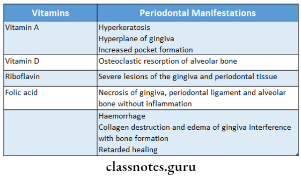
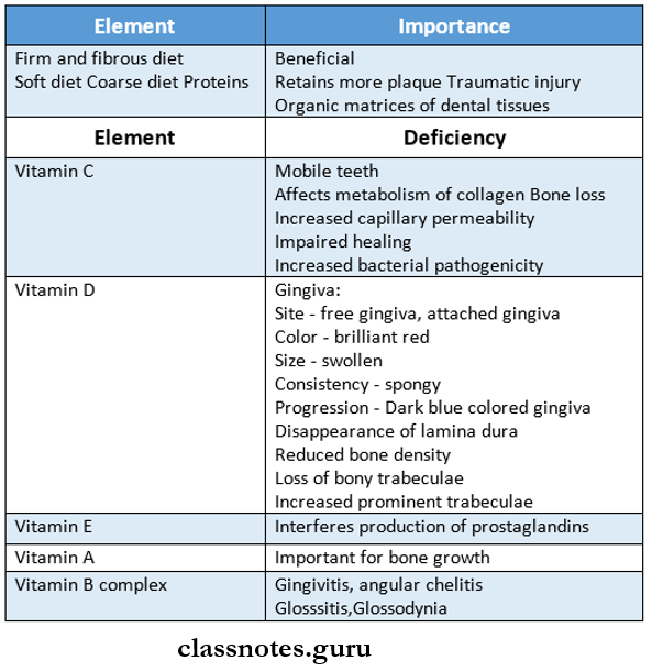
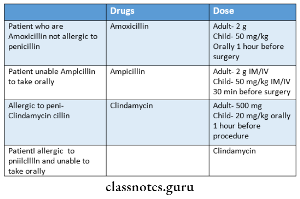
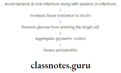
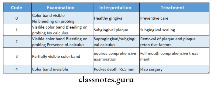
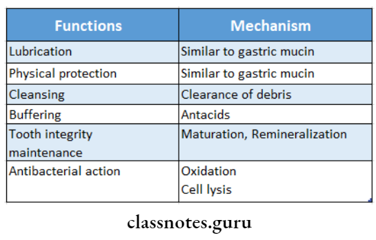
Trauma From Occlusion Short Answers

Question 1. Primary and secondary trauma from occlusion.
Answer:

1. Primary:

  • When trauma from occlusion results from alteration of occlusal forces, it results in primary trauma from occlusion
  • It does not alter the level of connective tissue attachment
  • It does not initiate pocket formation because su- parental gingival fibres are not affected which prevents apical immigration of junctional epithelium

2. Secondary:

  • It is trauma from occlusion that results due to re- reduced ability of tissues to resist occlusal forces
  • Adaptive capacity is impaired by bone loss resulting from marginal inflammation
  • Reduces periodontal attachment
  • Alters leverage on remaining tissues

Question 2. Lipping.
Answer:

  • Excessive forces lead to the resorption of bone
  • When bone is resorbed in such cases, the body attempts to reinforce trabeculae to form new bone
  • This process is called buttressing

Types:

1. Central buttressing:

  • In it, endosteal cells deposit new bone
  • It restores bony trabeculae
  • Reduces the size of bone marrow

2. Peripheral buttressing:

  • Occurs on facial and lingual surfaces of bone
  • May produce shelflike thickening of the alveolar margin called lipping
  • It is pronounced bulge in the contour of the facial or lingual bone

Question 3. Define trauma from occlusion.
Answer:

  • When occlusal forces exceed the adaptive capacity of the periodontal tissues, the tissue injury results
  • This resultant injury is termed trauma from occlusion

Question 4. Diagnosis of trauma from occlusion.
Answer:

1. Fremitus test:

Procedure:

Ask the patient to contact the teeth together in the maximum intercuspal position

Place dampened index finger over the buccal surface of maxillary teeth

Examine the teeth which are displaced

Result:

  • Class 1- Mild vibration
  • Class 2- Easily palpable but no visible movements
  • Class 3- Movements visible with the naked eye

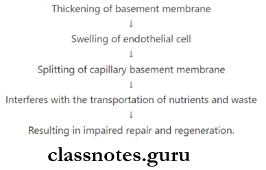
2. Radiographic features:

  • Increase in width of PDL space
  • Thickening of lamina dura along lateral borders of root, apical and bifurcation areas
  • Vertical bone loss
  • Radiolucency occurs due to condensation of alveolar bone
  • Root resorption

Question 5. Causes and changes produced by primary trauma from occlusion.
Answer:

Causes:

  • High filling restoration
  • Prosthetic replacement
  • Drifting movement or extrusion of teeth
  • Unreplaced missing teeth
  • Orthodontic movement of teeth

Changes Produced:

  • Does not alter the level of connective tissue attachment
  • Do not initiate the pocket formation
  • As suprarenal fibres are not affected it prevents apical migration of junctional epithelium

Question 6. Define acute and chronic trauma from occlusion
Answer:

1. Acute trauma from occlusion:

  • It results from an abrupt occlusal impact such as that produced by biting on a hard object

2. Chronic trauma from occlusion:

  • It develops from gradual changes in occlusion produced by tooth wear due to drifting movement and extrusion of teeth combined with parafunctional habits such as bruxism and clenching

Question 7. Facets.
Answer:

  • Facets are shiny and irregular
  • They indicate tooth-to-tooth wear that is associated with bruxism
  • These worn and abraded teeth are invariably firm with no sign of mobility

Question 8. Buttressing bone formation
Answer:

  • Excessive forces lead to the resorption of bone
  • When bone is resorbed in such cases, the body attempts to reinforce trabeculae to form new bone
  • This process is called buttressing

Types:

  1. Central buttressing
    • In it, endosteal cells deposit new bone
    • It restores bony trabeculae
    • Reduces the size of bone marrow
  2. Peripheral buttressing
    • Occurs on facial and lingual surfaces of bone
    • May produce shelf-like thickening of the alveolar margin called lipping
    • It is a pronounced bulge in the contour of the facial or lingual bone

Question 9. Primary and secondary trauma from occlusion
Answer:

Primary:

  • When trauma from occlusion results from alteration of occlusal forces, it results in primary trauma from occlusion
  • It does not alter the level of connective tissue attachment
  • It does not initiate pocket formation because supracre- stal gingival fibres are not affected which prevents apical migration of junctional epithelium

Secondary:

  • It is trauma from occlusion that results due to reduced ability of tissues to resist occlusal forces
  • Adaptive capacity is impaired by bone loss resulting from marginal inflammation
  • Reduces periodontal attachment Alters leverage on remaining tissues

Periodontics Question and Answers

Role Of Systemic Diseases In The Etiology Of Periodontal Disease Short Essay Question And Answers

Role Of Systemic Diseases In The Etiology Of Periodontal Disease Important Notes

1. Vitamin deficiency and periodontal manifestations

Role Of Systemic Diseases In The Etiology Of Periodontal Disease Vitamin deficiency and their periodonatal manifestations

Role Of Systemic Diseases In The Etiology Of Periodontal Disease Short Essays

Question 1. Role of nutrition in Periodontal diseases.
Answer:

  • Essential nutrients are required for the maintenance of healthy periodontium
  • They must be present in balanced quantities
  • Its deficiencies may cause adverse effects on the periodontium

Role Of Systemic Diseases In The Etiology Of Periodontal Disease Role of nutrition in periodontal diseases

Question 2. Periodontal management in Renal Transplant Patient.
Answer:

  • Consult physician
  • Monitor blood pressure
  • Laboratory Investigation
  • Bleeding time
  • Prothrombin time
  • Screen for hepatitis B
  • Provide antibiotic prophylaxis

Read And Learn More: Periodontics Question and Answers

  • Eliminate the source of infection on the day after dialysis
  • Frequent recall visit
  • Avoid administration of the nephrotoxic drug
  • Dental planning must be done to decide whether to re- Essential, biologically active constituents tain/extract teeth
  • Teeth with furcation involvements, periodontal abscess, and extensive surgical requirements should be extracted
  • Use of oral antibiotic mouthwash with nystatin

Question 3. Infective Endocarditis.
Answer:

It is a disease in which micro-organisms colonize the damaged endocardium/heart valves

Types of – Acute and sub-acute

Organisms involved – Streptococci, Staphylococci

Preventive Measures:

  • Define susceptible persons
  • Provide oral hygiene instruction
  • Antibiotic prophylaxis
  • Endodontic therapy instead of extraction
  • Elimination of source of infection
  • Avoid prolonged impingement on gingival tissues by retraction
  • Rinse with 0.12% chlorhexidine prior to surgery
  • Regular recall visits

Q.4. Antibiotic prophylaxis for medically compromised patients.
Answer:

Role Of Systemic Diseases In The Etiology Of Periodontal Disease Antibiotic prophylaxis for medically compromised patients

Question 5. Vitamin C and periodontal disease.
Answer:

Periodontal disease Vitamin C:

  • The essential, biological active constituent
  • Water soluble vitamin

Periodontal disease functions:

  • Vitamin C helps in tissue oxidation
  • It is needed for the formation of collagen

Periodontal disease Deficiency: Scurvy

Periodontal disease Features:

1. General:

  • Increased susceptibility to infection
  • Increased capillary permeability
  • Impaired wound healing
  • Sluggishness of blood flow

2. Oral – Gingiva:

  • Involved site – Free gingiva and attached gingiva
  • Size – Increased
  • Color Brilliant red
  • Consistency-spongy
  • Symptom – Tender, bleeding gums

Periodontal disease others:

  • Defective osteoblastic formation
  • Decreased bone formation
  • Impaired bone removal
  • Results in bone resorption and tooth mobility

Role Of Deficiency Of Vitamin C:

  • Influence the collagen metabolism
  • Affects tissue regeneration and repair
  • Interferes with bone remodeling
  • Allows passage of pathogens due to increased capillary permeability
  • Increases bacterial pathogenicity

Question 6. Diabetes mellitus and periodontal disease.
Answer:

Role Of Systemic Diseases In The Etiology Of Periodontal Disease.

Periodontal disease Pathogenesis:

1. Vascular changes:

Role Of Systemic Diseases In The Etiology Of Periodontal Disease Vascular changes

2. PMN”s function: Impaired adherence, phagocytosis, and intracellular killing

3. Crevicular fluid: Reduced cyclic AMP levels

4. Microflora: Reduced hyaluronidase activity

Diabetes And Periodontal Treatment:

  • Periodontal therapy decreases pathogens thereby inflammation
  • Results in improved metabolic control

Alterations In Treatment Plan:

  • Morning appointments
  • Altered post-operative dose
  • Atraumatic procedure
  • Antibiotic prophylaxis

Question 7. Periodontal therapy for pregnant women.
Answer:

1. Plaque control:

  • Reinforce oral hygiene techniques
  • Scaling, polishing, and root planning may be per- formed whenever necessary
  • Avoid the use of high alcohol content antimicrobial rinses

2. Elective dental treatment:

  • Avoid during 1st trimester and the last half of 3rd tri- semester because
    • First trimester- period of orga
    • Third trimester-risk of premature delivery
  • Avoid prolonged chair time
  • The pregnant patient is allowed to turn on her left side frequently to remove pressure on the vena cava Second trimester is the safest period
  • Major surgery should be postponed until delivery

3. Dental radiograph:

  • Avoided protecting the fetus from radiation
  • If necessary radiographs are taken along with a protective lead apron

4. Medications:

Medications Safe Antibiotics:

  • Penicillin Erythromycin
  • Clindamycin
  • Cephalosporin

Medications Contraindicated Antibiotics:

  • Tetracyclines
  • Ciprofloxacin
  • Metronidazole
  • Clarithromycin

Question 8. Periodontal infection is a cause of atherosclerosis.
Answer:

  • products, calcium, and other substances accumulate in the blood vessel wall forming plaque that elevates into the blood vessel blocking the blood flow
  • The rupture plaque fragments can release certain pro-coagulant chemicals that may cause platelet aggregation and in turn cause thrombus formation

Role Of Systemic Diseases In The Etiology Of Periodontal Disease Q8

  • They travel to distant sites and initiate pathology specific to that organ/system
  • Periodontitis influences atherosclerosis by
    • Microbes like Streptococcus samurais and P. gin- gives releases platelet aggregation associated pro- tein and aggregate platelet leading to thromboembolic events
    • Direct invasion of periodontal pathogens
    • Presence of periodontitis
    • Action of macrophages

Role Of Systemic Diseases In The Etiology Of Periodontal Disease Short Question And Answers

Question 1. Pregnancy gingivitis.
Answer:

Occurs during the first trimester of pregnancy

Pregnancy gingivitis Changes:

  • Mushroom-like flattened spherical mass
  • Protrusion from interdental papilla occurs

Pregnancy gingivitis Causes:

  • Aggregation of previous inflammation
  • Altered tissue metabolism

Pregnancy gingivitis Features:

1. Gingiva:

  • Marginal and interdental gingiva is edematous, pits on pressure
  • Color- bright red or magenta
  • Consistency- Sof and friable
  • Bleeding on probing is present
  • Size- increased
  • Loss of stippling
  • Shape- mushroom-like flattened spherical mass
  • Increased GCF

2. Other features:

  • Increased susceptibility to mechanical irritation
  • Increased GCF
  • Increased pocket depth
  • Increased mobility
  • Depression of maternal T-lymphocyte response

Question 2. Periotemp.
Answer:

  • It is a probe used to detect pocket temperature differences of 0.1 degrees C from the reference temperature
  • The temperature gradient exists between maxillary and mandibular teeth and between posterior and anterior teeth
  • Individual temperature differences are compared
  • Higher temperature pockets are signaled with a red-emitting diode
  • Periotemp is also used to identify loss of attachment
  • Red temperature indicates twice the risk of attachment loss

Question 3. Biopsy.
Answer:

Biopsy Uses:

  • Rule out malignancies
  • Detect local and systemic inter-relationship
  • Differentiate among different types of gingival enlargements
  • Helpful in the presence of desquamative gingivitis

Specimens Obtained Are:

Marginal and attach gingiva

Question 4. Periodontal care in patients with anticoagulant therapy.
Answer:

  • Consult the physician
    • To stop aspirin 7-14 days prior to surgery
    • Minimize trauma
    • Prophylactic antibiotics
    • Use pressure hemostasis
    • Complete stoppage of bleeding prior to placement of the periodontal pack
    • Avoid therapy if the patient has an acute infection
    • Recall after 3-5 days

Question 5. Stress and periodontal diseases.
Answer:

  • Stress may induce periodontal diseases in the oral cavity through
    • Development of habits that are injurious to the periodontium
    • Poor oral hygiene
    • Poor nutritional intake
    • Overeating of high-fat diet due to stress
    • All of the above increases susceptibility to bacterial infection and leads to periodontal diseases

Question 6. Periodontal care of patients with tuberculosis.
Answer:

  • The patient with tuberculosis should receive only. emergency care
  • If the patient has completed chemotherapy, the patient’s physician should be consulted regarding ineffective- city and results of sputum cultures for M. tuberculosis
  • When medical clearance has been given and sputum culture results are negative, these patients may be treated normally
  • Adequate treatment of tuberculosis requires a minimum of 18 months and thorough post-treatment follow-up should include chest radiographs, sputum cultures, and a review of the patient’s symptoms by the physician at least every 12 months.


Various Aids Periodontics Short Essay Question And Answers

Various Aids Important Notes

1. Types of probes

Various Aids Types of probes

2. Miller’s mobility grading

Various Aids Miller's mobility grading

3. Types of periodontal probes

Various Aids Types of periodontal probes

Various Aids Short Essays

Question 1. Enumerate advanced diagnostic aids and write. Determines mucogingival relationship
in detail about DNA probes.
Answer:

Advanced Diagnostic Aids:

1. Aids in radiographic techniques:

  • Digital radiography
  • Subtraction radiography
  • CADIA (Computer Assisted Densitometric Image Analysis)
  • Computerized tomography
  • Nuclear Medicine Bone scan

1. Aids in microbiological diagnosis

  • Jar technique
  • Anaerobic chamber technique
  • Enzyme reduction technique

Read And Learn More: Periodontics Question and Answers

2. Other Aids

  • GLC (Gas Liquid Chromatography)
  • DNA probe
  • PCR (Polymerase chain reaction)

Advanced Diagnostic Aids DNA Probe:

  • DNA probe uses segments of single-stranded nucleic acid labeled with an enzyme or radioisotope that is able to hybridize to the complementary nucleic acid sequence
  • It detects the presence of target micro-organisms causing periodontal diseases

Procedure:

Various Aids Procedure


Question 2. Periodontal probes.
Answer:

Periodontal probes Use:

  • Measure pocket depth
  • Determines bleeding on probing
  • Determines furcation involvement
  • Locate calculus
  • Identifies irregularities

Periodontal probes Types:

  1. Marquis color coded – Markings at 3 mm section
  2. UNC-15 probe
    • Length – 15 mm
    • Markings present – at each mm
    • Color coding-5th, 10th and 15mm
  3. The University of Michigan O probe
    • Markings 1, 2, 3, 5, 7, 8, 9
  4. Michigan ‘O’ probe
    • Markings – 3, 6, and 8 mm
  5. WHO probe
    • Tip-0.5 mm ball
    • Markings – 3.5, 8.5, 11.5 mm
    • Color coding – 3.5-5.5 mm
  6. Naber’s probe for furcation areas

Periodontal probes Generations:

  1. First Generation: Conventional probe
  2. Second Generation: Pressure-sensitive probe
  3. Third Generation: Computerized probes

Various Aids Florida probe

Various Aids Marquis color code probe calibration in 3 mm sections

Question 3. PSR.
Answer:

  • Periodontal Screening and Recording

PSR Probe:

  • 0.5 mm ball tip
  • Color coding-3.5.-5.5 mm

PSR Method:

  • Divide the patient’s mouth into 6 sextants
  • Examine six points around each tooth and code it.

Codings:

Various Aids Codings

Various Aids Short Answers

Question 1. ELISA test.
Answer:

  • ELISA is Enzyme-Linked Immunosorbent Assay
  • Diagnostic test for HIV
  • It is color based test

ELISA test Procedure:

ELISA test Apparatus: Separate wells containing

  • Antigen
  • Suspected antibodies
  • Controls
  • Antisera to antibody

Method:

Various Aids ELISA test method

ELISA test Result: Positive shows color change


Host Modulation Therapy Short Essay Question And Answers

Host Modulation Therapy Short Essays

Question 1. Host Modulation Therapy (HMT).
Answer:

1. Regulation of Immune and Inflammatory re- responses:

Host Modulation Therapy Regulation of Immune and Inflammatory re- responses

Protective mechanism:

  • Activation of neutrophils
  • Production of antibodies
  • Release of TGF-B, IL-4, IL-10, IL-12

Destructive mechanism:

  • Prolonged bacterial presence
  • Release of IL-1, IL-6, TNF-α, PGE2
  • Extracellular matrix destruction
  • Bone resorption
    • Thus for therapeutic purposes, a protective mechanism is introduced

2. Role of MMPs [Matrix metalloproteinases]:

Host Modulation Therapy Matrix metalloproteinases

  • Inhibited by…
  • A reduced dose of doxycycline (SSD) –
  • Subantimicro- by Dose of Doxycycline
  • Example: Periostat-20mg BID for 3 months
  • It has…

Read And Learn More: Periodontics Question and Answers

    • No antimicrobial effect
    • Has anti-collagenolytic activity
    • Decreased bone resorption
    • Increases lymphocytes and PMNs

3. Production of Arachidonic acid Metabolites:

Host Modulation Therapy Production of Arachidonic acid Metabolites

  • Cyclo-oxygenase
    • Metabolites into
    • Prostaglandin
    • Prostacyclin
    • Thromboxane
  • Lipoxygenase
    • Metabolites into
    • Leukotrienes
    • Hydroxyeicosate retinoic acid

Host Modulation Therapy Lipoxygenase

4. Regulation of Bone Metabolism Osteoclast:

Host Modulation Therapy Regulation of Bone Metabolism Osteoclast

Host Modulation Therapy Short Question And Answers

Question 1. Periostat.
Answer:

  • It is a reduced dose of doxycycline- sub antimicrobial dose of doxycycline
  • It is a 20 mg dose of doxycycline
  • Indicated as an adjuvant to scaling and root planning in the treatment of chronic periodontitis
  • It must not be used as monotherapy because it is based on the sub-antimicrobial dosage of doxycycline
  • It is taken twice daily or 3 months, up to a maximum of 9 months of continuous dosing
  • It exerts its therapeutic effect through enzyme, cytokine, and osteoclast inhibition

Periostat Effects:

  • No antimicrobial effect
  • Has anti-collagenolytic activity
  • Decreased bone resorption
  • Increases lymphocytes and PMNs

Periostat Mechanism:

  • Downregulates MMPs
  • Reduces cytokine levels
  • Upregulates collagen production
  • Stimulates osteoblastic activity and new bone formation

Host Modulation Therapy Viva Voce

  1. Host modulation therapy restores the balance between pro-inflammatory and anti-inflammatory mediators
  2. Host modulation therapy is used as an adjuvant to conventional periodontal therapy
  3. Host modulation therapy usually ameliorates excessive or pathologically elevated inflammatory processes
  4. Sub antimicrobial dose of Doxycycline is 20 mg
  5. Sub antimicrobial dose of Doxycycline can be pre-scribed continuously up to a maximum of 9 months


Periodontal Medicine Short Essays

Question 1. Periodontal disease and Coronary Heart disease.
Answer:

  • According to cross-sectional studies, myocardial infarction had significantly poor dental conditions. But is independent of the presence of risk factors
  • The study of Mattila performed by oral reading- raphy and diagnostic angiography, showed a correlation between the severity of periodontal disease and coronary atheromatous

Mechanism:

Periodontal Medicine Mechanism

Pathogenesis Of Periodontal Diseases Short Essay Question And Answers

Pathogenesis Of Periodontal Diseases Important Notes

1. Cytokines

  • The three proinflammatory cytokines that have a central role in periodontal tissue destruction are interleukin-1, interleukin-6, and tumor necrosis factor, TNF
  • The properties of these cytokines that relate to tissue destruction involve stimulation of bone resorption and in- duction of tissue degrading proteinases
  • IL-1 exists in alpha and beta forms
  • Both forms are the main constituents of the osteoclast activating factor
  • It is a potent stimulant of osteoclast proliferation, differentiation, and activation
  • TNF is also found in alpha and beta forms
  • IL-1 and TNF-a induce the production of proteinases in mesenchymal cells including matrix metalloproteinases which may contribute to tissue destruction

Pathogenesis Of Periodontal Diseases Short Essays

Question 1. Cytokines.
Answer:

Cytokines Definition:

‘Cell protein’ is used for molecules that transmit information/signals from one cell to another.

Cytokines Actions:

  • Acts as fibroblast, macrophages, keratinocytes, and PMNs
  • Release MMP’s
  • Degrade connective tissue matrix

Pathogenesis Of Periodontal Diseases Signals from one cell to another

Pathogenesis Of Periodontal Diseases Short Answers

Question 1. Define cytokine.
Answer: cytokine

Question 2. Interleukin 1.
Answer: Interleukin is cytokines

Interleukin 1 Secreted By:

  • Macrophages, lymphocytes
  • Fibroblasts, platelets
  • Keratinocytes and endothelial cells

Interleukin 1 Functions:

  • Increases neutrophils adhesion
  • Promotes antibody production
  • Causes tissue destruction

Read And Learn More: Periodontics Question and Answers

  • Secretes MMP’s
  • Releases PGE2

Question 3. Prostaglandin.
Answer:

  • Short range hormone
  • Present in inflammatory exudates, leukotrienes
  • Released from – Mast cells and basophils

Example: PGE2

Prostaglandin Functions:

  • Osteoclastic resorption
  • Degrades connection tissue matrix

Question 4. Bacterial Endotoxin.
Answer:

Location: Outer membrane of Gram-ve bacteria

Bacterial Endotoxin Pathogenesis:

  • Penetrate gingival epithelium
  • Produce fatty and organic acids, amines, VSCs, indole, ammonia, and glycans

Bacterial Endotoxin Effects:

  • Direct activation of host responses
  • Produce leukopenia
  • Activate Factor XII, Complement system
  • Cytotoxic effects on fibroblast
  • Tissue Necrosis
  • Bone resorption

Question 5. Virulence Factors.
Answer:

The properties that enable the bacterium to cause disease are termed virulence factor

  1. Fimbriae Helps in adherence of A. viscous (for example) on the saliva-coated tooth surface
  2. Exotoxin’s Toxic Effect on PMNs
  3. Lipopolysaccharides – Activates host response
  4. Peptidoglycan – Activates complement system
  5. Immunosuppressive activity
  6. Enzymes
    • Collagenase-Degrades collagen
    • Hyaluronidase – Alters gingival permeability
  7. Others Damage host cells
    • Degrade antibody
    • Damage keratinocytes


Defence Mechanism Of Gingiva Short Essay Question And Answers

Defence Mechanism Of Gingiva Important Notes

1. Cells in Gingival Crevicular Fluid

  • The predominant cell in GCF is PMNs- 92%
  • Mononuclear cells contribute to 8%

2. Methods of collecting Gingival Crevicular Fluid (GCF)

  • Placing filter paper into the sulcus – intra crevicular
  • Placing paper at the entrance of sulcus- extra crevicular
  • Placing preweighed twisted threads or micropipettes and crevicular washings

3. Methods of measuring the amount of Gingival Crevicular Fluid (GCF)

  • The wetted area of the filter paper is stained with Ninhydrin and is measured parametrically under a microscope
  • The electronic method using fluid collected on a blotter paper and employing perceptron

4. Glucose content of GCF is 3-4 times greater than in serum due to the metabolic activity of the adjacent tissues and the function of microbial flora. While the protein content of GCF is less than that of serum

Defence Mechanism Of Gingiva Gingival Crevicular Fluid

  1. Functions of saliva
  2. Significance of gingival crevicular fluid
  3. Methods of Gingival crevicular fluid
  4. Composition of Gingival crevicular fluid (GCF)
  5. Defense mechanism of gingival
  6. Circadian periodicity

Defense Mechanism Of Gingival Short Essays:

Question 1. Functions of Saliva.
Answer:

Defence Mechanism Of Gingiva Functions of Saliva

Question 2. Significance of Gingival Crevicular Fluid (GCF)
Answer:

1. Circadian periodicity:

  • There is a gradual increase in Gingival Crevicular Fluid (GCF) amount from 6:00 AM to 10.00 PM and decreases afterward
  • This is called Cicardian periodicity

2. Sex Hormones:

  • Female sex hormones increase the flow
  • Pregnancy, ovulation, and hormonal contraceptives increase gingival fluid

3. Smoking:

Causes an immediate transient increase in flow

4. Periodontal therapy:

Increase in gingival fluid during the healing period

Read And Learn More: Periodontics Question and Answers

Question 3. Methods of Collection of Gingival Crevicular Fluid (GCF)
Answer:

1. Absorbing paper strips:

Collection of Gingival Crevicular Fluid (GCF) Types:

  • Intracrevicular – Insert paper in sulcus
  • Extracurricular – Place at the entrance of sulcus

Collection of Gingival Crevicular Fluid (GCF) Evaluation:

  • Staining with alcohol
  • Pre and post-weighing the papers
  • Periotron electronic device

2. Micropipettes:

Analyze the amount of GCF collected in tubes by capillary action

3. Gingival washing:

A known amount of solution is introduced and removed into the Gingival sulcus

4. Other:

  • Plastic strips/Platinum loops
  • Inserted into sulcus and pressure is applied

Question 4. Composition of Gingival Crevicular Fluid (GCF)
Answer:

  1. Cells: Bacteria, Epithelial cells, Leukocytes (BEL)
  2. Electrolytes: Sodium, Potassium, Calcium
  3. Organic compounds: Carbohydrates, Proteins, Lipids
  4. Metabolic and Bacterial products: Lactic acid, Urea, Prostaglandins, Endotoxin
  5. Enzymes: Acid phosphatase, Alkaline phosphatase, Pyrophosphatase, lysozyme, Hyaluronate

Question 5. Defense Mechanism of Gingiva.
Answer:

Defence Mechanism Of Gingiva Non specific and Specific protective

1. Non-specific Protective Mechanisms:

  • Bacterial Balance:
    • Balance exists between different species of microorganisms
  • Surface Integrity:
    • Maintained by the persistent renewal of the epithelium from its base and desquamation of surface layers
    • This maintains the constant thickness of the epithelium
  • Surface Fluid and Enzymes:
    • Saliva contains antibacterial substances
    • Gingival Crevicular Fluid (GCF) contains phagocytic leukocytes and enzymes
  • Phagocytosis:
    • Monocytes act in chronic infections
    • PMNs act in acute infections
    • Complements induce smooth muscle contraction, increase vascular permeability, and release histamine from mast cells.
  • Inflammatory reaction:
    • Stimulated by tissue injury and infections
    • Results in hyperemia increased vascular permeability and formation of exudate

2. Specific Protective mechanism:

My immune system

Protective mechanism Types:

  • Humoral immunity
  • Cell-mediated immunity

Defense Mechanism Of Gingiva Short Question And Answers

Question 1. Cicardian Periodicity.
Answer:

  • There is a gradual increase in gingival fluid amount from 6:00 AM to 10.00 PM and decreases afterward
  • This is called Cicardian periodicity

Question 2. Intracellular drug delivery.
Answer:

  • Drugs that are excreted through the GCF may be used. This flow of neutrophils is important for protection from advantageously in periodontal therapy
  • Such drugs are tetracycline and metronidazole
  • These drugs are excreted in crevicular fluid when given systemically
  • When these drugs are given locally, higher concentrations are achieved in the desired area leading to faster action

Question 3. Name the defense mechanism of the gingiva.
Answer:

Defence Mechanism Of Gingiva:

  • Epithelial barrier
  • GCF
  • Saliva
  • Orogranulocytes

Question 4. Granulocytes.
Answer:

  • The viable neutrophils present in the saliva are termed orogranulocytes or salivary corpuscles
  • In normal individuals, 30,000 neutrophils per minute enter the oral cavity via the gingival sulcus
  • This flow of neutrophils is required for periodontal health
  • Any defect in neutrophil function and chemotaxis is associated with early-onset periodontal disease in children’s caries

Defence Mechanism Of Gingiva Viva Voce

  1. The ratio of T: B lymphocytes is about 1:3 in GCF
  2. Predominant immunoglobulin in GCF is IgG
  3. Predominant immunoglobulin in saliva is IgA

Dental Plaque Question and Answers

Dental Plaque Definitions

1. Dental plaque

  • It is soft deposits that form biofilm adhering to the tooth surface or other hard surfaces in the oral cavity including removable and fixed restoration

Dental Plaque Dental Plaque Of Biofilm

Dental Plaque Classification

1. Dental plaque

  • Dental plaque is classified into
    • Supra gingival plaque
    • Subgingival plaque
      • It is further divided into
        1. Tooth associated
        2. Epithelium associated

Dental Plaque Important Notes

  1. Early plaque contains streptococcus sanguis and Actinomyces viscosus. Late plaque contains p.gingivalis, p. media, fusobacterium and capnocytophaga
  2. Type of plaque and its outcome
  3. Stages of formation of plaque
    • Formation of pellicle
    • Initial colonization of the tooth surface
    • Secondary colonization and plaque maturation

Dental Plaque Type of plaque and its outcome

4. Socransky’s postulate

  • It is for the identification of causative agents
  • No. of etiologic organisms in the diseased site must be increased
  • While it should be reduced in healthy sites
  • If an etiologic organism is eliminated then disease progression ceases
  • Presence of antibodies in organisms
  • Presence of virulence factors
  • In vitro, experiments should demonstrate the disease process

5. Composition of plaque

  1. Micro-organisms
    • Bacteria
    • Mycoplasm
    • Fungi
    • Protozoa
    • Virus
  2. Intracellular matrix
    • Organic
    • Carbohydrates
    • Glycoproteins
    • Lipids
  3. Inorganic
    • Calcium
    • Phosphorous
    • Magnesium
    • Potassium
    • Sodium

6. Bacteria and periodontal health

Dental Plaque Bacteria and periontal health

7. Colonizers

  • They contribute to biofilm formation
  • Initial colonizers include co-aggregation of fusobacterium with s. sanguis, prevotella with A. viscosus
  • and capo-autophagy with A viscous

Read And Learn More: Periodontics Question and Answers

  • Secondary colonizers adhere to bacteria already present
  • Initial colonizers form yellow or purple complexes while secondary colonizers form green, orange, or red complexes

Dental Plaque Colonizers

Dental Plaque Long Essays

Question 1. Define and classify microbial plaque. Discuss the role of plaque in the etiology of gingival and periodontal disease.
Answer:

Definition:

  • It is soft deposits that form biofilm adhering to the tooth surface or other hard surfaces in the oral cavity including removable and fixed restoration

Microbial plaque Classification:

Dental Plaque Classification of microbial plaque

Role Of Plaque In Gingivitis And Periodontitis:

  • Gingivitis usually precedes periodontitis
  • In the early stage of gingivitis, bacteria in plaque build-up, causing the gingival to become inflamed and to easily bleed during tooth brushing.
  • No irreversible bone or other tissue damage has occurred at this stage.
  • When gingivitis is left untreated, it can advance to periodontitis. In a person with periodontitis, form pockets.
  • These small spaces between teeth and gingival collect debris and can become infected
  • As the disease progresses, the pockets deepen and more gingival tissue and bone are destroyed.
  • When this happens, teeth are no longer anchored in place, they become loose, and tooth loss occurs.

Question 2. Define and classify plaque. Write in detail about its composition and ill effects.
Answer:

Definition:

  • It is soft deposits that form biofilm adhering to the tooth surface or other hard surfaces in the oral cavity including removable and fixed restoration

Classification:

  • Dental plaque is classified into
    • Supra gingival plaque
    • Subgingival plaque
  • It is further divided into
    • Tooth associated
    • Epithelium associated

Composition:

1. Microorganisms:

  • Bacteria
  • Mycoplasma
  • Fungi
  • Protozoa
  • Virus

2. Intracellular matrix:

  • Organic:
    • Carbohydrates
    • Glycoproteins
    • Lipids
  • Inorganic:
    • Calcium
    • Phosphorous
    • Magnesium
    • Potassium
    • Sodium

Ill Effects Of Dental Plaque:

  • Predisposes to caries
  • Gingivitis
  • Supra gingival plaque matures and creates an environment for the development of subgingival plaque
  • Organisms get accumulated in this space
  • The disease advances to periodontitis with the pocket formation and bone loss
  • Due to the loss of tooth-supporting bone teeth becomes loose and get exfoliated

Question 3. Discuss the role of Local factors in the etiology of Periodontal disease.
Answer:

Local Factors In Etiology Of Periodontal Diseases:

1. Role of plaque

  • Gingivitis usually precedes periodontitis
  • In the early stage of gingivitis, bacteria in plaque build-up, causing the gingival to become inflamed and to easily bleed during tooth brushing.
  • No irreversible bone or other tissue damage has occurred at this stage.
  • When gingivitis is left untreated, it can advance to periodontitis. In a person with periodontitis, form pockets.
  • These small spaces between teeth and gingival collect debris and can become infected
  • As the disease progresses, the pockets deepen and more gingival tissue and bone are destroyed. When this happens, teeth are no longer anchored in place, they become loose, and tooth loss occurs.

2. Role of calculus

  • Calculus provides a fixed nidus for continuous plaque accumulation
  • It brings plaque bacteria close to the supporting tissues
  • Interferes with a local self-cleansing defense mechanism
  • Acts as a reservoir for irritating substances such as endotoxins, antigenic material, and bone-resorbing factors
  • Makes plaque removal more difficult for the patient

Dental Plaque Short Essays

Question 1. Dental plaque – its definition, classification, and composition.
Answer:

Definition:

  • It is soft deposits that form the biofilm adhering to the tooth surface or other hard surfaces in the oral cavity, including removable and fixed restoration.

Classification:

  • Dental Plaque
  • Supra gingival
  • Subgingival
  • Tooth – associated
  • Epithelium associated

Composition:

Microorganisms:

  • Bacteria
  • Mycoplasma
  • Fungi
  • Protozoa
  • Virus

Intracellular Matrix:

Organic:

  • Carbohydrates
  • Glycoproteins
  • Lipids

Inorganic:

  • Ca, P
  • Mg, K, Na

Question 2. Specific and non-specific plaque. (or) Elaborate on various plaque hypotheses.
Answer:

Specific Plaque Hypothesis:

  • The specific plaque hypothesis states that not all plaque is pathogenic, and its pathogenicity depends on the presence of certain specific microbial pathogens in plaque
  • Specific microorganisms responsible for periodontal dis-eases release certain damaging factors called virulence factors which lead to host tissue destruction

Example: A.a. contains causes aggressive periodontitis

Non-Specific Plaque:

  • It states that it is the total bulk of the plaque that determines the pathogenicity rather than the individual species in it
  • According to it, If a small amount of plaque is present, then the products released by it can be neutralized by the host
  • But if it is present in large quantity, then it alters the host response
  • It forms the basis of recent treatment and prevention modalities

Question 3. Structure of dental plaque.
Answer:

1. Supra gingival plaque:

  • It adheres to the tooth surface
  • It contains Gram-positive cocci and Gram-negative rods and filaments
  • The arrangement is described as Corncob arrangement- ment
  • The central core consists of rod-shaped bacterial cells like Fusobacterium nucleatum
  • Coccal cells like streptococci get attached to it

2. Sub gingival plaque:

  • It contains many large filaments with flagella
  • It is rich in spirochetes

Tooth-associated plaque:

  • Its structure is similar to supra gingival plaque

Tissue-associated plaque:

  • The extracellular matrix is not well-defined and contains numerous bristle brush formations
  • It forms test tube brush formation
  • It is characterized by large filaments that form the long axis
  • Short filaments or Gram-negative rods get embedded in the matrix

Dental Plaque subgingival plaque and supragingival plaque

Question 4. Differences between supra and subgingival plaque.
Answer:

Dental Plaque differences between supra and subgingival plaque

Dental Plaque Viva Voce

  1. 1 gm of plaque contains 2*1011 bacteria
  2. Organisms that predominate in early plaque are gram-positive cocci and rods
  3. In late plaque, organisms present are gram-negative anaerobic rods and filament
  4. Supra gingival plaque typically demonstrates corncob structures
  5. Plaque is the most common cause of gingivitis and periodontitis
  6. Material alba is a yellowish or white soft sticky deposit and is less adherent
  7. A. actinomycete contains is a facultative anaerobe
  8. The red complex is associated with bleeding on probing.

Trauma From Occlusion Treatment Question and Answers

Trauma From Occlusion Definitions

1. Trauma from occlusion

  • When occlusal forces exceed the adaptive capacity of the periodontal tissues, the tissue injury results
  • This resultant injury is termed trauma from occlusion.

2. Pathological migration of teeth

  • Pathological migration of teeth refers to tooth displacement that results when the balance among the factors that maintain physiologic tooth position is disturbed by periodontal disease

Trauma From Occlusion Important Notes

1. Types of trauma from occlusion

  • Primary trauma from occlusion
    • Occurs due to excessive occlusal forces
    • Changes produced by it do not alter the level of connective tissue
    • It does not initiate the pocket formation
    • As suprarenal fibres are not affected it prevents apical migration of junctional epithelium
  • Secondary trauma from occlusion
    • Occurs when the adaptive capacity of the tissues to withstand occlusal forces is impaired by bone loss re- resulting from marginal inflammation

2. Trauma from occlusion does not cause pockets or gingivitis as the marginal gingiva is unaffected by trauma from occlu- sion because of its rich blood supply

  • But trauma from occlusion may affect the progress and severity of periodontal pockets started by local irritation

3. Signs and symptoms of trauma from occlusion

  • Tooth pain, sensitivity to percussion
  • Increased tooth mobility
  • Cementum tears
  • Widening of periodontal space
  • Root resorption
  • Thickening of lamina dura
  • Vertical or angular bone defects

4. Buttressing bone formation

  • When excessive occlusal forces resorb bone, the bone attempts to reinforce the thinned bony trabeculae with new bone
  • This attempt to compensate for lost bone is called buttressing bone formation
  • Buttressing bone formation may be central or peripheral
  • Peripheral buttressing bone formation is also called lipping.

5. Increased tooth mobility is the most common finding of trauma from occlusion

  • This is due to the destruction of periodontal fibres in the injury stage and the widening of the periodontal ligament in the final adaptation stage of trauma from occlusion

Trauma From Occlusion Long Essays

Question 1. Define and classify trauma from occlusion. Discuss the stages of tissue response to increased occlusal forces.
Answer:

Definition:

  • When occlusal forces exceed the adaptive capacity of the periodontal tissues, the tissue injury results
  • This resultant injury is termed trauma from occlusion.

Classification:

1. According to the Onset:

Acute:

  • Due to abrupt occlusal forces.

Example: Biting on a hard object

Chronic:

  • Due to gradual changes in the periodontium

Example: Bruxism

2. According to the cause:

Primary:

  • When trauma from occlusion results from the alteration of occlusal forces
  • Do not alter the level of connective tissue attachment
  • Do not initiate pocket formation because supracre- stal gingival are not affected which prevents apical migration of junctional epithelium

Secondary:

  • When trauma from occlusion results due to the reduced ability of tissues to resist occlusal forces
  • Adaptive capacity is impaired by bone loss resulting from marginal inflammation
  • Reduces PDL attachment
  • Alters leverage on remaining tissues

Trauma From Occlusion Zone of irritation and zone of codestruction

Tissue Response:

Stage 1 – Injury:

Application of excessive occlusal forces

Distribution of periodontal tissues

Adaptation of periodontal structures to altered forces

On horizontal forces tooth rotates, 2 zones appear

  • Pressure zone
  • Tension zone

Stage 2 – Repair:

Excessive occlusal forces lead to the resorption of bone

Bone formation from thinned bony trabeculae is called “Buttressing Bone formation”

  • Within the jaw “Central Buttressing”
  • On bone surface “Peripheral Buttressing”
  • Shelf-like thickening of alveolar bone “Lipping”

Read And Learn More: Periodontics Question and Answers

Stage 3: Adaptive Remodelling of Periodontium:

  • Occurs when the repair cannot cope with the destruction
    • Results in
    • Thickening of periodontal ligament
    • Angular bone defects

Trauma From Occlusion Adptive remodelling of periodontium

Trauma From Occlusion Short Essays

Question 1. Clinical and Radiological features of TFO.
Answer:

Clinical Features:

1. Acute:

  • Excessive tooth pain
  • TOP positive
  • Mobility of teeth
  • Periodontal abscess
  • Pathologic migration of teeth
  • Pocket
  • If force is dissipated, symptoms subside

2. Chronic:

  • No pain is present
  • Deep bite
  • Symptom-free condition
  • Fremitus test-positive

Radiographic Changes:

  • Widening of PDL space
  • Vertical bone loss
  • Angular facets
  • Root resorption

Question 2. Glickman’s concept.
Answer:

Glickman explained the path of progression of disease along with its result

Trauma From Occlusion Primary and Result

Zones:

1. Zone of Irritation:

Marginal and interdental gingival inflammation

Passes apically to alveolar bone

Then to the PDL area

Results in horizontal bone loss

2. Zone of Co-destruction:

Trauma from occlusion

Involvement of PDL, cementum and bone

Spread of infection directly to PDL

Results in angular bone loss

Trauma From Occlusion Pathway of inflammatoy process

Question 3. Types of Trauma from Occlusion.
Answer:

Classification:

1. According to the onset:

  • Acute:
    • Due to abrupt occlusal forces. Ex- biting on a hard object
  • Chronic:
    • Due to gradual change in periodontium.

Example: bruxism

2. According to the cause:

Primary:

  • When trauma from occlusion results from al-iteration of occlusal forces, it results in primary trauma from occlusion
  • It does not alter the level of connective tissue attachments
  • It does not initiate pocket formation because suprarenal gingival fibres are not affected which prevents apical migration of junctional epithelium

Secondary:

  • It is trauma from occlusion that results due to reduced ability of tissues to resist occlusal forces
  • Adaptive capacity is impaired by bone loss re- resulting from marginal inflammation
  • Reduces periodontal attachment
  • Alters leverage on remaining tissues

Question 4. Tissue response to trauma from occlusion.
Answer:

Stage I-Injury:

Application of excessive occlusal forces

Distribution of forces to periodontal tissues

Adaptation of periodontal structures to altered forces

On application of horizontal forces tooth rotates

Two zones appear- The pressure zone and tension zone

Stage 2-Repair:

  • Excessive forces lead to the resorption of bone
  • When bone is resorbed in such cases, the body attempts to reinforce trabeculae to form new bone
  • This process is called buttressing

Types:

1. Central buttressing:

  • In it, endosteal cells deposit new bone
  • It restores bony trabeculae
  • Reduces the size of bone marrow

2. Peripheral buttressing:

  • Occurs on facial and lingual surfaces of bone
  • May produce shelflike thickening of the alveolar margin called lipping
  • It is a pronounced bulge in the contour of the facial or lingual bone

Stage 3- Adaptative Remodelling:

  • Adaptive remodelling of periodontium occurs
  • It occurs when the repair cannot cope with the destruction
  • It results in
    • Thickening of periodontal ligament
    • Angular bone defects
    • Loosening of teeth

Question 5. Pathological migration of teeth
Answer:

  • Pathological migration of teeth refers to tooth dis- placement that results when the balance among the factors that maintain physiologic tooth position is dis- turned by periodontal disease

Pathogenesis:

  • Factors that maintain the normal position of teeth are

1. Health and normal height of the periodontium:

  • A tooth with weakened periodontal support is un- able to withstand the forces and moves away from the opposing force
  • Forces that are acceptable to an intact period of- time become injurious when periodontal support is reduced

2. Forces exerted on the teeth:

  • Changes in the forces may occur as a result of
  1. Unreplaced missing teeth
    • It leads to the drifting of teeth into spaces created by unreplaced missing teeth
  2. Failure to replace first molars- It consists of
    • Tilting of second and third molars leading to the reduced vertical dimension
    • Premolars move distally
    • Mandibular incisors tilt or drift lingually -Increase in an anterior overbite
    • Maxillary incisors are pushed labially and laterally
    • Extrusion of anterior teeth
    • Diastema
  3. Other causes
    • Pressure from the tongue
    • Pressure from the granulation tissue of the periodontal pocket

Trauma From Occlusion Viva Voce

  1. Furcation areas are most susceptible to injury from trauma from occlusion
  2. Pathological migration occurs mostly in the anterior region
  3. The tooth moves towards the bone resorption area
  4. The body attempts to repair the injury when the forces are diminished or if the tooth drifts away from them
  5. Peripheral buttressing bone formation is called lip-ping
  6. Slightly excessive pressure stimulates resorption of the bone with a resultant widening of periodontal ligament space
  7. Slightly excessive tension causes elongation of the periodontal ligament fibres and apposition of the alveolar bone.
  8. Trauma from occlusion refers to an injury in the period- donation from occlusion
  9. Restoration or prosthetic appliances that alter the direction of Occlusal forces on teeth may induce acute trauma.
  10. Trauma from occlusion is reversible


Clinical Features Of Gingivitis Question and Answers

Clinical Features Of Gingivitis

Gingivitis Definitions

  1. Gingival bleeding
    • It refers to bleeding from any part of the gingival
  2. Gingival recession
    • The apical shift of the gingival margin to a position apical to the CEJ, with exposure of the root surface to the oral cavity

Clinical Features Of Gingivitis Important Notes

1. Gingival recession

Class 1:

  • Includes marginal gingival only
  • No loss of bone or soft tissue in the interdental area
  • It can be narrow or wide

Class 2:

  • Extension of recession beyond mucogingival junction
  • No loss of bone/soft tissue
  • Can be wide and narrow

Class 3:

  • Extension of marginal recession beyond the mucogingival junction
  • Loss of bone/soft tissue
  • Malpositioning of teeth

Class 4:

  • Extension of marginal recession beyond the mucogingival junction
  • Severe bone/soft tissue loss Severe
  • malpositioning of teeth

1. Two earliest signs of gingival inflammation are

  • Increased GCF production
  • Bleeding on probing

2. Etiological factors for gingival recession

  • Faulty tooth-brushing technique
  • High renal attachment
  • Gingival inflammation
  • Tooth malposition
  • Gingival ablation

Clinical Features Of Gingivitis Long Essays

Question 1. Define gingival bleeding. Describe its Suture around bleeding end causes and management.
Answer:

Gingival Bleeding:

  • It refers to bleeding from any part of the gingival

Gingival Bleeding Causes

1. Gingival Bleeding Local causes:

  • Toothbrush trauma
  • Food impaction
  • Presence of plaque and calculus
  • Biting into solid foods
  • Acute necrotizing ulcerative gingivitis
  • Gingival burns

2.  Gingival Bleeding Systemic causes:

  • Scurvy
  • vitamin K deficiency
  • Purpura
  • Hemophilia
  • Leukemia
  • Drug-induced like salicylates, heparin

Gingival Bleeding Significance:

  • It is the earliest sign of inflammation
  • It gives clues for the stage where
    • Active – readily bleeds
    • Inactive-no bleeding
  • Bleeding severity indicates the severity of inflammation

Read And Learn More: Periodontics Question and Answers

Gingival Bleeding Management:

  • Consult physician
  • Carry out blood tests to rule out bleeding disorders
  • Treat the cause
  • Operate gently and carefully
  • Use of sharp instruments
  • Application of pressure
  • Application of moist gauze soaked in sterile ice water for several minutes
  • Use of local anesthesia with vasoconstriction
  • Use of hemostatic agents
    • Gel foam
    • Oxidized cellulose
    • Surgical

Clinical Features Of Gingivitis Bledding Of Gingivitis

Clinical Features Of Gingivitis Short Essays

Question 1. Clinical Features of Gingivitis.
Answer:

Changes Seen In Gingival:

  1. Color: From coral pink to bright red
  2. Contour: rolled margins
    • Blunt papilla
  3. Consistency: Puffiness
    • Pits on pressure
    • Sloughing of gingival
    • Vesicle formation
  4. Size: Increased
  5. Surface Texture: Loss of stippling
  6. Position: Apically displaced – In the recession
    • Coronally displaced – In pseudo pockets
  7. Bleeding on probing: Present

Gingival Types:

  1. Acute: Sudden onset
    • Short duration, painful
  2. Subacute: Less severe than acute
  3. Recurrent: Reappears after treatment
  4. Chronic: Slow in onset
    • Longer duration
    • Painless

Question 2. Gingival Bleeding.
Answer:

Gingival Bleeding Definition:

  • It refers to bleeding from any part of the gingival

Gingival Bleeding Significance:

  • The earliest sign of inflammation
  • Clue for the stage where
  • Active-readily bleeds
  • Inactive – No bleeding
  • Bleeding severity indicates the severity of inflammation

Gingival Bleeding Etiology:

  1. Local causes
    • Toothbrush trauma
    • Food impaction
    • Presence of plaque and calculus
    • Biting into solid foods
    • Aug
    • Gingival burns
  2. Systemic causes
    • Scurvy
    • vitamin K deficiency
    • Purpura
    • Hemophilia
    • Leukemia
    • Drug-induced like salicylates, heparin

Question 3. Gingival Recession.
Answer:

Gingival Recession Definition:

  • The apical shift of the gingival margin to a position apical to the CEJ, with exposure of the root surface to the oral cavity

Gingival Recession Etiology:

1. Inflammatory:

  • Plaque-induced periodontal diseases
  • Toothbrush injury

2. Anatomic factors:

  • Development anomalies
  • Dehiscences

3. Iatrogenic factors:

  • Clasps and bars of partial dentures
  • Prolonged orthodontic treatment
  • Overhanging restoration

Classification: Miller’s classification

Class 1:

  • Includes marginal gingival only
  • No loss of bone or soft tissue in the interdental area
  • It can be narrow or wide

Class 2:

  • Extension of recession beyond mucogingival junction
  • No loss of bone/soft tissue
  • Can be wide and narrow

Class 3:

  • Extension of marginal recession beyond the mucogingival junction
  • Loss of bone/soft tissue
  • Malpositioning of teeth

Class 4:

  • Extension of marginal recession beyond the mucogingival junction
  • Severe bone/soft tissue loss
  • Severe malpositioning of teeth

Significance:

  • Gingival recession predisposes to
  • Accumulation of plaque
  • Exposure of root surface
  • Root caries
  • Hyperemia of pulp Unesthetic appearance

Treatment:

Objectives:

  • Widening of attached gingival
  • Esthetic correction

Procedures:

  • Colonially repositioned flap
  • Guided tissue regeneration

Clinical Features Of Gingivitis Treatment of Gingial recessions

Clinical Features Of Gingivitis Short Answers

Question 1. Classify gingival recession.
Answer:

Gingival Recession Classification:

  • Miller’s classification

1. Class1:

  • Includes marginal gingival only
  • No loss of bone or soft tissue in the interdental area
  • It can be narrow or wide

Class 2:

  • Extension of recession beyond mucogingival junction No loss of bone/soft tissue
  • Can be wide and narrow

Class 3:

  • Extension of marginal recession beyond the mucogingival junction
  • Loss of bone/soft tissue
  • Malpositioning of teeth

Class 4:

  • Extension of marginal recession beyond the mucogingival junction
  • Severe bone/soft tissue loss
  • Severe malpositioning of teeth

Question 2. Etiology of gingival recession.
Answer:

1. Inflammatory:

  • Plaque-induced periodontal diseases
  • Toothbrush injury

2. Anatomic factors:

  • Developmental anomalies
  • Dehiscences

3. Iatrogenic factors:

  • Clasps and bars of partial dentures
  • Prolonged orthodontic treatment
  • Overhanging restoration

Clinical Features Of Gingivitis Viva Voce

  1. Bleeding on probing is a more objective sign that requires less subjective estimation by the examiner
  2. In gingivitis, there is no true pocket formation
  3. Bacteria found in gingivitis are localized in the gingival sulcus
  4. Inflammation of the gingival margin and a portion of con-contiguous attached Gingival is regarded as marginal gin-gives
  5. The earliest symptoms of gingival inflammation are in-creased gingival reticular fluid production and bleeding on probing
  6. Most common cause of abnormal gingival bleeding on probing is chronic inflammation
  7. The severity of gingival recession is determined by the level of epithelial attachment
  8. Recession tends to be more frequent and severe in patients with good oral hygiene
  9. Bleeding on probing is a best clinical indicator for gingival inflammation