
Periodontics Normal Periodontium Long Essays
Question 1. Define Gingiva. Describe the microscopic and macroscopic features of the gingiva. Add a note on the importance of GCF.
Answer:
Gingiva: Gingiva is a part of oral mucosa that covers the alveolar process of the jaws and surrounds the neck of the teeth
Macroscopic: Gingiva is divided into
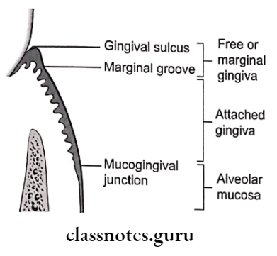
1. Marginal Gingiva:
- Border of the gingiva surrounding the teeth in the collar-like fashion
- Demarcated apically by a shallow depression called “free gingival groove”
Read And Learn More: Periodontics Question and Answers
2. Attached Gingiva:
- Part of the gingiva that is firm, resilient, and tightly bound to the underlying periosteum of the alveolar bone
- The width of the attached gingiva is the distance between the mucogingival junction and the projection on the external surface of the bottom of the gingival sulcus or periodontal pocket
3. Interdental Gingiva:
- Occupies gingival embrasure
Parts: Facial papilla, lingual papilla and col
- Lateral borders and tips of interdental papilla are formed by continuation of marginal gingiva.
- In diastema, interdental papilla is absent
Microscopic Features:
1. Oral/Outer Epithelium Layers:
- Outer Epithelium Basal layer:
- Cells are cylindrical/cuboidal
- Attach to the basement membrane
- Cells have the ability to divide
- Stratum Spinosum:
- Large cells with short processes called spines
- Cells have a prickled appearance
- Cells are attached to one another with the help of desmosomes
- Stratum Granulosum:
- Keratohyalin granules are seen
- Stratum Corneum:
- The cytoplasm of cells in this layer is filled with keratin
- It can be
- Orthokeratinized – In this cells are devoid of a nucleus
- Parakeratinized – In this cells contains the pinpoint nucleus
- Sulcular epithelium:
- Extends from the gingival margin to the junctional epithelium
- Made up of basal and prickle cell layer
- Junctional epithelium:
- It is the tissue that joins to the tooth on one side and to sul- color epithelium and connective tissue on the other
- It is attached to the tooth surface by the internal basal lamina and to the gingival connective tissue by an external basal lamina
Outer Epithelium Connective tissue:
- Termed as lamina propria
- Superficial papillary layer:
- Contains epithelial ridges
- Deeper Reticular layer:
- Contains collagen fibers
Outer Epithelium Cells:
- Fibroblast
- Mast cells
- Macrophages
- Inflammatory cells
Outer Epithelium Fibers present:
- Collagen fibers
- Reticulin fibers
- Oxytalan fibers
- Elastin fibers
Importance Of Gingival Crevicular Fluid:
1. Cicardian periodicity:
- There is a gradual increase in a gingival fluid amount from 6:00 am to 10:00 pm and decreases afterward
- This is called Cicardian periodicity
2. Sex hormones:
- Female sex hormones increase flow
- Pregnancy, ovulation, and hormonal contraceptives increase gingival fluid
3. Smoking:
- Causes an immediate transient increase in flow
4. Periodontal therapy:
- An increase in gingival fluid occurs during the healing period
Question 2. Define gingiva. Describe morphological, histological, and functional features of normal gingiva.
Answer:
Gingiva is a part of oral mucosa that covers the alveolar process of the jaws and surrounds the neck of the teeth.
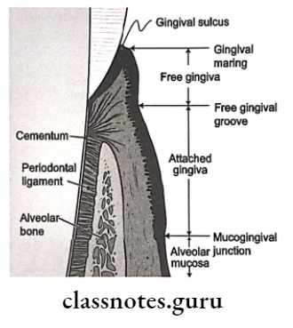
Morphological Features: Gingiva is divided into
- Marginal gingiva:
- Marginal gingiva is border of the gingiva surrounding the teeth in col- lar like fashion
- Marginal gingiva is demarcated apically by a shallow depression called a “free gingival groove”
- Attached Gingiva:
- Attached Gingiva is part of the gingiva that is firm, resilient, and tightly bound to the underlying periosteum of the alveolar hone
- The width of the attached gingiva is the distance between the mucogingival junction and the projection on the external surface of the bottom of the gingival sulcus or periodontal pocket
- Interdental gingiva:
- Interdental Gingiva occupies gingival embrasure
- Interdental Gingiva parts are facial papilla, lingual papilla, and col
- Lateral borders and tips of interdental papilla are formed by the continuation of the marginal gingiva
- In diastema, interdental papilla is absent

Gingiva Histological Features:
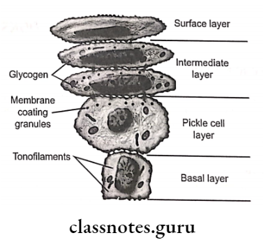
1. Epithelium Layers:
- Basal layer:
- Cells are cylindrical/cuboidal
- Attach to the basement membrane
- Cells have the ability to divide
- One of the divided cells migrates to the superficial layer
- Basal cells are separated from connective tissue by a basement membrane
- Beneath the basal cell, the electro-lucent zone can be seen called lamina lucida
- Beneath it, there is a dense zone called lamina densa
- Hemidesmosomes attach epithelium to the connective tissue.
- Stratum Spinosum:
- Large cells with short processes called spines
- Cells have a prickled appearance
- Cells are attached to one another with the help of desmosomes
- Stratum Granulosum:
- Keratohyalin granules are seen
- Stratum Corneum:
- The cytoplasm of cells in this layer are filled with keratin
- It can be
- Orthokeratinized – In this cells are devoid of a nucleus
- Parakeratinized – In this cells contains pinpoint nucleus
2. Sulcular epithelium:
- Extends from the gingival margin to the junctional epithelium
- Made up of basal and prickle cell layer
3. Junctional epithelium:
- It is the tissue that joins to the tooth on one side and to sul- color epithelium and connective tissue on the other
- It is attached to the tooth surface by an internal basal lamina and to the gingival connective tissue by an external basal lamina
Connective tissue:
- Termed as lamina propria
- Superficial papillary layer:
- Contains epithelial ridges
- Deeper Reticular layer:
Cells:
- Contains collagen fibers
- Fibroblast
- Mast cells
- Macrophages
- Inflammatory cells
Fibers Present:
- Collagen fibers
- Reticulin fibers
- Oxytalan fibers
- Elastin fibers
Importance Of Gingival Crevicular Fluid:
1. Cicardian periodicity:
- There is a gradual increase in gingival fluid amount from 6:00 am to 10:00 pm and decreases afterward
- This is called Cicardian periodicity
2. Sex hormones:
- Female sex hormones increase flow
- Pregnancy, ovulation, and hormonal contraceptives increase gingival fluid
3. Smoking:
- Causes an immediate transient increase in flow
4. Periodontal therapy:
- An increase in gingival fluid occurs during the healing period
Gingival Crevicular Fluid Functions:
1. Attached gingival:
- Attached gingival braces marginal gingiva
- Attached gingival allows for proper deflection of food
- Attached gingival provides room for proper placement of toothbrush
- Attached gingival is important for the overall maintenance of gingival health
2. Gingival crevicular fluid:
- Gingival Crevicular Fluid cleanses material from the sulcus
- Gingival Crevicular Fluid improves the adhesion of the epithelium to the tooth by plasma proteins
- Gingival Crevicular Fluid possesses antimicrobial properties
- Gingival Crevicular Fluid exerts antibody activity to defend gingiva
- Gingival Crevicular Fluid transports a variety of molecules
3. Gingival fibers:
- Provides support to the gingiva and attaches it to the bone
- It anchors the tooth to the bone
- Maintains relationship of adjacent teeth
- Secures alignment of teeth in the arch
Question 3. Define PDL. Write in detail about its structure and function. (or) Enumerate principal groups of periodontal ligament fibers. Add a note on the cellular elements and functions of PDL. Principal Fibres of PDL. (Extracellular components)
Answer:
Periodontal Ligaments are the connective tissue that surrounds the root and connects it with the bone
Periodontal Ligaments Structure:
Cells:
1. Synthetic Cells:
- Osteoblasts:
- Covers the periodontal surface of alveolar bone
- It actively synthesizes ribosomes
- Contains a largely open nucleus containing prominent nucleoli
- Fibroblasts, spindle-shaped cells:
- Most prominent cell
- Synthesizes chondroitin sulfates, heparin sulfate and hyaluronan sulfate
- Synthesizes connective tissue matrix
- Produces:
- Collagen fibers
- Reticulin fibers
- Oxytalan fibers
- Elastin fibers
- Cementoblast:
- Seen lining cementum
- Lay down cementum
2. Resorptive cells:
- Osteoclast:
- Multinucleated giant cell
- Lies adjacent to the bone
- Undergoes resorption of bone – Formed by monocytes
- Fibroblast:
- Contain fragments of collagen – These undergo digestion
- Results in resorption of bone
- Cementoclast:
- Located in Howships Lacunae
- Causes resorption of cementum
3. Progenitor cells:
- Formed in the basal cell layer
- Basal cells have the ability to divide
- One of the divided cells migrates to the superficial layer and the other remains as a progenitor cell
4. Epithelial cell rests of Malassez:
- Remnants of Hertwig’s epithelial root sheath are Present near and parallel to root surfaces
- Attached to one another by desmosomes
- During disease condition, they undergo proliferation
- Persists as a network strand, island, or tubule
- Exhibits tonofilaments
5. Mast Cell:
- Small, round, or oval cell
- Contains cytoplasmic granules
- Contains heparin and histamine
- During an inflammatory response, these releases- time causes antigen- antibody formation
6. Macrophages:
- Capable of phagocytosis
Extracellular Components
1. Fibres:
- Collagen:
- Synthesized by fibroblasts, chondroblasts, os
- neoblasts, odontoblast, and other cells
- Type 1,3 and 4 are common
- Oxytalin:
- Provide elastic properties to PDL
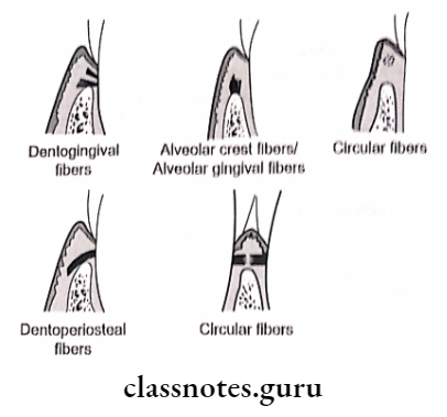
Principal Fibres:
- Trans-septal group:
- Connects cementum of one tooth with that of other
- Alveolar crest:
- Extends from cementum to alveolar crest Function: Retains tooth in the socket, Retains lateral tooth movement
- Horizontal group:
- Extends from cementum to alveolar bone
- Oblique group:
- Extends coronally from the cementum to bone Function: Resist axially directed forces
- Apical group:
- From cementum to the bone of alveolar fundus Function: Prevents tipping movement, Resists luxation
- Inter-radicular fibers:
- Presents between cementum of a multi-rooted tooth.
- Function: Resists luxation, Resists tipping and torquing
2. Ground substance:
- Glycosaminoglycan – hyaluronic acid, proteoglycan- cane
- Glycoproteins – fibronectin and laminin
Principal Fibres Functions:
1. Physical:
- Provide soft tissue casing
- Protect nerves and vessels from injury
- Transmit occlusal forces to the bone
- By stretching of oblique fibers of PDL
- Transmits tensional force to the bone
- Results in bone formation Attach tooth to the bone
- Maintains architecture of gingival tissue
- Shock absorbent
2. Formative and Remodelling function:
- Synthesis and resorption of cementum, PDL, and al- alveolar bone
- Old cells and fibers are replaced by a new one
3. Nutritional and Sensory function:
- Nutrition – Through blood supply
- Sensory Transmits sensation of touch, pressure, and pain to CNS
Neural Transmission
- Apical area – Ruffini
- Apex Pressure and vibration endings
- Mid root – Meissners corpuscles
Question 4. Discuss the role of alveolar bone in health and periodontal diseases.
Answer:
Alveolar Bone In Health:
- Alveolar Bone In Health is that portion of the maxilla and mandible that forms and supports the tooth socket
- The alveolar process is a thickened ridge of bone that contains tooth sockets that bear teeth
- The alveolar bone proper is the thin layer that provides attachment to principal fibers of the periodontal ligament Alveolar bone is perforated with numerous openings for intra-alveolar nerves and blood vessels
- Alveolar Bone In Health consists of:
1. Cells:
- Osteoblast:
- Cuboidal cell
- Contains
- Rough endoplasmic reticulum
- Large Golgi apparatus
- Secretory vesicles
- Functions:
- Synthesizes osteoid and collagen
- Regulates mineralization
- Precursor:
- Progenitor cell
- Osteoclasts:
- Multinucleated giant cells
- Precursor- Blood-borne monocytes
- Functions:
- Resorption of bone
- Secretes hydrolytic enzymes
- Osteocytes:
- These extend processes from lacunae to canaliculi
- Functions:
- Canaliculi bring oxygen and nutrients to osteocytes
2. Extracellular matrix:
- Inorganic:
- Calcium
- Hydroxyl
- Phosphate
- Carbonate
- Citrate
- Sodium
- Magnesium
- Fluorine
- Organic:
- Osteocalcin
- Osteonectin
- BMP
- Proteoglycans
- Glycoproteins
- Parathyroid hormone regulates bone removal- selling by both bone formation and bone resorption
Alveolar Bone In Disease:
- Fenestration and dehiscence are seen during disease in relation to alveolar bone
Bone-In Disease Fenestration:
- These are isolated areas in which the root surface is covered only by the periosteum and the gingiva Marginal bone is intact
Dehiscence:
- It is a defect involving the denudation of marginal bone
Bone-In Disease Etiology:
- Root prominence
- Malposition
- Teeth in labial version
Bone-In Disease Common Location:
- Site- Facial bone
- Teeth commonly involved- Anterior
Bone-In Disease Importance:
- Affects the outcome of surgical treatment