
Osseous Defects In Periodontic Disease Important Notes
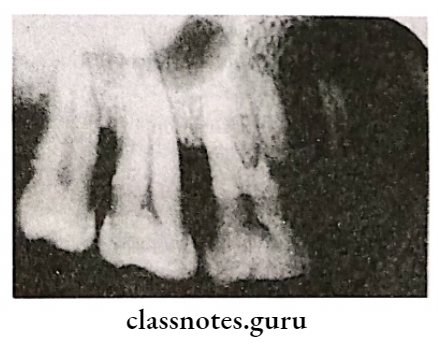
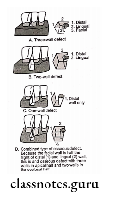
1. Angular Defects
- They are classified according to the number of walls remaining
- The fewer the number of walls remaining the poorer the prognosis
Angular Defects Types:
- One wall defect – One wall is present
- Also called hemiseptum
- The prognosis is poor regenerative procedures cannot be carried out in it
- Two walls defect-Two walls are present
- Three wall defect – Three walls are present
- Also called intrabody defect
- Prognosis is better
- Regenerative procedures can be easily carried out
- Appears most frequently on the mesial aspects of second and third maxillary and mandibular molars
- Combined osseous defect – No. of walls present in the apical portion are greater than that in the coronal portion
Read And Learn More: Periodontics Question and Answers
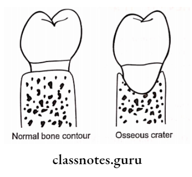
2. Osseous Craters
- Osseous Craters are concavities in the crest of interdental bone confined within facial and lingual walls
- Osseous Craters make up about one-third of all defects and two-thirds of all mandibular defects
3. Pathways Of Inflammation
- Interproximal from gingiva to bone, from bone to periodontal ligament
- Facially and lingually from gingiva to bone along the outer bone and from gingiva into the periodontal ligament
Osseous Defects In Periodontic Disease Short Essays
Question 1. Various Bone Destruction patterns.
Answer:
1. Horizontal bone loss:
- Reduction in bone height
- Bone margins are perpendicular to the tooth surface
- The common pattern of bone loss
2. Vertical/Angular Defects:
- Bone loss in an oblique direction
- The hallow trough in bone alongside the root
- The base of the defect apical to the surrounding bone
- Seen in trauma from occlusion
- Juvenile periodontitis
Vertical/Angular Defects Types:
- One wall defect: One wall is present
- Two wall defects: Two walls are present
- Three wall defect: Three walls are present
- Combined osseous defect: No. of walls present in the apical portion is greater than that in cor- the a portion
2. Osseous Craters:
- Concavities in a crest of interdental bone confined Answer: within facial and lingual walls
- Seen in TFO
3. Bulbous Bony Contours:
- Bony enlargements due to exostoses
4. Reversed bone architecture:
- Interdental bone loss without loss of radicular bone
- Seen in food impaction
Osseous Defects In Periodontic Disease Short Answers
Question 1. Reversed Architecture.
Answer:
- Interdental bone loss without loss of radicular bone
- Common in maxilla
- Seen in food impaction
Question 2. Osseous Craters.
Answer:
Concavities in the crest of the interdental bone are confined within facial and lingual walls
Osseous Craters Site:
- Mandibular teeth
- Common in posteriors
Osseous Craters Reason For Occurrence:
- Prone for plaque accumulation
- Difficult to clean
- Vascular patterns from gingiva
Question 3. Ledges.
Answer:
- Bone defect
- Plateau-like bone margins
Ledges Cause: Resorption of thickened bony plates
Question 4. Angular Defects.
Answer:
- Bone loss in the oblique direction
- A hallow trough in bone alongside root
- The base of the defect apical to the surrounding bone
- Seen in trauma from occlusion
- Juvenile periodontitis
Angular Defects Types
- One wall defect- one wall is present
- Two wall defects – Two walls are present
- Three wall defect – Three walls are present
- Common in mesial surfaces of upper and lower molars
- Combined osseous defect
- The number of walls present in the apical portion is greater than that in the coronal portion
Question 5. Enumerate bone destructive patterns in periodontal disease.
Answer:
Bone Destructive Patterns In Periodontal Disease:
- Horizontal bone loss
- Angular defects
- One wall defect
- Two wall defect
- Three wall defect
- Combined osseous defect
- Osseous craters
- Bulbous bony contours
- Reversed bone architecture
Osseous Defects In Periodontic Diseaseviva Voce
- Osseous craters are the most occurring defect in mandible
- The pathway of the spread of inflammation affects the pattern of bone destruction in periodontal disease
- When inflammation spreads directly from PDL to gin- give and from gingiva to PDL, it results in angular bone loss
- Angular defects are classified according to the number of walls remaining.
- Ledges are plateau-like bone margins caused by re- sorption of thickened bony plates.
- Combined osseous defect refers to the combination of angular defect with varying numbers of walls at the cervical and apical part
- Three walls are destroyed in one wall defect
- 50 gms of probing force is required to diagnose a periodontal osseous defect