
Wall Of Thorax And Thoracic Cavity Question And Answers
Question 1. What is the thoracic cage?
Answer:
It is an osseocartilaginous skeletal framework.
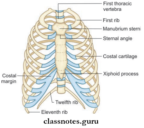
- Formed by
- Anteriorly: All 3 parts of the sternum (manubrium, body, and xiphoid)
- Posteriorly: All 12 thoracic vertebrae and intervertebral discs between them
- Laterally: 12 ribs and the costal cartilages to which the ribs are attached.
- The first 7 ribs (true ribs) articulate with the sternum through bicoastal cartilage.
- The 8th–10th ribs (false ribs) end by getting attached to the next higher cartilage.
- The anterior ends of 11th and 12th ribs are free, so-called as floating ribs.
- Ribs are attached posteriorly to the corresponding thoracic vertebrae.
- Atypical ribs: 1, 2, 10, 11, 12
- Typical ribs : 3, 4, 5, 6, 7, 8
- Costal cartilage : Hyaline cartilage.
Read And Learn More: Anatomy Question And Answers
Ossification centers of a typical rib
- Primary center: Forms at the completion of second month of fetal life
- Secondary center: Forms at puberty.
Thoracic Cage Applied Anatomy
- Accessory ribs may sometimes be present
- Rib attached to 7th cervical vertebrae: Cervical rib
- Rib attached to fist lumbar vertebrae: Lumbar rib
- Costal cartilages make thorax more elastic, in old age costal cartilages calcify and lose elasticity.
Thoracic Cage Joints
- First cartilage with manubrium sternum: Synchondrosis
- 2nd-7th cartilage with sternum: Synovial joint
- 8th, 9th, 10th cartilage with next higher cartilage: Synovial joint
Question 2. What is a thoracic inlet?
Answer:
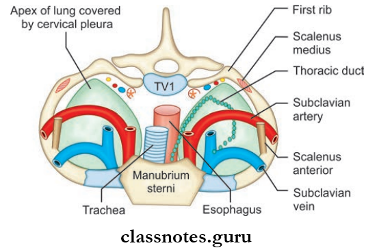
Thoracic Inlet
- It is the upper end of thorax
- Also known as superior thoracic aperture
- It is a kidney-shaped, narrow opening
- Its size corresponds to the diameter of neck
Thoracic Inlet Boundaries
- Anteriorly: Upper border of manubrium sterni
- Posteriorly: Upper end of the body of T1 vertebrae
- Laterally: Inner border of first rib and its cartilage
- The thoracic inlet is divided into two halves, each half is formed by suprapleural membrane (it is fused with conical pleura inferiorly)
- There is central cleft in between both halves.
Important structures passing through the inlet
- Trachea, Esophagus, lung apices
- Muscles: Sternohyoid, sternothyroid
- Blood vessels
- Brachiocephalic artery
- Left common carotid and left subclavian artery
- Internal thoracic artery
- Brachiocephalic veins
- Nerves: Phrenic nerves, vagus nerves, sympathetic trunk.
Question 3. What is a thoracic outlet?
Answer:
Thoracic Outlet
- It is the inferior aperture of thorax.
- It is separated from abdominal cavity by the diaphragm.
- Its size corresponds to diameter of upper part of the abdomen.
Thoracic Outlet Boundaries
- Anteriorly: 7th–10th costal cartilages
- Posteriorly: Lower border of 12th thoracic vertebrae
- Laterally: 11th and 12th pairs of ribs.
Since the inferior aperture is closed by the diaphragm, structures from the thorax to abdomen has to pass through large and small openings in the diaphragm.
Question 4. Write briefly about the boundaries and contents of the scalene triangle.
Answer:
Scalene Triangle
- Narrow triangular space
- Boundaries
- Anteriorly: Scalenus anterior
- Posteriorly: Scalenus medius
- Inferiorly: Upper surface of fist rib
- Contents
- Subclavian artery
- Lower trunk of brachial plexus.
Scalene Triangle Applied Anatomy
- Cervical rib syndrome: Due to the presence of a cervical rib or a congenitally hypertrophied scalenus anterior muscle.
- The scalene triangle gets narrowed and results in compression of the lower trunk of brachial plexus and subclavian artery.
Question 5. Describe in detail about the origin and insertion of the diaphragm. What is meant by dome of the diaphragm?
Answer:
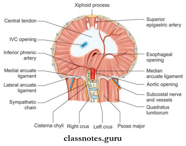
It is a large muscle that closes the thoracic outlet
Diaphragm Formation: It has 3 points of origin
- Sternal part: Arise from back of xiphoid process as two slips, right and left
- Costal part: Consisting of broad slips arising from the inner surface of 7th–12th ribs and their costal cartilage
- Lumbar part: Consist of right and left crura, which arises from
- The anterolateral surface of body of lumbar vertebrae
- Medial and lateral arcuate ligaments
- Medial arcuate ligament: Fascia covering psoas major
- Lateral arcuate ligament: Fascia covering quadratus lumborum
- Medial margins of two crura are joined to each other to form median (not medial) arcuate ligament at the level of the lower border of T-12 vertebrae
Diaphragm Insertion
All muscular fibers of the diaphragm runs upwards and converge to get inserted into flt central tendon (located below the pericardium)
Dome of diaphragm
- It is the upper convex part of the diaphragm
- The central part of the dome is made by central tendon
- And peripheral part by muscular fibers.
Question 6. What are the openings of the diaphragm? What are the structures passing through it?
Answer:
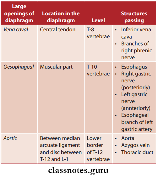
Mneumonic—Voice of America (VOA)
Structures Passing Through Small Openings
- Superior epigastric artery
- Musculophrenic artery
- 8th–11th intercostals nerves and vessels
- Subcostal nerves and vessels
- Sympathetic trunk
- Splanchnic nerves.
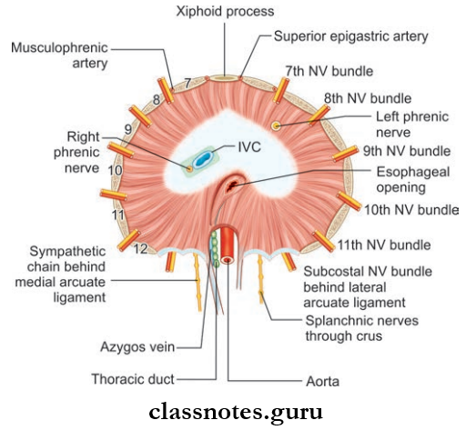
Abbreviation: IVC = Inferior vena cava; NV = Neurovascular
Question 7. What is the nerve supply of the diaphragm?
Answer:
Nerve Supply of Diaphragm
- Sensory supply: Lower six intercostals nerves, right and left phrenic nerves
- Motor supply: Right and left phrenic nerves.
Question 8. Write a note on the development of diaphragm.
Answer:
Development of Diaphragm
- Central tendon from septum transversum
- Dorsal paired portion of the diaphragm from the pleuroperitoneal membrane. A circumferential portion from lateral thoracic wall
- A dorsal unpaired portion from dorsal mesentery of the esophagus.
Question 9. Write a note on intercostal space–muscles, nerves, blood supply, and lymphatic drainage?
Answer:
Intercostal Space
- The space between two adjacent ribs and there costal cartilages is called intercostal space
- There are 11 intercostal spaces between 12 ribs on both sides:
- Intercostal space contains
- Intercostal muscles
- Intercostal nerves, vessels and lymphatics
- Intercostal space contains
- 3rd–6th intercostal spaces formed between the typical ribs are traversed by vessels and nerves which are confined only to the thorax. As a result, these intercostal spaces are called typical intercostal spaces.
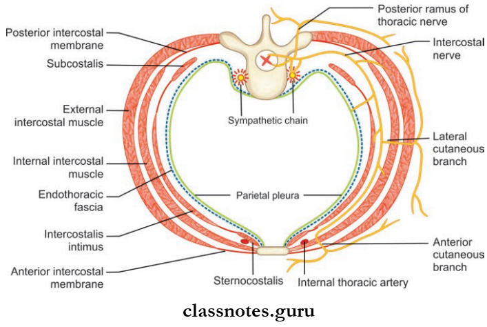
Intercostal Muscles They are arranged in three layers as:
- External intercostal muscles
- Internal intercostal muscles
- Innermost muscles: Sternocostalis, intercostalis intimi, subcostalis.
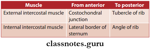
1. External Intercostal Muscles : Takes origin from the lower margins of ribs 1 to 11, and is inserted on the upper margin of the rib just below it.
- External Intercostal Muscles Parts It has 2 parts, namely:
- Fleshy interosseous part: Located between the ribs
- Membranous interchondral part: Located between the adjacent costal cartilages.
2. Internal Intercostal Muscles: Takes origin from the lower margins of ribs and costal cartilage and to the floor of the costal groove, and are inserted on the upper margin of the rib and costal cartilage just below it
- Internal Intercostal Muscles Parts: It also has 2 parts, namely:
- Fleshy part: Long part, located between anterior end of intercostal space to the angle of rib
- Membranous part: Short part, located beyond the angle of the rib.
- Internal Intercostal Muscles Extent
- External intercostal muscle is replaced by an external intercostal membrane, between the costochondral junction and sternum but posteriorly it is continuous with the posterior fibers of superior costotransverse ligament
- Internal intercostal muscle is continuous posteriorly as an internal intercostal membrane.
3. Innermost Intercostal Muscles
- Innermost Intercostal Muscles Sternocostalis:
- Anterior part of innermost muscles
- Lies behind sternum and costal cartilages
- Innermost Intercostal Muscles Intercostalis intimi
- Present on the middle two fourth of intercostal space
- Innermost Intercostal Muscles Subcostalis
- Present over the posterior part of intercostal space.
Diaphragm Nerve Supply All intercostal muscles are supplied by intercostal nerves of the corresponding space.
Intercostal Nerves
- They are derived from the anterior primary rami of T1 to T11 spinal nerves
- They are eleven in number
- Anterior primary rami of T 12 spinal nerve gives rise to the subcostal nerve (It runs in the abdominal wall below the 12th rib)
Classification Of Intercostal nerves are classified into:
- Typical Intercostal Nerves
- They are confined to their own intercostal spaces in the thoracic wall
- 3rd, 4th, 5th and 6th intercostal nerves.
- Atypical Intercostal Nerves
- They extend beyond the thoracic wall and partly or completely supply other areas
- 1st, 2nd, 7th, 8th, 9th, 10th and 11th intercostal nerves.
Course of a typical intercostal nerve
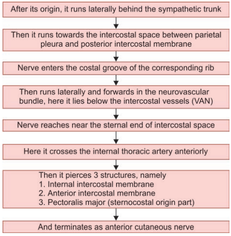
Branches of Typical Intercostal Nerves
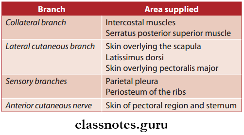
Intercostal Arteries
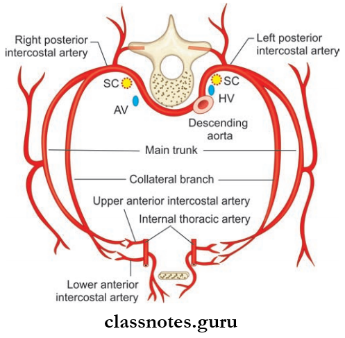
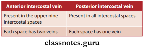
The upper nine intercostal spaces contain three arteries:
- Single large posterior intercostal artery
- Two small anterior intercostal arteries
- 10th and 11th spaces only contains a single posterior intercostal artery.
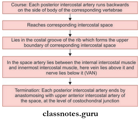
Posterior Intercostal Arteries
In the upper two intercostal spaces the posterior intercostal arteries are branches of superior intercostal artery.
Drainage Origin
- In the lower nine intercostal spaces the posterior intercostal arteries arises from the descending thoracic aorta
- Right intercostal arteries in the lower nine intercostal spaces are longer than left, since descending thoracic aorta lies near the left of vertebral column.
Drainage Branches
- Muscular branches: Supply intercostal, pectoral, and serratus anterior muscle.
- Dorsal branches: Supply vertebrae, spinal cord, muscles, and skin of the back.
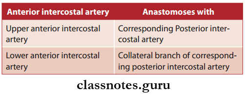
Anterior Intercostal Arteries: There are two anterior intercostal arteries each in the upper nine intercostal spaces.
Anterior Intercostal Arteries Origin
- Upper six spaces: Internal thoracic artery.
- 7th–9th spaces: Musculophrenic artery.
Anterior Intercostal Arteries Termination
Lymphatic Drainage of Intercostal Space
Lymphatics from the anterior part of intercostal space: Anterior intercostal nodes.
Lymphatics from the anterior part of intercostal space: Posterior intercostal nodes.
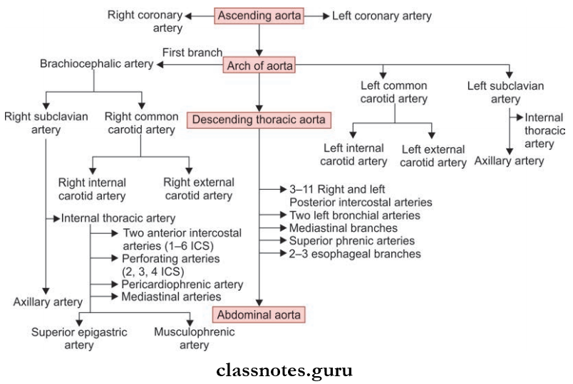
Question 10. Describe the origin, course, and branches of internal thoracic artery. Write brief on the internal thoracic vein.
Answer:
The internal thoracic artery is also known as the internal mammary artery.
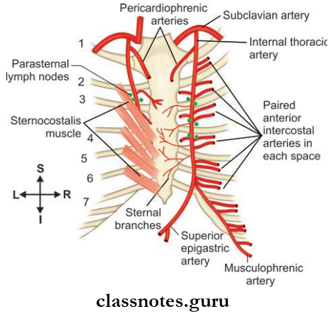
Internal Thoracic Artery Origin
Two cm above the sternal end of the clavicle, from the inferior part of the first part of the Subclavian artery.
Internal Thoracic Artery Course
- Runs medially and downwards behind the sternal end of the clavicle and fist costal cartilage
- The runs downwards behind the fist to sixth costal cartilage (about 1 cm lateral to sternal margin). And terminates in the 6th intercostal space by dividing into two arteries.
- Musculophrenic artery
- Superior epigastric artery
- In its course, the internal thoracic artery also gives several other branches, namely:
Anterior intercostal arteries
- Two anterior intercostal arteries are given of to each upper six intercostal spaces.
- In each intercostal space, two anterior intercostal arteries anastomoses with posterior intercostal artery.
Pericardiophrenic branches
- It is given of near the upper end of the internal thoracic artery.
- It runs downwards along with phrenic nerve and reaches the diaphragm, supplying the pericardium and pleura.
Perforating branches
- Given of in upper six intercostal spaces.
- Supplies Pectoralis major muscle and Breast.
- From 2nd to 4th intercostal spaces, these branches are found to be larger in females (to supply breasts).
1. Musculophrenic Artery
- Lateral terminal branch of internal thoracic artery.
- Course: From the origin, it runs downward and laterally deep in to the costal margin.
- Reaches the abdominal wall by passing through a small opening in the diaphragm.
- Branches: Anterior intercostal branches to 7th, 8th and 9th intercostal spaces.
2. Superior Epigastric Artery
- The medial terminal branch of internal thoracic artery
- Course: Runs downwards through the gap between costal and xiphoid origin of the diaphragm into the abdomen
- It supplies the anterior mediastinum, pericardium, and diaphragm.
3. Applied Anatomy
Internal thoracic artery is the commonly used coronary graft since it is less prone to develop atherosclerosis due to its histological peculiarity (its wall contains elastic tissue only).
Internal thoracic vein
- The internal thoracic artery is accompanied by a number of small veins.
- These veins eventually joins each other forming one large vessel, the internal thoracic vein.
- Course: Runs upwards through the medial aspect of the internal thoracic artery.
- Termination: Drain in to Brachiocephalic vein.
- Tributaries: Corresponds to branches of the internal thoracic artery.
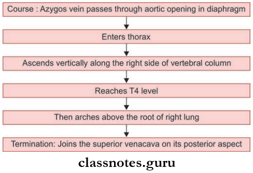
Question 11. Write a note on the azygos vein.
Answer:
Azygos Vein
- It is present only on right side (azygos means unpaired)
- Arises opposite to L1 or L2 vertebrae
Formation: Formed by union of lumbar azygos vein, right subcostal vein, and right ascending lumbar vein.
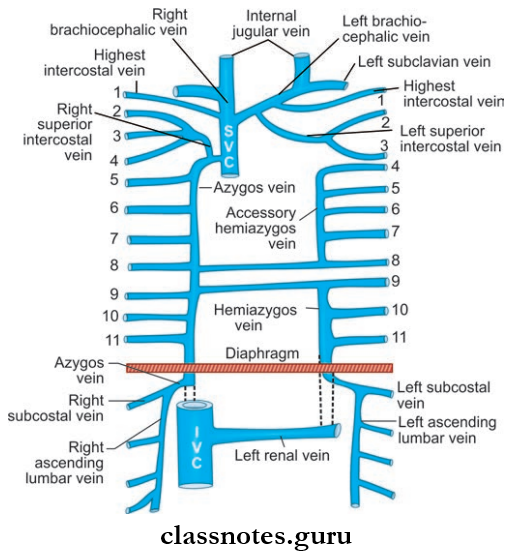
- Lumbar azygos vein:
- It lies right of lumbar vertebrae
- Its lower end communicates with the inferior vena cava
- Its upper end joins with other veins of azygos system to form azygos vein
- Right subcostal vein:
- It accompanies the corresponding artery
- Right ascending lumbar vein:
- It is a vertical channel connecting lumbar veins (Sometimes, the lumbar azygos vein may be absent, then azygos vein is formed by the subcostal vein and ascending lumbar vein)
- Applied Anatomy
- Since azygos vein forms a channel between the superior vena cava and inferior vena cava, in case of superior vena cava obstruction act as the main channel through which blood from upper half of the body is transmitted to inferior vena cava.
Question 12. Write a note on hemiazygos vein.
Answer:
Hemiazygos Vein
- Also known as inferior azygos vein, since it is the mirror image of lower part of azygos vein.
- Present only on the left side.
- Formation: By union of left ascending lumbar vein and left subcostal vein.
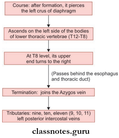
Question 13. Write a note on accessory hemiazygos vein.
Answer:
- Also known as superior azygos vein of, since it is the mirror image of the upper part of azygos vein.
- It is also connected superiorly to the left superior intercostal vein.
- Sometimes it joins hemiazygos vein, or have a connection to both the azygos and hemiazygos vein.
Tributaries:
4th to 8th left posterior intercostal veins.(Sometimes left bronchial veins also join with it).
Question 14. What is thoracic sympathetic trunk and what are its branches?
Answer:
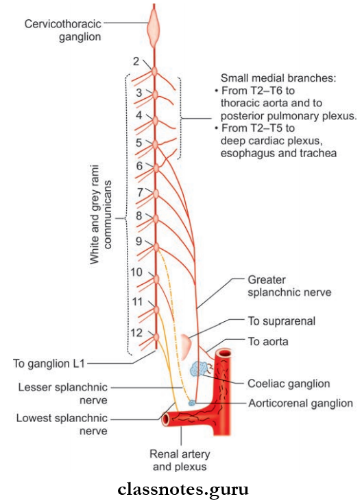
Thoracic Sympathetic Trunk
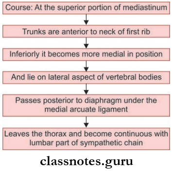
- Sympathetic trunk is a long nerve cord extending from the base of skull to the coccyx.
- Thracic part of the sympathetic trunk is situated on both sides of the thoracic vertebral column (one each per side), in front of the heads of ribs and is a ganglionated chain.
- It is considered as a component of the posterior mediastinum since they pass through it.
- There are 12 thoracic ganglia, but the fist thoracic ganglion is fused with the inferior cervical ganglion, to form a cervicothoracic ganglion
- So the lower eleven (T2-T12) thoracic ganglia are considered as true thoracic ganglia.
Arteries of Thorax
Thoracic Sympathetic Truck Branches
- Laterally: Sympathetic ganglia is connected to adjacent
- Thoracic spinal nerves by white and grey rami communicans.
Medially: It supplies the viscera.
Medial branches arising from T1-T5 ganglia are postganglionic, they are connected to nerve plexus supplying viscera
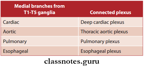
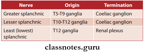
Medial branches from T6-T12 are preganglionic, they join together and forms three nerves, they are:
Wall Of Thorax And Thoracic Cavity Multiple Choice Questions
Question 1. Which is considered a ‘false’ rib?
- 1
- 2
- 3
- 11
Answer: 4. 11
Question 2. All the following structures pass through the superior aperture of thorax except:
- Left common carotid artery
- Left sympathetic trunk
- Right recurrent laryngeal nerve
- Thoracic duct
Answer: 3. Right recurrent laryngeal nerve
Question 3. Which rib usually articulates with more than one vertebral body?
- Rib 1
- Rib 2
- Rib 11
- Rib 12
Answer: 2. Rib 2
Question 4. Azygos vein:
- Formed by arch of aorta
- Arches the groove of the left lung
- Opens in to inferior vena cava
- Ascends through the aortic opening in the diaphragm
Answer: 4. Ascends through the aortic opening in the diaphragm
Question 5. The external intercostal membrane is a continuation of the:
- Internal intercostal muscle
- Posterior intercostal muscle
- External intercostal muscle
- None of the above
Answer: 3. External intercostal muscle