Scalp Temple And Face Question Ans Answers

Surface Landmarks And Features Introduction

Forehead: Part of the face between hairline of the adolescent scalp and eyebrows.

Frontal Eminence: Superolateral prominence of the forehead.

Dorsum Of The Nose: Prominent ridge separating right and left halves of the nose.

Root Of Nose: The upper narrow end of nose just below forehead.

Columella: The soft median partition that separates two nostrils.

Palpebral Fissure: An elliptical opening between two eyelids.

Canthi: The lateral and medial angles of the eye.

Oral Fissure: The opening between upper and lower lips.

Philtrum: Median vertical groove on the upper lip.

Auricle/pinna: The superficial projecting part of the external ear.

Supraorbital Margin: Lies beneath the upper margin of the eyebrow.

Supraorbital Notch: Palpable at the junction of medial one-third and lateral two thirds of supraorbital margin.

Glabella: Median elevation connecting the superciliary arches and corresponding to an elevation between two eyebrows.

Scalp Temple And Face Question Ans Answers

Question 1. Describe the layers of the scalp.
Answer:

The Layers Of The Scalp

Scalp Temple And Face Layers Of The Scalp(Schematic)

Scalp Temple And Face Layers Of The Scalp

Scalp Temple And Face Sagittal Section Of Cranial Vault Showing Continuity Between Dangerous Area Of Scalp With Upper Eyelid

Layers Of The Scalp: Scalp refers to the soft tissues covering the cranial vault.

Layers Of Scalp Extent:

  • Anteriorly: Up to the eyebrows
  • Posteriorly: Up to superior nuchal line and external occipital protuberance
  • Laterally: Superior temporal lines.

Layers Of Scalp Layers: Consist of 5 layers (from outside to inside)

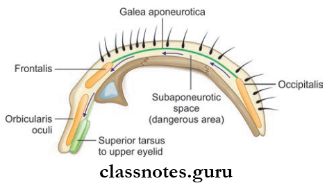
  • Skin: Thick and hairy, contain sebaceous glands and sweat glands, and is adherent to epicranial aponeurosis.
  • Superficial Fascia: Fibrous and dense in the center than in the periphery. Binds skin to the aponeurosis vessels and nerves are present in this layer.
  • The Third Layer Formed By:
    • Mainly the epicranial aponeurosis (Galea aponeurotic)—the aponeurosis of occipitofrontalis muscle
    • Occipitofrontalis Muscle Consisting Of:
      • Occipital bellies arising from superior nuchal line
      • Frontal bellies arising from the skin of the forehead
      • Supplied by branches of the facial nerve.
  • Fourth Layer Made Of Loose Areolar Tissue Which Extends
    • Anteriorly into eyelids
    • Posteriorly up to superior nuchal line
    • Laterally to superior temporal lines.
    • Emissary veins from the scalp to the sinuses traverse through this layer.
  • Fifth Layer: Pericranium, which is loosely attached to the surface of bones, and at the sutures, the sutural ligaments bind it firmly to the endocranium.
    • The first three layers are fully united to one another and they move together over the fourth layer.

Layers Of Scalp Applied

  • Scalp bleed profusely due to rich blood supply.
  • Loose areolar tissue layer is also known as a dangerous area of scalp because emissary veins that open here may transmit infection from the scalp to cranial venous sinuses.
  • Avulsed portions of the scalp can be stitched back into position and heal well due to adequate blood supply even through the narrow areas of attachment.
  • Due to the abundance of sebaceous glands, sebaceous cysts are common in the scalp.
  • Bleeding in the scalp can be arrested by applying direct pressure over the wound against the bone.
  • Bleeding in layer of loose areolar tissue forms a generalized swelling or spread widely reaching nuchal line posteriorly or temporal lines laterally, or can reach orbital margin and eyelids causing black eye.
  • Cephalohematoma refers to the subperiosteal collection of blood bound by suture lines and assumes the shape ofrelated bones because periosteum loosely covers the bones except at the suture lines.
  • Caput succedaneum is a subcutaneous edema occurring over the presenting part of the fetal head at delivery. It occurs due to interference of venous return during passage through the birth canal.

Mnemonics: SCALP

From Superficial To Deep:

  • Skin
  • Connective tissue
  • Aponeurosis
  • Loose areolar tissue
  • Pericranium

Question 2. Describe the blood supply of the scalp and superficial temporal region.
Answer:

The Blood Supply Of The Scalp

Scalp Temple And Face Arteries Of the Scalp

  • Arterial Supply
    • The scalp on each side of the midline is supplied by five arteries, three in front of the auricle and two behind the auricle.
    • These arteries enter from the periphery and freely anastomose with arteries of same side and cross anastomose with the other side.
    • The scalp is a potential site of collateral circulation between internal and external carotid arteries.
    • In front Of The Auricle
      • Internal carotid artery → Ophthalmic artery → Supratrochlear artery → Supraorbital artery → External carotid artery → Superficial temporal artery
    • Behind The Auricle
      • External carotid artery → Posterior auricular artery → Occipital arteries
  • Venous Drainage
    • The scalp on each side of the midline is drained by fine veins and these accompany the arteries and have similar names.
    • These Include:  Supratrochlear and supraorbital veins unite to form an angular vein at medial angle of eye and continue as facial vein.
  1. The superficial temporal vein and maxillary vein unite to form the retromandibular vein and its anterior division in turn joins with the facial vein to form common facial vein.
  2. The posterior division of the retromandibular vein unites with the posterior auricular vein to form the external jugular vein which drains into the subclavian vein.
  3. The occipital veins drain into the suboccipital venous plexus.
  4. Emissary veins connect extracranial veins with intracranial venous sinuses.
  5. The diploic veins like the frontal diploic vein open into a supraorbital vein, the anterior temporal diploic vein drains to the anterior deep temporal vein, the posterior temporal diploic vein drains to the transverse sinus, and the occipital diploic vein drains to either occipital vein or the transverse sinus.

Scalp Temple And Face Nerves And Arteries Of Anterior And Posterior Quadrants Of Scalp

Question 3. Describe the nerve supply of the scalp and superficial temporal region.
Answer:

The Nerve Supply Of The Scalp

Scalp Temple And Face Nerves Of The Scalp

The scalp and the temple is supplied by ten nerves on each side. Out of this 4 are sensory and 1 is motor and 5 enter in front of the auricle and 5 enter behind the auricle.

Scalp Temple And Face Temporial Region

Mnemonic: GLASSZ

  • Greater occipital/Greater auricular
  • Lesser occipital
  • Auriculotemporal
  • Supratrochlear
  • Supraorbital
  • Zygomaticotemporal

Scalp Temple And Face Facial Skeleton Seen From The Side To Show Some Muscle Attachments

Scalp Temple And Face The Orbicularis Oris And Its Relationship To Various Muscles Attached To The Lips

Scalp Temple And Face Muscles Of facial Expressions

Scalp Temple And Face Buccinator Muscle

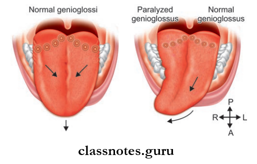
Since derived from the mesoderm of 2nd branchial arch they are supplied by the facial nerve

Topographically, Muscles Are Grouped Into Six Headings:
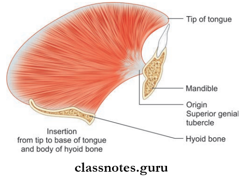
  1. Muscles Of Scalp
    • Occipito frontalis
  2. Muscles Of Auricle
    • Auricularis anterior
    • Auricularia superior
    • Auricularis posterior
  3. Muscles Of Eyelid
    • Orbicularis oculi
    • Corrugator supercilii
    • Levator palpebrae superioris
  4. Muscles Of Nose
    • Procerus
    • Compressor naris
    • Dilator naris
    • Depressor septi
  5. Muscles Of Neck
    • Platysma
  6. Muscles Around Mouth
    • Orbicularis oris
    • Levator anguli oris
    • Depressor anguli oris
    • Zygomaticus major
    • Zygomaticus minor
    • Levator labii superioris
    • Depressor labii inferioris
    • Levator labii superioris alaeque nasi
    • Mentalis
    • Risorius
    • Buccinator

Main Muscles And Their Attachments

Scalp Temple And Face Muscles And Their Attachments

Superficial Temporal Region Applied: The involuntary part (smooth part) of the levator palpebrae superioris or Muller’s muscle is supplied by fibers from superior cervical ganglion and the paralysis of this muscle leads to partial ptosis in Horner’s syndrome.

Question 5. Describe the nerve supply of face.
Answer:

The Nerve Supply Of Face

Scalp Temple And Face Sensory Nerves Of face, Scalp And Auricle

Scalp Temple And Face Nerve Supply Of Face

Nerve Supply Of Face Applied

  • In case of supranuclear lesions of the facial nerve associated with hemiplegia usually the lower part of the opposite side of face is paralyzed.
  • Bell’s palsy: It is the lower motor neuron paralysis of facial muscles due to compression of facial nerve in the facial canal. Here the whole of the face of the same side gets paralysed. As a result, wrinkles are absent on the forehead, the inability to close eye is present, deviation of angle of the mouth to the normal side, and food accumulates between the cheek and teeth.

Question 6. Describe the venous drainage of face.
Answer:

The Venous Drainage Of Face

Scalp Temple And Face Dangerous Are Of The Face Superior Ophthalmic Vein Connecting The Angular Vein To the Cavernous Sinus

Scalp Temple And Face Deep Facial Vein Connecting Dangerous Area Of The Cavernous Sinus Via Pterygoid Plexus-Sphenodial Emissary Vein

  • The veins accompany arteries of the face and drain into a common facial vein and retromandibular vein and they communicate with cavernous sinus
  • The vein on each side form a ‘W’ shaped arrangement and each corner of ‘W’ is prolonged upwards to the scalp and downwards to the neck
  • The deep connections include:
    • Communication between supraorbital and superior ophthalmic veins
    • Communication with pterygoid plexus through deep facial vein.

Venous Drainage Of Face

Scalp Temple And Face Drainage Of Face Venous Drainage Flow Chart

Venous Drainage Of Face Applied

  • Since, the facial vein communicates with the cavernous sinus, retrograde infection from face is possible and result in thrombosis of cavernous sinus.
  • Mostly infection in the upper lip, lower part of nose and adjoining cheek area can result in thrombosis. So these areas are called ‘dangerous area of face’.

Question 7. Describe the lymphatic drainage of the face.
Answer:

The Lymphatic Drainage Of The Face

It can be studied under three headings based on the territories of face.

  1. Upper territory: A greater part of the forehead, lateral halves of eyelids, conjunctiva, lateral part of cheek, and parotid area
  2. Middle territory: Median part of the forehead, external nose, upper lip, medial halves of the eyelid, medial part of the cheek, the greater part of jaw
  3. Lower territory: Central part of lower lip and chin.

Scalp Temple And Face Lymphatic Drainage Of Face Lower Territory Flowchart

Question 8. Write a note on the lacrimal apparatus.
Answer:

Lacrimal Apparatus

Scalp Temple And Face Parts Of The Lacrimal Apparatus The Arrows Indicates bThe Direction Of flow Of lacrimal Fluid

Scalp Temple And Face Components Of Lacrimal Apparatus

  • It consists of structures that are involved in the secretion and drainage of lacrimal fluid
  • Consist Of Mainly The Following Parts
    1. Lacrimal gland
    2. Lacrimal ducts
    3. Conjunctival sac
    4. Lacrimal puncta and lacrimal canaliculi
    5. Lacrimal sac
    6. Nasolacrimal duct.

1. Lacrimal Gland

  • Serous gland situated in the lacrimal fossa on the anterolateral part of the roof of the bony orbit and upper eyelid
  • Consists of an orbital part and a palpebral part
  • Involved in the secretion of lacrimal fluid along with accessory lacrimal gland
  • Supplied by lacrimal nerve and lacrimal branch of ophthalmic artery.

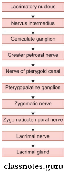
2. Secretomotor Fiers

Scalp Temple And Face Lacrimal Apparatus Secretomotor Fibers

  • Lacrimal Ducts: Arises from the lacrimal gland, pierce the conjunctiva of the upper lid, and opens into a conjunctival sac near the superior fornix.
  • Conjunctival Sac: The space between the palpebral and bulbar conjunctiva is known as the conjunctival sac.
  • Lacrimal Puncta And Canaliculi: These are small openings on the lid margin:
    • Each lacrimal canaliculi begin at lacrimal punctum and is 10 mm long
    • Has a vertical part 2 mm long and horizontal part 8 mm long
    • Both canaliculi open into the lateral wall of sac close to each other.
  • Lacrimal Sac:
    • Membranous sac situated in the lacrimal groove behind medial palpebral ligament
    • Upper end of the sac is blind and lower end is continuous with the nasolacrimal duct.
  • Nasolacrimal Duct:
    • It is an 18 mm long membranous passage
    • It begin at lower end of the lacrimal sac and opens into the inferior meatus of nose
    • A fold of mucous membrane forms an imperfect valve called the valve of Hasner at the lower end of duct.

Lacrimal Apparatus Applied

  • For the purpose of clinical evaluation, the palpebral conjunctiva is examined for anemia and bulbar conjunctiva for jaundice.
  • Excessive secretion of lacrimal fluid overflowing on the cheeks is called Epiphora and occurs as a result of an obstruction in the lacrimal fluid pathway.
  • Inflammation of the conjunctiva is known as conjunctivitis and is a common disease of the eye caused due to infection or allergy.
  • Inflammation of the lacrimal sac is known as dacryocystitis.
  • Ducts from the orbital part traverse the palpebral part to open into the conjunctival sac. So the removal of the palpebral part is equivalent to the removal of the entire gland.

Scalp Temple And Face Multiple Choice Question And Answers

Question 1. Which layer of scalp is regarded as the ‘dangerous layer’?

  1. Subcutaneous layer
  2. Aponeurotic layer
  3. Layer of loose areolar tissue
  4. Pericranium

Answer: 3. Layer of loose areolar tissue

Question 2. Regarding muscles of facial expression which of the following statements is incorrect:

  1. They are present in the superficial fascia
  2. They are developed from the first pharyngeal arch
  3. Their motor supply is derived from the facial nerve
  4. Morphologically they represent panniculus carnosus

Answer: 2. They are developed from first pharyngeal arch

Question 3. 26 October 2016 2:08 PMAll of the following arteries supply the anterior quadrant of the scalp except:

  1. Supratrochlear
  2. Supraorbital
  3. Posterior auricular
  4. Superficial temporal

Answer: 3. Posterior auricular

Question 4. All of the following innervate the posterior quadrant of the scalp except:

  1. Auriculotemporal
  2. Great auricular
  3. Greater occipital
  4. Lesser occipital

Answer: 1. Auriculotemporal

Question 5. Regarding the lacrimal gland which of the following statements is not correct?

  1. It consists of a larger orbital part and a smaller palpebral part
  2. It is a mucus gland
  3. It receives a secretomotor supply through the lacrimal nerve
  4. The two parts of the lacrimal glands are separated from each other by levator palpebrae superioris.
  5. Answer: 2. It is a mucus gland

Surface Landmarks Of Neck Question And Answers

Surface Landmarks Introduction

Surface Landmarks On the side

  • Sternocleidomastoid: Seen as a prominent raised ridge extending obliquely from clavicle and sternum to mastoid process.
  • Anterior Border Of Trapezius: Prominent on the elevation of the shoulder against resistance.
  • Mastoid Process: Large bony projection behind the lower part of the auricle.
  • Greater Supraclavicular Fossa: A depression above the middle one-third of the clavicle.
  • Lesser Supraclavicular Fossa: Small depression between sternal and clavicular part of the sternocleidomastoid.

Surface Landmarks Anteriorly

  • Hyoid Bone: A horseshoe-shaped bone felt just below and behind the chin. Lies at the level of C3 vertebra.
  • Thyroid Cartilage: Sharp protuberance in the median plane below the hyoid. Also called Adam’s apple/laryngeal prominence.
  • Cricoid Cartilage: Lies just below the thyroid cartilage. Lies at the level of lower border of C6 vertebrae.
  • Suprasternal Notch: Felt between anterior ends of clavicle. Lies at level of lower border of body of T2 vertebrae.

Surface Landmarks Back

  • Nuchal Furrow: A vertical groove in the midline on the back of neck.
  • External Occipital Protuberance: A bony projection felt in the upper end of the nuchal furrow.
  • The Spine Of C7 Vertebrae: A knob-like bony projection felt at the lower end of nuchal furrow.

Side Of Neck Questions And Answers

Question 1. Write a short note on deep cervical fascia (fascia colli).
Answer:

Deep Cervical Fascia (fFascia Colli)

The deep cervical fascia of neck consists of the following layers from outside inwards:

  • Investing layer of deep fascia
  • Pretracheal fascia
  • Prevertebral fascia.

It Is Also Condensed To Form:

  • Carotid sheath
  • Buccopharyngeal fascia
  • Pharyngobasilar fascia.

Investing Layer Or Deep Fascia: Lies deep to platysma and superficial fascia and encircles the neck like a collar

Deep Cervical Fascia Attachments.

  • Superiorly
    • External occipital protuberance
    • Superior nuchal line
    • Mastoid process
    • Base of mandible
  • Inferiorly
    • Spine of scapula
    • Acromion process
    • Clavicle
    • Manubrium
  • Anteriorly
    • Symphysis menti
    • Hyoid bone
  • Posteriorly
    • Ligamentum nuchae
    • Spine of C7 vertebrae.

Side Of Neck Transverse Section Through Lower Part Of neck To Show Various Muscles And Parts Of the Deep Cervical Fascia

Side Of Neck Cross-section Of Neck At Lower Level To Deplict Disposition Of Layer Of Cervical Fascia

Deep Cervical Fascia Features

  • The superior portion of the fascia splits to enclose the submandibular salivary gland.
  • The superficial layer that covers the submandibular gland is attached to the base of the mandible and the deep layer to the mylohyoid line.
  • Posterior to the submandibular gland and at lower pole of the parotid gland it splits to enclose the parotid gland.
  • The superficial layer that covers the parotid gland is known as the parotid masseteric fascia and is attached to the zygomatic arch and the deep layer is attached to the lower border of the tympanic plate and styloid process.
  • Part of layer between the styloid process and the angle of the mandible condenses to form the stylomandibular ligament and separates the parotid gland from the submandibular gland.

Inferiorly, The Fascia Splits To Enclose 2 Spaces

  1. Above Suprasternal Notch Splits To Enclose Suprasternal Space (Of Burns) Which Consists Of:
    • Sternal head of right and left sternocleidomastoid
    • Jugular venous arch
    • Lymph node
    • Interclavicular ligament.
  2. Above Middle Of Clavicle, It Splits To Enclose Supraclavicular Space Which Consist Of:
    • External jugular vein
    • Supraclavicular nerves
    • Cutaneous vessels and lymphatics.
  • It also forms pulleys to bind the tendon of the omohyoid and digastric muscles.

Deep Cervical Fascia Applied

Fascia covering parotid is very thick and strong, so in infections of the parotid gland, the gland cannot expand, as a result, the nerves are compressed and result in pain.

Deep Cervical Fascia TIP: Rule of 2

  • It Encloses 2 Muscles: Trapezius and sternocleidomastoid
  • Forms Roof Of 2 Triangles: Anterior and posterior
  • Split To Enclose 2 Glands: Parotid and submandibular
  • Split To Enclose 2 Spaces: Suprasternal and supraclavicular
  • Forms Two Fascial Slings: For the inferior belly of omohyoid and intermediate tendon of digastric.

Pretracheal Fascia

  • Covers front and side of trachea
  • Splits to enclose thyroid gland and form its false capsule
  • Ligament of Berry, a firous band which connects the capsule of lateral lobe of thyroid to cricoid cartilage.

Pretracheal Fascia Attachments

  • Superiorly: Hyoid bone in median plane
    • Oblique line of thyroid cartilage
    • Cricoid cartilage.
  • Inferiorly: Encloses the inferior thyroid veins and passes behind brachiocephalic vein and blends with arch of aorta.
  • On Either Side: Fuses with front of carotid sheath.

Pretracheal Fascia Applied

The thyroid gland is connected to the hyoid bone, thyroid, and cricoid cartilages via the ligament of Berry. As a result, thyroid gland moves up and down with the larynx during swallowing.

Prevertebral Fascia

  • Lies in front of the prevertebral muscles
  • Forms the flor of the posterior triangle of neck

Prevertebral Fascia Attachments

  • Superiorly: Base of skull
  • Inferiorly: Anterior longitudinal ligament and body of 4th thoracic vertebrae in superior mediastinum
  • Anteriorly: Separated by retropharyngeal space from posterior aspect of pharynx and buccopharyngeal fascia
  • Laterally: Present deep to trapezius.

Prevertebral Fascia Features

  • It covers scalene muscles, levator scapulae, splenius capitus, and form fascial carpet of the posterior triangle. The roots of cervical spinal nerves lie deep to it.
  • As the trunks of the brachial plexus and subclavian artery emerge from the scalenus muscles and move towards the axilla, they carry a tubular sheath of fascia around them called as axillary sheath.
  • Fascia provides a field base for movement of the pharynx, esophagus, and carotid sheath during movements of neck and swallowing.

Prevertebral Fascia Applied

  • In case of infection of retropharyngeal lymph nodes, the acute abscess formed bulges forward in the paramedian position.
  • This occurs because the retropharyngeal lymph nodes are present in between the prevertebral fascia (behind) and buccopharyngeal fascia (in front) and these fascia are adherent to each other along the midline.
    • Carotid Sheath: Tubular condensation of deep cervical fascia around the main vessels of neck.

Buccopharyngeal Fascia

  • Covers the constrictor muscles of the pharynx and
  • It extend from base of the skull to esophagus.

Pharyngobasilar Fascia

  • It extend from base of the skull to upper border of the superior constrictor muscle
  • Lies deep to the constrictor muscles and cover the internal surface of constrictor muscles and thereby closing gaps in the muscular wall of pharynx.

Question 2. Write a short note on the carotid sheath.
Answer:

The Carotid Sheath

Side Of Neck Contents And Relations Of Carotid Sheath As Seen In Transverse Section

  • Tubular condensation of deep cervical fascia around the main vessels of the neck
  • It covers the common carotid artery and internal carotid, internal jugular vein, and vagus nerve
  • It extends from base of the skull above to the arch of aorta below
  • It is thick around the arteries and thin around the vein to allow expansion of the vein during increased venous return
  • The vagus nerve lies posteriorly between arteries and veins.

Carotid Sheath Relations

  • Anteriorly: Ansa cervicalis is embedded in the anterior wall of the carotid sheath
  • Posteriorly: Cervical sympathetic chain lies close to the posterior wall, plastered to the prevertebral fascia.

Carotid Sheath Applied: In case of tuberculosis, cervical lymph nodes adhere to internal jugular vein, so in block dissection of neck carotid sheath is exposed to remove lymph nodes.

Mnemonic: ‘I See 10 CCs in the IV’

  • I See (IC) = Internal Carotid artery
  • 10 = CN 10 (Vagus nerve)
  • CC = Common Carotid artery
  • IV = Internal Jugular Vein

Question 3. Write a note on posterior triangle of neck.
Answer:

Posterior Triangle Of Neck

It is a triangular space on the side of the neck behind the sternocleidomastoid.

Posterior Triangle Of Neck Boundaries: Boundaries of the posterior triangle

  • Anteriorly: Posterior border of sternocleidomastoid
  • Posteriorly: Anterior border of trapezius
  • Apex: Meeting point of sternocleidomastoid with trapezius at the superior nuchal line
  • Base: Middle third of clavicle
  • Roof: Formed by investing layer of deep fascia
  • Floor: Formed by muscles above to downwards:
    • Semispinalis capital
    • Splenius capitis
    • Levator scapulae
    • Scalenus medius

Side Of Neck Triangles Of the Neck

Side Of Neck Boundaries Of Posterior Triangle Of Neck And Muscles In Its Floor

Side Of Neck Superficial Nerves Of The Posterior Triangle Of Neck

Side Of Neck Deeper Contents Of lower part Of Posterior Triangle

Posterior Triangle Of Neck Features

  • The Superficial Fascia Overlying The Roof Consists Of:
    • Platysma
    • External jugular and posterior jugular veins
    • Cutaneous nerves and vessels
  • Structures Piercing Roof:
    • Cutaneous branches of the cervical plexus
    • The external jugular vein pierces at the anteroinferior angle of the triangle to open into the subclavian vein.
  • It Is Divided Into Two Parts By The Inferior Belly Of Omohyoid:
    1. The larger upper part is called the occipital triangle
    2. The smaller lower part called the subclavian/supraclavicular triangle
  • Named because they contain occipital and subclavian arteries respectively.

Contents Of Posterior Triangle

  • In the Occipital Triangle
    • Occipital Triangle Nerves
      • Spinal Accessory Nerve: Four cutaneous branches of the cervical plexus: Greater auricular nerve, lesser occipital nerve, transverse cervical nerve, supraclavicular nerve
      • Muscular branches supplying trapezius, levator scapulae, rhomboids
      • C5, C6 roots of brachial plexus
    • Occipital Triangle Vessels: Transverse cervical artery and vein, occipital artery
    • Occipital Triangle Lymph nodes: Occipital nodes and supraclavicular nodes.
  • In the Subclavian Triangle:
    • Subclavian Triangle Nerves
      • 3 trunks of brachial plexus
      • Nerve to serratus anterior (C5, C6, C7)
      • Nerve to subclavius (C5, C6)
      • Suprascapular nerve (C5, C6).
    • Subclavian Triangle Vessels
      • Third part of the subclavian artery and vein
      • Suprascapular artery and vein
      • Transverse cervical artery and vein
      • The lower part of the external jugular vein.
    • Subclavian Triangle Lymph node: Supraclavicular nodes.

Features Of Contents Of Posterior Triangle

  • Spinal Accessory Nerve: Present between roof and floor mainly adhered to the fascia of the roof
    • It supplies sternocleidomastoid and trapezius.
    • Four cutaneous branches of cervical plexus pierce the floor of the triangle and pass the triangle and pierce deep fascia to become cutaneous.
  • Muscular Branches To
    • Trapezius and levator scapula (C3, C4)—to levator scapula end in it and to trapezius runs parallel to spinal accessory nerve and cross the triangle
    • To rhomboideus ( C5)—proprioceptive.
  • Nerve To Serratus Anterior (C5, C6, and C7): The nerve passes behind the branchial plexus and descends over the serratus anterior in axilla and branches to digitations of muscle.
  • Nerve To Subclavius (C5, C6 ): Descends in front of the brachial plexus and subclavian vessels behind omohyoid, suprascapular vessels, and clavicle to reach subclavius muscle
  • Sometimes, it gives accessory phrenic nerve.
  • Suprascapular Nerve (C5, C6): Supplies supraspinatus and infraspinatus muscle.
  • Three trunks of the brachial plexus along with subclavian artery carry an axillary sheath around them.
  • The transverse cervical artery is a branch of the thyrocervical trunk and divides superficial and deep branches.
  • The suprascapular artery which is also a branch of the thyrocervical trunk pass behind clavicle.
  • The subclavian artery and vein pass behind the tendon of scalenus anterior over the first rib.

Posterior Triangle Of Neck Applied: Pus collected in posterior triangle deep to fascia tracks downwards and may appear fist in axilla or arm.

Question 4. Write a short note on sternocleidomastoid.
Answer:

Sternocleidomastoid

Side Of Neck Attachments Of the Sternocleidomastoid Muscle

Sternocleidomastoid: One of the superficial muscles of the neck

Sternocleidomastoid Origin: Sternal head from the superolateral part of the manubrium sterni and clavicular head from the medial one-third of superior surface of clavicle

Sternocleidomastoid Insertion:

  • To lateral surface of mastoid process by thick tendon
  • To lateral half of superior nuchal line by thin aponeurosis.

Sternocleidomastoid Nerve supply: Motor supply—spinal accessory

  • Proprioceptive: Ventral rami of C2

Sternocleidomastoid Blood supply: Arterial supply—two branches from occipital artery and one branch each from superior thyroid artery and suprascapular artery.

Sternocleidomastoid Action

  • When muscle of one side contracts
    • Turns chin to the opposite side
    • Tilts head towards the shoulder of same side
  • When muscles of both sides contracts together
    • Draws the head forward as in eating and in lifting the head from the pillow
    • Also, help in forced inspiration.

Sternocleidomastoid Relations

  • Superficial
    • Skin
    • Superficial fascia
    • Investing layer of deep cervical fascia
    • Platysma
    • External jugular vein
    • Cutaneous nerves
    • Parotid gland
  • Deep
    • Mastoid process and sternoclavicular joint
    • Carotid sheath
    • Muscles of posterior triangle
    • Common carotid artery and its branches
    • Subclavian artery and its branches
    • The internal jugular vein and its tributaries
    • Vagus nerve, spinal accessory nerve part of branchial plexus, and ansa cervicalis.

Sternocleidomastoid Applied

Torticollis or wry neck is a deformity in which the head is bent to one side and chin points to the other side. This occurs due to spasms of the sternocleidomastoid and trapezius supplied by the spinal accessory nerve.

Side Of Neck Multiple Choice Question And Answers

Question 1. All of the following are contents of carotid sheath except:

  1. Internal carotid artery
  2. External carotid artery
  3. Internal jugular vein
  4. Vagus nerve

Answer: 3. Internal jugular vein

Question 2. All of the following are derivatives of deep cervical fascia except:

  1. Pretracheal fascia
  2. Prevertebral fascia
  3. Stylomandibular ligament
  4. Sphenomandibular ligament

Answer: 2. Prevertebral fascia

Question 3. Select the incorrect statement about the carotid sheath:

  1. It extends from the base of the skull to the clavicle
  2. The ansa cervicalis is embedded in its anterior wall
  3. It is ill-defied over the internal jugular vein
  4. The cervical sympathetic chain is posterior to it

Answer: 3. It is ill-defied over the internal jugular vein

Question 4. All of the following muscles forms the floor of the posterior triangle except:

  1. Splenius capitis
  2. Levator scapulae
  3. Scalenus medius
  4. Scalenus anterior

Answer: 1. Splenius capitis

Question 5. All of the following structures lie deep to fascial carpet of the posterior triangle except:

  1. Trunks of brachial plexus
  2. Spinal accessory nerve
  3. Occipital artery
  4. The third part of the subclavian artery

Answer: 2. Spinal accessory nerve

Structures In Anterior Median Region Of Neck Question And Answers

Structures In Anterior Median Region Of Neck Question And Answers

Question 1. Write short note on structures in anterior median region of neck.
Answer:

Structures In Anterior Median Region Of Neck

  • The region extends from chin to sternum
  • Structures From Superficial To Deep Include:
    • Skin: Freely movable over deeper structures
    • Superficial Fascia Consisting Of:
      • Upper decussating fibers of platysma
      • Anterior jugular vein just above the hyoid bone
      • Submental lymph nodes below the chin
      • The transverse cutaneous nerve of neck.
    • Deep Fascia
      • Above the hyoid bone, it is single-layered in the median plane but in sides, it splits to enclose the submandibular gland
      • Between the hyoid bone and cricoid cartilage, it is a single layer
      • Below the cricoid cartilage, it splits and encloses suprasternal space.
  • Deeper structures can be studied under the following heading
    • Lying Above The Hyoid Bone
      • Mylohyoid muscle
      • Anterior belly of digastric and its tendon
      • Mylohyoid nerve and its vessel
      • Submental branch of facial artery
      • Anterior inferior part of hyoglossus muscle
      • Stylohyoid muscle
      • Subhyoid bursa.

Lying Below The Hyoid Bone Structures Are Grouped Into 3 Planes.

  • Superficial Plane
    • Infrahyoid Muscles
      • Sternohyoid
      • Sternothyroid
      • Thyrohyoid
      • Superior belly of omohyoid
  • Middle Plane
    • pretracheal fascia
    • Thyrohyoid membrane
    • Thyroid cartilage
    • Thyroid gland
    • Cricothyroid membrane
    • Cricoid cartilage
    • Cricothyroid muscle
  • Deep Plane
    • Larynx
    • Trachea
    • Associated structures

Anterior Median Region Of Applied

  • Skin incisions in the neck are made parallel to natural creases/Langer’s line so that they heal easily and will not be visible.
  • Cutthroat wound are commonly present just above or below the hyoid bone and the great vessels are usually spared since the voluntary extension of neck causes these vessels to be pushed backward to a deeper plane.
  • Tracheostomy is the process in which the trachea is opened a tube in inserted for ventilation it is usually done in the retro thyroid region after retracting the isthmus of the thyroid gland.

Question 2. Write a note on the anterior triangle of neck.
Answer:

The Anterior Triangle Of Neck

Structures In Anterior Median Region Of Neck Lateral View Of The Neck Depicting Boundaries Of Triangles Of Neck

It is a triangular space on each side of the anterior midline of neck in front of the sternocleidomastoid

Anterior Triangle Of Neck Boundaries:

  • Anterior: The anterior median line of the neck from symphysismenti to the suprasternal notch
  • Posterior: Anterior border of sternocleidomastoid
  • Apex: Suprasternal notch where anterior border of the sternocleidomastoid and anterior median line meets
  • Base: Lower border of body of the mandible and the imaginary line joining angle of mandible to the mastoid process
  • Roof: Investing layer of deep cervical fascia

For Descriptive Purposes, The Anterior Triangle Has Been Subdivided By Digastric Muscle And Superior Belly Of Omohyoid Into:

  • Submental triangle
  • Digastric/submandibular triangle
  • Carotid triangle
  • Muscular triangle.
  • The digastric muscle has two bellies, the posterior and anterior belly—the posterior belly arises from the mastoid process and runs towards the hyoid bone whereas the anterior belly arises from base of the mandible near the symphysismenti which again runs towards the hyoid bone.
  • And just above the bone, both bellies are connected by a tendon which is anchored to the hyoid bone.

Submental Triangle: Median triangle, which is unpaired, and half of it lies on each side of the midline of neck.

Submental Triangle Boundaries:

  • Apex: Lies at chin
  • Base: Formed by body of the hyoid bone
  • Laterally: Anterior belly of digastric muscles on each side
  • Floor: Right and left mylohyoid muscles

Submental Triangle Contents:

  • 2–4 small submental lymph nodes in the superficial fascia between anterior bellies of digastric muscles.
  • They drain superficial tissues of the chin, lower lip, gums and anterior portion of floor of mouth, and tip of tongue and drain into the submandibular lymph node
  • Small submental veins joins and form anterior jugular veins.

Digastric Triangle Or Submandibular Triangle: Boundaries of digastric triangle

  • Anteroinferiorly: Anterior belly of digastric
  • Posteroinferiorly: Posterior belly of digastric and stylohyoid
  • Base: Base of the mandible and line joining the angle of the mandible to the mastoid process
  • Apex: Intermediate tendon of digastric which is attached to hyoid bone
  • Floor:
    • Mylohyoid muscle anteriorly
    • Hyoglossus muscle, small part of middle constrictor muscle posteriorly
  • Roof: Investing layer of deep cervical fascia which encloses submandibular gland superficial fascia above it, consisting of Platysma, cervical branches of the facial nerve

The triangle is divided into anterior and posterior parts by the stylomandibular ligament which arises from tip of the styloid process and attaches to angle of mandible.

Digastric Triangle Or Submandibular Triangle Contents:

  • Contents in anterior part of the triangle.
    • Submandibular gland
    • Submandibular lymph node
    • Facial artery
    • Facial vein
    • Hypoglossal nerve
    • Mylohyoid nerve and vessels
    • Submental artery.

Digastric Triangle Or Submandibular TriangleTip: Digastric triangle is in the submandibular region of face where arteries are deep and veins superficial with nerves to flor muscles

  • Contents in posterior part of triangle
    • External carotid artery
    • Carotid sheath and its contents
    • Styloid process
    • Styloglossus and stylopharyngeus muscle
    • Glossopharyngeal nerve.
  • Contents include external carotid arteries and structures that pass between external and internal carotid artery.

Carotid Triangle: Boundaries of Carotid Triangle

  • Superiorly: Posterior belly of digastric and stylohyoid
  • Anteroinferiorly: Superior belly of omohyoid
  • Posteriorly: Anterior border of sternocleidomastoid muscle
  • Roof: Investing layer of deep cervical fascia, superficial tissues, and its contents
  • Floor: Formed by 4 muscles
    • Thyrohyoid
    • Hyoglossus
    • Middle and inferior constrictor.

Carotid Triangle Contents:

  • Common carotid artery and its branches
  • Carotid sinus and its body
  • Internal jugular vein
  • Last 3 cranial nerves vagus, spinal accessory, and hypoglossal nerves
  • Ansa cervicalis and cervical sympathetic chain
  • Deep cervical lymph nodes.

Muscular Triangle: Boundaries Of Muscular Triangle

  • Anterior: Anterior median line of neck
  • Anterosuperior: Superior belly of omohyoid
  • Posteroinferiorly: Anterior border of sternocleidomastoid
  • Floor: Sternothyroid, sternohyoid muscle
  • Roof: investing layer of deep cervical fascia, superficial fascia

Muscular Triangle Contents:

  • Infrahyoid Muscles (Ribbon Muscles):
    • Superficial
      • Sternohyoid
      • Omohyoid
    • Deep
      • Sternothyroid
      • Throhyoid
  • Supplied by ventral rami of 1, 2, and 3 cervical spinal nerves and are involved in the movement of hyoid bone and thyroid cartilage.

Anterior Triangle Of Neck Applied

  • The submandibular triangle is a common site of swellings due to the presence of submandibular lymph nodes and submandibular salivary gland.
  • The carotid sinus may be hypersensitive in some persons and in such cases, the sudden rotation of head causes the slowing down of the heart known as carotid sinus syndrome.
  • In cases of supraventricular tachycardia, carotid sinus massage can be done to control heart rate due to the inhibitory effects of vagus nerve on the heart.

Structures In Anterior Median Region Of Neck Multiple Choice Question And Answers

Question 1. The subclavian artery is divided into 3 parts:

  1. Scalenus posterior muscle
  2. Scalenus medius muscle
  3. Scalenus anterior muscle
  4. Scalenus minimus muscle

Answer: 3. Scalenus anterior muscle

Question 2. All of the following arises from the thyrocervical trunk except:

  1. Inferior thyroid artery
  2. Suprascapular artery
  3. Superficial cervical artery
  4. Dorsal scapular artery

Answer: 3. Superficial cervical artery

Question 3. The cervical part of the facial artery gives all of the following branches:

  1. Inferior alveolar artery
  2. Tonsillar artery
  3. Glandular branches to submandibular gland
  4. Submental artery

Answer: 1. Inferior alveolar artery

Question 4. The cerebral part of the internal carotid artery gives all of the following branches:

  1. Anterior cerebral artery
  2. Posterior communicating artery
  3. Posterior choroidal artery
  4. Middle cerebral artery

Answer: 3. Posterior choroidal artery

Question 5. All of the arteries arise from first part of the subclavian artery except:

  1. Throcervical trunk
  2. Dorsal scapular artery
  3. Internal mammary artery
  4. Vertebral artery.

Answer: 2. Dorsal scapular artery

Deep Structure Of Neck Question And Answers

Deep Structure Of Neck Question And Answers

Question 1. Write a note on the thyroid gland.
Answer:

Thyroid Gland

Deep Structure Of Neck Location And Parts Of Thyroid Gland

Deep Structure Of Neck True And False Capsules Of Thyroid Gland And The Relation Of Venous Plexus To the True Capsule

Deep Structure Of Neck Relations Of the Surface Of The Lobes Of Thyroid Gland

  • Thyroid Gland is an endocrine gland situated in the lower part of neck
  • Thyroid Gland consists of right and left lobes which are connected to each other by isthmus
  • Sometimes, a third lobe may project from the isthmus upwards known as pyramidal lobe
  • The Gland Secretes Hormone T3 And T4 And Calcitonin Which Are Responsible For
    • Regulation of basal metabolic rate
    • Stimulation of somatic and psychic growth
    • Important role in calcium metabolism.

Thyroid Gland Location And Extent

  • Lies in the lower part in front and sides of neck against the C5, C6, C7, and T1 vertebrae embracing the upper part of trachea.
  • Each lobe extends from middle of thyroid cartilage above to the fourth or fifth tracheal ring below. The isthmus lies in front of the 2nd, 3rd, and 4th tracheal rings across the midline.

Thyroid Gland Weight And Dimensions

  • The gland weighs about 25 g, which it is larger in females than males and further increases during pregnancy and menstruation.
  • Each lobe measures about 5 cm × 2.5 cm × 2 cm and isthmus measures about 1.2 cm × 1.2 cm.

Thyroid Gland Capsules Of Thyroid

  • Consist of an inner true capsule and outer false capsule
  • True capsule is the peripheral condensation of the firous stroma of the gland
  • False capsule is derived from the pretracheal fascia and is thin on the posterior surface of lobes and thick on the medial surface and forms suspensory ligament of Berry which attaches to the cricoid cartilage.

Thyroid Gland Relations

  • Each lobe of thyroid gland is conical shape and has apex, base, 3 surfaces (lateral, medial, and posterolateral), and 2 borders (anterior and posterior).
  • Apex is directed upwards and laterally and extends up to the oblique line of thyroid cartilage limited by attachment of sternothyroid.
  • Base lies on 4th or 5th tracheal ring.
  • The lateral surface is convex and is covered by strap muscle (the sternothyroid, sternohyoid, superior belly of omohyoid) and anterior border of the sternocleidomastoid.
  • The medial surface is related to the trachea and esophagus, inferior constrictor and cricothyroid muscle, cricoid and thyroid cartilage, and nerves external and recurrent laryngeal nerves.
  • The posterolateral surface is related to the carotid sheath and its contents.
  • The anterior border is thin and related to the anterior branch of superior thyroid artery. The posterior border is thick and separates medial and posterior surfaces and is related to the inferior thyroid artery, arterial anastomoses between superior and inferior thyroid arteries, and parathyroid glands.
  • The anterior surface of the isthmus is related to strap muscles (sternothyroid and sternohyoid), and anterior jugular vein.
  • The posterolateral surface is related to the 2nd, 3rd, and 4th tracheal rings.

Deep Structure Of Neck Cervical Fascia

Thyroid Gland Blood Supply

Deep Structure Of Neck Arteries Supplying The Thyroid Gland

  • Arterial Supply: Mainly supplied by superior and inferior thyroid arteries.
    • Superior Thyroid Artery:
      • A branch of external carotid artery
      • It runs downwards and forwards along with external laryngeal nerve and pierces pre tracheal fascia to reach upper pole of the lobes and divides to anterior and posterior branches.
      • Anterior branch descends along anterior border and continues along upper border of isthmus to anastomose with its fellow of opposite side.
      • Posterior branch descends along posterior border and anastomose with the ascending branch of inferior thyroid artery.
    • Inferior Thyroid Artery:
      • A branch of thyrocervical trunk which runs along the medial border of the scalenus anterior and passes medially behind the carotid sheath to reach back of the gland.
      • It divides into 4–5 glandular branches which pierce fascia and reach the lower part of the gland.
      • One ascending branch anastomoses with the posterior branch of the superior thyroid artery and supplies the parathyroid gland.
        • In 3% of individuals, an artery arises from the brachiocephalic trunk/directly from arch of aorta called thyroid ima artery which enters into the lower part of isthmus.
        • Accessory thyroid arteries arising from esophageal and tracheal arteries also supply the gland.

Thyroid Gland Venous Drainage: Drained by superior, middle, and inferior thyroid veins.

Deep Structure Of Neck Venous Drainage Of Thyroid Gland

  • Superior Thyroid Vein: Usually emerges at the upper pole and passes along with superior thyroid artery and drains into the internal jugular vein.
  • Middle Thyroid Vein: Emerges at middle of the lobe and drains to internal jugular vein.
  • Inferior Thyroid Vein: Emerge at the lower border of isthmus and in front of the trachea they form a plexus and drain to left brachiocephalic vein.
  • Throid veins of Kocher emerge between middle and inferior thyroid veins drain to internal jugular vein.

Thyroid Gland Lymphatic Drainage

Deep Structure Of Neck Lymphatic Drainage Of thyroid Gland

Lymphatic Drainage Of Thyroid Gland

Deep Structure Of Neck Lymphatic Drainage Of Thyroid Gland Flow Chart

Thyroid Gland Nerve Supply

  • Nerves are derived from middle cervical ganglia mainly.
  • The gland also receives supply from superior and inferior cervical ganglia.

Thyroid Gland Microscopy: Presence of two types of secretory cells:

  1. Follicular Cells: Responsible for secretion of T3, T4.
  2. Parafollicular Cells: Responsible for secretion of calcitonin.

Thyroid Gland Development

  • After the formation of pharyngeal arches, the medial ends of mandibular arches are separated by a midline swelling called tuberculum impar.
  • Just behind the tuberculum, the flor of the pharynx shows a thickening in the middle line which soon gets depressed to form a diverticulum called the thyroglossal duct.
  • The site of origin of the thyroglossal duct is seen as a depression called foramen cecum.
  • The thyroglossal duct growns down in midline into the neck and its tip bifurcates.
  • The cells of the bifid end proliferate to form the two lobes of thyroid gland.
  • The parafollicular cells develop from the caudal pharyngeal complex which is derived from fourth and fifth pharyngeal pouches.

Thyroid Gland Applied

  • The ligament of Berry holds the thyroid gland firmly in contact with larynx and prevents the sliding even when it is enlarged.
  • The thyroid gland moves up and down during swallowing because of its fascial capsule attached to the thyroid cartilage.
  • The venous plexus lies deep to the true capsule. So, in surgeries thyroid gland is removed along with true capsule to avoid injuries to the venous plexus.
  • Thyroid gland can be found at an abnormal position along the course of the thyroglossal duct. This can be under the mucosa of the dorsum of tongue (lingual thyroid), within substance of the tongue, above the hyoid bone, or may even be intrathoracic.
  • In case of hyperthyroidism, the thyroid gland is removed with true capsule, and the procedure is known as a thyroidectomy. The thyroidectomy can be total, subtotal, or near-total thyroidectomy.
  • Dense venous plexus are present deep to the true capsule, so in thyroidectomy gland is removed along with the true capsule to avoid hemorrhage.
  • During thyroidectomy, superior thyroid artery is ligated near the gland to save the external laryngeal nerve and the inferior thyroid artery is ligated away from the gland to save the recurrent laryngeal nerve.
  • Simple goiter is the enlargement of the gland without any signs of hyperthyroidism and occurs due to defiiency of iodine in diet.
  • Tumors of the gland can spread to neighboring structures and may cause compression of structures like trachea and nerves.

Question 2. Write a short note on parathyroid glands.
Answer:

Parathyroid Glands

Deep Structure Of Neck Thyroid And Parathyroid Glands Seen From Behind

  • Two pairs of small endocrine glands present on the posterior border of thyroid gland
  • Responsible for secretion of hormone parathormone which controls the metabolism of calcium and phosphorous along with calcitonin.

Deep Structure Of Neck Posterior View Near Pharyngoesophageal Junction To Show Locations Of Parathyroid Glands

Parathyroid Glands Location

  • Superior parathyroid usually lies on the middle of posterior border of thyroid gland dorsal to the recurrent laryngeal nerve.
  • Position of the inferior parathyroid is variable as they can be present:
  • Within the false capsule below the inferior thyroid artery
  • Behind and outside false capsule just above the inferior thyroid artery
  • Within substance of thyroid gland.

Parathyroid Glands Weight And Dimension

  • Each gland weighs about 50 mg
  • It is oval in shape and measures 6 mm × 4 mm × 2 mm.

Parathyroid Glands Blood Supply

  • Arterial Supply
    • Mainly by inferior thyroid artery
    • Also from anastomosis between superior and inferior thyroid artery.
  • Venous Drainage: By veins associated with the thyroid gland, i.e. superior, middle, and inferior thyroid vein.

Parathyroid Glands Lymphatics: Lymph nodes associated with the thyroid gland drains the parathyroid.

Parathyroid Glands Nerve Supply: Vasomotor nerves derived from middle and superior cervical ganglia.

Parathyroid Glands Development

  • Superior Parathyroid: Endoderm of the fourth pharyngeal pouch.
  • Inferior Parathyroid: Endoderm of third pharyngeal pouch.
  • During development, the thymus gland (which develops from third pouch) descends downwards to thorax.
  • Since the parathyroid glands derived from third pouch are in close relation with thymus, it is carried with the thymus for some distance. On the other hand, the parathyroid glands derived from the fourth pouch are prevented from descending caudally.
  • As a result, the parathyroids derived from the fourth pouch become the superior parathyroid and those derived from third pouch become inferior parathyroid glands.

Parathyroid Glands Applied

  • Hypoparathyroidism occurs as a result of the inadvertent removal of parathyroid glands during thyroidectomy.
  • Parathyroid insufficiency results in tetany which is characterized by carpopedal spasms, convulsions, and low serum calcium.
  • Hyperparathyroidism occurs as a result of tumors of parathyroid glands.
  • Excess of parathormone causes rise in blood calcium leading to the fragility of bones and proneness to fractures.

Question 3. Write A Short Note On The Thymus.
Answer:

Thymus

  • Thymus is a lymphoid organ situated in the anterior and superior mediastinum of thorax extending to the lower part of neck.
  • Thymus is well­developed at birth and grows up to puberty and undergoes gradual atrophy thereafter and is later replaced by fat.

Thymus Weight And Dimension

  • Thymus Weight And Dimension is a bilobed structure, made of two pyramidal lobe of unequal size connected by areolar tissue.
  • Thymus Weight And Dimension weighs about 10–15 g at birth, 30–40 g at puberty, and 10 g after mid­adult life.

Thymus Blood Supply

  • Supplied by internal thoracic and inferior thyroid artery.
  • Drained by left brachiocephalic, internal thoracic, and inferior thyroid vein.

Thymus Nerve Supply: Vasomotor nerves are derived from stellate ganglion.

Thymus Function

  • Involved in lymphopoiesis, as this is a primary lymphoid organ.
  • Site of maturation of T lymphocytes.

Thymus Development: Develops from the third pharyngeal pouch.

Thymus Applied

  • In patients with thymic aplasia, agenesis or hypoplasia lymphopenia occurs and leads to decreased immunity and death early due to infections.
  • One of the severe forms of T cell immunodeficiency is DiGeorge syndrome.
  • The epithelial cell tumor of the thymus is known as thymoma and when large in size, can compress structures like the trachea, esophagus, and large veins in the superior mediastinum.

Question 4. Write a short note on the lymph nodes of head and neck.
Answer:

The lymph Nodes Of Head And Neck

Deep Structure Of Neck Superficial Lymph Nodes Of Head And Neck

Deep Structure Of Neck Deep Lymph Nodes Of Neck

  • The lymph nodes of head and neck can be studied under two headings:
    1. Peripheral nodes or superficial nodes
    2. Deep cervical nodes
  • The entire lymph from head and neck region ultimately drain into deep cervical nodes either directly or through peripheral nodes.

Peripheral Or Superficial Nodes: The peripheral nodes can be arranged as superficial circles or deep circle.

The superficial Circle Consists Of:

  • Submental Nodes
    • Lie deep to chin
    • Drain from tip of the tongue and the anterior part of the floor of mouth
  • Submandibular Nodes: Drain lateral surface of the tongue, lower gum, teeth, and central area of the forehead
  • Buccal And Mandibular Nodes
    • Buccal nodes lie on the buccinator and mandibular node at the lower border of mandible near the anteroinferior angle of the masseter
    • They drain part of the cheek and lower eyelids
    • Efferent pass to the superior group of deep cervical nodes.
  • Preauricular Nodes: Drain parotid gland, temporal region, and middle ear.
  • Postauricular Nodes
    • Lie on mastoid process
    • Drain a part of the scalp above, behind the auricle, posterior wall of the external acoustic meatus.
  • Occipital Nodes
    • Lie at the apex of the posterior triangle
    • Drain occipital region of the scalp.
  • Anterior Cervical Nodes
    • Lie along anterior jugular vein
    • Drain skin of anterior part of neck.
  • Superficial Cervical Nodes
    • Lie along external jugular vein
    • Drain lobule of the auricle, flor of external acoustic meatus and skin over parotid, and angle of jaw.

The Deep Circle Consists Of:

  • Prelaryngeal And Paratracheal Nodes:
    • Lie deep to investing layer of deep fascia on the cricothyroid membrane and in front of trachea respectively
    • Drain the larynx, trachea, and isthmus of thyroid
  • Paratracheal Nodes
    • Lie on the sides of the trachea and esophagus
    • Drain esophagus, trachea, and larynx
  • Retropharyngeal Nodes
    • Lie in between the prevertebral and buccopharyngeal fascia covering the posterior wall of the pharynx
    • Drain pharynx, soft palate, part of the hard palate, and auditory tube.

Deep Nodes

For convenience, deep cervical lymph nodes can be grouped as superior and inferior group, even though there is no sharp/clear demarcation between them.

  • Superior Group
    • Lie above omohyoid
    • Main lymph node of the group is the jugulodigastric node present below the posterior belly of digastric and between the angle of the mandible and the anterior border of the sternocleidomastoid
    • It drains from the palatine tonsil.
  • Inferior Group
    • Present below omohyoid muscle
    • The main lymph node of the group is the jugulo-omohyoid lymph node which is present just above the intermediate tendon of the omohyoid deep to the sternocleidomastoid
    • It mainly receives lymph from the tongue.
  • The supraclavicular nodes are present in the lower part of the posterior triangle and are related to subclavian vessels.
  • The left supraclavicular lymph nodes are known as Virchow’s nodes.
  • The deep cervical lymph nodes receive lymph from the entire head and neck.
  • The efferents from the deep group form the jugular lymph trunk on each side and terminate into the right lymphatic duct on the right side and thoracic duct on the left side.

Lymph Nodes Of Head And Neck Applied

  • Cervical lymph nodes are enlarged in diseases of the lymphatic system, like Hodgkin’s disease, inflmmatory conditions like tuberculosis, and other malignancies.
  • In carcinomas of the stomach, colon, testis, etc. causes enlargement of Virchow’s node due to retrograde spread through thoracic duct.
  • Confirmation of clinical diagnosis is made by doing a cervical node biopsy.

Question 5. Write a short note on scalene muscles.
Answer:

Scalene Muscles

Deep Structure Of Neck Attachments Of The Scalenus Anterior And Scalenus Medius Muscles

Deep Structure Of Neck Attachments Of The Scalenus Posterior And Scalenus Minimus Muscles

  • These are the paravertebral muscles of neck
  • These muscles extend from the transverse process of cervical vertebrae to the first two ribs
  • They elevate ribs or bend the cervical part of the vertebral column laterally.

Scalenus Anterior

  • Key Muscle Of Neck
    • Origin: Anterior tubercles of transverse processes of 3rd to 6th cervical vertebrae.
    • Insertion
      • Scalene tubercles on the inner margin of 1st rib
      • Adjoining ridge on the superior surface of 1st rib.
    • Nerve Supply: Ventral rami of C4–C6 nerves.
    • Action
      • When both act together—the muscles fi the first rib during quiet and forced inspiration
      • When acting singly—it bends neck forwards or laterally turning face to the opposite side.

Scalene Muscles Relations

  • Anterior
    • Phrenic nerve covered by prevertebral fascia
    • Lateral part of carotid sheath
    • Sternocleidomastoid
    • Clavicle.
  • Posterior
    • Brachial plexus
    • Subclavian artery
    • Scalenus medius
    • Cervical pleura.
  • Medial Border: Forms the lateral margin of scalenovertebral triangle which contains vertebral artery thyrocervical trunk, sympathetic chain, brachiocephalic vein, and thoracic duct on left side.
  • Lateral Border: Related to trunks of brachial plexus and subclavian artery.

Scalene Muscles Scalenus Medius

  • Origin
    • Posterior tubercles of 2nd–7th cervical vertebrae
    • Transverse process of the axis.
  • Insertion: Superior surface of the first rib posterior to groove for subclavian artery.
  • Nerve Supply: Ventral rami of C3–C8 nerves.
  • Action
    • Lateral flexion of the cervical spine
    • Elevates first rib during forceful inspiration.

Scalene Muscles Scalenus Posterior

  • Origin: Posterior tubercles of transverse process of C4– C6 vertebrae
  • Insertion: Outer surface of 2nd rib behind tubercle for serratus anterior
  • Nerve Supply: Ventral rami of C6–C8 nerves
  • Action
    • Lateral flexion of cervical spine
    • Elevation of 2nd rib during forceful inspiration.

Scalene Muscles Scalenus Minimus: Also known as Scalenus pleural

  • Origin: Transverse process of 7th cervical vertebra
  • Insertion: Inner border of fist rib few fibers of the muscle are continuous with the subpleural membrane (Sibson’s fascia) and cervical pleura. The subpleural membrane is regarded as the flattened tendon of this muscle.
  • Action: Pulls the dome of the cervical pleura.

Scalene Muscles Applied

  • Scalenius Anterior Syndrome: Hypertrophy or spasm of scalenus anterior muscle may compress the contents of scalene triangle.
  • Division of the muscle is done to relieve the symptoms.

Question 6. Write A Short Note On Cervical Pleura.
Answer:

Cervical Pleura: Covers the apex of lung.

Cervical Pleura Extent

  • Rises into the root of the neck 5 cm above the 1st costal cartilage and 2.5 cm above the medial one­third of clavicle.
  • Cervical Pleura Extent is strengthened by a subpleural membrane so that the root of neck is not puffed up and down during respiration.

Cervical Pleura Relations: Relations of cervical pleura

  • Anterior
    • Subclavian artery and branches
    • Scalenus anterior
  • Posterior
    • Sympathetic trunk
    • 1st posterior intercostal vein
    • Superior intercostal artery
    • First thoracic nerve
  • Lateral
    • Scalenus medius
    • Lower trunk of brachial plexus
  • Medial
    • Vertebral bodies
    • Esophagus
    • Trachea
    • Left recurrent laryngeal nerve
    • The thoracic duct is on the left side.

Question 7. Write a short note on the styloid apparatus.
Answer:

Styloid Apparatus

Deep Structure Of Neck Structures Attached To the Styloid Process

  • The stylohyoid ligament is attached to the tip, the styloglossus muscle to the anterior aspect (lower part), the stylohyoid muscle to the posterior aspect (upper part), and the stylopharyngeus to the medial aspect
  • The styloid process with its attached structures is called the styloid apparatus.

The Attached Structures Include:

  • 3 muscles: Stylohyoid, styloglossus, stylopharyngeus
  • 2 ligaments: Stylohyoid, stylomandibular.

Styloid Apparatus Origin

  • Syloid process, stylohyoid ligament, and muscle from 2nd branchial arch.
  • Stylopharyngeus: 3rd arch
  • Styloglossus: Occipital myotomes
  • Stylomandibular ligament from deep fascia of neck.

Styloid Apparatus Features

  • Styloid Process
    • Long slender bony process projecting downwards, forwards, and medially from the temporal bone
    • Descends between external and internal carotid arteries to reach side of pharynx
    • Interposed between the parotid gland and internal jugular vein
    • The attachments resemble the reins of the chariot of which ligaments are nonadjustable and muscles are adjustable.
  • Styloglossus Muscle: Arises from the anterior surface of the styloid process and is inserted to side of the tongue.
  • Stylopharyngeus Muscle: Arises from the medial surface of base of the styloid process and is inserted to the posterior border of the lamina of thyroid cartilage.
  • Stylohyoid Muscle: Extends between the posterior surface of the styloid process and muscles to the hyoid bone.
  • Stylohyoid Ligament: Extended from the tip of the styloid process to lesser cornu of the hyoid bone.
    • The stylomandibular ligament attaches laterally to the styloid process above and angle of mandible below.

Styloid Apparatus Applied

  • In case of some individuals elongation of the styloid process and stylohyoid ligament, calcification occurs and this condition is known as Eagle syndrome.
  • The tissues in the throat rub on the styloid process during the act of swallowing with resulting in pain along the glossopharyngeal nerve. There is also pain upon turning the head or extending the tongue.

Deep Structure Of Neck Multiple Questions And Answers

Question 1. Select the incorrect statements regarding internal jugular vein.

  1. It begins as the direct continuation of the sigmoid sinus
  2. It presents 2 dilatations
  3. It is crossed by 2 muscles on its superficial aspect
  4. Its lower part lies in the greater supraclavicular fossa

Answer: 4. Its lower part lies in the greater supraclavicular fossa

Question 2. The inferior thyroid artery is a branch of:

  1. External carotid artery
  2. Internal carotid artery
  3. Throcervical trunk
  4. Brachiocephalic trunk

Answer: 3. Throcervical trunk

Question 3. The thyroid venous plexus lies:

  1. Between true and false capsules of the thyroid gland
  2. Deep to true capsule
  3. Outside false capsule
  4. Within the substance of the gland

Answer: 2. Deep to true capsule

Question 4. Select the true statement about the inferior parathyroid gland:

  1. It develops from the third pharyngeal pouch
  2. It develops from the fourth pharyngeal pouch
  3. It is more constant in position compared to the superior parathyroid gland
  4. It is closely related to the external laryngeal nerve

Answer: 1. It develops from the third pharyngeal pouch

Question 5. Select the incorrect statement about the cervical plexus:

  1. It is formed of the ventral rami of C1 to C4 cervical spinal nerves
  2. It is called the plexus of loops
  3. It is superfiial to prevertebral fascia
  4. Its superficial branches are cutaneous in nature.

Answer: 3. It is superficial to the prevertebral fascia

Back Of Neck Anatomy Question And Answers

Back Of Neck Question And Answers

Question 1. Write a short note on the muscles of the back.
Answer:

Muscles Of Back

  • These are complex groups of muscles extending from the skull to the sacrum.
  • The postural tone of the muscles is responsible for maintaining normal curvatures of the spine.

The Muscles Can Be Grouped Into Four Layers From Superficial To Deeper.

Back Of Neck Anatomy The muscles can be grouped into four layers from superficial to deeper.

Splenius Muscle: Splenius means bandage, muscle wraps around other deep muscles like a bandage.

Consists Of Two Muscles:

  1. Splenius capitis originates from the lower half of ligamentum nuchae and spines of C3, C4, and C7 vertebrae and is inserted into the mastoid process.
  2. Splenius cervicis originates from the spine of the C3, and C6 vertebrae and is inserted to the posterior tubercle of the transverse process of the C2, and C3 vertebrae.

Splenius Muscle Action: When acting together, they draw the head backward, and when acting singly the muscle causes lateral flexion.

Erector Spinae Or Sacrospinalis: Long complex muscle extending from sacrum to cranium.

Erector Spinae Or Sacrospinalis Origin

  • U­shaped origin
  • The lateral limb is attached to the posterior segment of the iliac crest and lateral sacral crest.
  • The medial limb is attached to the median crest of the sacrum, lumbar and lower thoracic spines, and supraspinous ligaments.

Erector Spinae Or Sacrospinalis Features: In the lumbar region, muscles expand to form a thick fleshy mass, which divides into three columns and each consists of three muscles.

  • Iliocostocervicalis: Lateral column
    • Consist of iliocostalis lumborum, iliocostalis thoracis and iliocostalis cervicis.
  • Longissimus: Middle column
    • Consist of Longissimus thoracis, longissimus cervicis, longissimus capitis.
  • Spinalis: Medial column
    • Consists of spinal lumborum, spinalis thoracis, spinalis cervicis.

Semispinalis

  • Arises from the transverse process of C3–T4 vertebrae and inserted into the area between the superior and inferior nuchal line
  • Consists Of Three Muscles:
    • Semispinalis thoracic
    • Semispinalis services
    • Semi spinalis capitis.
  • The Multifidus is a deep muscle that arises from lumbar vertebrae and is inserted into 2–3 higher spinous processes.
  • Rotatores: Deepest group
  • Interspinales: Lies between adjacent spines of vertebrae
  • Intertransversarii: Connect transverse process of adjacent vertebrae.

Muscles Of Back Applied

  • Neck rigidity caused as a result of meningitis is due to spasms of extensor muscles caused due to irritation of nerve roots.
  • Passive flexion of the neck produces pain as the nerves are stretched.

Question 2. Write a note on the suboccipital triangle.
Answer:

Suboccipital Triangle

The Suboccipital Triangle is a triangular muscular space situated deep in the suboccipital region of the neck.

Suboccipital Triangle Boundaries: Boundaries of suboccipital triangle

  • Superomedially
    • Rectus capitis posterior major
    • Rectus capitis posterior minor
  • Superolaterally: Superior oblique muscle/obliquus capitis muscle
  • Inferiorly: Inferior oblique muscle/obliquus capitis inferior
  • Roof
    • Medially: Dense fibrous tissue covered by semispinalis capitis
    • Laterally: Longissimus capitis and splenius capitis
  • Floor:
    • Posterior arch of atlas
    • Posterior atlanto-occipital membrane

Suboccipital Triangle Contents

  • Suboccipital plexus of veins
  • Greater occipital nerve
  • Dorsal ramus of 1st cervical nerve/suboccipital nerve
  • The third part of the vertebral artery.

Suboccipital Muscles

  • These are deep muscles in the suboccipital region.
  • They connect the atlas to the axis and both vertebrae to the base of the skull.
  • These muscles cause the extension of the head at atlantooccipital joints and rotation of the head and atlas on the axis.
  • They mainly function as postural muscles.
  • On Each Side, Four Muscles Are Present Which Include The Following:
    • Rectus capitis posterior major
    • Rectus capitis posterior minor
    • Obliquus capitis inferior
    • Obliquus capitis superior.
  • These muscles are supplied by the dorsal ramus of the C1 spinal nerve.
  • The two recti muscles cause extension at the atlantooccipital joint and on the other hand, the obliquus muscles cause rotation of the head to the same side.

Suboccipital Venous Plexus

  • Suboccipital Venous Plexus lies in and around the suboccipital triangle.
  • It Connects The Following Veins:
    • Occipital veins
    • Internal vertebral venous plexus
    • Deep cervical vein
    • Emissary vein from sigmoid sinus
    • Muscular veins from six neighboring muscles.

Suboccipital Triangle Applied: The connection between the suboccipital venous plexus and internal vertebral venous plexus serves as a path of intracranial infection in the carbuncles of the neck.

Back Of Neck Multiple Choice Question And Answers

Question 1. All of the following nerves cutaneous supply on the back of the neck except:

  1. Lesser occipital nerve
  2. Greater occipital nerve
  3. Third occipital nerve
  4. Cutaneous branches of C4 and C5

Answer: 1. Lesser occipital nerve

Question 2. The contents of the suboccipital triangle include all except:

  1. The third part of the vertebral artery
  2. Suboccipital nerve
  3. Occipital artery
  4. Suboccipital venous plexus

Answer: 3. Occipital artery

Question 3. The dorsal ramus of the first cervical nerve supplies all of the following muscles except:

  1. Rectus capitis posterior major
  2. Rectus capitis posterior minor
  3. Semispinalis capital
  4. Splenius capitis

Answer: 4. Splenius capitis


Parotid Gland Anatomy Question And Answers

Parotid Region Question And Answers

Question 1. Write a note on the parotid gland.
Answer:

Parotid Gland

Parotid Region Location And Parts Of Parotid Gland

Parotid is derived from ‘para’, which means beside, and ‘it’ means earIt is the largest salivary gland of the body.

Situation And Extent

  • Weighs about 15–25 g and resembles the shape of an inverted pyramid
  • Situated below the external acoustic meatus and between the sternocleidomastoid and ramus of the mandible
  • Anteriorly, it overlaps the masseter and posteriorly, it overlaps the sternocleidomastoid.

Capsule Of Parotid Gland

  • The investing layer of deep fascia splits to enclose the gland and form a capsule for the gland
  • The fascia splits to superficial and deep lamina at the lower end of the parotid
  • The superficial lamina is thick and adherent to gland and is attached above to the zygomatic arch
  • The deep lamina is thin and is attached to the styloid process, tympanic plate, and mandible
  • A part of the deep lamina is thickened to form the stylomandibular ligament and it separates the parotid gland from the submandibular gland.

Parotid Region Horizontal Section To Show Relations Of Parotid Gland

Parotid Gland Parts, Surfaces, And Borders: It has a narrow base and rounded apexFour surfaces: Superfiial, superior, anteromedial, posteromedial

Three Borders: Medial, Anterior, And Posterior

  • The medial border separates the anteromedial and posteromedial surface
  • The anterior border separates the superficial and anteromedial surfaces
  • The posterior border separates the superficial and posteromedial surfaces.

Parotid Gland Relations: Apex is related to the posterior belly of the digastric and cervical branch of the facial nerve

  • Superior Surface Or Base Forms The Upper End Of The Gland And Is Related To:
    • The cartilaginous part of the external acoustic meatus
    • The posterior surface of the temporomandibular joint
    • Superfiial temporal vessel
    • Auriculotemporal nerve.
  • The Superficial Surface Is Related To:
    • Skin
    • Superficial fascia
    • Parotid fascia
    • Parotid lymph nodes
  • The Anteromedial Surface Is Related To:
    • Masseter
    • The lateral surface of the temporomandibular joint
    • Medial pterygoid
    • Branches of the facial nerve.
  • The Posteromedial Surface Is Related To:
    • Mastoid process
    • Styloid process
  • The Anterior Border Is Related To:
    • Parotid duct
    • Terminal branches of the facial nerve
    • Transverse facial vessel
    • Accessory parotid gland
  • The posterior Border is related to the sternocleidomastoid
  • The Medial Border is related to the lateral wall of the pharynx.

Contents Or Structures Within Gland

  • Arteries:
    • External carotid artery enters through the posteromedial surface
    • The maxillary artery leaves through the anteromedial surface.
  • Veins:
    • The retromandibular vein is formed inside the gland
    • It divides into anterior and posterior divisions and emerges through the apex.
  • Facial Nerve: Enters through the upper part of the posteromedial surface and divides to terminal branches which leave through the anteromedial surfaceParotid lymph node.

Parotid Duct or Stenson’s Duct

  • It is a 3 cm long structure, thick­walledEmerges from the middle of the anterior border and runs forwards and downwards on masseter
  • Then it runs forwards for a short distance between the buccinator and oral mucosa and pierces the buccal pad of fat and buccopharyngeal fasciaThn it opens at the vestibule of mouth at the level of crown of the upper 2nd molar tooth.

Parotid Gland Blood Supply

  • Arterial Supply: External carotid artery and its branches within the gland
  • Venous Drainage: Into external jugular and internal jugular veins.

Parotid Gland Nerve Supply

  • Parasympathetic nerves are secret motor and reach glands through the auriculotemporal nerve
  • Sympathetic fibers are vasomotor and are derived from the plexus around the middle meningeal artery
  • Sensory nerves are mainly from the auriculotemporal nerve.
  • It is also innervated by fibers of the great auricular nerve.

Parotid Gland Lymphatic Drainage: Drain into parotid nodes and to upper deep cervical nodes from there.

Parotid Gland Development: Developed from buccal epithelium lateral to the angle of mouth.

Parotid Gland Applied

  • Frey’s Syndrome: As a result of penetrating wounds of the parotid gland, the auriculotemporal and greater auricular nerves may be damaged.
  • Since the auriculotemporal nerves contain secretomotor, sensory, and sympathetic fibers and greater auricular nerves contain sensory and pseudo-motor fibers when the nerves are damaged and regenerated, secretomotor fiers may regrow into the sensory nerve fibers supplying pain, touch, and temperature, and sympathetic fibers supplying sweat glands and blood vessels.
  • This, stimulation for salivation causes cutaneous hyperesthesia, sweating, and flushing.
  • During eating the ipsilateral cheek becomes red, hot, and painful and is associated with sweating (gustatory sweating).

Mumps (Viral Parotitis): It is a contagious disease caused by myxovirus and presents as an acute inflammation and swelling of the gland associated with pain and fever.

  • The pain is aggravated with the movement of the jaw.
  • The parotid swellings may be due to inflammation of the gland or may be due to tumors that may be benign or malignant.
  • The parotid abscess presents with slight swelling with redness of the surface accompanied by excruciating pain.
  • The parotid abscess does not show fluctuation due to its unyielding capsule.

Parotid Region Multiple Choice Question And Answers

Question 1. All of the structures are pierced by parotid ducts except:

  1. Buccal pad of fat
  2. Buccinator muscle
  3. Pharyngobasilar fascia
  4. Buccopharyngeal fascia

Answer: 3. Pharyngobasilar fascia

Question 2. The parotid duct opens in the vestibule of the mouth opposite the crown of which tooth?

  1. Upper fist molar
  2. Upper second molar
  3. Upper third molar
  4. Upper fist premolar

Answer: 2. Upper second molar


Ear Tympanic Membrane Question And Answers

Ear Question And Answers

Question 1. Write a short note on the tympanic membrane (eardrum).
Answer:

Tympanic Membrane (eardrum)

Ear Medial Surface Of Tympanic Membrane As Seen Through The Middle Ear

Ear Lateral Surface Of Tympanic Membrane

The semitransparent membrane which forms the partition between external acoustic meatus and middle ear.

Tympanic Membrane Layers: Made of three layers

  1. Outer cuticular layer
  2. Middle fibrous layer
  3. Inner mucosal layer.

Tympanic Membrane Parts: Consists of two parts

  1. Pars Tensa: Forms the most part of the tympanic membrane:
    • It is thickened at the periphery and is called annulus tympanicus
    • The handle of malleus is attached to the inner surface and tensor tympani muscle is attached to the root of handle of the malleus.
  2. Pars Florida: Small triangular area above the lateral process of malleus between anterior and posterior malleal fold.

Tympanic Membrane Consists Of Two Surfaces:

  • Lateral Surface: Concave surface directed downwards, forwards laterally towards the meatus
  • Medial Surface: Convex and bulges towards middle ear.

Tympanic Membrane Blood Supply

  • By deep auricular artery, anterior and posterior tympanic artery
  • Veins drain into the external jugular vein and transverse sinus.

Tympanic Membrane Nerve Supply

  • By auriculotemporal nerve
  • Auricular branch of vagus nerve
  • Tympanic branch of the glossopharyngeal nerve.

Tympanic Membrane Applied

  • Traumatic rupture of the tympanic membrane occurs commonly as a result of trauma due to the insertion of sharp objects into ear for removing wax or foreign body or may be due to a sudden change of air pressure as in a slap on the ear.
  • Tympanic Membrane Perforations of the tympanic membrane can be either central or marginal and are associated with chronic suppurative otitis media.
  • Herpes zoster oticus refers to a viral infection involving the geniculate ganglion of the facial nerve. It is characterized by the appearance of vesicles on the tympanic membrane, deep meatus, etc.
  • Tympanosclerosis refers to the hyalinization and calcification of firous layer of tympanic membrane. It causes interference in the conduction of sound.

Question 2. Write a note on the middle ear (tympanic cavity).
Answer:

Middle Ear (Tympanic Cavity)

Middle Ear is a narrow air­filed cavity present within the petrous part of temporal bone between external ear and internal ear.

Ear Medial wall Of Middle Ear

Abbreviations: IJV = Internal jugular vein; ICA = Internal carotid artery

Middle Ear Size And Shape

  • Middle Ear Size And Shape is a cube­like structure having large lateral and medial walls and narrow other walls
  • Middle Ear Size And Shape is biconcave in coronal section.

Middle Ear Communications

  • Anteriorly with nasopharynx through Eustachian tube
  • Posteriorly with mastoid antrum through aditus.
    • Middle Ear Communications Subdivisions: Divided into three parts
      1. Epitympanum (attic): Part above tympanic membrane and contains head of malleus, body, and short process of incus
      2. Mesotympanum: Part opposite to tympanic membrane and contains head of malleus, long process of incus and stapes
      3. Hypotympanum: Part below tympanic membrane.
    • Middle Ear Communications Contents
      • Three ear ossicles: Malleus, incus, and stapes
      • Two muscles: Tensor tympani and stapedius
      • Two nerves: Chorda tympani, tympanic plexus
      • Ligaments of ear ossicles
      • Vessels supplying and draining middle ear
      • Air.

Middle Ear Boundaries

  • Roof: Formed by a thin plate of bone—tegmen tympani
    • Separates middle ear from middle cranial fossa
  • Floor: Formed by thin plate of bone and separate middle ear cavity from jugular bulb
    • The tympanic branch of the glossopharyngeal nerve pierces the flor and takes part in the tympanic plexus
  • Anterior Wall: Formed by thin plate of bone
    • The lower part separates the cavity from the internal carotid artery
    • In the upper part, two openings are present—one for tensor tympani muscle (upper) and one for the auditory tube (lower)
    • The bony partition between two openings extends along medial wall as curved lamina—processus cochleariformis.
  • Posterior Wall: Separates cavity from the mastoid antrum
    • It has an opening in the upper part which communicates with mastoid antrum called aditus
    • Fossa includes a depression close to aditus
    • The pyramid is a conical bony projection below aditus and the tendon of stapedius muscle passes through its summit
    • Vertical part of facial nerve runs just behind the pyramid through facial canal.
  • Lateral Wall: Formed by tympanic membrane
    • Chorda tympanic nerve passes across the tympanic membrane lateral to long process of incus and medial to handle of malleus.
  • Medial Wall: Separates the tympanic cavity from the internal ear. It has the following features:
    • Promontory: A rounded prominence produced by basal (fist turn) of cochlea
    • Oval Window: A kidney­shaped opening above and behind the promontory and is closed by base of stapes and annular ligament
    • Round Window: A small round opening below the promontory and is closed by a fibrous membrane and separates the middle ear from scala tympani
    • Sinus Tympani: A depression behind promontory between round and oval window and indicates the position of the ampulla of the semicircular canal
    • Prominence Of The Facial Canal: Extends backward and downwards above oval window and joins vertical part in the posterior wall
    • The prominence of the semicircular canal.

Middle Ear Blood Supply

  • Arterial: Mainly by anterior tympanic branch of maxillary artery and posterior tympanic branch of posterior auricular artery
  • Venous: Drainage into pterygoid plexus of veins and superior petrosal sinus.

Middle Ear Lymphatic: To preauricular and retropharyngeal lymph node.

Middle Ear Nerve Supply: Derived from tympanic plexus.

Middle Ear Functions

  • Transmission of sound waves from the external ear to internal ear
  • Increase in intensity of sound waves.

Ear Tympanic Membrane

Middle Ear Applied

  • Acute suppurative otitis media refers to the acute inflammation of the middle ear by pyogenic organisms.
  • Serous otitis media refers to a condition characterized by accumulation of nonpurulent effusion in the middle ear.
  • The presence of keratinizing squamous epithelium in the middle ear or mastoid is known as cholesteatoma.
  • Glomus tumor is a benign neoplasm of middle ear and arises from the glomus bodies.

Question 3. Write a short note on ear ossicles and muscles.
Answer:

Ear Ossicles And Muscles

Ear Ossicles Of The Ear As Seen From The Medial Side

Three ear ossicles are present inside the middle ear connected to each other and are involved in conduction of sound vibration from the tympanic membrane to oval window.

Malleus

  • Resembles a hammer
  • Consists of head, neck, and handle, lateral and anterior process
  • Head and neck are present in the epitympanum and the handle is attached to tympanic membrane
  • Anterior and posterior malleal folds are attached to the lateral process
  • The head articulates to the body of incus to form incudomalleolar joint.

Incus

  • Resembles a premolar tooth
  • Consists of a large body and a long and a short process
  • The body and short process lie in atlas; the long process hangs behind the handle of the malleus
  • Its tip articulates with head of stapes to form incudostapedial joint.

Stapes

  • Resembles stirrup
  • Consists of the head, neck, anterior and posterior areas, and footplate
  • The footplate is attached to oval window.

Intratympanic Muscles

  • Tensor tympani originates from the cartilaginous part of the auditory tube and is inserted to the handle of the malleus
  • The Stapedius muscle arises from pyramidal eminence on the posterior wall of the tympanic cavity and inserted to the posterior aspect of neck.

Ear Ossicles And Muscles Applied

  • Otosclerosis or otospongiosis refers to the replacement of the normal endochondral layer of bony otic capsule by irregularly laid spongy bone.
  • Most commonly otosclerotic focus affects the stapes region and causes stapes fixation and conductive deafness.
  • Surgically, it is treated by removal of stapes (stapedectomy) and placement of the prosthesis.

Question 4. Write a short note on mastoid antrum.
Answer:

Mastoid Antrum

Ear Boundaries Of The Suprameatal Triangle Of Macewen

  • Supramastoid crest above,
  • The posterosuperior margin of external acoustic meatus in front,
  • Vertical tangent to posterior margin of the external meatus behind
  • Large air space in the upper part of mastoid process
  • Communicates directly with the tympanic cavity through aditus.

Mastoid Antrum Relations

  • The roof is formed by tegmen anti which is a continuation of tegmen tympani
  • The floor receives openings of mastoid air cells
  • The medial wall is formed by the petrous temporal bone
  • The lateral wall is formed by a plate of the squamous temporal bone and it is marked on surface by suprameatal triangle
  • The posterior wall is related to sigmoid sinus
  • Anteriorly, it communicates with epitympanic recess through aditus.

Mastoid Air Cells

  • These are a series of intercommunicating spaces within the mastoid process
  • Their number, size, and distribution vary from region to region of the mastoid process
  • Usually, they are confined to the mastoid process but they can extend into squamous and petrous parts of the temporal bone.

Mastoid Antrum Blood Supply

  • Arterial supply is by the posterior tympanic artery, derived from the stylomastoid branch of a posterior auricular artery
  • Veins drain into mastoid emissary vein, posterior auricular vein, and sigmoid sinus.

Mastoid Antrum Nerve Supply: Antrum is supplied by branches from the tympanic plexus, which is formed by the glossopharyngeal nerve and meningeal branch of mandibular nerve.

Mastoid Antrum Lymphatic Drainage: Lymph is drained into postauricular and upper deep cervical lymph nodes.

Mastoid Antrum Applied

  • Chronic suppurative otitis media (CSOM) is a long­standing infection of the middle ear cleft and is characterized by ear discharge and permanent perforation.
  • The CSOM can be tubotympanic or atticoantral
  • The atticoantral type involves the mastoid antrum and the attic and is characterized by purulent foul­smelling discharge and associated with conductive hearing loss.

Question 5. Write a note on the internal ear.
Answer:

Internal Ear Consists Of:

  • Bony labyrinth
  • Membranous labyrinth.
  • Bony Labyrinth

Consists Of A Series Of Intercommunicating Bony Cavities And Canal: It consists of three parts

  1. Cochlea
  2. Vestibule
  3. Three Semicircular Canals.

1. Cochlea

  • Resembles the shell of a common snail
  • Consists of an apex called the cupula, which is directed towards the medial wall of the middle ear, and a base directed to the internal acoustic meatus
  • Cochlea consists of a central pillar called modiolus and a bony cochlear canal.
  • Modiolus is the axis around which the cochlear canal spirals. It has a base and an apex. Base lies at the fundus of the internal acoustic meatus and the apex is overlaid by the apical turn of the cochlea
  • The cochlear canal is arranged spirally around the modiolus
  • Spiral lamina, a ridge of bone projects to the cochlear canal and has a lamina
  • The vestibular membrane extends from the upper lip of the lamina to the outer wall of the cochlea
  • The basilar membrane extends from the lower lip of the lamina to outer wall of the cochlea
  • The cochlear duct (scala media) is a triangular area enclosed by a vestibular and basilar membranes and outer walls of cochlear canal
  • The spiral lamina divides the cochlear canal to the scala vestibule above and scala tympani below and each communicates with
  • one other through helicotrema
  • Near the basal turn of the cochlea, a round window and aqueduct of cochlea are present
  • The perilymph of cochlea communicates with the cerebrospinal fluid of the subarachnoid space through the aqueduct of the cochlea.

Ear Bony Labyrinth Seen From The Lateral Side

Ear Interior Of The Bony Labyrinth As Seen From The Lateral Side

Ear Parts Of Bony Or Osseous Labyrinth And Membranous Labyrinth Of Internal Ear

2. Vestibule

  • Central oval cavity between cochlea and semicircular canal
  • The lateral wall communicates with middle ear through oval window
  • The medial wall contains two recesses—spherical recess and elliptical recess
  • The spherical recess lodges saccule whereas the elliptical recess lodges utricle
  • Below the elliptical recess, an aqueduct of the vestibule is present
  • The anterior wall contains an opening that communicates with scala vestibuli and the posterior wall bears fie openings of semicircular canals.

3. Semicircular Canals

  • Three in number—anterior, posterior, and lateral, and they lie in planes right-angled to each other
  • Each canal is dilated at one end to form an ampulla
  • Anterior semicircular canal lies at the vertical plane right-angled to long axis of the petrous temporal bone.
  • The anterior end (ampullated) communicates with the vestibule anterolaterally. The posterior end unites with the nonampullated end of posterior semicircular canal and opens to the vestibule.
  • Posterior semicircular canal lies in vertical plane parallel to long axis of petrous temporal bone. Th ampullated end communicates with vestibule.
  • The lateral semicircular canal lies in horizontal plane. Its anterolateral end is ampullated. Both ends open into the vestibule.

Internal Ear Membranous Labyrinth: Consists of four parts

  1. Cochlear Duct Which Lies Within Bony Cochlea
  2. Saccule Lies Wwithin Bony Vestibule
  3. Utricle Lies Within Bony Vestibule
  4. Three Semicircular Ducts Lie Within The Bony Semicircular Canal.
  • The cochlear duct is connected to the saccule by ductus reunions
  • The saccule and utricle are connected to each other by ‘Y’­ shaped utriculosaccular duct which expands to form ductus and saccus endolymphatic
  • Utricle is connected to three semicircular ducts through fie openings.

Ear Parts Of The Membranous Labyrinth

1. Cochlear Duct

  • Spiral anterior part of membranous labyrinth
  • Lies in the middle part of cochlear canal between scala vestibule and scala tympani
  • The duct contains the spiral organ of Corti, the sensory receptor for hearing
  • The structure is studied in a cross­section of the cochlear canal
  • The cochlear duct is bounded by the outer wall of the cochlear canal laterally, the roof is bounded by Reissner’s membrane, and base formed by osseous spinal lamina and basilar membrane
  • The organ of Corti is the peripheral organ of hearing in the cochlear duct, situated on the basilar membrane. It consists of the following:
    • Tunnel Of Corti: Formed by inner and outer rod cells and containing corticolymph
    • Hair Cells: Receptor cells located on basilar membrane and contain stereocilia overlaid by tectorial membrane. There are outer hair cells and inner hair cells.
    • Supporting Cells: Include Deiter’s cells and Hansen’s cells and superior outer hair cells
    • Tectorial Membrane: Made of a gelatinous substance that overlies the hair cells and is attached to spiral lamina. The shearing force between the hair cells and the tectorial membrane stimulates hair cells.

2. Saccule And Utricle

  • Saccule is a small globular membranous sac present in the anteroinferior part of the vestibule
  • Utricle is a larger membranous sac in the posterosuperior part of vestibule
  • Saccule is connected to basal turn of the cochlear duct through ductus reuniens and with utricle through utriculosaccular duct. The vertical limb of duct forms a blind terminal end called saccus endolymphaticus.
  • The utricle receives the three semicircular ducts posteriorly through fie openings.

3. Semicircular Ducts

  • Thee Ducts: Anterior, posterior, and lateral ducts lie within the corresponding canal
  • Each duct has a dilated end called the ampulla, which corresponds to the ampulla of the corresponding semicircular canal.
  • Each ampullar end has a raised crest called crista ampullar and is responsible for kinetic balance and responds to angular acceleration
  • The medial wall of saccule and utricle contains macula, which are sensory receptors responsible for static balance and sense the position of head in response to gravity and linear acceleration.

Ear Transverse Section Through One Turn Of The Cochlea

Internal Ear Applied

  • Meniere’s disease of the inner ear is caused as a result of distension of the endolymphatic system by endolymph.
  • It is characterized by vertigo, sensory neural hearing loss, tinnitus, and aural fullness.

Question 6. Write a short note on the auditory tube/Eustachian tube.
Answer:

Auditory Tube

  • Connects nasopharynx with middle ear
  • Responsible for aeration of middle ear and maintenance of pressure in the middle ear
  • Measures about 36 mm in adult and is directed downwards medially and forwards to reach lateral wall of the nasopharynx and open 1 cm behind the posterior end of the inferior turbinate.

Internal Ear Parts: Consists of three parts

  • Outer Bony Part: About 12 mm in length, extends from the outer wall of middle ear to the junction between squamous and petrous parts of temporal bone
  • Isthmus: Junction between bony and cartilaginous parts
  • Inner cartilaginous Part: About 24 mm fied to sulcus tubae and bounded by petrous part of temporal bone and greater wing of sphenoid. The cartilaginous portion at the pharyngeal end presents as an elevation called torus tuberous.

Ear Bony And Cartilaginous Parts Of Auditory Tube

Auditory Tube Or Eustachian Tube Muscles Attached

  • The Various Muscles Attached Include
    • Salpingopharyngeus
    • Levator veli palatini
    • Tensor palate
    • Tensor tympani.

Auditory Tube Or Eustachian Tube Muscles Nerve Supply

  • Glossopharyngeal nerve supplies the lateral part
  • Pharyngeal branch of sphenopalatine ganglion supplies the pharyngeal part.

Auditory Tube Or Eustachian Tube Muscles Blood Supply

  • Arterial Supply
    • Ascending pharyngeal artery
    • Middle meningeal artery
    • Artery to the pterygoid canal.
  • Venous Drainage: To pharyngeal plexus and pterygoid venous plexus.

Auditory Tube Or Eustachian Tube Muscles Lymphatic Drainage: To retropharyngeal and upper deep cervical lymph nodes.

Auditory Tube Or Eustachian Tube Muscles Applied

  • Mechanical obstruction of the auditory tube as a result of inflammation of allergy can cause earache, tinnitus, vertigo, and even disturbances of equilibrium.
  • Adenoids can cause tubal obstruction and can lead to otitis media with effusion.

Ear Multiple Choice Question And Answers

Question 1. Embryologically the tympanic membrane is derived from:

  1. Ectoderm
  2. Mesoderm
  3. Endoderm
  4. All of the above

Answer: 4. All of the above

Question 2. Th contents of middle ear include all except:

  1. Tensor tympani
  2. Tegmen tympani
  3. Chorda tympani
  4. Tympanic plexus

Answer: 2. Tegmen tympani

Question 3. All of the following structures are of adult size at birth except:

  1. External ear
  2. Middle ear
  3. Ear ossicles
  4. Internal ear

Answer: 1. External ear

Question 4. Medial wall of middle ear presents all of the following except:

  1. Oval window
  2. Round window
  3. Pyramid
  4. Lateral semicircular canal prominence

Answer: 3. Pyramid

Question 5. The membranous labyrinth of internal ear consists of all except:

  1. Cochlear canal
  2. Utricle
  3. Saccule
  4. Cochlear duct

Answer: 1. Cochlear canal

Laryngopharynx Question And Answers

Pharynx Question And Answers

Question 1. Write a short note on the pharynx.
Answer:

Pharynx

Pharynx Median Section Of Pharynx Is Nasopharynx, Oropharynx, Laryngopharynx

  • Pharynx is a funnel-shaped muscular tissue extending from the base of skull to esophagus
  • Pharynx is a common channel for both food and air.

Pharynx Dimension And Location

  • Pharynx Dimension And Location measures approximately 12–14 cm in length and is 1.5–3.5 cm in width
  • Pharynx Dimension And Location is located behind the cavity of the nose, mouth, and larynx.

Pharynx Boundaries And Relation

  • Superiorly: Base of the skull, including posterior part of sphenoid and basilar part of the occipital bone
  • Inferiorly: Continuous with esophagus at the level of C6 vertebra
  • Anteriorly: Nasal cavity, mouth, and larynx
  • Posteriorly: Prevertebral fascia
  • Laterally: Neurovascular bundle of neck and styloid apparatus

Pharynx Parts: Divided into three parts

  1. Nasopharynx: Lying behind the nose
  2. Oropharynx: Behind oral cavity
  3. Laryngopharynx: Behind the larynx.

1. Nasopharynx

  • The nasopharynxis situated behind nose and extends from the base of skull to soft palate
  • Nasopharynx communicates anteriorly through nose and posteriorly through choana and inferiorly with the oropharynx
  • Lined by ciliated columnar epithelium
  • The nasopharynxacts as a passage for air
  • The Main Features Seen In This Part Include:
    • Nasopharyngeal tonsil/adenoids
    • Opening of Eustachian tube
    • Pharyngeal recess/fossa of Rosenmüller
  • Supplied by pharyngeal branches of the pterygopalatine ganglion.

2. Oropharynx

  • Situated behind oral cavity and extends from soft palate to upper border of the epiglottis
  • Oropharynx communicates anteriorly with oral cavity through the oropharyngeal isthmus, above with nasopharynx through the nasopharyngeal isthmus inferiorly with the laryngopharynx
  • Oropharynx is lined by stratified squamous non-keratinized epithelium
  • Oropharynx acts as a common passage for food and air
  • The Main Features Seen In This Part Include:
    • Palatine tonsil
    • Palatoglossal arch
    • Palatopharyngeal arch
    • Lingual tonsil
    • The free end of the epiglottis
    • Valleculae
    • Glossoepiglottic folds.

3. Laryngopharynx

  • Situated behind larynx and extends from the upper border of the epiglottis to lower border of the cricoid cartilage
  • Laryngopharynx communicates anteriorly with the laryngeal cavity through the laryngeal inlet and inferiorly with the esophagus at the pharyngoesophageal junction
  • Laryngopharynx is lined by stratified squamous non-keratinized epithelium
  • Laryngopharynx mainly acts as a passage for food
  • The Main Features Seen In This Part Include:
    • Laryngeal inlet
    • Piriform fossa.
    • Supplied by 9 and 10th cranial nerves.

Blood Supply Of Pharynx

  • Arterial
    • Ascending pharyngeal artery
    • Ascending palatine artery
    • Ascending tonsilar artery
    • Greater palatine artery
    • Lingual artery
  • Venous drainage: Into pharyngeal venous plexus and ultimately to internal jugular vein.

Pharynx Lymphatic Drainage: Drain into upper and lower deep cervical nodes.

Pharynx Nerve Supply

  • Sensory
    • Nasophaynx: Pharyngeal branch of the pterygopalatine ganglion
    • Oropharynx: Glossopharyngeal nerve
    • Laryngopharynx: Internal laryngeal nerve
  • Motor: All muscles are supplied by the cranial root of the accessory nerve except stylopharyngeus which is supplied by the glossopharyngeal nerve.

Pharynx Applied

  • Pharyngeal tonsil or adenoids usually regress by puberty.
  • Enlargement of adenoids can lead to obstruction of the posterior nasal aperture and can interfere with respiration and speech.
  • Pharyngitis refers to the inflammation of the pharynx and can be caused as a result of viral, bacterial, or fungal infections.
  • In Faucial Diphtheria, a grayish-white membrane forms over the tonsil which later extends to the soft palate and posterior pharyngeal wall and causes bleeding when removed.
  • Retropharyngeal Space is the space between buccopharyngeal fascia covering the pharyngeal constrictor muscles and prevertebral fascia. The suppuration of retropharyngeal lymph nodes can result in retropharyngeal abscess.

Laryngopharynx Route Of AIr Through Human Lungs

Question 2. Write a short note on the palatine tonsil.
Answer:

Palatine Tonsil

Pharynx Relations Of Tonsil

Pharynx Sources Of Arterial Supply Of Palkatine Tonsil

  • Palatine Tonsil is an almond-shaped mass of lymphoid tissue.
  • Palatine Tonsil is situated in the tonsillar fossa of the lateral wall of the oropharynx between anterior and posterior pillars.
  • The anterior pillar is formed by the palatoglossal arch and the posterior pillar is formed by the palatopharyngeal arch.

Palatine Tonsil Boundaries And Relation

  • Anteriorly: Anterior pillar with palatoglossal muscle
  • Posteriorly: Posterior pillar with palatopharyngeus muscle
  • Apex: Soft palate
  • Base: Dorsal surface of posterior 1/3rd of tongue
  • Laterally (Tonsillar Bed):
    • Pharyngobasilar fascia
    • Superior constrictor muscle
    • Buccopharyngeal fascia
    • Styloglossus
    • Glossopharyngeal nerve

Palatine Tonsil Features: Consist of 2 surfaces, 2 borders, and 2 poles

  • Medial Surface: Free and bulge to the oropharynx and lined by non-keratinized stratified squamous epithelium
  • Tonsillar crypts are present and the largest and deepest crypt called crypto magna is present
  • Lateral Surface: Covered by fibrous tissue and forms a capsule of tonsil and is loosely attached to the pharyngeal wall and anteroinferior it is firmly adhered to side of the tongue
  • Anterior Border: Related to palatoglossal arch and muscle
  • Posterior Border: Related to palatopharyngeal arch and muscle
  • Upper pole is related to the soft palate and lower pole to the tongue.

Blood Supply Of Tonsil

  • Arterial Supply: Mainly by a tonsillar branch of the facial artery
    • Dorsal lingual branch of lingual artery
    • Ascending palatine artery
    • Ascending pharyngeal artery
    • Descending palatine artery
    • Greater palatine artery
  • Venous Drainage: Drains into peritonsillar vein which in turn drain into pharyngeal venous plexus.

Palatine Tonsil Lymphatic Drainage: Drains into upper deep cervical nodes mainly jugulodigastric nodes.

Palatine Tonsil Nerve Supply: Supplied by glossopharyngeal nerve and branches from sphenopalatine ganglion.

Palatine Tonsil Applied

  • In children, tonsils are the frequent sites of infection.
  • Acute infections of the tonsil can lead to acute tonsillitis. It can be membranous or follicular or parenchymatous.
  • Spread of infection from tonsils to surrounding areas can lead to peritonsillar abscess.
  • Tonsillectomy refers to the removal of tonsils and is indicated when the tonsils interfere with speech, swallowing, or respiration and cause recurrent attacks.

Question 3. Write a short note on the constrictor muscles of the pharynx.
Answer:

Constrictor Muscles Of The Pharynx

Pharynx Simplified Depiction Of Constrictor Muscles Of Pharynx

Pharynx Flower-pot Arrangement Of The Constrictor Muscles In The Wall Of The Pharynx

  1. Simplified Depiction Of Constrictor Muscles Of The Pharynx
    1. Ascending pharyngeal artery;
    2. Ascending palatine artery;
    3. Pterygomandibular raphe;
    4. Internal laryngeal nerve;
    5. Superior laryngeal artery);
  2. Flower­pot arrangement of the constrictor muscles in the wall of the pharynx (Structures passing through the gaps in the pharyngeal wall)
  • These muscles form the bulk of the muscular coat of the pharyngeal wall.
  • The origin of the constrictor is situated anteriorly in relation to the posterior openings of the nose, mouth, and larynx.
  • From there the fibers pass backward to lateral walls in a fan-shaped manner and get inserted to the median raphe of the pharynx.
  • The muscles are arranged like a flower pot without a base placed one above the other and open in front for communicating with nasal, oral, and laryngeal cavities.
  • The fibers of the inferior constrictor overlap the middle constrictor which in turn overlaps the superior constrictor.

Detail Of Muscles

  • Superior Constrictor
    • Superior Constrictor Origin:
      • Pterygoid hamulus
      • Pterygomandibular raphe
      • Medial surface of mandible at upper end of the mylohyoid line
      • Side of the posterior part of the tongue
    • Superior Constrictor Insertion: Pharyngeal tubercle
      • Median raphe
    • Superior Constrictor Nerve supply: Pharyngeal plexus through a pharyngeal branch of the vagus
    • Superior Constrictor Action: Helps in deglutition
  • Middle Constrictor
    • Middle Constrictor Origin:
      • The lower part of the stylohyoid ligament
      • Lesser cornua and greatercornua of hyoid bone
    • Middle Constrictor Insertion: Median raphe
    • Middle Constrictor Nerve supply: Pharyngeal plexus through a pharyngeal branch of the vagus
    • Middle Constrictor Action: Helps in deglutition
  • Inferior Constrictor: Consists of two parts
    1. Thyropharyngeus
      • Inferior Constrictor Origin:
        • The oblique line on the lamina of thyroid cartilage
        • Tendinous band attached to inferior tubercle of thyroid cartilage
      • Inferior Constrictor Insertion: Median raphe
      • Inferior Constrictor Nerve supply:
        • External laryngeal nerve
        • Pharyngeal plexus
  • Cricopharyngeus
    • Cricopharyngeus Origin: Cricoid cartilage
    • Cricopharyngeus Insertion: Median raphe
    • Cricopharyngeus Nerve supply:
      • Recurrent laryngeal nerve
      • Pharyngeal plexus
    • Cricopharyngeus Action: Helps in deglutition

Structures Passing through Space Between Constrictors

  • Between Base Of Skull And Superior Constrictor: Auditory tube
    • Levator veli palatini
    • Ascending palatine artery
    • Palatine branch of ascending pharyngeal artery
  • Between Superior And Middle Constrictor: Stylopharyngeus muscle
    • Glossopharyngeal nerve
  • Between Middle And Inferior Constrictor: Internal laryngeal nerve
    • Superior laryngeal vessel
  • Between Inferior Constrictor And Esophagus: Recurrent laryngeal nerve
    • Inferior laryngeal vessels

Question 4. Write a short note on Killian’s dehiscence.
Answer:

Killian’s Dehiscence

  • The gap between the thyropharyngeus and cricopharyngeus muscle is called Killian’s dehiscence or pharyngeal dimple.
  • The mucosa and submucosa of the pharynx can bulge through this area to form a pharyngeal pouch.

Laryngopharynx Multiple Questions And Answers

Question 1. The pharyngeal wall consists of all the following except:

  1. Mucous membrane
  2. Pharyngobasilar fascia
  3. Buccopharyngeal fascia
  4. Prevertebral fascia

Answer: 4. Prevertebral fascia

Question 2. All of the following are features of the nasopharynx except:

  1. Pharyngeal tonsil
  2. Tubal tonsil
  3. Pharyngeal recess
  4. Piriform recess

Answer: 4. Piriform recess

Question 3. The Pas savant’s ridge is formed by:

  1. Salpingopharyngeus
  2. Stylopharyngeus
  3. Palatopharyngeus
  4. Thropharyngeus

Answer: 3. Palatopharyngeus

Question 4. Motor nerve supply of pharyngeal muscles is derived from:

  1. Vago-accessory complex
  2. Glossopharyngeal nerve
  3. External laryngeal nerve
  4. All of the above

Answer: 4. All of the above

Question 5. The inferior constrictor of the pharynx is supplied by all of the following nerves except:

  1. Pharyngeal plexus
  2. Glossopharyngeal nerve
  3. External laryngeal nerve
  4. Recurrent laryngeal nerve

Answer: 2. Glossopharyngeal nerve

Larynx Question And Answers

Larynx Question And Answers

Question 1. Write a short note on larynx.
Answer:

Larynx

  • It is the upper expanded part of lower respiratory tract.
  • It is modified for the production of voice; hence it is called as voice box or organ of phonation.

Larynx Features

  • It is located in the anterior midline of upper part of neck in front of the laryngopharynx.
  • It extends from root of the tongue to the trachea and lies at the level of the C3, C4, C5, and C6 vertebrae.
  • Till puberty, its size is more or less the same in males and females.
  • But during puberty, male larynx grows larger and thus makes low pitched loud voice compared to high pitched voices in females and children.

Larynx Formation

  • Larynx is formed by paired and unpaired cartilages.
  • The paired cartilages include:
    • Arytenoid cartilage
    • Corniculate cartilage
    • Cuneiform cartilage
  • The unpaired cartilages include:
    • Thyroid cartilage
    • Cricoid cartilage
    • Epiglottis

Muscles Of Larynx

  • The muscles of the larynx include intrinsic and extrinsic muscles.
  • Extrinsic muscles attach larynx to its surroundings and are responsible for movement of the larynx as a whole.
  • Intrinsic muscles attach the laryngeal cartilages to each other.

Joints of Larynx: The joints include

  • The cricothyroid joint between the inferior cornua of the thyroid cartilage and side of the cricoid cartilage. It allows rotatory movements around transverse axis.
  • The cricoarytenoid joint between the base of the arytenoid cartilage and the upper border of the lamina of the cricoid cartilage. It permits rotatory movements around a vertical axis.

Larynx Blood Supply

  • Arterial supply: Up to the vocal folds by the superior laryngeal artery, a branch of superior thyroid artery, and below the vocal folds by the inferior laryngeal artery, a branch of the inferior thyroid artery.
  • Venous drainage: Up to the vocal folds, the superior laryngeal vein drain into the superior thyroid vein, and below the vocal folds, the inferior laryngeal vein drains into the inferior thyroid vein.

Larynx Nerve Supply

  • Sensory supply: Above vocal cords is supplied by the superior laryngeal nerve and below vocal cords by the recurrent laryngeal nerve
  • Motor supply: All muscles of the larynx are supplied by recurrent laryngeal nerve except cricothyroid which is supplied by external laryngeal nerve.

Larynx Lymphatic Drainage: Lymph from parts above vocal folds are drained into an anterior-superior group of deep cervical nodes and parts below vocal folds is drained into a posteroinferior group of deep cervical nodes.

Larynx Applied

  • Laryngomalacia is the commonest congenital anomaly of the larynx.
  • It results in excessive flaccidity of the supraglottic larynx and causes stridor on inspiration and may lead to cyanosis.
  • The laryngeal web refers to the web seen between the vocal cords and occurs as a result of the incomplete recanalization of larynx.

Question 2. Write a note on the cartilages of larynx.
Answer:

Cartilages of larynx

Larynx Anterior View Of The Framework Of Larynx

Larynx Structures Attached bTo Internal Aspect Of Tyroid Cartilage And To The Back Of The Lamina Of Cricoid Cartilage

  • Larynx is made up of nine cartilages.
  • Among the nine cartilages, three are paired and three are unpaired.
  • The paired cartilages include:
    • Arytenoid cartilage
    • Corniculate cartilage
    • Cuneiform cartilage
  • The unpaired cartilages include:
    • Thyroid cartilage
    • Cricoid cartilage
    • Epiglottis

Thyroid Cartilage

  • Thyroid Cartilage is the most prominent cartilage of the larynx.
  • Thyroid Cartilage consists of two quadrilateral laminae which meet in front and form thyroid angle.
  • The thyroid angle is prominent in males and measures about 90 degrees in males and 120 degrees in females.
  • The posterior surface provides attachment for thyroepiglottic ligament, vestibular ligaments, and vocal ligaments.
  • The posterior border of each lamina has upper and lower prolongations known as superior and inferior cornu or horn.
  • The salpingopharyngeus, palatopharyngeus, and stylopharyngeus are attached to the cornu.
  • There is an oblique line in the outer surface of each lamina where thyrohyoid, sternothyroid, and inferior constrictor muscles are attached.

Cricoid Cartilage

  • Cricoid Cartilage is signet ring-shaped cartilage with a narrow anterior arch and broad posterior laminae.
  • It is situated at the level of C6 vertebrae and completely encircles the lumen of the larynx.
  • A median ridge is present on the posterior surface of the lamina.

Larynx Of Cartilage Epiglottis

  • Larynx Of Cartilage Epiglottis is a leaf-like cartilage of the larynx that is present behind the hyoid bone and base of the tongue.
  • Larynx Of Cartilage Epiglottis has a free upper end which is broad and forms the upper boundary of the laryngeal inlet and the lower end is connected to the posterior surface of the angle between thyroid cartilages.
  • The anterior surface is connected to the base of the tongue by median and lateral glossoepiglottic folds.
  • The depression on each side of the median folds is known as vallecula.

Arytenoid Cartilages

  • These articulates with the upper border of the cricoid lamina.
  • Arytenoid cartilage is pyramidal in shape and has an apex, base, three surfaces (posterior, medial, and anterolateral), and two processes (muscular and vocal).
  • Apex is curved posteromedially and articulates with corniculate cartilage.
  • Base is concave and articulates with the upper border of the lamina of the cricoid cartilage.

Corniculate Cartilages Of Santorini

  • There are two conical nodules that articulate with apices of arytenoid cartilages.
  • They lie in the posterior part of the aryepiglottic folds.
  • Cuneiform Cartilages of Weisberg
  • These are rod-shaped cartilages lying in the posterior part of aryepiglottic folds above corniculate cartilages.

Larynx Of Cartilage Applied

  • Acute epiglottitis refers to the acute inflammation of the epiglottis and is confined to the supraglottic structures.
  • As a result of the inflammation, there is marked edema of these structures, leading to airway obstruction.

Question 3. Write a note on the muscles of larynx.
Answer:

Muscles Of Larynx

The muscles of the larynx include intrinsic and extrinsic muscles.

Larynx Intrinsic Mudcles Of Larynx

Extrinsic Muscles

  • They attach the larynx to the surroundings and are responsible for the movement of the larynx as a whole.
  • They are paired in nature.
  • They Include:
    • Palatopharyngeus
    • Salpingopharyngeus
    • Stylopharyngeus
    • Throhyoid
    • Sternothyroid

Intrinsic Muscles

  • They attach the laryngeal cartilage to each other.
  • They Are Involved In:
    • Opening and closing of laryngeal inlet.
    • Increasing and decreasing tension of vocal cords.
    • Adduction and abduction of vocal cords.

According To The Actions, They Are Grouped Into The Following:

  • Muscles That Open Or Close Laryngeal Inlets
    • Opening Of Laryngeal Inlet: Throepiglotticus
    • Closing Of Laryngeal Inlet: Oblique arytenoids
      • Aryepiglotticus
  • Muscles That Adduct Or Abduct The Vocal Cords
    • Adductors: Lateral cricoarytenoids
      • Transverse arytenoids
    • Abductor: Posterior cricoarytenoid
  • Muscles That Decrease Or Increase The Tension Of Vocal Cords
    • Tensors: Cricothyroid
    • Relaxer: Throarytenoid and vocalis.

Cricothyroid Muscle

  • Cricothyroid Muscle attaches the cricoid cartilage to the thyroid cartilage.
  • Cricothyroid Muscle causes tensing and adduction of vocal cords.
  • Cricothyroid Muscle is supplied by the external laryngeal nerve.

Vocalis Muscle

  • The Vocalis Muscle is the detached medial part of the thyroarytenoid and lies within the vocal fold lateral to vocal ligament.
  • Vocalis Muscle originates from the thyroid angle and is inserted to the vocal process.
  • Vocalis Muscle causes the tensing of vocal ligaments and thereby modulation of voice.
  • Vocalis Muscle is supplied by the recurrent laryngeal nerve.

Posterior Cricothyroid

  • Posterior Cricothyroid is a triangular muscle extending from the posterior surface of the cricoid lamina to muscular process of the arytenoid.
  • Posterior Cricothyroid causes abduction of vocal cords.
  • Posterior Cricothyroid is supplied by the recurrent laryngeal nerve.

Muscles Of Larynx Applied

  • Unilateral recurrent laryngeal nerve paralysis can lead to ipsilateral paralysis of all intrinsic muscles except cricothyroid and vocal cords assuming a median or paramedian position. This condition is usually undetected in the majority and others may present with airway obstruction.
  • Bilateral recurrent laryngeal paralysis can result in paralysis of all intrinsic muscles of the larynx and vocal cords lie in the midline and also results in dyspnea and stridor.
  • Muscles Of Larynx is treated as an emergency and tracheostomy is usually done.

Laeynx RLN recurrent laryngeal Nerve

Question 4. Write a note on the cavity of the larynx.
Answer:

Cavity Of Larynx

Cavity Of Larynx extends from the inlet of larynx to the lower border of the cricoid cartilage.

Larynx Subdivisions Of Laryngeal Cavity

Cavity Of Larynx Boundaries

  • Anteriorly: Epiglottis
  • Posteriorly: Interarytenoid fold of mucous membrane
  • Laterally: Aryepiglottic fold of mucous membrane

Cavity Of Larynx Features

  • Within the cavity of the larynx, two pairs of folds of mucous membrane extend posteroanterior from arytenoid cartilage to thyroid cartilage.
  • The upper folds are produced by the vestibular ligament and are called vestibular folds or false vocal cords and the space between them is known as rima vestibuli.
  • They mainly prevent aspiration of food and liquids.
  • The lower folds are produced by vocal ligaments and vocalis muscle and are called vocal folds or true vocal cords and the space between them is called rima glottidis.
  • The rima glottidis is the narrowest part of the larynx and is longer in males than in females.
  • The rima glottidis consists of an anterior intermembranous part and a posterior intercartilaginous part.
  • The rima glottidis is limited posteriorly by an interarytenoid fold of mucous membrane.

Cavity Of Larynx Subdivisions: The laryngeal cavity is divided into three

  • Supraglottic compartment
  • Glottic compartment
  • Infraglottic compartment.

Cavity Of Larynx Applied

  • The process of visualization of the laryngeal cavity is known as laryngoscopy. It can be direct or indirect.
  • Direct laryngoscopy is done by using a direct laryngoscope and indirect laryngoscopy is done by using a laryngeal mirror.
  • Acute laryngitis refers to the inflammation of the larynx and is usually associated with upper respiratory tract infection.
  • Reinke’s edema refers to the edema of the subepithelial space (Reinke space) of vocal cords.
  • It is characterized by bilateral swelling of the membranous part of vocal cords, leading to hoarseness of voice and low-pitched voice.

Larynx Multiple Choice And Answers

Question 1. Which of the following cartilages completely encircles the laryngeal cavity?

  1. Thyroid
  2. Cricoid
  3. Epiglottis
  4. Arytenoid

Answer: 2. Cricoid

Question 2. All of the following cartilages of the larynx are composed of hyaline cartilage except:

  1. Epiglottis
  2. Thyroid
  3. Cricoid
  4. Arytenoid

Answer: 1. Epiglottis

Question 3. All of the following structures form the boundaries of the laryngeal inlet except:

  1. Epiglottic fold
  2. Interarytenoid fold
  3. Lamina of thyroid cartilage
  4. Aryepiglottic fold

Answer: 3. Lamina of thyroid cartilage

Question 4. The space between the right and left vocal folds is called:

  1. Vestibule
  2. Vallecula
  3. Rima vestibuli
  4. Rima glottidis

Answer: 4. Rima glottidis

Question 5. All of the following muscles adduct vocal folds except:

  1. Lateral cricoarytenoid
  2. Posterior cricoarytenoid
  3. Throarytenoid
  4. Interarytenoid

Answer: 2. Posterior cricoarytenoid

Oral Cavity And Tongue Question And Answers

Oral Cavity And Tongue Question And Answers

Question 1. Write a short note on hard palate.
Answer:

Hard Palate

Oral Cavity And Tongue Inferior View Of Bony And Soft Palate

  • Partition between oral and nasal cavities
  • Anterior 2/3rd is formed by the palatine process of the maxilla and posterior 1/3rd by horizontal plates of palatine bones
  • Anterolateral margins are continuous with alveolar arches and gums
  • The posterior margin gives the attachment to the soft palate
  • Superior surface forms flor of nasal cavity
  • Inferior surface forms roof of oral cavity and presents the following features:
    • Incisive Fossa
    • Greater Palatine Foramen
    • Lesser Palatine Foramen
    • Posterior Nasal Spine
    • Palatine Crest
    • Masticatory Mucosa.

Hard Palate Blood Supply

  • Arterial Supply: By greater palatine arteries
  • Venous Drainage: Drains into pterygoid venous plexus and pharyngeal venous plexus.

Hard Palate Nerve Supply

  • By Greater Palatine Nerve: Whole of palate except premaxilla
  • Nasopalatine Nerve: Premaxilla.

Hard Palate Lymphatics: Lymph is drained into upper deep cervical nodes and to retropharyngeal lymph nodes.

Hard Palate Development

  • Palate Is Developed From:
    • Maxillary process, which is two in number
    • Frontonasal process.
  • From each maxillary process, a plate-like shelf grows medially, known as the palatal process.
  • The primitive palate is formed by the fusion of medial nasal folds.
  • Medial nasal folds are folds of the frontonasal process.
  • Now palate proper is formed by the fusion of these three components, i.e. two palatal processes and one primitive palate.
  • Each palatal process fuses with the posterior margin of the primitive palate.
  • Each palatal process fuses with the other at the midline.
  • Also palatal process fuses with lower free edge of the nasal septum separating nasal cavity into two.
  • Most of the palate gets ossified to form hard palate and unossified posterior part forms a soft palate.

Hard Palate Applied

  • Cleft Palate: Cleft Palate is a developmental anomaly due to defective fusion of various components of palate.
    • Bilateral Complete Cleft Palate:
      • Occurs due to complete nonfusion
      • Gives rise to a Y-shaped cleft
      • Associated with bilateral cleft lip.
    • Unilateral Complete Cleft Palate
      • Due to the complete arrest of fusion of palatine process of one side with primitive palate and nasal septum
      • Associated with unilateral hair lip.
    • Partial Midline Cleft: Due to complete failure of fusion of palatine process with each other.
    • Cleft Of Soft Palate: Due to complete failure of fusion of palatine process with each other in the dorsal ¼ part.

Question 2. Write a short note on soft palate.
Answer:

Soft Palate

  • Soft Palate is a mobile muscular fold suspended from the posterior border of hard palate
  • Soft Palate separates the nasopharynx from the oropharynx

Oral Cavity And Tongue Soft Palate As Seen Through The Mouth The Dotted Line Indicates Its Upper And Lateral Limits

Soft Palate External Features

  • Consists Of Two Surfaces: Anterior and posterior
    • The anterior surface is concave and marked by median raphe
    • Posterior surface is convex and continuous with the floor of the nasal cavity.
  • Consists Of Two Borders: Superior and inferior
    • The superior border is attached to the posterior border of the hard palate
    • The inferior border is free and bound to the pharyngeal isthmus.
  • A small tongue-like projection hangs down from the middle and is called uvula
  • From each side of base of the uvula, two curved folds of mucous membrane extend laterally and downwards
  • The anterior fold is called the palatoglossal fold and contains palatoglossal muscle and merges inferiorly with side of the tongue
  • The posterior fold is known as the palatopharyngeal fold which merges inferiorly with lateral wall of the pharynx.

Soft Palate Structure: It is a fold of mucous membrane containing the following parts:

  • Palatine Aponeurosis: Flattened tendon of tensor veli palatini
  • Levator veli palatini and palatopharyngeus above palatine aponeurosis
  • Palatoglossal on inferior surface of palatine aponeurosis.

Soft Palate Blood Supply

  • Arterial Supply
    • Lesser palatine branch of the maxillary artery
    • Ascending palatine branch of facial artery
    • Branch of ascending pharyngeal artery.
  • Venous: Drained by pterygoid and pharyngeal plexus of veins.

Soft Palate Lymphatics: Drains into upper deep cervical and retropharyngeal
nodes.

Soft Palate Functions

  • Separates oropharynx and nasopharynx while swallowing so that food does not enter in nose
  • Helps in modifiation of quality of voice
  • Protects against the damage of nasal mucosa during sneezing by directing blast of air through nasal and oral cavities
  • Prevents entry of sputum into nose during coughing.

Soft Palate Applied: Paralysis of soft palate in lesions of vagus nerve causes the following

  • Nasal regurgitation of liquids
  • Nasal twang in voice
  • Flattening of the palatal arch
  • Deviation of uvula to normal side.

Question 3. Write a short note on muscles of soft palate.
Answer:

Muscles Include The Following:

  1. Tensor Veli Palatini
  2. Levator veli Palatini
  3. Musculus Uvulae
  4. Palatoglossus
  5. Palatopharyngeus

Oral Cavity And Tongue Schematic Coronal Section Through Palate To Show Arrangements Of Muscles

Oral Cavity And Tongue Extrinsic Muscles Of Soft Palate And Musculus uvulae As Seen In Coronal Section

1. Tensor Veli Palatini

  • Thin triangular muscle
  • Tensor Veli Palatini Origin: Lateral side of Eustachian tube, adjoining part of greater wing of the sphenoid
  • Tensor Veli PalatiniInsertion: Forms a tendon and expands to form palatine aponeurosis and attaches to:
    • Posterior border of hard palate
    • Inferior surface of hard palate behind palatine crest
  • Tensor Veli Palatini Action:
    • Tightening of soft palate
    • Opening of Eustachian tube.

2. Levator Veli Palatini: Cylindrical muscle presents deep to tensor veli palatini

  • Levator Veli PalatiniOrigin:
    • Inferior aspect of Eustachian tube
    • Adjoining part of inferior surface of petrous temporal bone
  • Levator Veli Palatini Insertion: It spreads out and inserts to upper surface of palatine aponeurosis
  • Levator Veli Palatini Action:
    • Elevation of soft palate to close pharyngeal isthmus.
    • Opening of Eustachian tube.

3. Musculus Uvulae: Longitudinal strip on both sides of median plane within palatine aponeurosis

  • Levator Veli Palatini Origin:
    • Posterior nasal spine
    • Palatine aponeurosis
  • Levator Veli Palatini Insertion: Mucous membrane of uvula
  • Levator Veli Palatini Action: Pull uvula forwards to side of action.

4. Palatoglossus

  • Palatoglossus Origin: Oral surface of palatine aponeurosis
  • Palatoglossus Insertion: To side of tongue at junction of oral and\ pharyngeal parts
  • Palatoglossus Action:
    • Pulls the root of tongue up
    • Approximation of palatoglossal arches.

5. Palatopharyngeus: Consists of two fasciculi separated by levator veli palatini.

  • Palatopharyngeus Origin:
    • Anterior Fasciculus: From posterior border of hard palate
    • Posterior Fasciculus: Palatine aponeurosis.
  • Palatopharyngeus Insertion:
    • Median raphe of pharyngeal wall
    • Posterior border of lamina of thyroid cartilage.
  • Palatopharyngeus Action: Raise the walls of the pharynx during swallowing.

Question 4. Write a short note on Passavant’s ridge.
Answer:

Passavant’s Ridge

  • The upper fibers of the palatopharyngeus muscle pass deep to the mucous membrane of the pharynx in a circular form and form a sphincter internal to the superior constrictor and form Passavant’s muscle.
  • Contraction of this muscle raises a ridge called
  • Passavant’s ridge on posterior wall of nasopharynx.
  • When soft palate is elevated, the ridge comes in contact with soft palate and closes the pharyngeal isthmus.

Question 5. Write a short note on tongue.
Answer:

Tongue

  • Mobile muscular organ in the oral cavity which is responsible for taste, speech, mastication, and deglutition
  • Tongue is conical in shape and elongated posteroanteriorly and flttened dorsoventrally and is separated into right and left halves by firous septum
  • Consists of root, tip, and body
  • The root is attached to styloid process and soft palate above and mandible and hyoid bone below.
  • The tip is the anterior free end and body is the back of tongue
  • The body consists of dorsal surface and ventral surface

Oral Cavity And Tongue Macroscopic Or gross Appearance Of Dorsum Of Tongue

Oral Cavity And Tongue Blood Supply Of Tongue

Oral Cavity And Tongue Lympahtic Drainage Of Tongue

The ventral surface consists of the following features:

  • Frenulum: A median fold of mucous membrane connecting tongue to the flor of mouth
  • Deep Lingual Vein: Seen on either side of the frenulum
  • Plica Fimbriate: A fimbriated fold of mucous membrane lateral to lingual vein
  • Dorsal surface is divided into two parts by V-shaped sulcus terminals:
  1. Anterior 2/3rd Or Oral Part Which Presents:
    • Median furrow
    • Large number of papillae.
  2. Posterior 1/3rd or Pharyngeal Part Which Presents:
    • Large number of lymphoid follicles which constitute palatine tonsil
    • Large number of mucous and serous glands.

Papillae Of Tongue: These are projections of lamina propria of mucous membrane covered with epithelium. Mainly four types of papillae are seen:

  • Vallate Papillae/Circumvallate Papillae
    • Largest is 1–2 mm in diameter
    • Arranged in a V-shape row in front of sulcus terminalis
    • Resembles a truncated cone surrounded by a circular sulcus bounded by a wall
    • The duct opens into sulcus and taste buds are found in the papillae and wall.
  • Filiform Papillae
    • Narrowest and largest in number
    • Minute conical projections with pointed tips
    • Located abundantly on the dorsum of tongue and is responsible for velvety appearance of tongue.
  • Fungiform Papillae
    • It has a rounded head and narrow base
    • I present mainly at apex and margins of tongue
    • Seen as discrete pink pin heads due to red rounded heads.
  • Foliate Papillae
    • Consists of vertical grooves and ridges near margin in front of sulcus
    • Usually rudimentary in human.

Blood Supply Of Tongue

  • Arterial Supply: Mainly by lingual arteries, a tonsillar branch of the facial artery and ascending pharyngeal artery.
  • Venous Drainage: Drained by deep lingual vein and venae comitantes accompanying lingual artery and hypoglossal nerve.

Lymphatic Drainage Of Tongue

  • Apical vessels drain tip and inferior surface of tongue to submental lymph nodes
  • Marginal Vessels: Drain marginal portion of anterior 2/3rd of tongue to submandibular, lower deep cervical nodes
  • Lateral vessels drain anterior 2/3rd of tongue and posterior 1/3rd to upper deep cervical nodes.

Nerve Supply Of Tongue

  • Motor Supply: All muscles are supplied by hypoglossal nerve except the palatoglossus muscle, which is supplied by cranial root of accessory.
  • Sensory Supply
    • Anterior 2/3rd
      • Lingual nerve for general sensation
      • Chorda tympani for special sensation.
    • Posterior 1/3rd: Glossopharyngeal—both general and special sensations
    • Internal laryngeal branch of superior laryngeal nerve:
    • Posterior-most part—special sensation.

Development Of Tongue: Medial-most part of the fist pharyngeal arches proliferates and forms two lingual swellings.

  • Lingual swellings are partially separated by a median swelling called tuberculum impar.
  • Another midline swelling called hypobranchial eminence is also formed from the mesoderm of the 2nd, 3rd, and 4th arches.
  • Anterior 2/3rd of the tongue is formed by the fusion of two lingual swellings with tuberculum impar.
  • The cranial part of hypobranchial eminence (3rd arch) gives rise to posterior one-third of tongue.
  • Posterior-most part is formed from fourth arch.
  • Connective tissue is derived from mesenchyme.
  • Tongue muscles are derived from occipital myotomes.

Applied Of Tongue

  • Ankyloglossia or tongue tie is a condition where the frenulum extends to the tip of tongue and, as a result, the tongue becomes anchored to flor of the mouth.
  • The geographical tongue is characterized by an erythematous area, devoid of papillae, surrounded by an irregular keratotic outline.
  • Fissured tongue is a condition seen in cases of syphilis, vitamin B complex deficiency, or even can be congenital.
  • Hairy tongue refers to a clinical condition where there is excessive formation of keratin, leading to elongation of filiform papillae which gets colored brown or black due to chromogenic bacteria and looks like hair.
  • Carcinoma of the tongue usually affects the anterior 2/3rd of the tongue and is commonly seen in young men.

Question 6. Write a short note on muscles of tongue.
Answer:

Muscles Of Tongue

Consists of extrinsic and intrinsic muscles.

Oral Cavity And Tongue Movements Of Tongue Four Extrinsic Muscles Of Tonue

Oral Cavity And Tongue Narrow Origin And Wide Insertion Of Genioglossus

Intrinsic Muscles

  • Intrinsic muscles are muscles located within the tongue and have no attachment outside and are responsible for changing shape of tongue
  • Each half of tongue contains four intrinsic muscles, which are as follows:
    • Superior longitudinal muscle
    • Inferior longitudinal muscle
    • Transverse muscle
    • Vertical muscles
  • These muscles are arranged in several planes and run in three directions—longitudinal, horizontal, and vertical
  • These muscles occupy the upper part of tongue and are attached to submucous firous layer.

Extrinsic Muscles

  • They attach tongue to the mandible, the hyoid, the styloid process and the palate
  • They move the tongue as well as alter the shape
  • These Include:
    • Genioglossus
    • Hyoglossus
    • Styloglossus
    • Palatoglossus.

Movements Of Tongue

Oral Cavity And Tongue Movements Of Tongue

  • Alteration Of Shape Of Tongue By Intrinsic Muscles
    • Superior Longitudinal:
      • Shortening the tongue
      • Making the dorsum concave
    • Inferior Longitudinal:
      • Shortening the tongue
      • Making the dorsum convex
    • Transverse Muscle: Narrowing and elongation
    • Vertical Muscle: Broadening and flattening.
  • Proper Movements
    • Protrusion: Genioglossus—both muscles
    • Retraction: Styloglossus—both muscles
    • Depression: Hyoglossus—both muscles
    • Elevation: Palatoglossus—both muscles.

Alteration Of Shape By Extrinsic Muscles

  • Hyoglossus:
    • Depresses sides of tongue
    • Makes the dorsal surface convex.
  • Styloglossus: Draws the side of the tongue upwards and downwards
  • Palatoglossus: Pulls the root of tongue and approximation of palatoglossal arches.

Oral Cavity And Tongue Multiple Choice Question And Answers

Question 1. All the muscles of the tongue are supplied by the hypoglossal nerve except:

  1. Palatoglossus
  2. Styloglossus
  3. Hyoglossus
  4. Genioglossus

Answer: 1. Palatoglossus

Question 2. All of the following nerves carry taste sensations from the tongue except:

  1. Facial nerve
  2. Glossopharyngeal nerve
  3. Vagus nerve
  4. Hypoglossal nerve

Answer: 4. Hypoglossal nerve

Question 3. The most abundant papillae on the dorsum of the tongue are:

  1. Filiform
  2. Fungiform
  3. Foliate
  4. Vallate

Answer: 1. Filiform

Question 4. The pain from the tongue is referred to the ear through:

  1. Mandibular nerve
  2. Facial nerve
  3. Glossopharyngeal nerve
  4. Hypoglossal nerve

Answer: 1. Mandibular nerve

Question 5. All of the following glands pour their secretion into the vestibule of the mouth except:

  1. Parotid glands
  2. Sublingual glands
  3. Labial glands
  4. Molar glands

Answer: 2. Sublingual glands