
Pharynx Question And Answers
Question 1. Write a short note on the pharynx.
Answer:
Pharynx
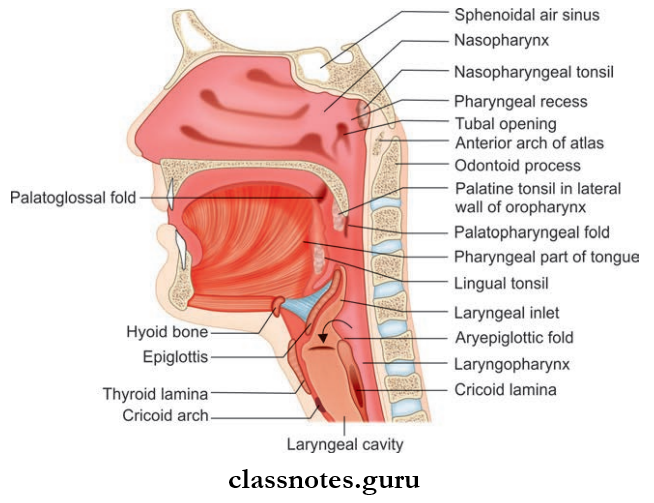
- Pharynx is a funnel-shaped muscular tissue extending from the base of skull to esophagus
- Pharynx is a common channel for both food and air.
Pharynx Dimension And Location
- Pharynx Dimension And Location measures approximately 12–14 cm in length and is 1.5–3.5 cm in width
- Pharynx Dimension And Location is located behind the cavity of the nose, mouth, and larynx.
Pharynx Boundaries And Relation
- Superiorly: Base of the skull, including posterior part of sphenoid and basilar part of the occipital bone
- Inferiorly: Continuous with esophagus at the level of C6 vertebra
- Anteriorly: Nasal cavity, mouth, and larynx
- Posteriorly: Prevertebral fascia
- Laterally: Neurovascular bundle of neck and styloid apparatus
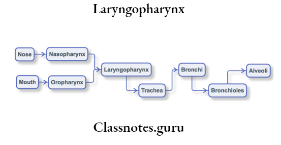
Pharynx Parts: Divided into three parts
- Nasopharynx: Lying behind the nose
- Oropharynx: Behind oral cavity
- Laryngopharynx: Behind the larynx.
1. Nasopharynx
- The nasopharynxis situated behind nose and extends from the base of skull to soft palate
- Nasopharynx communicates anteriorly through nose and posteriorly through choana and inferiorly with the oropharynx
- Lined by ciliated columnar epithelium
- The nasopharynxacts as a passage for air
- The Main Features Seen In This Part Include:
- Nasopharyngeal tonsil/adenoids
- Opening of Eustachian tube
- Pharyngeal recess/fossa of Rosenmüller
- Supplied by pharyngeal branches of the pterygopalatine ganglion.
2. Oropharynx
- Situated behind oral cavity and extends from soft palate to upper border of the epiglottis
- Oropharynx communicates anteriorly with oral cavity through the oropharyngeal isthmus, above with nasopharynx through the nasopharyngeal isthmus inferiorly with the laryngopharynx
- Oropharynx is lined by stratified squamous non-keratinized epithelium
- Oropharynx acts as a common passage for food and air
- The Main Features Seen In This Part Include:
- Palatine tonsil
- Palatoglossal arch
- Palatopharyngeal arch
- Lingual tonsil
- The free end of the epiglottis
- Valleculae
- Glossoepiglottic folds.
3. Laryngopharynx
- Situated behind larynx and extends from the upper border of the epiglottis to lower border of the cricoid cartilage
- Laryngopharynx communicates anteriorly with the laryngeal cavity through the laryngeal inlet and inferiorly with the esophagus at the pharyngoesophageal junction
- Laryngopharynx is lined by stratified squamous non-keratinized epithelium
- Laryngopharynx mainly acts as a passage for food
- The Main Features Seen In This Part Include:
- Laryngeal inlet
- Piriform fossa.
- Supplied by 9 and 10th cranial nerves.
Blood Supply Of Pharynx
- Arterial
- Ascending pharyngeal artery
- Ascending palatine artery
- Ascending tonsilar artery
- Greater palatine artery
- Lingual artery
- Venous drainage: Into pharyngeal venous plexus and ultimately to internal jugular vein.
Pharynx Lymphatic Drainage: Drain into upper and lower deep cervical nodes.
Pharynx Nerve Supply
- Sensory
- Nasophaynx: Pharyngeal branch of the pterygopalatine ganglion
- Oropharynx: Glossopharyngeal nerve
- Laryngopharynx: Internal laryngeal nerve
- Motor: All muscles are supplied by the cranial root of the accessory nerve except stylopharyngeus which is supplied by the glossopharyngeal nerve.
Pharynx Applied
- Pharyngeal tonsil or adenoids usually regress by puberty.
- Enlargement of adenoids can lead to obstruction of the posterior nasal aperture and can interfere with respiration and speech.
- Pharyngitis refers to the inflammation of the pharynx and can be caused as a result of viral, bacterial, or fungal infections.
- In Faucial Diphtheria, a grayish-white membrane forms over the tonsil which later extends to the soft palate and posterior pharyngeal wall and causes bleeding when removed.
- Retropharyngeal Space is the space between buccopharyngeal fascia covering the pharyngeal constrictor muscles and prevertebral fascia. The suppuration of retropharyngeal lymph nodes can result in retropharyngeal abscess.
Question 2. Write a short note on the palatine tonsil.
Answer:
Palatine Tonsil
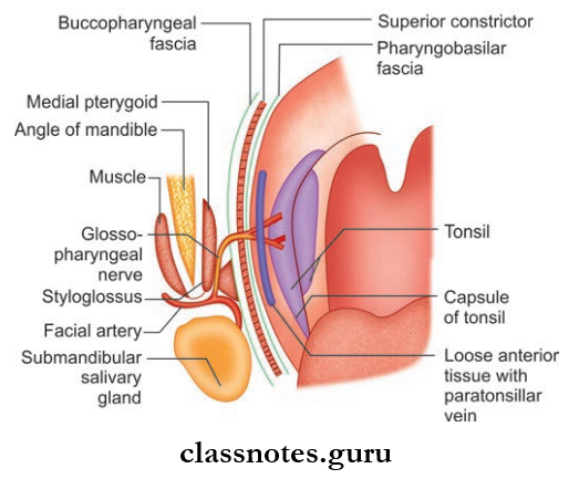
- Palatine Tonsil is an almond-shaped mass of lymphoid tissue.
- Palatine Tonsil is situated in the tonsillar fossa of the lateral wall of the oropharynx between anterior and posterior pillars.
- The anterior pillar is formed by the palatoglossal arch and the posterior pillar is formed by the palatopharyngeal arch.
Palatine Tonsil Boundaries And Relation
- Anteriorly: Anterior pillar with palatoglossal muscle
- Posteriorly: Posterior pillar with palatopharyngeus muscle
- Apex: Soft palate
- Base: Dorsal surface of posterior 1/3rd of tongue
- Laterally (Tonsillar Bed):
- Pharyngobasilar fascia
- Superior constrictor muscle
- Buccopharyngeal fascia
- Styloglossus
- Glossopharyngeal nerve
Palatine Tonsil Features: Consist of 2 surfaces, 2 borders, and 2 poles
- Medial Surface: Free and bulge to the oropharynx and lined by non-keratinized stratified squamous epithelium
- Tonsillar crypts are present and the largest and deepest crypt called crypto magna is present
- Lateral Surface: Covered by fibrous tissue and forms a capsule of tonsil and is loosely attached to the pharyngeal wall and anteroinferior it is firmly adhered to side of the tongue
- Anterior Border: Related to palatoglossal arch and muscle
- Posterior Border: Related to palatopharyngeal arch and muscle
- Upper pole is related to the soft palate and lower pole to the tongue.
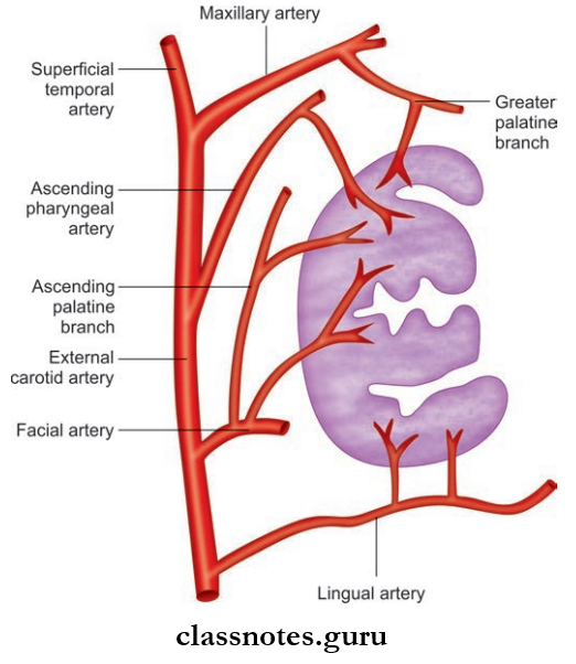
Blood Supply Of Tonsil
- Arterial Supply: Mainly by a tonsillar branch of the facial artery
- Dorsal lingual branch of lingual artery
- Ascending palatine artery
- Ascending pharyngeal artery
- Descending palatine artery
- Greater palatine artery
- Venous Drainage: Drains into peritonsillar vein which in turn drain into pharyngeal venous plexus.
Palatine Tonsil Lymphatic Drainage: Drains into upper deep cervical nodes mainly jugulodigastric nodes.
Palatine Tonsil Nerve Supply: Supplied by glossopharyngeal nerve and branches from sphenopalatine ganglion.
Palatine Tonsil Applied
- In children, tonsils are the frequent sites of infection.
- Acute infections of the tonsil can lead to acute tonsillitis. It can be membranous or follicular or parenchymatous.
- Spread of infection from tonsils to surrounding areas can lead to peritonsillar abscess.
- Tonsillectomy refers to the removal of tonsils and is indicated when the tonsils interfere with speech, swallowing, or respiration and cause recurrent attacks.
Question 3. Write a short note on the constrictor muscles of the pharynx.
Answer:
Constrictor Muscles Of The Pharynx
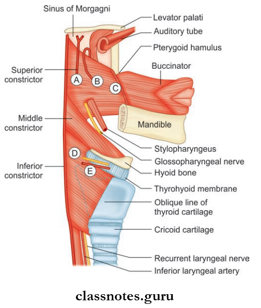
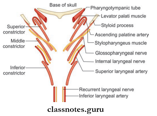
- Simplified Depiction Of Constrictor Muscles Of The Pharynx
- Ascending pharyngeal artery;
- Ascending palatine artery;
- Pterygomandibular raphe;
- Internal laryngeal nerve;
- Superior laryngeal artery);
- Flowerpot arrangement of the constrictor muscles in the wall of the pharynx (Structures passing through the gaps in the pharyngeal wall)
- These muscles form the bulk of the muscular coat of the pharyngeal wall.
- The origin of the constrictor is situated anteriorly in relation to the posterior openings of the nose, mouth, and larynx.
- From there the fibers pass backward to lateral walls in a fan-shaped manner and get inserted to the median raphe of the pharynx.
- The muscles are arranged like a flower pot without a base placed one above the other and open in front for communicating with nasal, oral, and laryngeal cavities.
- The fibers of the inferior constrictor overlap the middle constrictor which in turn overlaps the superior constrictor.
Detail Of Muscles
- Superior Constrictor
- Superior Constrictor Origin:
- Pterygoid hamulus
- Pterygomandibular raphe
- Medial surface of mandible at upper end of the mylohyoid line
- Side of the posterior part of the tongue
- Superior Constrictor Insertion: Pharyngeal tubercle
- Median raphe
- Superior Constrictor Nerve supply: Pharyngeal plexus through a pharyngeal branch of the vagus
- Superior Constrictor Action: Helps in deglutition
- Superior Constrictor Origin:
- Middle Constrictor
- Middle Constrictor Origin:
- The lower part of the stylohyoid ligament
- Lesser cornua and greatercornua of hyoid bone
- Middle Constrictor Insertion: Median raphe
- Middle Constrictor Nerve supply: Pharyngeal plexus through a pharyngeal branch of the vagus
- Middle Constrictor Action: Helps in deglutition
- Middle Constrictor Origin:
- Inferior Constrictor: Consists of two parts
- Thyropharyngeus
- Inferior Constrictor Origin:
- The oblique line on the lamina of thyroid cartilage
- Tendinous band attached to inferior tubercle of thyroid cartilage
- Inferior Constrictor Insertion: Median raphe
- Inferior Constrictor Nerve supply:
- External laryngeal nerve
- Pharyngeal plexus
- Inferior Constrictor Origin:
- Thyropharyngeus
- Cricopharyngeus
- Cricopharyngeus Origin: Cricoid cartilage
- Cricopharyngeus Insertion: Median raphe
- Cricopharyngeus Nerve supply:
- Recurrent laryngeal nerve
- Pharyngeal plexus
- Cricopharyngeus Action: Helps in deglutition
Structures Passing through Space Between Constrictors
- Between Base Of Skull And Superior Constrictor: Auditory tube
- Levator veli palatini
- Ascending palatine artery
- Palatine branch of ascending pharyngeal artery
- Between Superior And Middle Constrictor: Stylopharyngeus muscle
- Glossopharyngeal nerve
- Between Middle And Inferior Constrictor: Internal laryngeal nerve
- Superior laryngeal vessel
- Between Inferior Constrictor And Esophagus: Recurrent laryngeal nerve
- Inferior laryngeal vessels
Question 4. Write a short note on Killian’s dehiscence.
Answer:
Killian’s Dehiscence
- The gap between the thyropharyngeus and cricopharyngeus muscle is called Killian’s dehiscence or pharyngeal dimple.
- The mucosa and submucosa of the pharynx can bulge through this area to form a pharyngeal pouch.
Laryngopharynx Multiple Questions And Answers
Question 1. The pharyngeal wall consists of all the following except:
- Mucous membrane
- Pharyngobasilar fascia
- Buccopharyngeal fascia
- Prevertebral fascia
Answer: 4. Prevertebral fascia
Question 2. All of the following are features of the nasopharynx except:
- Pharyngeal tonsil
- Tubal tonsil
- Pharyngeal recess
- Piriform recess
Answer: 4. Piriform recess
Question 3. The Pas savant’s ridge is formed by:
- Salpingopharyngeus
- Stylopharyngeus
- Palatopharyngeus
- Thropharyngeus
Answer: 3. Palatopharyngeus
Question 4. Motor nerve supply of pharyngeal muscles is derived from:
- Vago-accessory complex
- Glossopharyngeal nerve
- External laryngeal nerve
- All of the above
Answer: 4. All of the above
Question 5. The inferior constrictor of the pharynx is supplied by all of the following nerves except:
- Pharyngeal plexus
- Glossopharyngeal nerve
- External laryngeal nerve
- Recurrent laryngeal nerve
Answer: 2. Glossopharyngeal nerve