
Ear Question And Answers
Question 1. Write a short note on the tympanic membrane (eardrum).
Answer:
Tympanic Membrane (eardrum)
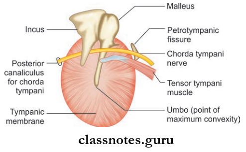
The semitransparent membrane which forms the partition between external acoustic meatus and middle ear.
Tympanic Membrane Layers: Made of three layers
- Outer cuticular layer
- Middle fibrous layer
- Inner mucosal layer.
Tympanic Membrane Parts: Consists of two parts
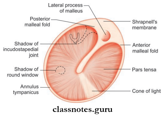
- Pars Tensa: Forms the most part of the tympanic membrane:
- It is thickened at the periphery and is called annulus tympanicus
- The handle of malleus is attached to the inner surface and tensor tympani muscle is attached to the root of handle of the malleus.
- Pars Florida: Small triangular area above the lateral process of malleus between anterior and posterior malleal fold.
Tympanic Membrane Consists Of Two Surfaces:
- Lateral Surface: Concave surface directed downwards, forwards laterally towards the meatus
- Medial Surface: Convex and bulges towards middle ear.
Tympanic Membrane Blood Supply
- By deep auricular artery, anterior and posterior tympanic artery
- Veins drain into the external jugular vein and transverse sinus.
Tympanic Membrane Nerve Supply
- By auriculotemporal nerve
- Auricular branch of vagus nerve
- Tympanic branch of the glossopharyngeal nerve.
Tympanic Membrane Applied
- Traumatic rupture of the tympanic membrane occurs commonly as a result of trauma due to the insertion of sharp objects into ear for removing wax or foreign body or may be due to a sudden change of air pressure as in a slap on the ear.
- Tympanic Membrane Perforations of the tympanic membrane can be either central or marginal and are associated with chronic suppurative otitis media.
- Herpes zoster oticus refers to a viral infection involving the geniculate ganglion of the facial nerve. It is characterized by the appearance of vesicles on the tympanic membrane, deep meatus, etc.
- Tympanosclerosis refers to the hyalinization and calcification of firous layer of tympanic membrane. It causes interference in the conduction of sound.
Question 2. Write a note on the middle ear (tympanic cavity).
Answer:
Middle Ear (Tympanic Cavity)
Middle Ear is a narrow airfiled cavity present within the petrous part of temporal bone between external ear and internal ear.

Abbreviations: IJV = Internal jugular vein; ICA = Internal carotid artery
Middle Ear Size And Shape
- Middle Ear Size And Shape is a cubelike structure having large lateral and medial walls and narrow other walls
- Middle Ear Size And Shape is biconcave in coronal section.
Middle Ear Communications
- Anteriorly with nasopharynx through Eustachian tube
- Posteriorly with mastoid antrum through aditus.
- Middle Ear Communications Subdivisions: Divided into three parts
- Epitympanum (attic): Part above tympanic membrane and contains head of malleus, body, and short process of incus
- Mesotympanum: Part opposite to tympanic membrane and contains head of malleus, long process of incus and stapes
- Hypotympanum: Part below tympanic membrane.
- Middle Ear Communications Contents
- Three ear ossicles: Malleus, incus, and stapes
- Two muscles: Tensor tympani and stapedius
- Two nerves: Chorda tympani, tympanic plexus
- Ligaments of ear ossicles
- Vessels supplying and draining middle ear
- Air.
- Middle Ear Communications Subdivisions: Divided into three parts
Middle Ear Boundaries
- Roof: Formed by a thin plate of bone—tegmen tympani
- Separates middle ear from middle cranial fossa
- Floor: Formed by thin plate of bone and separate middle ear cavity from jugular bulb
- The tympanic branch of the glossopharyngeal nerve pierces the flor and takes part in the tympanic plexus
- Anterior Wall: Formed by thin plate of bone
- The lower part separates the cavity from the internal carotid artery
- In the upper part, two openings are present—one for tensor tympani muscle (upper) and one for the auditory tube (lower)
- The bony partition between two openings extends along medial wall as curved lamina—processus cochleariformis.
- Posterior Wall: Separates cavity from the mastoid antrum
- It has an opening in the upper part which communicates with mastoid antrum called aditus
- Fossa includes a depression close to aditus
- The pyramid is a conical bony projection below aditus and the tendon of stapedius muscle passes through its summit
- Vertical part of facial nerve runs just behind the pyramid through facial canal.
- Lateral Wall: Formed by tympanic membrane
- Chorda tympanic nerve passes across the tympanic membrane lateral to long process of incus and medial to handle of malleus.
- Medial Wall: Separates the tympanic cavity from the internal ear. It has the following features:
- Promontory: A rounded prominence produced by basal (fist turn) of cochlea
- Oval Window: A kidneyshaped opening above and behind the promontory and is closed by base of stapes and annular ligament
- Round Window: A small round opening below the promontory and is closed by a fibrous membrane and separates the middle ear from scala tympani
- Sinus Tympani: A depression behind promontory between round and oval window and indicates the position of the ampulla of the semicircular canal
- Prominence Of The Facial Canal: Extends backward and downwards above oval window and joins vertical part in the posterior wall
- The prominence of the semicircular canal.
Middle Ear Blood Supply
- Arterial: Mainly by anterior tympanic branch of maxillary artery and posterior tympanic branch of posterior auricular artery
- Venous: Drainage into pterygoid plexus of veins and superior petrosal sinus.
Middle Ear Lymphatic: To preauricular and retropharyngeal lymph node.
Middle Ear Nerve Supply: Derived from tympanic plexus.
Middle Ear Functions
- Transmission of sound waves from the external ear to internal ear
- Increase in intensity of sound waves.

Middle Ear Applied
- Acute suppurative otitis media refers to the acute inflammation of the middle ear by pyogenic organisms.
- Serous otitis media refers to a condition characterized by accumulation of nonpurulent effusion in the middle ear.
- The presence of keratinizing squamous epithelium in the middle ear or mastoid is known as cholesteatoma.
- Glomus tumor is a benign neoplasm of middle ear and arises from the glomus bodies.
Question 3. Write a short note on ear ossicles and muscles.
Answer:
Ear Ossicles And Muscles

Three ear ossicles are present inside the middle ear connected to each other and are involved in conduction of sound vibration from the tympanic membrane to oval window.
Malleus
- Resembles a hammer
- Consists of head, neck, and handle, lateral and anterior process
- Head and neck are present in the epitympanum and the handle is attached to tympanic membrane
- Anterior and posterior malleal folds are attached to the lateral process
- The head articulates to the body of incus to form incudomalleolar joint.
Incus
- Resembles a premolar tooth
- Consists of a large body and a long and a short process
- The body and short process lie in atlas; the long process hangs behind the handle of the malleus
- Its tip articulates with head of stapes to form incudostapedial joint.
Stapes
- Resembles stirrup
- Consists of the head, neck, anterior and posterior areas, and footplate
- The footplate is attached to oval window.
Intratympanic Muscles
- Tensor tympani originates from the cartilaginous part of the auditory tube and is inserted to the handle of the malleus
- The Stapedius muscle arises from pyramidal eminence on the posterior wall of the tympanic cavity and inserted to the posterior aspect of neck.
Ear Ossicles And Muscles Applied
- Otosclerosis or otospongiosis refers to the replacement of the normal endochondral layer of bony otic capsule by irregularly laid spongy bone.
- Most commonly otosclerotic focus affects the stapes region and causes stapes fixation and conductive deafness.
- Surgically, it is treated by removal of stapes (stapedectomy) and placement of the prosthesis.
Question 4. Write a short note on mastoid antrum.
Answer:
Mastoid Antrum

- Supramastoid crest above,
- The posterosuperior margin of external acoustic meatus in front,
- Vertical tangent to posterior margin of the external meatus behind
- Large air space in the upper part of mastoid process
- Communicates directly with the tympanic cavity through aditus.
Mastoid Antrum Relations
- The roof is formed by tegmen anti which is a continuation of tegmen tympani
- The floor receives openings of mastoid air cells
- The medial wall is formed by the petrous temporal bone
- The lateral wall is formed by a plate of the squamous temporal bone and it is marked on surface by suprameatal triangle
- The posterior wall is related to sigmoid sinus
- Anteriorly, it communicates with epitympanic recess through aditus.
Mastoid Air Cells
- These are a series of intercommunicating spaces within the mastoid process
- Their number, size, and distribution vary from region to region of the mastoid process
- Usually, they are confined to the mastoid process but they can extend into squamous and petrous parts of the temporal bone.
Mastoid Antrum Blood Supply
- Arterial supply is by the posterior tympanic artery, derived from the stylomastoid branch of a posterior auricular artery
- Veins drain into mastoid emissary vein, posterior auricular vein, and sigmoid sinus.
Mastoid Antrum Nerve Supply: Antrum is supplied by branches from the tympanic plexus, which is formed by the glossopharyngeal nerve and meningeal branch of mandibular nerve.
Mastoid Antrum Lymphatic Drainage: Lymph is drained into postauricular and upper deep cervical lymph nodes.
Mastoid Antrum Applied
- Chronic suppurative otitis media (CSOM) is a longstanding infection of the middle ear cleft and is characterized by ear discharge and permanent perforation.
- The CSOM can be tubotympanic or atticoantral
- The atticoantral type involves the mastoid antrum and the attic and is characterized by purulent foulsmelling discharge and associated with conductive hearing loss.
Question 5. Write a note on the internal ear.
Answer:
Internal Ear Consists Of:
- Bony labyrinth
- Membranous labyrinth.
- Bony Labyrinth
Consists Of A Series Of Intercommunicating Bony Cavities And Canal: It consists of three parts
- Cochlea
- Vestibule
- Three Semicircular Canals.
1. Cochlea
- Resembles the shell of a common snail
- Consists of an apex called the cupula, which is directed towards the medial wall of the middle ear, and a base directed to the internal acoustic meatus
- Cochlea consists of a central pillar called modiolus and a bony cochlear canal.
- Modiolus is the axis around which the cochlear canal spirals. It has a base and an apex. Base lies at the fundus of the internal acoustic meatus and the apex is overlaid by the apical turn of the cochlea
- The cochlear canal is arranged spirally around the modiolus
- Spiral lamina, a ridge of bone projects to the cochlear canal and has a lamina
- The vestibular membrane extends from the upper lip of the lamina to the outer wall of the cochlea
- The basilar membrane extends from the lower lip of the lamina to outer wall of the cochlea
- The cochlear duct (scala media) is a triangular area enclosed by a vestibular and basilar membranes and outer walls of cochlear canal
- The spiral lamina divides the cochlear canal to the scala vestibule above and scala tympani below and each communicates with
- one other through helicotrema
- Near the basal turn of the cochlea, a round window and aqueduct of cochlea are present
- The perilymph of cochlea communicates with the cerebrospinal fluid of the subarachnoid space through the aqueduct of the cochlea.


2. Vestibule
- Central oval cavity between cochlea and semicircular canal
- The lateral wall communicates with middle ear through oval window
- The medial wall contains two recesses—spherical recess and elliptical recess
- The spherical recess lodges saccule whereas the elliptical recess lodges utricle
- Below the elliptical recess, an aqueduct of the vestibule is present
- The anterior wall contains an opening that communicates with scala vestibuli and the posterior wall bears fie openings of semicircular canals.
3. Semicircular Canals
- Three in number—anterior, posterior, and lateral, and they lie in planes right-angled to each other
- Each canal is dilated at one end to form an ampulla
- Anterior semicircular canal lies at the vertical plane right-angled to long axis of the petrous temporal bone.
- The anterior end (ampullated) communicates with the vestibule anterolaterally. The posterior end unites with the nonampullated end of posterior semicircular canal and opens to the vestibule.
- Posterior semicircular canal lies in vertical plane parallel to long axis of petrous temporal bone. Th ampullated end communicates with vestibule.
- The lateral semicircular canal lies in horizontal plane. Its anterolateral end is ampullated. Both ends open into the vestibule.
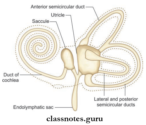
Internal Ear Membranous Labyrinth: Consists of four parts
- Cochlear Duct Which Lies Within Bony Cochlea
- Saccule Lies Wwithin Bony Vestibule
- Utricle Lies Within Bony Vestibule
- Three Semicircular Ducts Lie Within The Bony Semicircular Canal.
- The cochlear duct is connected to the saccule by ductus reunions
- The saccule and utricle are connected to each other by ‘Y’ shaped utriculosaccular duct which expands to form ductus and saccus endolymphatic
- Utricle is connected to three semicircular ducts through fie openings.
1. Cochlear Duct
- Spiral anterior part of membranous labyrinth
- Lies in the middle part of cochlear canal between scala vestibule and scala tympani
- The duct contains the spiral organ of Corti, the sensory receptor for hearing
- The structure is studied in a crosssection of the cochlear canal
- The cochlear duct is bounded by the outer wall of the cochlear canal laterally, the roof is bounded by Reissner’s membrane, and base formed by osseous spinal lamina and basilar membrane
- The organ of Corti is the peripheral organ of hearing in the cochlear duct, situated on the basilar membrane. It consists of the following:
- Tunnel Of Corti: Formed by inner and outer rod cells and containing corticolymph
- Hair Cells: Receptor cells located on basilar membrane and contain stereocilia overlaid by tectorial membrane. There are outer hair cells and inner hair cells.
- Supporting Cells: Include Deiter’s cells and Hansen’s cells and superior outer hair cells
- Tectorial Membrane: Made of a gelatinous substance that overlies the hair cells and is attached to spiral lamina. The shearing force between the hair cells and the tectorial membrane stimulates hair cells.
2. Saccule And Utricle
- Saccule is a small globular membranous sac present in the anteroinferior part of the vestibule
- Utricle is a larger membranous sac in the posterosuperior part of vestibule
- Saccule is connected to basal turn of the cochlear duct through ductus reuniens and with utricle through utriculosaccular duct. The vertical limb of duct forms a blind terminal end called saccus endolymphaticus.
- The utricle receives the three semicircular ducts posteriorly through fie openings.
3. Semicircular Ducts
- Thee Ducts: Anterior, posterior, and lateral ducts lie within the corresponding canal
- Each duct has a dilated end called the ampulla, which corresponds to the ampulla of the corresponding semicircular canal.
- Each ampullar end has a raised crest called crista ampullar and is responsible for kinetic balance and responds to angular acceleration
- The medial wall of saccule and utricle contains macula, which are sensory receptors responsible for static balance and sense the position of head in response to gravity and linear acceleration.

Internal Ear Applied
- Meniere’s disease of the inner ear is caused as a result of distension of the endolymphatic system by endolymph.
- It is characterized by vertigo, sensory neural hearing loss, tinnitus, and aural fullness.
Question 6. Write a short note on the auditory tube/Eustachian tube.
Answer:
Auditory Tube
- Connects nasopharynx with middle ear
- Responsible for aeration of middle ear and maintenance of pressure in the middle ear
- Measures about 36 mm in adult and is directed downwards medially and forwards to reach lateral wall of the nasopharynx and open 1 cm behind the posterior end of the inferior turbinate.
Internal Ear Parts: Consists of three parts
- Outer Bony Part: About 12 mm in length, extends from the outer wall of middle ear to the junction between squamous and petrous parts of temporal bone
- Isthmus: Junction between bony and cartilaginous parts
- Inner cartilaginous Part: About 24 mm fied to sulcus tubae and bounded by petrous part of temporal bone and greater wing of sphenoid. The cartilaginous portion at the pharyngeal end presents as an elevation called torus tuberous.

Auditory Tube Or Eustachian Tube Muscles Attached
- The Various Muscles Attached Include
- Salpingopharyngeus
- Levator veli palatini
- Tensor palate
- Tensor tympani.
Auditory Tube Or Eustachian Tube Muscles Nerve Supply
- Glossopharyngeal nerve supplies the lateral part
- Pharyngeal branch of sphenopalatine ganglion supplies the pharyngeal part.
Auditory Tube Or Eustachian Tube Muscles Blood Supply
- Arterial Supply
- Ascending pharyngeal artery
- Middle meningeal artery
- Artery to the pterygoid canal.
- Venous Drainage: To pharyngeal plexus and pterygoid venous plexus.
Auditory Tube Or Eustachian Tube Muscles Lymphatic Drainage: To retropharyngeal and upper deep cervical lymph nodes.
Auditory Tube Or Eustachian Tube Muscles Applied
- Mechanical obstruction of the auditory tube as a result of inflammation of allergy can cause earache, tinnitus, vertigo, and even disturbances of equilibrium.
- Adenoids can cause tubal obstruction and can lead to otitis media with effusion.
Ear Multiple Choice Question And Answers
Question 1. Embryologically the tympanic membrane is derived from:
- Ectoderm
- Mesoderm
- Endoderm
- All of the above
Answer: 4. All of the above
Question 2. Th contents of middle ear include all except:
- Tensor tympani
- Tegmen tympani
- Chorda tympani
- Tympanic plexus
Answer: 2. Tegmen tympani
Question 3. All of the following structures are of adult size at birth except:
- External ear
- Middle ear
- Ear ossicles
- Internal ear
Answer: 1. External ear
Question 4. Medial wall of middle ear presents all of the following except:
- Oval window
- Round window
- Pyramid
- Lateral semicircular canal prominence
Answer: 3. Pyramid
Question 5. The membranous labyrinth of internal ear consists of all except:
- Cochlear canal
- Utricle
- Saccule
- Cochlear duct
Answer: 1. Cochlear canal