
Surface Landmarks Introduction
Surface Landmarks On the side
- Sternocleidomastoid: Seen as a prominent raised ridge extending obliquely from clavicle and sternum to mastoid process.
- Anterior Border Of Trapezius: Prominent on the elevation of the shoulder against resistance.
- Mastoid Process: Large bony projection behind the lower part of the auricle.
- Greater Supraclavicular Fossa: A depression above the middle one-third of the clavicle.
- Lesser Supraclavicular Fossa: Small depression between sternal and clavicular part of the sternocleidomastoid.
Surface Landmarks Anteriorly
- Hyoid Bone: A horseshoe-shaped bone felt just below and behind the chin. Lies at the level of C3 vertebra.
- Thyroid Cartilage: Sharp protuberance in the median plane below the hyoid. Also called Adam’s apple/laryngeal prominence.
- Cricoid Cartilage: Lies just below the thyroid cartilage. Lies at the level of lower border of C6 vertebrae.
- Suprasternal Notch: Felt between anterior ends of clavicle. Lies at level of lower border of body of T2 vertebrae.
Surface Landmarks Back
- Nuchal Furrow: A vertical groove in the midline on the back of neck.
- External Occipital Protuberance: A bony projection felt in the upper end of the nuchal furrow.
- The Spine Of C7 Vertebrae: A knob-like bony projection felt at the lower end of nuchal furrow.
Side Of Neck Questions And Answers
Question 1. Write a short note on deep cervical fascia (fascia colli).
Answer:
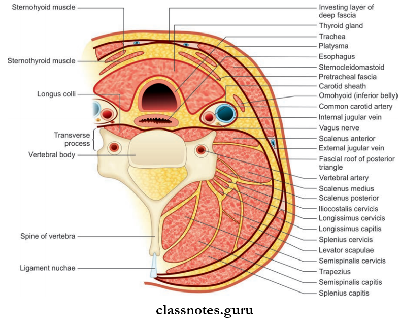
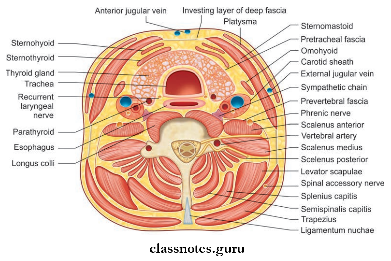
Deep Cervical Fascia (fFascia Colli)
The deep cervical fascia of neck consists of the following layers from outside inwards:
- Investing layer of deep fascia
- Pretracheal fascia
- Prevertebral fascia.
It Is Also Condensed To Form:
- Carotid sheath
- Buccopharyngeal fascia
- Pharyngobasilar fascia.
Investing Layer Or Deep Fascia: Lies deep to platysma and superficial fascia and encircles the neck like a collar
Deep Cervical Fascia Attachments.
- Superiorly
- External occipital protuberance
- Superior nuchal line
- Mastoid process
- Base of mandible
- Inferiorly
- Spine of scapula
- Acromion process
- Clavicle
- Manubrium
- Anteriorly
- Symphysis menti
- Hyoid bone
- Posteriorly
- Ligamentum nuchae
- Spine of C7 vertebrae.
Deep Cervical Fascia Features
- The superior portion of the fascia splits to enclose the submandibular salivary gland.
- The superficial layer that covers the submandibular gland is attached to the base of the mandible and the deep layer to the mylohyoid line.
- Posterior to the submandibular gland and at lower pole of the parotid gland it splits to enclose the parotid gland.
- The superficial layer that covers the parotid gland is known as the parotid masseteric fascia and is attached to the zygomatic arch and the deep layer is attached to the lower border of the tympanic plate and styloid process.
- Part of layer between the styloid process and the angle of the mandible condenses to form the stylomandibular ligament and separates the parotid gland from the submandibular gland.
Inferiorly, The Fascia Splits To Enclose 2 Spaces
- Above Suprasternal Notch Splits To Enclose Suprasternal Space (Of Burns) Which Consists Of:
- Sternal head of right and left sternocleidomastoid
- Jugular venous arch
- Lymph node
- Interclavicular ligament.
- Above Middle Of Clavicle, It Splits To Enclose Supraclavicular Space Which Consist Of:
- External jugular vein
- Supraclavicular nerves
- Cutaneous vessels and lymphatics.
- It also forms pulleys to bind the tendon of the omohyoid and digastric muscles.
Deep Cervical Fascia Applied
Fascia covering parotid is very thick and strong, so in infections of the parotid gland, the gland cannot expand, as a result, the nerves are compressed and result in pain.
Deep Cervical Fascia TIP: Rule of 2
- It Encloses 2 Muscles: Trapezius and sternocleidomastoid
- Forms Roof Of 2 Triangles: Anterior and posterior
- Split To Enclose 2 Glands: Parotid and submandibular
- Split To Enclose 2 Spaces: Suprasternal and supraclavicular
- Forms Two Fascial Slings: For the inferior belly of omohyoid and intermediate tendon of digastric.
Pretracheal Fascia
- Covers front and side of trachea
- Splits to enclose thyroid gland and form its false capsule
- Ligament of Berry, a firous band which connects the capsule of lateral lobe of thyroid to cricoid cartilage.
Pretracheal Fascia Attachments
- Superiorly: Hyoid bone in median plane
- Oblique line of thyroid cartilage
- Cricoid cartilage.
- Inferiorly: Encloses the inferior thyroid veins and passes behind brachiocephalic vein and blends with arch of aorta.
- On Either Side: Fuses with front of carotid sheath.
Pretracheal Fascia Applied
The thyroid gland is connected to the hyoid bone, thyroid, and cricoid cartilages via the ligament of Berry. As a result, thyroid gland moves up and down with the larynx during swallowing.
Prevertebral Fascia
- Lies in front of the prevertebral muscles
- Forms the flor of the posterior triangle of neck
Prevertebral Fascia Attachments
- Superiorly: Base of skull
- Inferiorly: Anterior longitudinal ligament and body of 4th thoracic vertebrae in superior mediastinum
- Anteriorly: Separated by retropharyngeal space from posterior aspect of pharynx and buccopharyngeal fascia
- Laterally: Present deep to trapezius.
Prevertebral Fascia Features
- It covers scalene muscles, levator scapulae, splenius capitus, and form fascial carpet of the posterior triangle. The roots of cervical spinal nerves lie deep to it.
- As the trunks of the brachial plexus and subclavian artery emerge from the scalenus muscles and move towards the axilla, they carry a tubular sheath of fascia around them called as axillary sheath.
- Fascia provides a field base for movement of the pharynx, esophagus, and carotid sheath during movements of neck and swallowing.
Prevertebral Fascia Applied
- In case of infection of retropharyngeal lymph nodes, the acute abscess formed bulges forward in the paramedian position.
- This occurs because the retropharyngeal lymph nodes are present in between the prevertebral fascia (behind) and buccopharyngeal fascia (in front) and these fascia are adherent to each other along the midline.
- Carotid Sheath: Tubular condensation of deep cervical fascia around the main vessels of neck.
Buccopharyngeal Fascia
- Covers the constrictor muscles of the pharynx and
- It extend from base of the skull to esophagus.
Pharyngobasilar Fascia
- It extend from base of the skull to upper border of the superior constrictor muscle
- Lies deep to the constrictor muscles and cover the internal surface of constrictor muscles and thereby closing gaps in the muscular wall of pharynx.
Question 2. Write a short note on the carotid sheath.
Answer:
The Carotid Sheath
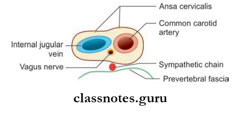
- Tubular condensation of deep cervical fascia around the main vessels of the neck
- It covers the common carotid artery and internal carotid, internal jugular vein, and vagus nerve
- It extends from base of the skull above to the arch of aorta below
- It is thick around the arteries and thin around the vein to allow expansion of the vein during increased venous return
- The vagus nerve lies posteriorly between arteries and veins.
Carotid Sheath Relations
- Anteriorly: Ansa cervicalis is embedded in the anterior wall of the carotid sheath
- Posteriorly: Cervical sympathetic chain lies close to the posterior wall, plastered to the prevertebral fascia.
Carotid Sheath Applied: In case of tuberculosis, cervical lymph nodes adhere to internal jugular vein, so in block dissection of neck carotid sheath is exposed to remove lymph nodes.
Mnemonic: ‘I See 10 CCs in the IV’
- I See (IC) = Internal Carotid artery
- 10 = CN 10 (Vagus nerve)
- CC = Common Carotid artery
- IV = Internal Jugular Vein
Question 3. Write a note on posterior triangle of neck.
Answer:
Posterior Triangle Of Neck
It is a triangular space on the side of the neck behind the sternocleidomastoid.
Posterior Triangle Of Neck Boundaries: Boundaries of the posterior triangle
- Anteriorly: Posterior border of sternocleidomastoid
- Posteriorly: Anterior border of trapezius
- Apex: Meeting point of sternocleidomastoid with trapezius at the superior nuchal line
- Base: Middle third of clavicle
- Roof: Formed by investing layer of deep fascia
- Floor: Formed by muscles above to downwards:
- Semispinalis capital
- Splenius capitis
- Levator scapulae
- Scalenus medius


Posterior Triangle Of Neck Features
- The Superficial Fascia Overlying The Roof Consists Of:
- Platysma
- External jugular and posterior jugular veins
- Cutaneous nerves and vessels
- Structures Piercing Roof:
- Cutaneous branches of the cervical plexus
- The external jugular vein pierces at the anteroinferior angle of the triangle to open into the subclavian vein.
- It Is Divided Into Two Parts By The Inferior Belly Of Omohyoid:
- The larger upper part is called the occipital triangle
- The smaller lower part called the subclavian/supraclavicular triangle
- Named because they contain occipital and subclavian arteries respectively.
Contents Of Posterior Triangle
- In the Occipital Triangle
- Occipital Triangle Nerves
- Spinal Accessory Nerve: Four cutaneous branches of the cervical plexus: Greater auricular nerve, lesser occipital nerve, transverse cervical nerve, supraclavicular nerve
- Muscular branches supplying trapezius, levator scapulae, rhomboids
- C5, C6 roots of brachial plexus
- Occipital Triangle Vessels: Transverse cervical artery and vein, occipital artery
- Occipital Triangle Lymph nodes: Occipital nodes and supraclavicular nodes.
- Occipital Triangle Nerves
- In the Subclavian Triangle:
- Subclavian Triangle Nerves
- 3 trunks of brachial plexus
- Nerve to serratus anterior (C5, C6, C7)
- Nerve to subclavius (C5, C6)
- Suprascapular nerve (C5, C6).
- Subclavian Triangle Vessels
- Third part of the subclavian artery and vein
- Suprascapular artery and vein
- Transverse cervical artery and vein
- The lower part of the external jugular vein.
- Subclavian Triangle Lymph node: Supraclavicular nodes.
- Subclavian Triangle Nerves
Features Of Contents Of Posterior Triangle
- Spinal Accessory Nerve: Present between roof and floor mainly adhered to the fascia of the roof
- It supplies sternocleidomastoid and trapezius.
- Four cutaneous branches of cervical plexus pierce the floor of the triangle and pass the triangle and pierce deep fascia to become cutaneous.
- Muscular Branches To
- Trapezius and levator scapula (C3, C4)—to levator scapula end in it and to trapezius runs parallel to spinal accessory nerve and cross the triangle
- To rhomboideus ( C5)—proprioceptive.
- Nerve To Serratus Anterior (C5, C6, and C7): The nerve passes behind the branchial plexus and descends over the serratus anterior in axilla and branches to digitations of muscle.
- Nerve To Subclavius (C5, C6 ): Descends in front of the brachial plexus and subclavian vessels behind omohyoid, suprascapular vessels, and clavicle to reach subclavius muscle
- Sometimes, it gives accessory phrenic nerve.
- Suprascapular Nerve (C5, C6): Supplies supraspinatus and infraspinatus muscle.
- Three trunks of the brachial plexus along with subclavian artery carry an axillary sheath around them.
- The transverse cervical artery is a branch of the thyrocervical trunk and divides superficial and deep branches.
- The suprascapular artery which is also a branch of the thyrocervical trunk pass behind clavicle.
- The subclavian artery and vein pass behind the tendon of scalenus anterior over the first rib.
Posterior Triangle Of Neck Applied: Pus collected in posterior triangle deep to fascia tracks downwards and may appear fist in axilla or arm.
Question 4. Write a short note on sternocleidomastoid.
Answer:
Sternocleidomastoid
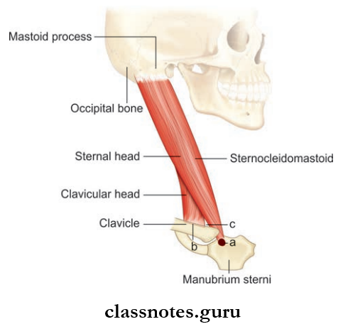
Sternocleidomastoid: One of the superficial muscles of the neck
Sternocleidomastoid Origin: Sternal head from the superolateral part of the manubrium sterni and clavicular head from the medial one-third of superior surface of clavicle
Sternocleidomastoid Insertion:
- To lateral surface of mastoid process by thick tendon
- To lateral half of superior nuchal line by thin aponeurosis.
Sternocleidomastoid Nerve supply: Motor supply—spinal accessory
- Proprioceptive: Ventral rami of C2
Sternocleidomastoid Blood supply: Arterial supply—two branches from occipital artery and one branch each from superior thyroid artery and suprascapular artery.
Sternocleidomastoid Action
- When muscle of one side contracts
- Turns chin to the opposite side
- Tilts head towards the shoulder of same side
- When muscles of both sides contracts together
- Draws the head forward as in eating and in lifting the head from the pillow
- Also, help in forced inspiration.
Sternocleidomastoid Relations
- Superficial
- Skin
- Superficial fascia
- Investing layer of deep cervical fascia
- Platysma
- External jugular vein
- Cutaneous nerves
- Parotid gland
- Deep
- Mastoid process and sternoclavicular joint
- Carotid sheath
- Muscles of posterior triangle
- Common carotid artery and its branches
- Subclavian artery and its branches
- The internal jugular vein and its tributaries
- Vagus nerve, spinal accessory nerve part of branchial plexus, and ansa cervicalis.
Sternocleidomastoid Applied
Torticollis or wry neck is a deformity in which the head is bent to one side and chin points to the other side. This occurs due to spasms of the sternocleidomastoid and trapezius supplied by the spinal accessory nerve.
Side Of Neck Multiple Choice Question And Answers
Question 1. All of the following are contents of carotid sheath except:
- Internal carotid artery
- External carotid artery
- Internal jugular vein
- Vagus nerve
Answer: 3. Internal jugular vein
Question 2. All of the following are derivatives of deep cervical fascia except:
- Pretracheal fascia
- Prevertebral fascia
- Stylomandibular ligament
- Sphenomandibular ligament
Answer: 2. Prevertebral fascia
Question 3. Select the incorrect statement about the carotid sheath:
- It extends from the base of the skull to the clavicle
- The ansa cervicalis is embedded in its anterior wall
- It is ill-defied over the internal jugular vein
- The cervical sympathetic chain is posterior to it
Answer: 3. It is ill-defied over the internal jugular vein
Question 4. All of the following muscles forms the floor of the posterior triangle except:
- Splenius capitis
- Levator scapulae
- Scalenus medius
- Scalenus anterior
Answer: 1. Splenius capitis
Question 5. All of the following structures lie deep to fascial carpet of the posterior triangle except:
- Trunks of brachial plexus
- Spinal accessory nerve
- Occipital artery
- The third part of the subclavian artery
Answer: 2. Spinal accessory nerve