Median Nerve

Median Nerve Anatomy Notes

Explain in detail about the Median Nerve under headings—origin, root value, course, branches, and innervation. Write a note on the injury to the nerve.
Answer:

  • The median nerve is called the median because it runs in the median plane of the forearm.
  • Median Nerve is called the eye of the hand because it gives sensory supply to the pulp of the thumb and index fingers.
  • Median Nerve is also known as the laborers’ nerve.

Median Nerve Origin

  • By the union of the medial and lateral cords of the brachial plexus.

Median Nerve Root Value

  • C5, C6, C7, C8, T1.

Median Nerve Function

Median Nerve Course

  1. In the axilla:
    • The median nerve goes down to enter in the arm from the lateral side of 3rd part of the axillary artery.
  2. In the arm:
    • The median nerve lies lateral to the brachial artery.
    • But at the level of the middle of the arm, it crosses the brachial artery and runs medial to enter the cubital fossa.
  3. In the cubital fossa:
    • It gives of muscular branches to flexor carpi radialis, flxor digitorum superfiialis and palmaris longus.
    • The median nerve leaves the cubital fossa between two heads of pronator teres and then deep to the fibrous arch of the flexor digitorum superficial.
  4. In the forearm:
    • In the forearm, it is adhered to the deep surface of the flexor digitorum superficial and leaves the muscle along its lateral border.
    • It runs deep to the palmaris longus and gives of the palmar cutaneous branch before getting under the carpal tunnel.
  5. In the palm:
    • After passing through the carpal tunnel, the median nerve divides into lateral and medial divisions.
    • Lateral division supplies 3 out of 4 thenar muscles, 1st and second lumbricals.
    • Medial division along with the remaining fingers of lateral division provides cutaneous supply to the lateral 3 and half digits and their nail beds including the skin of distal phalanges on their dorsal aspect.

Median Nerve Anatomy

Median Nerve Branches and Innervations

Nerves Of Upper Limb Median Nerve Branches And Innervations

Mnemonic: Median Nerve: Hand Muscles Innervated

‘The LOAF muscles’:

  • lumbrical 1 and 2
  • Opponents pollicis
  • Abductor pollicis brevis
  • Flexor pollicis brevis
  • Alternatively: LLOAF, with 2 L’s, to recall there is 2 lumbricals.
  • To remember that these are the Median nerve muscles, think ‘Meat LOAF’.

Median Nerve Clinical Anatomy

  • Injury to the median nerve can occur at various levels and the clinical features vary accordingly.
  1. Injury At The Level Of The Elbow
    • Median Nerve can be due to:
      • Supracondylar fracture of humerus.
      • Entrapment of nerve between the two heads of pronator teres during its course.
      • Tight tourniquet usage during venipunctures.
    • The clinical features are:
      • Loss of pronation as pronator teres is paralyzed.
      • Weak wrist flexion due to paralysis of flexors supplied by the median nerve.
      • The wrist will be adducted due to weakening of the flexor carpi radialis and unopposed action of the flexor carpi ulnaris and medial half of flexor digitorum profundus.

Median Nerve Anatomy Notes

  • Benedicts Deformity:
    • Median nerve injury leads to no flexion at interphalangeal joints of the index and middle fingers due to paralysis of the flexor digit running the superficial and lateral half of the flexor digitorum profundus. It results in placing of hand in a position comparable to Benedict’s hand.

Nerves Of Upper Hand Of Benediction When Patient Is Asked To Make A Fist

  • Ape thumb deformity:
    • It is characterized by flttening of thenar eminence with lateral rotation and adduction of thumb due to paralysis of thenar muscles supplied by the median nerve

Nerves Of Upper Limb Ape Thumb

Median Nerve Injury

1. Injury At The Level Of The Forearm

2. Injury At The Carpal Tunnel:

  • Median nerve can get compressed in the tightly packed carpal tunnel due to:
    • Myxedema
    • Tenosynovitis of flxor tendons
    • Dislocation of lunate bone following fracture
    • Retention of fluid in pregnancy
    • Osteoarthritis in the wrist joint
  • Median Nerve is presented with:
    • Burning sensation over the area of sensory supply of median nerve in the hand (lateral 3½ digits) more during the night
    • Weakening of thenar muscles
    • Ape thumb deformity, if left untreated
    • Reduced conduction velocity in nerve conduction studies
    • Phalen’s test and Tinel’s test were positive.

Nerves Of Upper Limb Sensory Loss In lesion Of Median Nerve

Median Nerve Pathway

Mnemonic: Carpal Tunnel Syndrome Causes

MEDIAN TRAP

  • Myxoedema
  • Edema premenstrually
  • Diabetes
  • Idiopathic
  • Acromegaly
  • Neoplasm
  • Trauma
  • Rheumatoid arthritis
  • Amyloidosis
  • Pregnancy

Mnemonic fits nicely since the median nerve is trapped.

Ulnar Nerve

Ulnar Nerve Anatomy Notes PDF

Explain in detail about the ulnar nerve under headings— origin, root value, course, branches, and innervation. Write a note on the injury to the nerve.
Answer:

  • The Ulnar Nerve runs along the ulnar side of the upper limb, hence the name.
  • Ulnar Nerve is the nerve of fine movement and is called the musician’s nerve.

Ulnar Nerve Origin

  • Medial cord of brachial plexus.

Ulnar Nerve Root Value

  • C8 and T1 spinal segments mainly. C7 also contributes.

Upper Limb Anatomy – Ulnar Nerve Explained

Ulnar Nerve Course

  1. In the axilla, Ulnar nerve lies medial to the third part of the brachial artery.
  2. In the arm:
    • It enters the arm along the medial side of the brachial artery and runs up to the level of the mid-arm.
    • The ulnar nerve pierces the medial intermuscular septum to reach the back of the arm.
    • From there, the ulnar nerve descends to pass through the cubital tunnel formed by the medial epicondyle and the fibrous band extending from the medial epicondyle to the olecranon process.
    • The ulnar nerve has no branches in the axilla as well as in the arm.
  3. In the forearm:
    • In the upper 1/3rd, the ulnar nerve passes between the two heads of flexor carpi ulnaris to reach underneath the muscle and runs vertically down.
    • In the lower 2/3rd, it becomes superficial and runs downwards along with the ulnar artery being on the lateral side.
    • Branches of the ulnar artery in the forearm are:
    • Proximal forearm Muscular branch:
      • Flexor carpi ulnaris
      • Medial half of flxor digitorum profundus
    • Mid forearm
      • Cutaneous branch: Palmar cutaneous branch
    • Distal forearm
      • Cutaneous branch: Dorsal cutaneous branch

Ulnar Nerve

Ulnar Nerve In the Hand

  • The ulnar nerve enters the hand superficial to the carpal tunnel through the ulnar tunnel and divides into superficial and deep terminal branches.
  • The superficial branch gives sensory supply to the palmar aspect of the medial ½ digits and motor supply to the palmaris brevis muscle.
  • Deep branches give off
    • Articular branches to the wrist
    • Muscular branches to:
      • 2 medial lumbricals
      • 4 palmar interossei
      • 4 dorsal interossei
      • Flexor digiti minimi (hypothenar muscle)
      • Abductor digiti minimi (hypothenar muscle)
      • Opponents digiti minimi (hypothenar muscle)
      • Adductor pollicis (thenar muscle)

Ulnar Nerve Branches and Supply

Nerves Of Upper Limb Ulnar Nerve Branches And Supply Flow Chart

Ulnar Nerve Clinical Anatomy: Injury to the ulnar nerve can occur in the elbow or wrist.

  1. Injury To The Ulnar Nerve At The Elbow
    • Common causes are:
      • Compression of the nerve in between the two heads of flexor carpi ulnaris
      • Fracture and dislocation of medial epicondyle
      • Valgus deformity of the elbow
      • Thickening of the fibrous roof of the cubital tunnel (cubital tunnel syndrome).

Nerves Of Upper Limb Ulnar Nerve

Ulnar Nerve Injury and Claw Hand – Essay

      • Claw hand deformity affecting ring and little fingers where the metacarpophalangeal joints are extended while the interphalangeal joints are fixed (it’s not a complete claw hand, complete claw hand is seen when both the ulnar nerve and medial nerve are injured simultaneously, resulting in the hyperextended wrist and metacarpophalangeal joints and fixed interphalangeal joints).
      • Flattening of the hypothenar eminence due to atrophy.
      • Adduction and abduction of figers are affcted.
      • Thmp can not be adducted.
      • Loss of sensation over the palmar and dorsal surface of the medial 1/3rd of the hand and medial 1-and-a-half
      • fingers
      • Foments sign will be positive when the integrity of the palmar interossei is tested.
      • The ulnar nerve can also get injured in the wrist. Here, the clawing is more pronounced known as the ulnar paradox.

Nerves Of Upper Limb Ulnar Claw Hand

Ulnar Nerve MCQs with Answers

Nerves Of Upper Limb Sensory Loss In Lesion Of Ulnar Nerve

Radial Nerve

Radial Nerve Anatomy Notes PDF

Explain in detail about the Radial Nerve under headings—origin, root value, course, branches, and innervation. Write a note on the injury to the nerve.
Answer:

  • Radial Nerve is the largest nerve of the brachial plexus.
  • In general, the Radial Nerve is the nerve of the dorsum of the arm, forearm, and hand.

Radial Nerve Root Value

  • C5–C8, T1 spinal segments.

Radial Nerve Origin

  • Radial Nerve is the continuation of the posterior cord of the brachial plexus.

Radial Nerve Course and Branches

Radial Nerve Course

  • From the posterior cord, the radial nerve descends downwards to enter the axilla.
  • In the axilla radial nerve has three branches:
  • The posterior cutaneous nerve of the arm
  • Nerve to the long head of triceps
  • Nerve to medial head of triceps
  • From the axilla, the radial nerve enters the posterior compartment of the arm through the intermuscular space between the medial and long head of triceps.
  • The radial nerve reaches the spiral groove of the humerus and gives 5 branches:
    1. The lower lateral cutaneous nerve of arm
    2. The posterior cutaneous nerve of arm
    3. Nerve to the lateral head of triceps
    4. Nerve to medial head of triceps
    5. Nerve to anconeus.
  • At the lower end of the spiral groove, the radial nerve pierces the lateral intermuscular septum to reach in between the brachialis and brachioradialis.
  • Radial Nerve descends further down to run between the brachialis and extensor carpi radialis longus and enters the cubital fossa.
  • In the anterior compartment of the arm, above the level of the lateral epicondyle, it gives of 3 branches:
    1. Brachialis
    2. Nerve to brachioradialis
    3. Nerve to extensor carpi radialis longus
  • Radial Nerve then divides into superficial and deep branches at the level of the lateral condyle of the humerus in the cubital fossa.
  • The deep branch or posterior interosseous nerve supplies 2 muscles:
    1. Extensor carpi radialis brevis
    2. Supinator
  • The deep branch further descends to enter the posterior compartment and supplies:
    • All extensors of the forearm
    • Distal radioulnar, wrist, and carpal joints.
  • The superficial branch passes down to the hand over the tendons of the anatomical snuff box to terminate as a cutaneous branch.

Radial Nerve Injuries – Essay and MCQs

Radial Nerve Branches And Innervation

Nerves Of Upper Limb Radial Nerve

Radial Nerve

Mnemonic: Radial Nerve: Muscles Innervated

  • ‘Try A Big Chocolate Chip Sundae, Double Dip Cherries And Peanuts Preferably Included’:
  • In order of their innervation, proximal to distal:
    • Triceps
    • Anconeus
    • Brachioradialis
      • Extenstion Carpi radialis longus
      • Extension Carpi radialis brevis
    • Supinator
      • Extension Digitorum
      • Extension Digiti minimi
      • Extension Carpi ulnaris
    • Abductor poll. longus
      • Extension Poll. brevis
      • Extension Poll. longus
      • Extension Indicis
  • For the neighboring words that start with the same letter (for example, chocolate and chip), notice that the longer word is in the mnemonic.
  • Corresponds to the longer of the two muscle names (example: extensor carpi radialis longus and ext. carpi radialis brevis).

Nerves Of Upper Limb Radial Nerve Flowchart

Upper Limb Nerves – Radial Nerve Explained

Radial Nerve Clinical Anatomy

  • Radial nerves can get injured at various sites.
  1. Injury to the radial nerve in the axilla can occur due to pressure from the upper end of crutch called as crutch palsy.
  2. Injury to radial nerve in the radial or spiral groove can occur due to:
    • Midshaft fracture of the humerus
    • The pressure was applied on the radial nerve against the humerus by a drunker who is asleep with the medial aspect of his arm resting over the chair (Saturday night palsy).
      • In such situations, the presentation will be:
        • Loss of extension of the wrist and fingers (wrist drop).
        • Loss of supination when the arm is extended.
        • Sensory loss is restricted only to a small area over the dorsum of the hand between the first and second metacarpals.
  3. Injury to the radial nerve at the elbow
    • It is called radial tunnel syndrome.

Nerves Of Upper Limb Wrist Drop Due To Inaction Of Carpal Extensor Muscles

Radial Nerve Viva Questions for MBBS Students

Nerves Of Upper Limb Coin Shaped Are Of Sensory Loss On Dorsum Of Hand

Musculocutaneous Nerve

Musculocutaneous Nerve Anatomy Notes PDF

Explain in detail about the Musculocutaneous Nerve under headings—origin, root value, course, branches, and innervation. Make a note on the injury to the nerve.
Answer:

Upper Limb Nerves – Musculocutaneous Nerve Essay

Musculocutaneous Nerve is the nerve of the front of the arm.

Musculocutaneous Nerve Origin

  • Lateral cord of the brachial plexus in the axilla.

Musculocutaneous Nerve Root Value

  • C5, C6, and C7 spinal segments.

Musculocutaneous Nerve Injury and Symptoms

Musculocutaneous Nerve Course

  • From the lateral cord of the brachial plexus, it runs downwards and laterally pierces the coracobrachialis by supplying it.
  • From there, it further descends downwards between the biceps brachii and brachialis muscles by innervating them.
  • It reaches the lateral aspect of the biceps tendon to pierce the deep fascia just above the elbow joint.
  • From there, it continues down as the lateral cutaneous nerve of the forearm.

Musculocutaneous Nerve

Musculocutaneous Nerve Branches and Innervation

  • Muscular branches supplying
    • Biceps brachii
    • Coracobrachialis
    • Brachialis
  • Cutaneous branch (lateral cutaneous nerve of the forearm)
    • Supplies skin over the front and lateral aspect of the forearm.
  • Articular branch
    • To the elbow joint

Nerves Of Upper Limb Musculocutaneous Nerve

Musculocutaneous Nerve Viva Questions and Answers

Musculocutaneous Nerve Clinical Anatomy

  • Injury to the musculocutaneous nerve, though rare, can cause loss of
    • Flexion of the elbow
    • Biceps tendon reflex
    • Loss of sensation over the lateral aspect of the forearm.

Class 10 Biology Our Environment Topic 5.1 What Happens When He Add Our Waste To The Environment

Our Environment

Environment or something is the totality of all external factors, conditions, substances, and organisms living in it, It has both living and nonliving components with each of them having several subcomponents.

All of them are delicately balanced and regulated. Disturbance in any one of them affects the whole environment.

The environment is also our resource. However due to the rapid increase in human population, development of technology, and industrialization, there has been increased use of natural resources, increased consumerism, and a high amount of waste generation. The regulatory factors of nature cannot cope with this.

Global summits between developed and developing countries are being regularly held to find out ways and means to undo the harm to the environment and bring it back to health.

Class 10 Biology Waste and Environment

What Happens When We Add Our Waste To The Environment

What is waste? Define biodegradable waste.

Waste is discarded or nonusable material. It can be gaseous, liquid, or solid. Solid waste includes garbage from homes, vegetable cum fruit markets, and food industries, rubbish or trash both from commercial sites and homes, farm waste, industrial waste, etc. According to its pulrcscibility, the ability to decay waste is classified into two types biodegradable and non-biodegradable.

Biodegradable Waste

It is that waste that can be degraded naturally with the help of microbes like bacteria and fungi. Waste-degrading microbes are saprophytes or decomposers. They secrete enzymes over the waste and bring about its degradation.

Biodegradable waste is organic, Example food leftovers, spoiled or stale food, vegetable peels, fruit peels, used lea leaves, livestock waste, crop residue, waste paper, waste cotton clothes, waste wool, jute articles, broken wooden articles, leather articles, etc.

Biodegradable waste should not be allowed to accumulate because after some time it begins to stink. It becomes a breeding and feeding place for rats, flies, and mosquitoes. The site also becomes a source of the spread of several pathogens.

It is, therefore, essential that the biodegradable waste be neatly collected and dumped into spaces for the production of manure, compost, and waste treatment plants.

Class 10 Biology Our Environment What Happens When We Add Waste

Non-biodegradablo Waste

Why non-biodegradable waste cannot be degraded? How they can be managed? It is that waste that cannot be degraded by decomposers because the latter do not have enzymes for their breakdown. For example, coal cannot be used as an article of diet because of the lack of enzymes to digest it.

Most of the nonbiodegradable waste is man-made, for Example plastic, polythene, glass, crockery, cans, ball pen refills, synthetic fibers, metallic waste articles, aluminum foil, tailing of metal industries, waste chemicals, persistent pesticides like DDT. Some of these non-biodegradable wastes can be broken down by heat, pressure, and oxidation.

However, under ambient conditions, they remain undegraded for several years. Heavy metals, toxins, and other synthetic chemicals slowly pass into groundwater making it unfit for human use. They also enter the plants through the soil and make the food toxic. Persistent pesticides also enter the food chain.

They undergo biomagnification or rise in concentration with the rise in trophic level. They, therefore, become quite harmful to higher forms of life including humans. Similarly burning of combustible articles, to reduce the bulk of the waste, releases toxic fumes.

Stacking them in the open makes them dirty and polluted. Some non-biodegradable waste can be reused and recycled. The remaining is used in landfills.

Our Environment What Happens When He Add Our Waste To The Environment Difference Between iodegradable And Non-Biodegradable Waste

Activity 5.1 Biodegradable and Non-biodegradable Waste

Collect all the waste generated during the day from your house. It consists of Kitchen Waste. Food leftovers, stale food, vegetable and fruit peels, used tea leaves, milk pouches, and empty cartons.

Non-Kitchen Waste. Polythene bags, waste paper, empty medicine bottles/strips/bubble packs, old and tom clothes, broken footwear, worn out glass, broken crockery, and household sweepings. Dig a 40-50 cm. deep pit in a corner of your school garden. Place all the waste at its bottom. Moisten it.

Cover the waste with soil which is also moistened. Sprinkle water over it on alternate days. Alternatively, the waste can be dumped in a bucket or empty flower pot, moistened, and covered with moist soil.

The waste is checked at intervals of 15 days. After about two months several articles get decomposed and form an amorphous mass. They are called biodegradable articles, for example, food leftovers, stale bread, vegetable and fruit peels, used tea leaves, paper, and some clothes. Worn-out footwear takes a little longer period for total decay.

Waste articles that do not decompose are polythene bags, plastics, glass and crockery items, metallic cans, and milk pouches. They are non-biodegradable articles.

Effects of Waste on Environment Class 10

Activity 5.2 Find out Biodegradable and Non-biodegradable Waste Articles

With the help of the school library or the internet, prepare a list of biodegradable and nonbiodegradable articles. Also, record the time various biodegradable articles take to get fully decomposed.

How much time will non-biodegradable articles last if left as such? Find the latest disposable articles made of biodegradable plastic. Where is it being manufactured and what is its present acceptance? Is there any harm to the environment from them?

Class 10 Biology Notes For Structure, Functions, Units and Types of Ecosystem

Ecosystem- What Are Its Components

How do living organisms and physical beings interact with each other to maintain a balance in nature?

An ecosystem (Tansley, 1935) is a self-sustaining nature that consists of a distinct Vegetation community of living organisms and physical components of the environment, all interacting and exchanging materials amongst themselves.

An ecosystem is the structural and functional unit of the biosphere. For its sustenance, each ecosystem requires a regular input of energy and circulation of materials.

Ecosystem may be small or large, natural or artificial, permanent or temporary, terrestrial or aquatic. Two large natural ecosystems are ocean and forest The largest artificial ecosystem is agro-ecosystem.

An artificial ecosystem requires human support for its persistence. Visit your school garden which is also an artificial ecosystem. See that the gardener has grown grass, trees, bushes, flower-bearing plants like rose, jasmine, sunflower, etc.

Some animals are also found in the garden, Example ants, frogs, butterflies, honey bees, squirrels, birds, etc. Aquarium is also human human-supported artificial system.

Environment Ecosystem What Are Its Components Aquarium

Activity 5.3 Aquarium— An Artificial Ecosystem

A common aquarium kept in a household is a large-sized glass box in which small-sized fish are reared in freshwater. The aquarium has an aerating pump and light.

Many small water plants like Hydrilla are also planted to augment the availability of oxygen. Some small aquatic animals like snails may also be added. Fishes are provided with fish food at regular intervals.

It is available in the market. After 3-4 weeks the aquarium is cleaned of debris as the decomposing system cannot cope with the decaying materials.

Components of an Ecosystem

An ecosystem has two types of components-abiotic and biotic.

Class 10 Biology Ecosystem Structure and Functions

Abiotic Components

They are nonliving substances and physical factors of the ecosystem. Among the physical factors are climatic factors, edaphic factors, and topographic factors.

  1. Climatic Factors. They include duration and intensity of light, temperature, periodicity rainfall, humidity, and air currents.
  2. Edaphic Factors. They are soil factors like the type of soil, its, nutrients, and soil water.
  3. Topographic Factors. They are the nature of the area like plains, mountains, valleys, etc.
  4. Non-living Substances. They include inorganic nutrients and organic remain

Biotic Components

Biotic components are various types of living organisms present in an ecosystem. They show many types of interactions and interdependence.

The whole group or assemblage of living organisms found in an ecosystem is called a biotic community. It consists of three groups— producers, consumers, and decomposers.

While producers are autotrophs the Other two are heterotrophs. HeterotroPhs obtain readymade food while autotrophs manufacture their food.

1. Producers. They are autotrophs that manufacture their food from inorganic raw materials with the help of solar energy absorbed by chlorophyll. The process is called photosynthesis.

Photosynthesis is performed by green plants, algae, phytoplankton, blue-green algae, and some bacteria.

Two things occur in photosynthesis the conversion of solar energy into chemical energy and the formation of organic food. Organic food is used by autotrophs as well as heterotrophs for bodybuilding and the release of chemical energy.

Autotrophs are called producers because they manufacture food not only for themselves but for all other living beings or heterotrophs.

Functions.

  1. Producers convert solar energy into chemical or usable forms of energy. They are, therefore, also called transducers or converters,
  2. Producers manufacture food for themselves as well as other organisms.
  3. They pick up CO2 and release oxygen in their photosynthesis.

Consumers. They are heterotrophic organisms that feed on other organisms. Consumers are divisible into four categories-herbivores, carnivores, omnivores and parasites. Except for some parasites, plants all others are animals.

  1. Herbivores. They are first-order or primary consumers who directly feed on plants and their seeds or fruits. They have special enzymes or symbionts to digest the cellulose of plants, for Example cow, buffalo, goat, sheep, horse, camel, deer, rabbit, mice, squirrels, grasshoppers, etc. Herbivores of aquatic ecosystems are small fish, mollusks, and zooplankton.
  2. Carnivores. They are animals that feed on other animals. Primary carnivores or second-order consumers feed on herbivores, for Example frogs, jackals, foxes, wildcats, some fishes, birds, and snakes.
  3. Secondary carnivores or third-order consumers prey upon primary carnivores, for Example, peacocks, and snakes feeding on frogs, owls, tigers, and lions, Tigers and lions feeding on deer are, however, primary carnivores.
  4. Top carnivores are those carnivores that are not preyed upon by other animals, for Example tigers, lions, sharks, and eagles.
  5. Omnivores. They are animals including humans which feed on both plant and animal matter, for Example cockroaches, crows, dogs, bears, and ants.
  6. Parasites. They feed on living hosts without killing them. Parasites belong to different groups and attack nearly all types oforganisms. They can be ectoparasites or external parasites (for example Cuscuta, lice) and endoparasites or internal parasites (For example Plasmodium, Ascaris).
  7. Functions of Consumers, (z) They exert biological control over different types oforganisms so that the population of all the organisms remains checked, Many consumers help producers in their pollination and fruit dispersal.

Class 10 Biology Notes on Ecosystem Structure, Functions, and Types

Environment Ecosystem What Are Its Components Difference Between Herbivores And Carnivores

Environment Ecosystem What Are Its Components Difference Between Producers And Consumers

Decomposers. They are microscopic saprotrophic organisms (bacteria, fungi) that feed on organic remains. Decomposers secrete enzymes to degrade the organic remains.

The process of decomposition yields two types of substances:

  1. Simple soluble organic substances that are absorbed by the saprotrophs.
  2. Inorganic nutrients. The release of inorganic nutrients is called mineralization. They are absorbed by plants for the manufacture of food. Being microscopic, decomposers are also called microconsumers.
  3. Functions. They are regularly cleaning the earth by feeding on organic remains, They help in the recycling of inorganic nutrients or biogeochemicals.

Environment Ecosystem What Are Its Components Difference Between Producers And Decompers

NCERT Class 10 Biology Ecosystem Functions and Structure

Activity 5.4 Who Eats Whom?

Study the various populations of a biotic community. It consists of producers, herbivores, and carnivores. Producers manufacture food.

Their number is the largest in an ecosystem. Producers are eaten by herbivores. They keep the population of producers under check. Herbivores are eaten by carnivores.

They keep the population of herbivores under check. Otherwise, the herbivores can destroy the population of producers. The population of carnivores depends upon the population of herbivores. If the herbivore population is small, the carnivores get starved and die.

In an aquarium, if you happen to add a carnivorous fish, the fish population of the aquarium will disappear after some time.

The carnivorous fish will also die after some time. An ecosystem will remain healthy and sustainable if the number of individuals at the lower trophic level is large as compared to some individuals at the higher trophic level.

Class 10 Biology Notes For Effect of Human Activities on The Environment

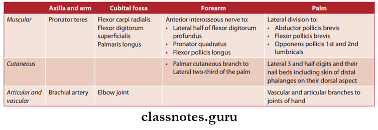
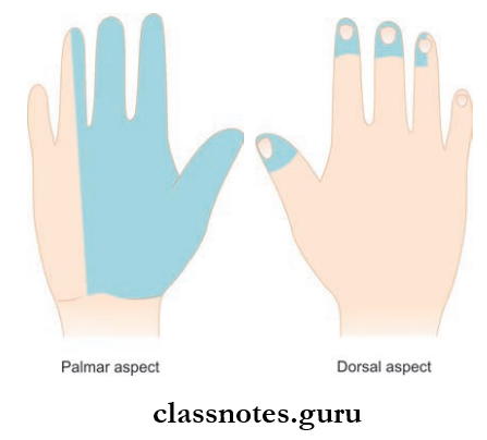
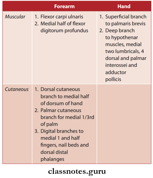
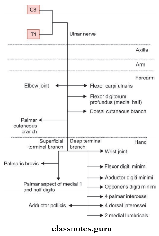
How Do Our Activities Affect The Environment?

Human beings are part ofthe environment. Our activities affect the environment. Changes in the environment affect us. Sahara and Thar deserts are the creation of excessive exploitation of resources by human beings.

By our activities, we are polluting air, water, and soil. Two of the major problems faced by humanity are ozone layer depletion and the disposal of waste we generate.

Ozone Layer And How Is It Getting Depleted?

How do our activities cause a depletion of the ozone layer?

Ozone (O3) is a triatomic molecule which is formed of 3 atoms of oxygen. Normal oxygen is diatomic (O2). It is essential for aerobic organisms. It is also essential for combustion or burning. However, ozone is a deadly poison. Luckily very little ozone is present in the lower part atmosphere or troposphere where we live.

A good quantity of ozone occurs in the upper part of the atmosphere called the stratosphere. Part of the stratosphere where ozone occurs in good quantity is called ozonosphere or ozone layer.

It lies at a height of 11-16 km above the poles and 23-25 km above the equator. As discovered by Molina (1992), ozone is formed in the stratosphere through the action of higher energy UV rays on oxygen.

Oxygen molecule splits up to form nascent or atomic oxygen [0]. Nascent or free oxygen atoms are highly reactive. They combine with diatomic normal oxygen molecules to form ozone molecules.

⇒ \(\begin{gathered}
\mathrm{O}_2 \xrightarrow{\mathrm{UV}}[\mathrm{O}]+[\mathrm{O}] \\
\mathrm{O}_2+[\mathrm{O}] \longrightarrow \mathrm{O}_3 \text { (ozone) }
\end{gathered}\)

Stratospheric ozone is highly protective. It filters out, rather than dissipates the energy of harmful UV radiations t 100 – 280 nm, UVB 280- 320 nm). UVA radiations (320- 390 nm) are allowed to pass down from the ozone layer an the earth.

They have some use in sun tanning and synthesis of vitamin D in the skin but excessive exposure is harmful to ozone Depletion. The amount of ozone present in the stratosphere began to drop sharply in the 1980s.

A big 0 thinned ozone layer was discovered by Farman et al (1985) over Antarctica. By 2000, it covered an area of 28.3 million km2. Ozone has also thinned out in other areas. Some Ozone Depleting Substances (ODS).

They are substances that react with ozone and destroy the same, common ozone-depleting substances are chlorofluorocarbons (CFCs) and halon. Out of them, chlorofluorocarbon is used as refrigerants, coolants, foam-producing agents, solvents for cleaning, aerosol spray, propellants, tuning, etc. Halon is used as a fire extinguisher.

ODS rise from the surface of the earth and reach the stratosphere. By the action of UV radiations, chlorofluorocarbon splits up to release chlorine. Chlorine reacts with ozone to form chlorine monoxide and oxygen.

Class 10 Biology Effect of Human Activities on the Environment

Chlorine monoxide breaks to release oxygen and chlorine. The released chlorine reacts with fresh molecules of ozone. It is estimated that a single atom of chlorine can destroy one hundred thousand ozone molecules. It can persist in the stratosphere for upto 100 years.

⇒ \(\begin{aligned}
\mathrm{CCl}_2 \mathrm{~F}_2 & \longrightarrow \mathrm{CCIF}_2+\mathrm{Cl} \\
2 \mathrm{Cl}^2+\mathrm{O}_3 & \longrightarrow \mathrm{Cl}_2 \mathrm{O}+\mathrm{O}_2 \\
2 \mathrm{Cl}_2 \mathrm{O} & \longrightarrow 4 \mathrm{Cl}+\mathrm{O}_2
\end{aligned}\)

It is estimated that a 1% reduction in the ozone layer increases the incidence of 2% more UV radiation reaching the earth.

Harmful Effects of Ozone Depletion.

  1. Skin Cancers. High-energy UV radiation reaching the earth will result in an increased number of skin cancers, skin aging, and herpes.
  2. Blinding. Cornea becomes swollen leading to what is called snow blindness. There will be an increased incidence of photobombing, dimming of eyesight, and cataracts.
  3. Immune System. It is weakened so that there is an increased incidence of diseases.
  4. Photosynthesis. It is reduced by 10-25%.
  5. Larval Deaths. They will increase especially in the aquatic animals.
  6. Mutations. The incidence of mutations shall increase. Most mutations are harmful.

International Efforts to Check Ozone Layer Damage

Montreal Protocol. In Montreal in 1987, UNEP (United Nations Environment Programme) succeeded in getting an agreement from industrialized nations to limit the production of chlorofluorocarbons to the level of 1986.

Helsinki Declaration. In 1989, it was agreed to phase out the production of CFCs by the end of the 20th century.

Beijing Conference. In 1999, a fund was created with the help of UNEP, UNDP, and World Bank to help developing countries to phase out ozone-depleting substances.

Kigali (Rwanda) Conference (2016). All countries have given a time frame for phasing out ODS.

Effect of International Efforts

In 2004, the ozone hole over Antarctica was 28.3 million km2. It decreased to 27.5 million km2 in 2006, 25.0 million km2 in 2011, 21.0 million km2 in 2013 and 19.1 million km2 in 2017. The data indicates that the health of the ozone layer is improving every year.

Activity 5.6 Study of Ozone Layer Depletion.

With the help of the internet, newspapers, and libraries prepare a list of ozone-depleting chemicals.

What efforts have been made to control the emission ofthese chemicals? Have they been replaced or phased out? Have the efforts to reduce the damage to the ozone layer borne any fruit? The collected data shows that efforts at the international level to phase out ODS have borne fruit.

The ozone hole over Antarctica which was 28.3 million km2 in 2000 A.D., was reduced to 19.1 million km2 in 2017.

Chlorofluorocarbons are being replaced by hydrofluorocarbons (HFCs) and Hydrochlorofluorocarbons (HCDFCs). They have shorter atmospheric life and deliver less active chlorine.

Managing the Garbage We Produce

What is Garbage? Suggest some measures to manage the garbage we produce. Garbage is solid waste from homes, vegetable markets, fruit markets, and food industries. Another source of garbage is disposables and packaging.

Disposables are used to check contamination. Decorative packagings are used to attract customers. Tourist spots are littered all around with such garbage.

It is estimated that an average Indian produces 0.5 kg of garbage per day while the higher group of individuals produces about 1.5 kg of waste. Garbage management is clean non-polluting and non-contaminating disposal of garbage.

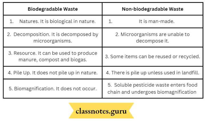
It consists of collection, transport, and disposal of garbage. Stress is being laid to segregate the garbage into biodegradable (green bins) and nonbiodegradable (blue bins) waste. In some localities, even recyclable articles are also separated (in red bins).

  1. Pig and Cattle Feeding. Pigs and stray cattle feed on waste food particles and reduce the bulk of garbage.
  2. Rag Pickers. They pick up recyclable articles like polythene, plastic, glass, metallic waste, paper, cardboard, rags, and e-waste. The articles are sold to factories for recycling.
  3. Composting. Biodegradable waste is used in the preparation of manure and compost.
  4. Biogas. Farm and cattle waste is used to generate biogas and prepare manure.
  5. Incineration. Hazardous wastes (For example hospital wastes) are burnt in chambers fitted with devices to control gaseous and particulate emissions. Ash and unbumt matter are used in landfilling.
  6. Land Filling. Solid nonbiodegradable waste is dumped in a low-lying area. It is pulverized by machines and covered with soil, lime, or bleaching powder.

Activity 5.7 Waste Disposal

Watch the collection of your domestic waste. Which agency (Panchayat, Municipal Corporation, or some resident welfare Association) is involved in the collection, transport, and disposal of waste?

Does segregation of biodegradable (green bins) and nonbiodegradable (blue bins) occur in your house? If not where is the segregation carried out? Find out the sites and methods of their disposal (biodegradable by composting and nonbiodegradable by dumping or landfilling).

Class 10 Biology Notes on Environmental Impact of Human Activities

Activity 5.8 Amount of Waste

Daily weigh the garbage generated in your home for about two weeks. Make it a point to weigh the biodegradable mid-nonbiodegradable wastes separately. Calculate the waste generated per person. Similarly weigh tire waste generated by your class daily for about 15 days. Weigh the biodegradable and nonbiodegradable waste separately. Suggest some methods of their disposal, Biodegradable for compost formation. Nonbiodegradable tomunicipal vans for disposal.

Activity 5.9 Sewage and Industrial Waste

Sewage. Study the sewage treatment in your locality, town, city, and village. Is there a regular sewage treatment plant (STP)? Where is wastewater passed? How are waterbodies ofthe area protected from contamination by raw sewage?

Industrial Waste. Industries generate a lot of waste. Some of the wastes are recyclable. They are sent for recycling. Others are toxic and harmful to the environment. Find out modes of checking pollution by at least one industry. How are the soil and water bodies protected from pollution?

Disposable Cups

In older times tea was served in trains in reusable glasses. The problems of cleanliness and contamination are always there. They were solved with the introduction of disposable plastic cups. However, used cups could not be burnt as they would emit toxic fumes.

There is not enough space to bury millions of cups daily. The railway ministry then introduced burnt clay cups or hills as they could be broken and pulverized.

However, the manufacture of a very large number of kulaks and their transport posed a big problem. The use of fertile topsoil left many fields barren. The practice was, therefore, discontinued. Soon disposable paper cups became available.

They were the best option as used paper cups could be recycled, burnt down, and allowed to decompose. The present trend is now to use disposable plates, spoons, glasses, etc. at parties, religious functions, marriages, etc.

These functions also generate a lot of leftover food. Proper and hygienic disposal of these items causes a minimum adverse impact on the environment.

NCERT Class 10 Biology Human Impact on Environment

Activity 5.10 e-waste

e-waste is discarded electrical and electronic devices. Search the internet and library to prepare a list of commonly produced e-waste. Find out how these items are broken down to obtain useful articles.

Also find out the hazardous or toxic substances, a worker engaged in e-waste breakdown comes in contact with, for example, nickel, arsenic, cadmium, mercury, selenium, chromium, silicon, lead, beryllium, etc.

Plastic Recycling. Polythene and plastic can be recycled. However, the process of recycling involves the release of hydrocarbons and cancer-causing dioxin.

Class 10 Biology Notes For Platelets In Human Body

Transportation in Human Beings

Human beings have a circulatory system for transportation. It consists of a pumping heart, circulatory fluids blood and lymph and tubes. Depending upon the fluid being circulated, the circulatory system is of two types, blood blood-vascular system and the lymphatic system. The blood-vascular system comprises blood, blood vessels and the heart.

Blood

What is a fluid connective tissue? Name its different components.

It is a reddish, opaque, slightly alkaline fluid connective tissue that is constantly circulating in the body. An adult human has 5-6 litres of blood. Blood consists of two parts, plasma and blood cells.

  1. Plasma. It is a pale yellow, fluid matrix of blood which constitutes 55% of it. It is slightly alkaline (pH 7.4). 92% of plasma is water. The rest 8% are proteins, waste products, nutrients, hormones, inorganic salts and anticoagulant heparin.
  2. Blood Cells. They form 45% of blood. Blood cells are of three types— red blood corpuscles, white blood cells and blood platelets.

Red Blood Corpuscles (RBOs). They are biconcave, circular, enucleate cells of 7 pm in diameter, 2 pm in thickness on the sides and put in the centre, Most of the cell organelles are absent. This is a specialisation to contain a maximum number of reddish respiratory pigments called haemoglobin. Its content is 14 -lo mg 100 n\L of blood in adult human males and 12-10 mg/mL of blood in adult human females.

The number of RBCs is 5 – 5.5 million/ml. in males and 4.5 – 5.0 million/ml. of blood in females. Old RBCs are destroyed in the spleen and liver while new ones are formed in red bone marrow.

White Blood Corpuscles (WBCs). They are colourless, rounded to irregular, nucleated blood corpuscles with a size of 8 -15 pm. The number is 6000-8000/ mL of blood. White blood corpuscles are of two types, granulocytes and agranulocytes. Granulocytes possess granules and lobulated nuclei. They are of 3 kinds— eosinophils, basophils and neutrophils.

Transportation Types Of Blood Cells

Agmnulocytes have granules of cytoplasm and a non-lobulated nucleus. They are of two kinds, monocytes and lymphocytes. The major function of WBC is ingestion of germs, secretion of antibodies and heparin.

Class 10 Biology Platelets in Human Body

Blood Platelets. They are non-nucleated, colourless cells of 2- 3 pm which possess different shapes due to their formation from cell fragments in red bone marrow. The number is 1,45,000 to 4,50,000/ mL of blood. Blood platelets take part in blood clotting and sealing the area of injury.

Blood Functions :

  1.  Transport of Gases. Both oxygen and carbon dioxide are carried by blood from the area of availability to the area of their release, that is, respiratory surface to tissues and vice-versa.
  2.  Transport of Nutrients. They are taken by blood from the intestine and passed to different parts of the body for absorption, assimilation and storage.
  3. Transport of Waste Products. Nitrogenous wastes formed by the liver are taken by the blood to the kidneys for separation and later elimination.
  4. Hormones. Hormones reach the target sites through circulating blood.
  5.  Heat. By its circulation, blood distributes heat uniformly in the whole body.
  6. Maintenance of Water Balance. The blood maintains a favourable water and ion balance by providing the same in the area of deficiency and removing them from the area of excess.
  7. Protection. Components of blood protect the body from pathogens and plug the area of injury.

Collection Of Data On Haemoglobin

Visit a clinical laboratory or a Health Centre. Collect the data on the haemoglobin content of adult males, adult females, adolescents and children. It will range between :

1. Adult human male: 14-16 mg/100 mL

2. Adult human female: 12-13 mg/100 mL

3. Adolescents : 12-13 mg/100 mL

4. Children: 11 -12 mg/100 mL

5. Infants : 11 mg/100 mL

You can compare the values with the values obtained in your locality or any other place.

Now visit a veterinary clinic and obtain data on the haemoglobin content of the cow, bull/bullock and calves. The data range is:

Cow : 11 – 12mg/100 mL

Bull: 13 -14 mg/100 mL

Calf: 10-11 mg/100 mL

Comparison of haemoglobin content of different age groups and sexes of humans and cattle indicates that there is not much difference between them.

NCERT Class 10 Biology Platelets and Their Functions

Our Pump: The Heart Heart is a conical muscular double pump structure that lies ventrally in the thoracic cavity in between the two lungs. It is reddish in colour. The size is 12cm long and 4cm broad.

Transportation External View Of Heart 1

  • The broader end is towards the upper side while the pointed end rests over the diaphragm. The tip belongs to the left ventricle. This gives the feeling of the heart being on the left side.
  • The heart is covered by a thin fluid-filled sac called pericardium. This provides protection from shock and allows frictionless movement of the heart. The heart has a coronary sulcus that separates the thin-walled upper auricular region from the lower thick-walled ventricular region.
  • Both regions have vertical septa to divide the auricular region into two atria (= auricles) and the ventricular region into two ventricles. The arrangement is such that oxygenated blood remains separate from the deoxygenated blood.
  • The two auricles are small and thin-walled while the two ventricles are larger and thick-walled. The left ventricle is bigger and thicker than the right ventricle.
  • The right auricle or atrium receives deoxygenated blood from a prenatal (superior vena cava), a post caval (inferior vena cava) and a coronary sinus. The left auricle or atrium receives oxygenated blood from two pairs of pulmonary veins. The right auricle opens into the right ventricle through a right auriculo-ventricular aperture.
  • It is guarded by a tricuspid valve supported by inelastic cords called chordae tendineae present in the right ventricle. The left auricle opens into the left ventricle through a left auriculo-ventricular aperture guarded by a bicuspid or mitral valve supported by chordae tendineae based in the left ventricle.
  • The right ventricle opens into a pulmonary trunk through an opening guarded by a semilunar pulmonary valve. The left ventricle opens into the aorta by an opening guarded by a semilunar aortic valve.

Platelets Function in Human Body Class 10 Biology

Working. Deoxygenated blood from the whole body enters the right atrium. After getting filled, the right auricle contracts and pushes the blood into the right ventricle. As the right ventricle gets filled, it undergoes systole or contraction to push the blood into the pulmonary trunk through the semilunar pulmonary valve.

Transportation Sectional View Of Human Heart`

  • Blood does not go back into the auricle due to the straightening of the tricuspid valve. The pulmonary trunk divides into two pulmonary arteries. They pass into the lungs. Here blood is oxygenated. The oxygenated blood then moves into the left auricle by means of two pulmonary veins from each lung.
  • On getting filled, the left auricle contracts and pushes the oxygenated blood into the left ventricle. The left ventricle contracts upon getting filled and pumps the blood into the aorta for supply to the whole body except the lungs. Reverse flow is not possible due to the presence of valves.
  • The occurrence of four-chambered heart and partitions to separate deoxygenated blood on the right and oxygenated blood on the left side ensures :
  • Complete oxygenation of deoxygenated blood and Supply of only oxygenated blood to all body parts for obtaining maximum energy. This is important for all mammals and birds which spend a good amount of energy for keeping their body warm at constant temperature.
  • However, there is no such requirement for reptiles and amphibians. Here the temperature varies with the temperature of the environment. The heart is three chambered in them. Some mixing of oxygenated and deoxygenated blood occurs.
  • Mixed blood produces lesser energy due to reduced oxygen supply. The efficiency is further decreased in fishes with two-chambered venous hearts. It pumps blood to the gills for oxygenation.
  • The oxygenated blood passes into body parts but is deoxygenated along the pathway so that a very reduced oxygen supply occurs in many parts.

Immunology