Ulnar Nerve Anatomy Notes PDF
Explain in detail about the ulnar nerve under headings— origin, root value, course, branches, and innervation. Write a note on the injury to the nerve.
Answer:
- The Ulnar Nerve runs along the ulnar side of the upper limb, hence the name.
- Ulnar Nerve is the nerve of fine movement and is called the musician’s nerve.
Ulnar Nerve Origin
- Medial cord of brachial plexus.
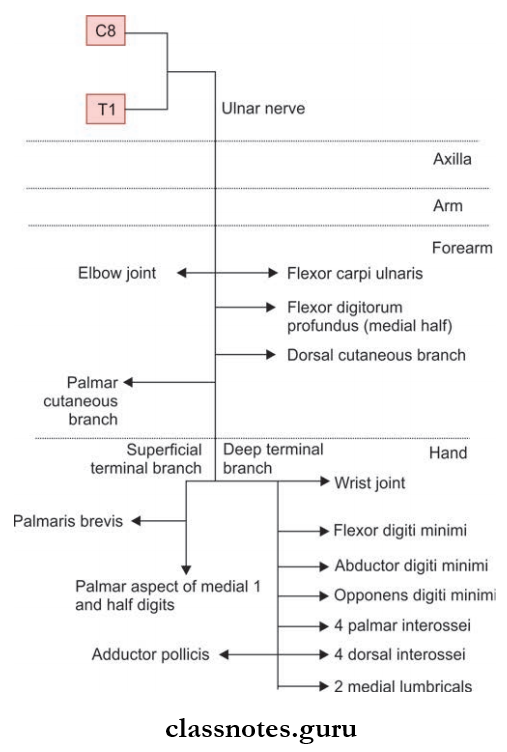
Ulnar Nerve Root Value
- C8 and T1 spinal segments mainly. C7 also contributes.
Upper Limb Anatomy – Ulnar Nerve Explained
Ulnar Nerve Course
- In the axilla, Ulnar nerve lies medial to the third part of the brachial artery.
- In the arm:
- It enters the arm along the medial side of the brachial artery and runs up to the level of the mid-arm.
- The ulnar nerve pierces the medial intermuscular septum to reach the back of the arm.
- From there, the ulnar nerve descends to pass through the cubital tunnel formed by the medial epicondyle and the fibrous band extending from the medial epicondyle to the olecranon process.
- The ulnar nerve has no branches in the axilla as well as in the arm.
- In the forearm:
- In the upper 1/3rd, the ulnar nerve passes between the two heads of flexor carpi ulnaris to reach underneath the muscle and runs vertically down.
- In the lower 2/3rd, it becomes superficial and runs downwards along with the ulnar artery being on the lateral side.
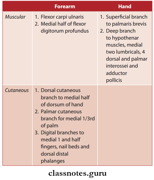
- Branches of the ulnar artery in the forearm are:
- Proximal forearm Muscular branch:
- Flexor carpi ulnaris
- Medial half of flxor digitorum profundus
- Mid forearm
- Cutaneous branch: Palmar cutaneous branch
- Distal forearm
- Cutaneous branch: Dorsal cutaneous branch

Ulnar Nerve In the Hand
- The ulnar nerve enters the hand superficial to the carpal tunnel through the ulnar tunnel and divides into superficial and deep terminal branches.
- The superficial branch gives sensory supply to the palmar aspect of the medial ½ digits and motor supply to the palmaris brevis muscle.
- Deep branches give off
- Articular branches to the wrist
- Muscular branches to:
- 2 medial lumbricals
- 4 palmar interossei
- 4 dorsal interossei
- Flexor digiti minimi (hypothenar muscle)
- Abductor digiti minimi (hypothenar muscle)
- Opponents digiti minimi (hypothenar muscle)
- Adductor pollicis (thenar muscle)
Ulnar Nerve Branches and Supply
Ulnar Nerve Clinical Anatomy: Injury to the ulnar nerve can occur in the elbow or wrist.
- Injury To The Ulnar Nerve At The Elbow
- Common causes are:
- Compression of the nerve in between the two heads of flexor carpi ulnaris
- Fracture and dislocation of medial epicondyle
- Valgus deformity of the elbow
- Thickening of the fibrous roof of the cubital tunnel (cubital tunnel syndrome).
- Common causes are:
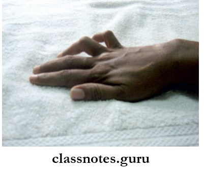
Ulnar Nerve Injury and Claw Hand – Essay
- Claw hand deformity affecting ring and little fingers where the metacarpophalangeal joints are extended while the interphalangeal joints are fixed (it’s not a complete claw hand, complete claw hand is seen when both the ulnar nerve and medial nerve are injured simultaneously, resulting in the hyperextended wrist and metacarpophalangeal joints and fixed interphalangeal joints).
- Flattening of the hypothenar eminence due to atrophy.
- Adduction and abduction of figers are affcted.
- Thmp can not be adducted.
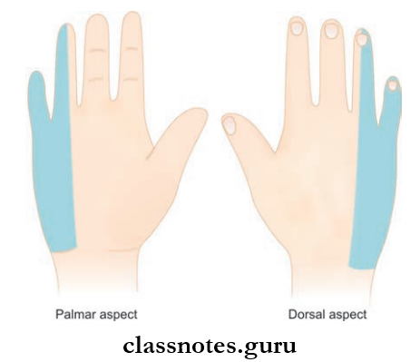
- Loss of sensation over the palmar and dorsal surface of the medial 1/3rd of the hand and medial 1-and-a-half
- fingers
- Foments sign will be positive when the integrity of the palmar interossei is tested.
- The ulnar nerve can also get injured in the wrist. Here, the clawing is more pronounced known as the ulnar paradox.
Ulnar Nerve MCQs with Answers