
Transportation in Human Beings
Human beings have a circulatory system for transportation. It consists of a pumping heart, circulatory fluids blood and lymph and tubes. Depending upon the fluid being circulated, the circulatory system is of two types, blood blood-vascular system and the lymphatic system. The blood-vascular system comprises blood, blood vessels and the heart.
Blood
What is a fluid connective tissue? Name its different components.
It is a reddish, opaque, slightly alkaline fluid connective tissue that is constantly circulating in the body. An adult human has 5-6 litres of blood. Blood consists of two parts, plasma and blood cells.
- Plasma. It is a pale yellow, fluid matrix of blood which constitutes 55% of it. It is slightly alkaline (pH 7.4). 92% of plasma is water. The rest 8% are proteins, waste products, nutrients, hormones, inorganic salts and anticoagulant heparin.
- Blood Cells. They form 45% of blood. Blood cells are of three types— red blood corpuscles, white blood cells and blood platelets.
Red Blood Corpuscles (RBOs). They are biconcave, circular, enucleate cells of 7 pm in diameter, 2 pm in thickness on the sides and put in the centre, Most of the cell organelles are absent. This is a specialisation to contain a maximum number of reddish respiratory pigments called haemoglobin. Its content is 14 -lo mg 100 n\L of blood in adult human males and 12-10 mg/mL of blood in adult human females.
The number of RBCs is 5 – 5.5 million/ml. in males and 4.5 – 5.0 million/ml. of blood in females. Old RBCs are destroyed in the spleen and liver while new ones are formed in red bone marrow.
White Blood Corpuscles (WBCs). They are colourless, rounded to irregular, nucleated blood corpuscles with a size of 8 -15 pm. The number is 6000-8000/ mL of blood. White blood corpuscles are of two types, granulocytes and agranulocytes. Granulocytes possess granules and lobulated nuclei. They are of 3 kinds— eosinophils, basophils and neutrophils.
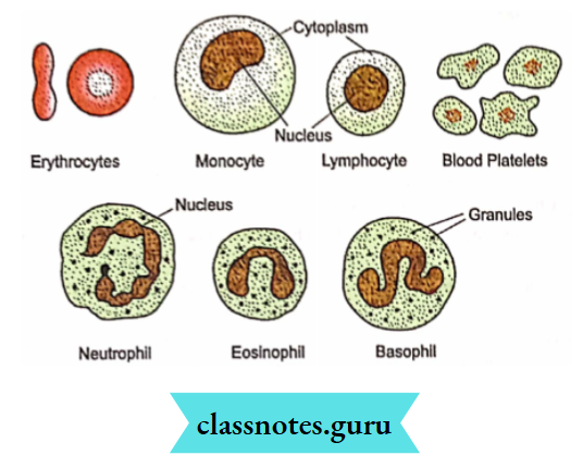
Agmnulocytes have granules of cytoplasm and a non-lobulated nucleus. They are of two kinds, monocytes and lymphocytes. The major function of WBC is ingestion of germs, secretion of antibodies and heparin.
Class 10 Biology Platelets in Human Body
Blood Platelets. They are non-nucleated, colourless cells of 2- 3 pm which possess different shapes due to their formation from cell fragments in red bone marrow. The number is 1,45,000 to 4,50,000/ mL of blood. Blood platelets take part in blood clotting and sealing the area of injury.
Blood Functions :
- Transport of Gases. Both oxygen and carbon dioxide are carried by blood from the area of availability to the area of their release, that is, respiratory surface to tissues and vice-versa.
- Transport of Nutrients. They are taken by blood from the intestine and passed to different parts of the body for absorption, assimilation and storage.
- Transport of Waste Products. Nitrogenous wastes formed by the liver are taken by the blood to the kidneys for separation and later elimination.
- Hormones. Hormones reach the target sites through circulating blood.
- Heat. By its circulation, blood distributes heat uniformly in the whole body.
- Maintenance of Water Balance. The blood maintains a favourable water and ion balance by providing the same in the area of deficiency and removing them from the area of excess.
- Protection. Components of blood protect the body from pathogens and plug the area of injury.
Collection Of Data On Haemoglobin
Visit a clinical laboratory or a Health Centre. Collect the data on the haemoglobin content of adult males, adult females, adolescents and children. It will range between :
1. Adult human male: 14-16 mg/100 mL
2. Adult human female: 12-13 mg/100 mL
3. Adolescents : 12-13 mg/100 mL
4. Children: 11 -12 mg/100 mL
5. Infants : 11 mg/100 mL
You can compare the values with the values obtained in your locality or any other place.
Now visit a veterinary clinic and obtain data on the haemoglobin content of the cow, bull/bullock and calves. The data range is:
Cow : 11 – 12mg/100 mL
Bull: 13 -14 mg/100 mL
Calf: 10-11 mg/100 mL
Comparison of haemoglobin content of different age groups and sexes of humans and cattle indicates that there is not much difference between them.
NCERT Class 10 Biology Platelets and Their Functions
Our Pump: The Heart Heart is a conical muscular double pump structure that lies ventrally in the thoracic cavity in between the two lungs. It is reddish in colour. The size is 12cm long and 4cm broad.
- The broader end is towards the upper side while the pointed end rests over the diaphragm. The tip belongs to the left ventricle. This gives the feeling of the heart being on the left side.
- The heart is covered by a thin fluid-filled sac called pericardium. This provides protection from shock and allows frictionless movement of the heart. The heart has a coronary sulcus that separates the thin-walled upper auricular region from the lower thick-walled ventricular region.
- Both regions have vertical septa to divide the auricular region into two atria (= auricles) and the ventricular region into two ventricles. The arrangement is such that oxygenated blood remains separate from the deoxygenated blood.
- The two auricles are small and thin-walled while the two ventricles are larger and thick-walled. The left ventricle is bigger and thicker than the right ventricle.
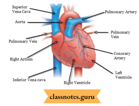
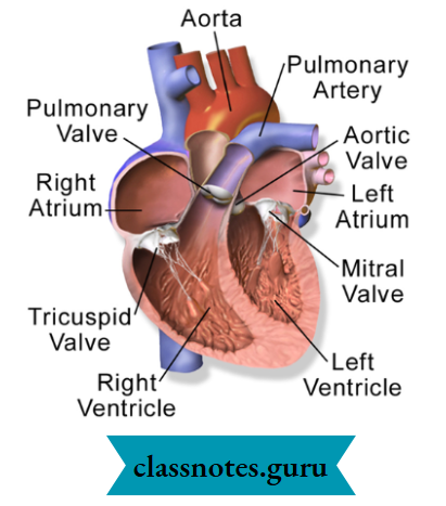
- The right auricle or atrium receives deoxygenated blood from a prenatal (superior vena cava), a post caval (inferior vena cava) and a coronary sinus. The left auricle or atrium receives oxygenated blood from two pairs of pulmonary veins. The right auricle opens into the right ventricle through a right auriculo-ventricular aperture.
- It is guarded by a tricuspid valve supported by inelastic cords called chordae tendineae present in the right ventricle. The left auricle opens into the left ventricle through a left auriculo-ventricular aperture guarded by a bicuspid or mitral valve supported by chordae tendineae based in the left ventricle.
- The right ventricle opens into a pulmonary trunk through an opening guarded by a semilunar pulmonary valve. The left ventricle opens into the aorta by an opening guarded by a semilunar aortic valve.
Platelets Function in Human Body Class 10 Biology
Working. Deoxygenated blood from the whole body enters the right atrium. After getting filled, the right auricle contracts and pushes the blood into the right ventricle. As the right ventricle gets filled, it undergoes systole or contraction to push the blood into the pulmonary trunk through the semilunar pulmonary valve.
 `
`
- Blood does not go back into the auricle due to the straightening of the tricuspid valve. The pulmonary trunk divides into two pulmonary arteries. They pass into the lungs. Here blood is oxygenated. The oxygenated blood then moves into the left auricle by means of two pulmonary veins from each lung.
- On getting filled, the left auricle contracts and pushes the oxygenated blood into the left ventricle. The left ventricle contracts upon getting filled and pumps the blood into the aorta for supply to the whole body except the lungs. Reverse flow is not possible due to the presence of valves.
- The occurrence of four-chambered heart and partitions to separate deoxygenated blood on the right and oxygenated blood on the left side ensures :
- Complete oxygenation of deoxygenated blood and Supply of only oxygenated blood to all body parts for obtaining maximum energy. This is important for all mammals and birds which spend a good amount of energy for keeping their body warm at constant temperature.
- However, there is no such requirement for reptiles and amphibians. Here the temperature varies with the temperature of the environment. The heart is three chambered in them. Some mixing of oxygenated and deoxygenated blood occurs.
- Mixed blood produces lesser energy due to reduced oxygen supply. The efficiency is further decreased in fishes with two-chambered venous hearts. It pumps blood to the gills for oxygenation.
- The oxygenated blood passes into body parts but is deoxygenated along the pathway so that a very reduced oxygen supply occurs in many parts.