
- Antibodies Immunoglobulins Short And Long Essays Question And Answers
- Antigen Antibody Reaction Short Question And Answers
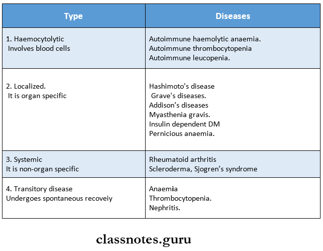
- Autoimmunity Immunology Short Question And Answers
- Immune Response Short Essay Question And Answers
- Immunity Short And Long Essays Question And Answers
- Immunology Complement System Short Essays Question And Answers
- Immunology Hypersensitivity Short Question And Answers
- Immunology Transplantation Short Question And Answers
- Structure And Functions Of Immune System Short Question And Answers
Immunology
Immunology Transplantation Short Question And Answers
Transplantation Short Question And Answers
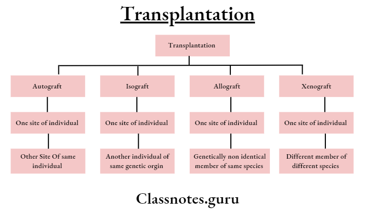
Question 1. Types of transplants.
Answer:
Types Of Transplants
Question 2. Allograft reaction.
Answer:
Allograft Reaction
Rejection of the graft by the recipient is called an allograft reaction.
Read And Learn More: Microbiology Question and Answers
Tissue Responses:
1. First Set Response.
- When allograft is applied following changes occur.
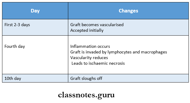
- This sequence of events is called the first set response.
2. Second Set Response.
- Second Set Response occurs when a second allograft from the same donor is applied to the same recipient.
- During this graft is rejected in an accelerated fashion.
- Early necrosis occurs and the graft sloughs off by the sixth day.
- This accelerated rejection is called the second set response.
Question 3. Graft-versus. Host reaction.
Answer:
Graft-Versus. Host reaction
The graft may cause an immune response against the antigens of the host.
- This is known as the graft-versus-host reaction.
- Graft-Versus. Host reaction is a cell-mediated reaction.
Graft-Versus Conditions:
- It occurs in the following conditions.
- Graft containing immune competent T-lymphocytes.
- HLA antigens of the recipient are different from the graft.
- Destroyed or impaired recipient’s immunological response.
Graft-Versus Manifestations:
- Splenomegaly
- Fever
- Rash
- Anaemia.
- Weight loss.
- Rarely death occurs.
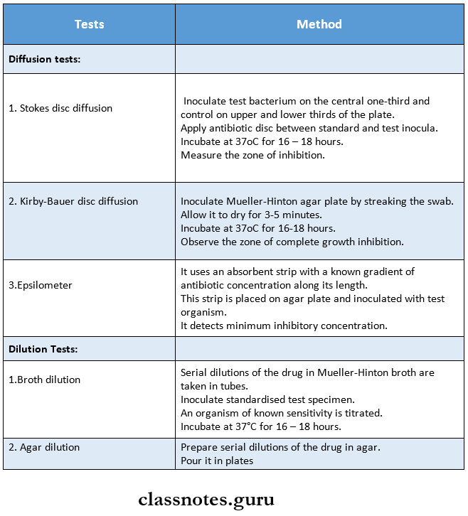
Question 4. Antibiogram.
Answer:
Antibiogram
- The overall profile of antimicrobial susceptibility is known as an antibiogram.
- An antibiogramis a chart produced by clinical laboratories which detect the percentage of microbial isolates that are sensitive to particular antibiotics.
- Every 6-12 months the hospitals typically generate antibiograms.
Antibiogram Uses:
- Guide the doctors in antibiotic selection.
- Helps to monitor resistance patterns throughout a region.
- Enables to compare patterns across regions and with other regions.
Transplantation Viva Voce
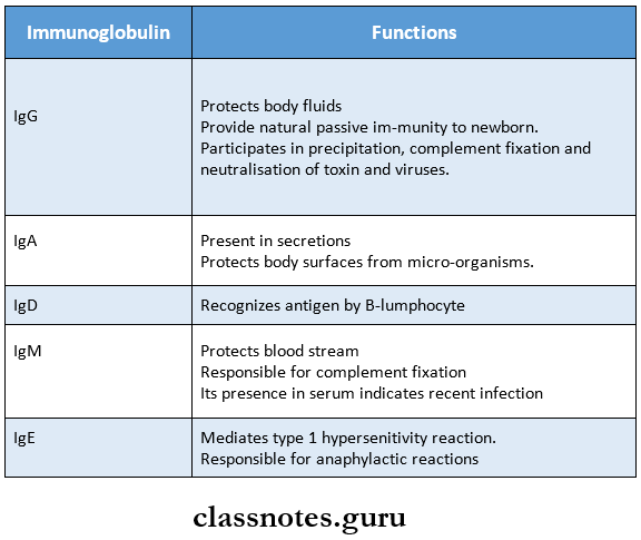
- IgG is the only immunoglobulin that crosses the placenta
- IgA is predominant immunoglobulin in saliva
- IgA and IgG are secreted in milk
- IgG protects body fluids
- IgA protects body surfaces
- IgM protects bloodstream
- T and B lymphocytes, plasma cells, and antigen-presenting cells are primarily concerned with immune response
- Plasma cells are antibody-secreting cells
- Dendritic cells and macrophage act as antigen-presenting cells
- Most antibodies are produced in the spleen and lymph nodes
- Lysozyme is present in tears, eggs, saliva, and nearly in all secretions except in CSF, sweat, and urine
- BGG is given by intradermal route
- C3a and C5a are anaphylactic and chemotactic
- Smallest unit of antigenicity is known as epitope
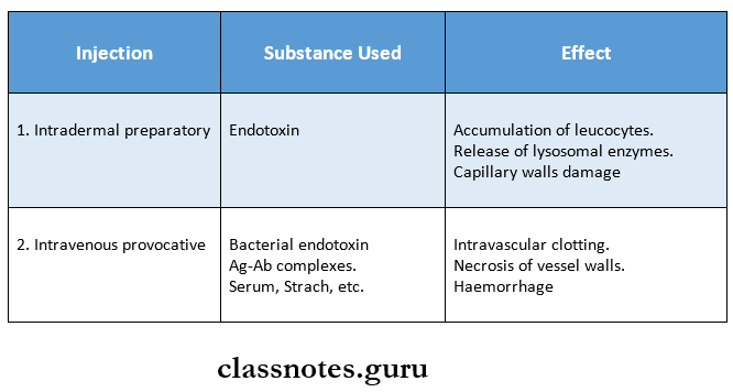
- In serum sickness, a single dose of injection can serve both as a sensitizing and shocking dose
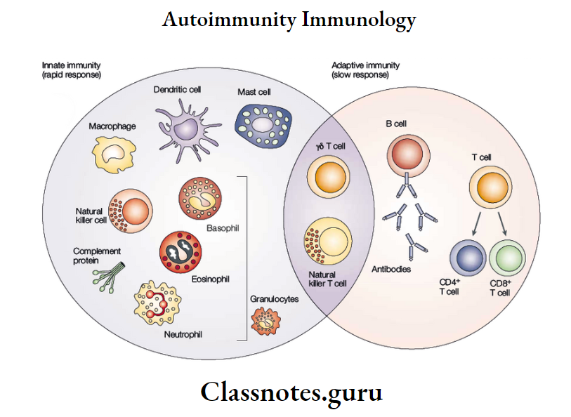
Autoimmunity Definition and Mechanisms
Autoimmunity
Autoimmunity Definition
- Autoimmunity is a condition in which structural or functional damage is produced by the action of immunologically competent cells or antibodies against normal components of the body.
- Autoimmunity is a state in which the body’s immune system fails to distinguish between self & non-self & reacts by the formation of autoantibodies against one’s own tissues’ antigens.
Read And Learn More: Microbiology Question and Answers
Autoimmune diseases
Autoimmunity Mechanisms
Autoimmunity can occur due to the following factors.
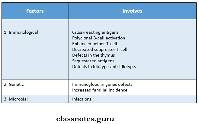
Mechanisms of autoimmunity
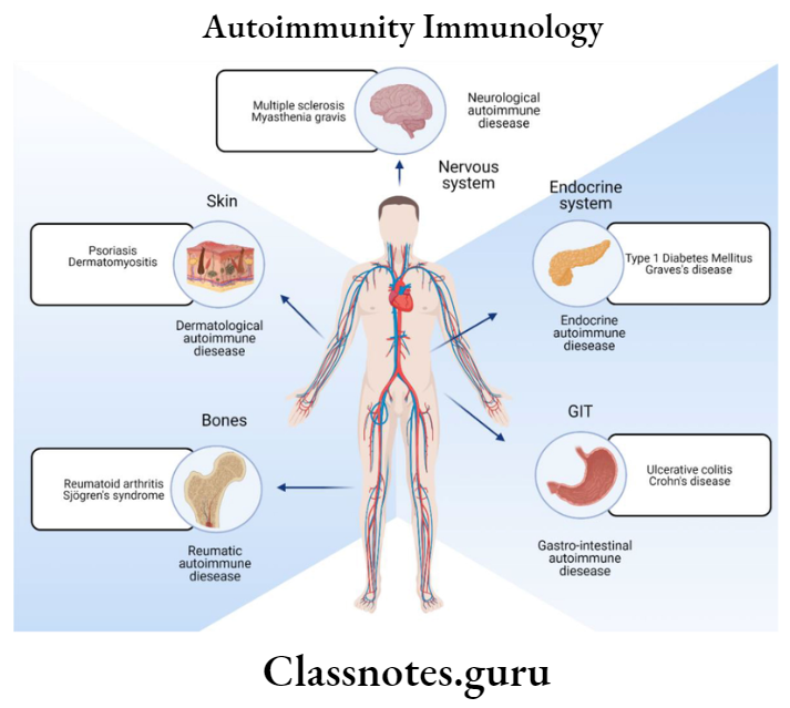
Autoimmune Diseases
Depending upon the site of involvement and nature of lesions, autoimmune diseases are classified as follows.
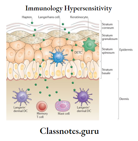
Immunology Hypersensitivity Short Question And Answers
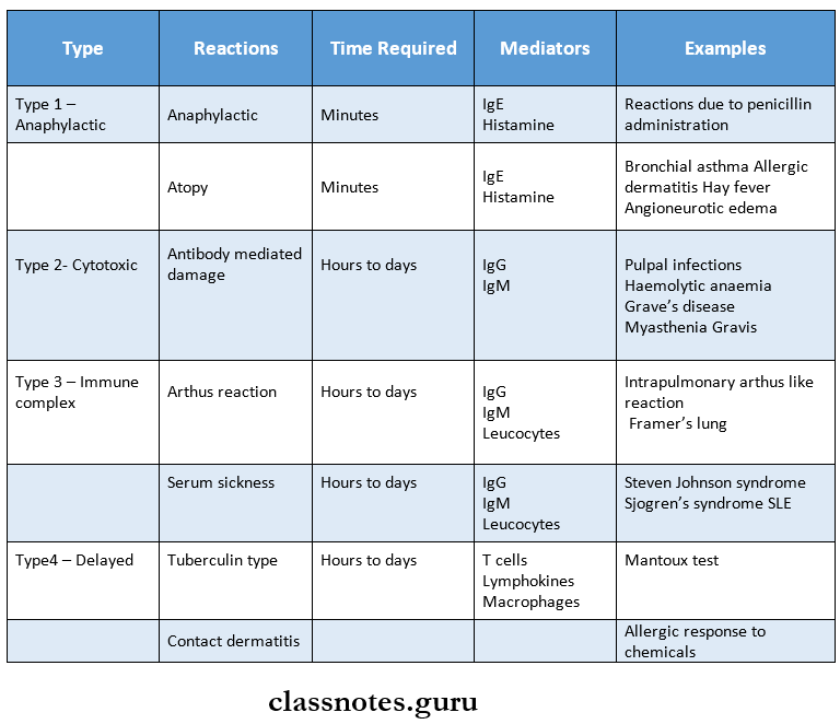
Hypersensitivity
Question 1. Define and classify hypersensitivity reactions. Describe in detail the principle and mechanism of hypersensitivity reaction.
Answer:
Hypersensitivity Reaction Definition:
Hypersensitivity refers to a condition in which an immune response results in excessive reactions leading to tissue damage, diseases, or even death in the sensitized host.
Hypersensitivity Reaction Classification:
- Based on the time required for the immune response.
- Immediate hypersensitivity.
- Delayed hypersensitivity.
- Coomb and gel classification.
- Type 1 – Anaphylactic reaction.
- Type 2 – Cytotoxic reaction.
- Type 3 – Immune complex.
- Type 4 – Delayed or cell-mediated.
Read And Learn More: Microbiology Question and Answers
Hypersensitivity Reaction Principle And Mechanism
1. Type 1 Reaction.
- Cytotropic IgE antibody is the major antibody responsible for it.
- This antibody is induced by an allergen and thus binds to mast cells and basophils.
- When exposed to the allergen again, the bound IgE is cross-linked.
- This induces degranulation and release mediators like histamine.
2. Type 2 – Reaction.
- Antigens on a cell surface combine with antibodies.
- This leads to complement-mediated lysis.
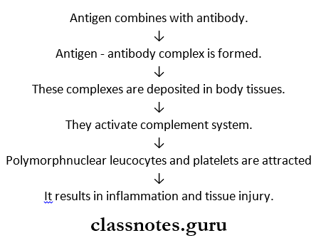
3. Type 3 Reaction.
- Antigen-antibody immune complexes are deposited in tissues.
- By this, the complement is activated and polymorphonuclear.
- As a result, they release lysosomal enzymes. Causing tissue damage.
Short notes on hypersensitivity
4. Type 4 Reaction.
- Helper T lymphocytes sensitized by antigen release lymphokines upon second contact with the same antigen.
- This lymphokine induced inflammation.
Anaphylaxis
- Anaphylaxis is the typical classical immediate hypersensitivity reaction.
- The term ‘anaphylaxis’ was coined by Richet in 1902.
- Anaphylaxis is a type of IgE-mediated hypersensitivity reaction, which develops quickly after the introduction of a large shocking dose of antigen following one or more small sensitizing doses.
Anaphylaxis Mechanism
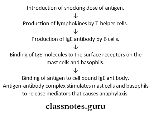
Anaphylaxis Types
- Anaphylaxis in vitro.
- Cutaneous anaphylaxis.
Anaphylaxis Features
- The lung is the principal shock organ.
- Bronchospasm.
- Laryngeal edema.
- Respiratory distress.
- Shock
- Death.
Hypersensitivity Definition
Hypersensitivity refers to a condition in which immune response results in excessive reactions leading to tissue damage, diseases, or even death in the sensitized host.
Type 3 – Hypersensitivity:
- Type 3 – Hypersensitivity is also called an immune complex or toxic complex disease.
- Type 3 – Hypersensitivity is a type of humoral antibody-mediated hypersensitivity reaction.
- In it, tissue damage is caused by antigen-antibody complexes.
Hypersensitivity Pathogenesis
Hypersensitivity Types
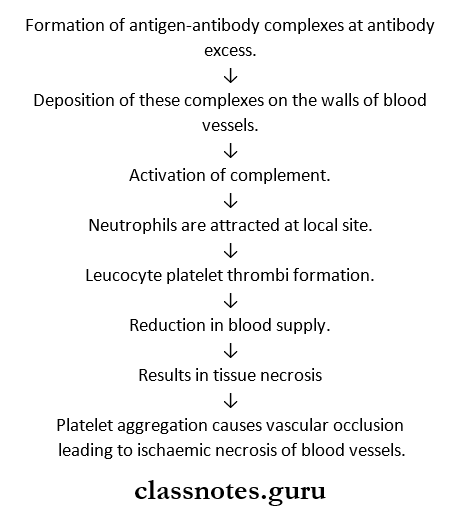
1. Arthus Reactions – Localized.
- Arthus Reactions – Localized results in intense local edema and hemorrhage in persons with repeated administration of the same antigen.
2. Serum Sickness – Generalized.
- Serum Sickness – Generalized appears in 7 – 12 days following administration of a high concentration of antigen.
- Its clinical features include.
- Fever
- Lymphadenopathy.
- Splenomegaly
- Arthritis.
- Glomerulonephritis.
- Endocarditis.
- Vasculitis.
- Urticarial rashes -Abdominal pain.
- Nausea and vomiting.
Type 1 Hypersensitivity Reaction
- An anaphylactic reaction is a type 1 hypersensitivity reaction.
- It is B -a cell-mediated immediate type of hypersensitivity
Type 1 Hypersensitivity Reaction Forms:
1. Anaphylaxis
- Acute
- Potentially fatal
- Systemic form
2. Atopy
- Recurrent
- Non-fata
- Localized form.
Hypersensitivity MCQs
Type 2 Hypersensitivity Reaction
- The cytotoxic reaction is a type 2 hypersensitivity reaction.
- Type 2 Hypersensitivity Reaction is an IgG antibody-mediated reaction.
- Type 2 Hypersensitivity Reaction is intermediate between hypersensitivity and autoimmunity.
Type 2 Hypersensitivity Reaction Pathogenesis
- IgG antibodies bind to an antigen on the cell surface.
- This causes.
- Phagocytosis of the cell.
- Cytotoxicity by natural killer cells.
- Cell lysis.
- In some reactions, the antibody in combination with the cell surface receptors disrupts the normal functioning of the cell.
- Uncontrolled activation.
- Blocking of receptors.
- Examples:
- Autoimmune anemias
- Hemolytic disease
- Drug reactions.
Delayed hypersensitivity or type 4 reaction
Delayed Hypersensitivity
- Delayed hypersensitivity reaction is mediated by sensitized T – T-lymphocytes but not by antibodies.
- Thus, it cannot be passively transferred by serum but can be transferred by lymphocytes or the transfer factor.
Delayed Hypersensitivity Pathogenesis
- Type 4 reaction occurs within 48 – 72 hours of antigen contact.
- Delayed Hypersensitivity Pathogenesis is provoked by intracellular infections or haptens – like simple chemicals applied on the skin.
- These sensitize T-lymphocytes.
- Sensitized T-lymphocytes on contact with specific antigen release lymphokines.
- This causes biological effects on macrophages, leucocytes, and tissue cells.
Delayed Hypersensitivity Types
- Tuberculin type.
- Contact dermatitis type.
- Examples:
- Tuberculosis.
- Typhoid fever
- Allergic contact dermatitis.
- Graft rejection.
Delayed hypersensitivity
Immediate Hypersensitivity Reaction
Based on the time required for a sensitized host to develop clinical reactions on re-exposure to the antigen, hypersensitivity is classified into.
- Immediate hypersensitivity.
- Delayed hypersensitivity.
1. Immediate Hypersensitivity:
- Immediate Hypersensitivity is a B-cell or antibody-mediated reaction.
- Immediate Hypersensitivity appears and recedes rapidly.
- Immediate Hypersensitivity is passively by antigens or haptens.
- Immediate Hypersensitivity is subdivided into the following clinical types.
- Anaphylaxis.
- Atopy
- Antibody-mediated cell damage.
- Arthus phenomenon.
- Serum sickness.
Atopy
Atopy is a form of type 1 hypersensitivity.
- The antigens commonly involved in it are pollens, house dust, and foods.
- These induce IgE antibodies.
Atopy Features:
- Atopy shows marked familial distribution.
- Atopy sensitization is developed spontaneously following natural contact with opens.
- Reactions occur at the site of entry of antigen.
- For example.
- Inhalation of pollens affects the lungs.
- Contact leads to local allergy.
Atopy Manifestations:
- Conjunctivitis.
- Rhinitis.
- Bronchospasm
- GI symptoms.
- Dermatitis.
- Cutaneous eruptions.
Immune system hypersensitivity
Arthus Reaction
Arthus reaction is a localized form of type 3 hypersensitivity reaction.
- When an antigen is injected subcutaneously or intradermally in an animal in whom there are repeated administrations of the same antigen previously, there occurs intense local edema and hemorrhage which reaches a peak in 3 – 6 hours, this is called the Arthus reaction.
Arthus Reaction Mechanism:
Serum Sickness
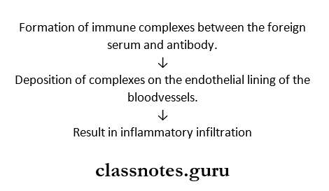
Serum Sickness is a systemic form of type 3 hypersensitivity that appears 7-12 days following a single injection of a high concentration of foreign serum such as diphtheria anti-toxin.
Serum Sickness Pathogenesis:
Tuberculin reaction
Tuberculin reaction is a type of delayed hypersensitivity reaction.
- When a small dose of tuberculin is injected intradermally in an individual sensitized to tuberculoprotein by prior infection or immunization, an indurated inflammatory reaction develops within 48 – 47 hours.
- The injection site is infiltrated by a large number of lymphocytes and 10 – 12% macrophages.
- It occurs in infections caused by bacteria, fungi, and parasites.
Tuberculin Test
- The Tuberculin test is a useful indicator for delayed hypersensitivity.
- In it, purified protein derivative (PPD) is used.
Tuberculin Test Indicates:
- Hypersensitivity to tubercle bacilli.
- Infectious with bacteria, fungi, and parasites.
- Subacute and chronic infections.
- Intracellular pathogen.
Tests Used For Delayed Hhypersensitivity
Tests that are used for delayed hypersensitivity are.
1. Tuberculin Tests.
- Positive for M tuberculosis.
2. Lepromin Test
- Positive in tuberculoid leprosy.
- Negative in lepromatous leprosy.
3. Freitest.
- Positive in lymphogranuloma venereum.
Shwartzman Reaction
Shwartzman in 1928 observed that if a culture of salmonella typhi is injected intradermally in a rabbit, followed by intravenous injection of the same filtrate after 24 hours, a hemorrhagic necrotic lesion develops at the site of in-dermal injection.
- This is called the Shwartzman reaction.
- Because of the absence of specificity and the short interval between the two doses, there is no immunological basis for the reaction.
Hypersensitivity questions and answers
Antibiotic Sensitivity Test
Cytokines Functions and Regulation
Immune Response Short Essays
Interferon
Interferon was originally identified as an antiviral agent but now it is classified as a more general regulatory peptide belonging to cytokines.
Read And Learn More: Microbiology Question and Answers
Interferon Types
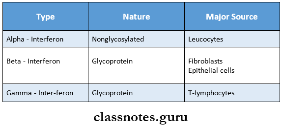
Interferon Function
1. Interferon Is An Antiviral Agent.
- Produces antiviral effects by induction of resistance to infections.
2. Antimicrobial Effect.
- Induces resistance to intracellular parasitic infections.
3. Cellular Effects.
- Inhibits cell growth and proliferation.
- Inhibits DNA and Protein synthesis.
- Increases expression of MHC antigens.
4. Immunoregulatory Effects.
- Enhances the cytotoxic activity of natural killer cells and T cells.
- Activates macrophage activity.
- Modulates antibody formation.
- Activates suppressor T cells.
Cytokines short notes
Cytokines
- Cytokines are peptide mediators or intercellular messengers.
- They are biologically active, hormone-like substances.
Cytokines Functions
- Regulates immunological, inflammatory, and reparative host responses.
- They have multiple effects on the growth and differentiation of various cells.
- They have multiple effects on the growth and differentiation of various cells.
- They act locally near the producing cells called paracrine effects.
- They produce an autocrine effect by acting directly on the cells producing them.
- Cytokines and their agonists and antagonists are used in the management of inflammatory, infectious, autoimmune, and neoplastic conditions.
Cytokines Sources
- Cytokines are produced by.
- Lymphocytes
- Macrophages
- Platelets
- Fibroblasts.
Cytokines Regulation
- Cytokine production is regulated by.
1. Exogenous Stimuli.
- Antigens
- Mitogens
2. Endogenous Factors.
- Neuroendocrine hormonal peptides.
3. Products Of Lipoxygenase And Cycloxygenase Pathways.
Cytokines Regulation Examples
- Important cytokines are:
- Interleukins.
- Colony stimulating factors.
- Tumor necrosis factor.
- Interferons.
- Transforming growth factor – p
- Leukemia inhibitory factor.
Cytokines functions and regulation in immunology
Lymphokines
Lymphokines are biologically active substances released by activated T – T-lymphocytes.
Lymphokine Functions:
- Responsible for cell-mediated immunity (CMI)
- Macrophages under the effect of lymphokines destroy micro-organisms involved in CMI.
- Participate in many functions of T-cells.
- Transmit various growth, differentiation, and behavior-neural signals between the cells of the immune system.
Lymphokines Examples
- Lymphokines affecting lymphocytes
- Lymphokines affecting macrophages
- Lymphokines affecting granulocytes
- Lymphokines affecting cultured cells
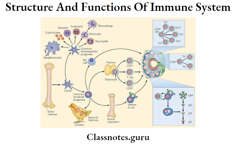
Structure And Functions Of Immune System Short Question And Answers
Structure And Functions Of The Immune System Short Questions And Answers
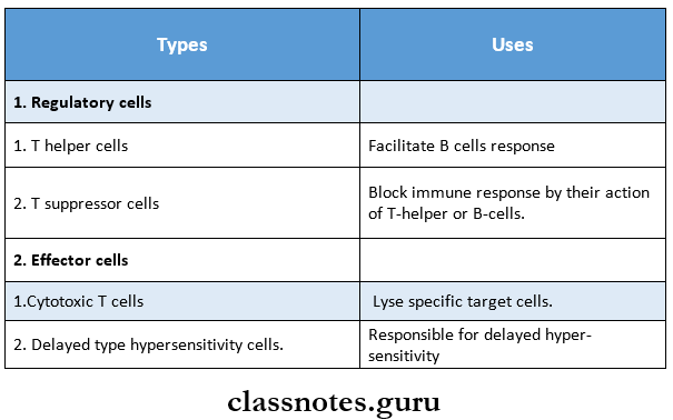
Question 1. T-Lymphocytes.
Answer:
T-Lymphocytes
- T-lymphocytes constitute 70% of total lymphocytes.
- They are derived from the thymus, thus called T- T-lymphocytes.
Read And Learn More: Microbiology Question and Answers
T-Lymphocyte Types:
Immune system short notes
Question 2. B-lymphocytes.
Answer:
B-lymphocytes
B-lymphocytes constitute 20% of total lymphocytes.
- They are derived from ‘Bursa’ or bone marrow, thus the name B-lymphocytes.
- Antigenically stimulated B-lymphocytes undergo blast transformation to become plasma cells.
B-lymphocytes Functions:
B cells produce an antibody-mediated immune response by specific differentiation and proliferation of plasma cells, which produce antigen-specific antibodies
Question 3. Major histocompatibility complex (MHC).
Answer:
Major Histocompatibility Complex (MHC)
The cell surface antigens that induce an immune response leading to rejection of allografts are known as histocompatibility antigens.
- The group of such antigens is called the major histocompatibility complex (MHC).
- MHC in humans is known as the human leucocyte antigen (HLA complex.
- The HLA complex of genes is located on the short arm of chromosome 6 and is grouped into 3 classes.
1. Class 1 MHC Antigens (A, B, C]
- Present on the surface of all nucleated cells.
- Involved in graft rejection and cell-mediated cytolysis.
Structure and function of the immune system
2. Class 1 MHC Antigens (DR, DQ, And DP]
- Found on the surface of macrophages, monocytes, activated T-lymphocytes, and B-lymphocytes.
- Responsible for graft-versus-host response.
3. Class 3 MHC Antigens.
- Encode C2 and C4 complement components of the classical pathway and properdin factor B of the alternative pathway.
Immunology Complement System
Immunology Complement System
Complement refers to a system of some non-specific proteins present in normal human and animal serum, which are activated characteristically by antigen-antibody reactions and subsequently lead to a number of biologically significant consequences.
Complement System Components
There are 9 components of complement: C1, C2, C3, C4, C5, C6, C7, C8 and C9.
Read And Learn More: Microbiology Question and Answers
Complement System Pathways
There are two pathways through which the complement system acts.
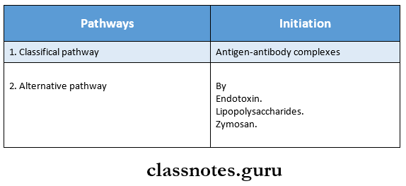
Complement system pathways
Complement System Effects Of Complement
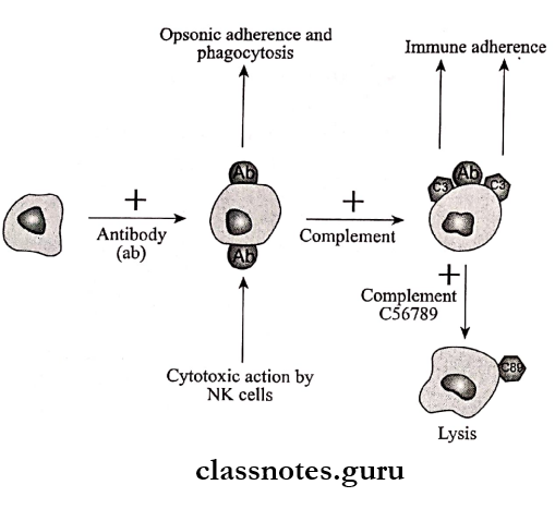
1. Complement leads to bacteriolysis and cytolysis through membrane damage.
2. C3a and C5a cause degranulation of mast cells to release histamine.
- This leads to an inflammatory reaction.
Hypersensitivity Reaction
- Complement participates in type 2 and type 3 hypersensitivity reactions.
4. Endotoxic Shock.
- Excessive C3 activation and platelet adherence lead to endotoxic shock.
5. Immune Adherence.
- Complement bound to antigen-antibody complexes adheres to erythrocytes or to platelets.
- This is called immune adherence.
6. Opsonisation
- Complement helps in the destruction of pathogens through opsonization.
Complement system short notes
7. Autoimmune Diseases.
- Decreased levels of serum complement lead to autoimmune diseases.
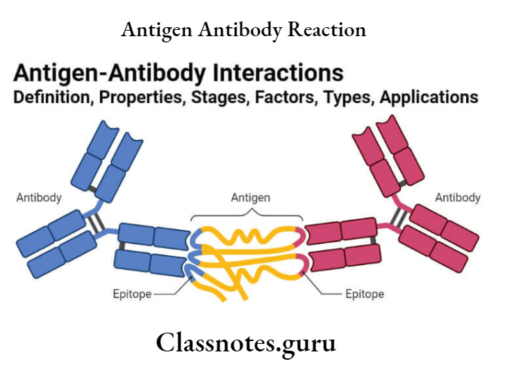
Antigen Antibody Reaction
Antigen-Antibody Reaction
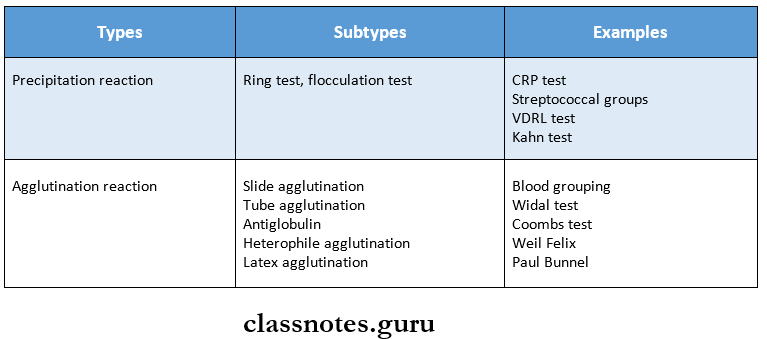
Question 1. Precipitation reaction.
Answer:
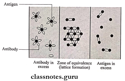
Precipitation Reaction Definition:
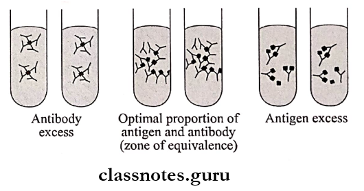
- When a soluble antigen reacts with its antibody in the presence of electrolytes at an optimal temperature and pH, the antigen-antibody complex forms an insoluble precipitate.
- This process is called precipitation.
Read And Learn More: Microbiology Question and Answers
Precipitation Reaction Mechanism
- Marrack in 1934 proposed the lattice hypothesis to explain the mechanism of precipitation.
- According to it, multivalent antigens combine with bivalent antibodies.
- Precipitation occurs only when a large lattice is formed.
- This is possible only in the zone of equivalence.
- Zone Of Equivalence.
- Antigen and antibodies are present in optimal proportion.
- Thus, a large lattice is formed and precipitation occurs.
- Zone Of Antigen Excess.
- The valencies of the antibody are fully satisfied.
- Thus lattice is not formed.
- Zone Of Antibody Excess.
- Valencies of the antigen are taken up by antibodies and a lattice is not formed.
- Zone Of Equivalence.
Antigen antibody reaction and its types
Precipitation Reaction Types
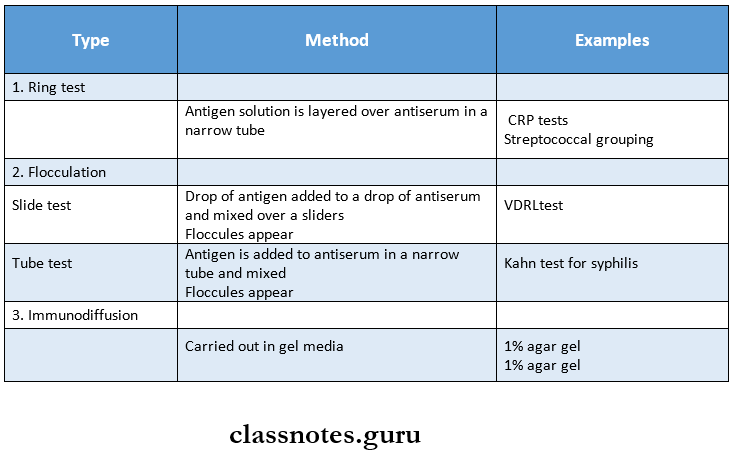
Immunodiffusion Test
Immunodiffusion tests are precipitation tests that occur in gel media.
Immunodiffusion Test Types:
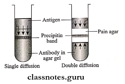
1. Single Diffusion In One Dimension.
Single Diffusion In One Dimension Steps:
- Mix antibodies in agar gel in a test tube
- Pour antigen over it.
Single Diffusion In One Dimension Result:
- Antigen diffuses downward.
- A line of precipitation is formed.
Short note on antigen-antibody interaction
Double Diffusion In One Dimension
Double Diffusion In One Dimension Steps:
- Mix antibodies in agar gel in a test tube.
- Place a column of plain agar over it.
- Pour antigen over the plain agar.
Double Diffusion In One Dimension Result:
- Antigen and antibodies move towards each other
- A precipitation band is formed.
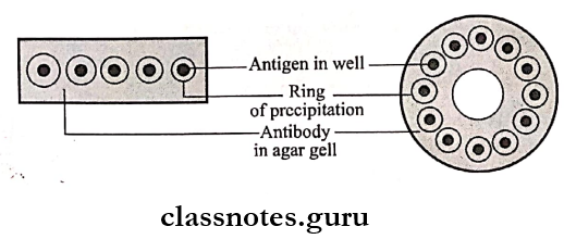
Single Diffusion In One Dimension
Single Diffusion In One Dimension Steps:
- Mix antibodies in agar gel on a slide.
- Cut wells on the surface of the gel.
- Add antigens to these wells.
Single Diffusion In One Dimension Results:
- Antigen diffuses radially,
- Ring-shaped bands of precipitation are formed.
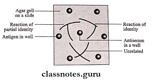
Double Diffusion In Two Dimensions
Double Diffusion In Two Dimensions Steps:
- Place agar gel on a slide.
- Cut wells over it.
- Fill antibodies in the central well and antigens in other wells.
Double Diffusion In Two dimensions Results:
- Lines of precipitate formed between two identical adjacent antigens fuse.
Immunoelectrophoresis
Immunoelectrophoresis Steps:
- Combines electrophoresis and immunodiffusion.
- Semisolid agar is layered on a slide,
- Cut wells over it and fill it with antigen
- Electrophoresis of the antigen is carried out for 1 hour,
- Cut a rectangular through in the agar and fill it with antibodies.
- Allow diffusion for 18 – 24 hours.
Immunoelectrophoresis Results:
- Precipitation lines develop with each separated antigen.
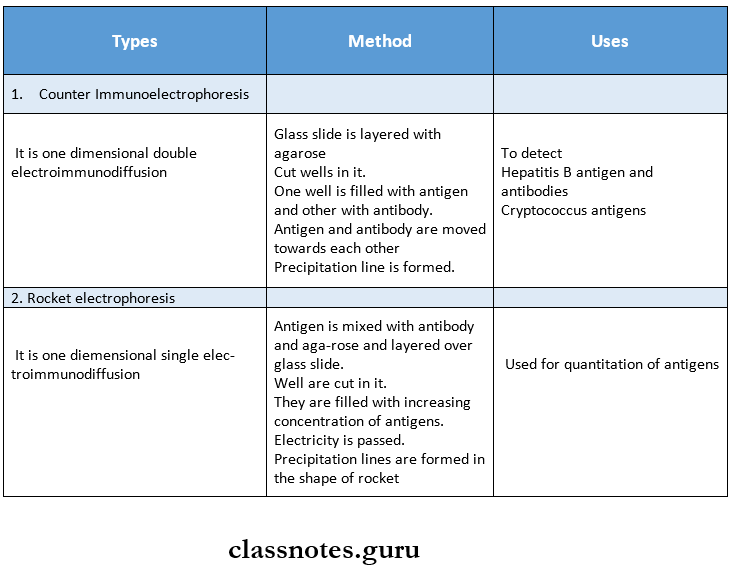
Electroimmunodiffusion
- Electroimmunodiffusion is a combination of electrophoresis and diffusion.
- Electroimmunodiffusion includes.
- Counter immunoelectrophoresis.
- Rocket electrophoresis.

Applications of antigen-antibody complex in lab tests
Agglutination
Agglutination Definition:
- Agglutination is an antigen-antibody reaction in which a particular antigen combines with its antibody in the presence of electrolytes at an optimal temperature and pH
- Agglutination results in clump formation.
Agglutination Properties
- Occurs with a particulate antigen.
- Agglutination is more sensitive.
- Agglutination takes place better with IgM antibodies.
Agglutination Principle
- When antigen and antibody are present in optimal proportions, lattice formation occurs.
- This results in agglutination.
Diagnostic uses of antigen-antibody reaction
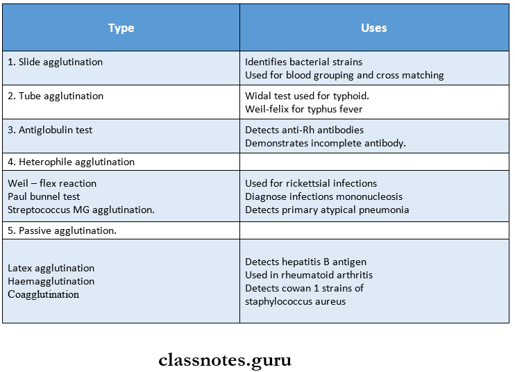
Agglutination Types
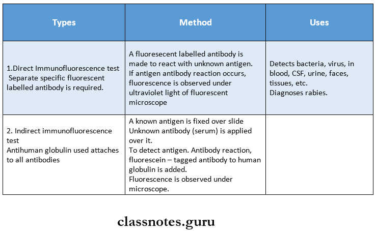
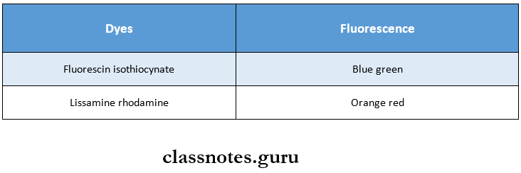
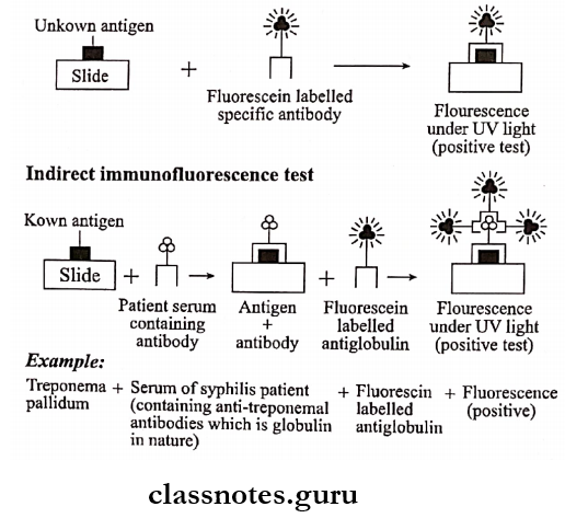
Immunofluorescence
Immunofluorescence
The dyes used are:
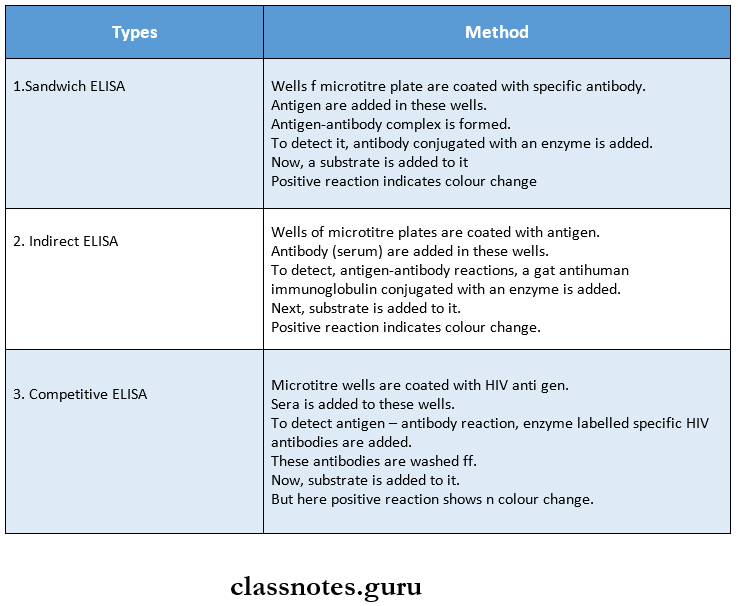
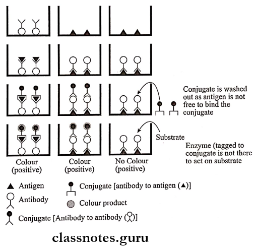
ELISA test
ELISA is an abbreviated form of Enzyme-Linked ImmunoSorbent Assay.
- ELISA test is widely used.
- ELISA test is simple and sensitive.
- The test can be done in polystyrene tubes or polyvinyl microlitre plates.
ELISA test Types:
Antigen-Antibody Reaction
Question 1. Define precipitation and flocculation.
Answer:
Precipitation Definition
- When a soluble antigen reacts with its antibody in the presence of electrolytes at an optimal temperature and pH, the antigen-antibody complex forms an insoluble precipitate.
- This process is called precipitation.
Flocculation Definition
- When a particulate antigen reacts with its antibody in the presence of electrolytes at an optimal temperature and pH, the antigen-antibody reaction shows visible clumps of particles.
- This process is called flocculation.
Question 2. Zone of equivalence.
Answer:
Zone Of Equivalence
- When antigen and antibody are present in optimal or equivalent proportion, a large lattice is formed.
- This is results in precipitation.
- This zone where precipitation occurs in called the zone of equivalence.
Question 3. Types of electro immunodiffusion.
Answer:
Types Of Electro Immunodiffusion
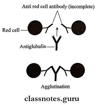
Coombs Test
The Coombs test was devised by Coombs, Mourant, and Race in 1945.
Coombs Test Method:
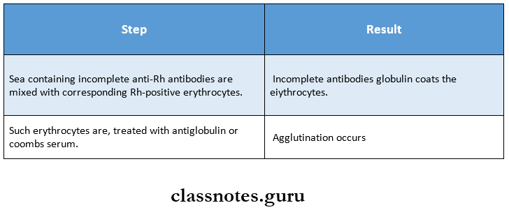
Coombs Test Types
- Direct Coombs test – in-vivo test
- Indirect Coombs test – in-vitro test.
Coombs Test Uses
- Detect anti – Rh antibodies.
- Demonstrates incomplete antibody.
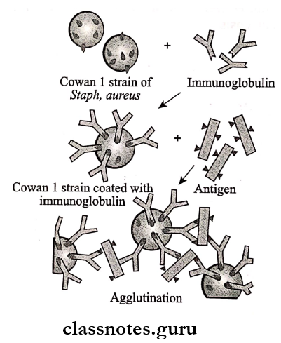
Question 5. Coaggiutination.
Answer:
Coaggiutination
- Coaggiutination is a type of passive agglutination test.
- Coaggiutination is based on the presence of protein A on Cown I strains of staphylococcus aureus.
- Specific IgG is coated on these bacteria.
- IgG has two portions.
- Fc portion – Binds to protein A
- Fab terminal – remains free.
- When the corresponding antigen is mixed with it, the Fab terminal binds to it.
- This results in agglutination.
Coaggiutination Uses
- Detects bacterial antigens in blood, urine, and CSF.
- Detects N.gonorrhoea, strep pyrogens, and H. influenzae antigens.
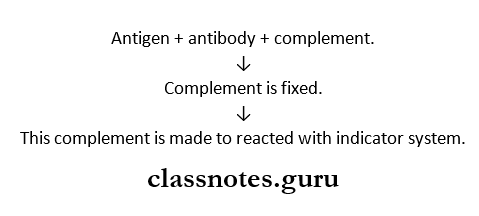
Complement fixation test
Complement Fixation Test Principle:
The antigen-antibody complexes are able to fix complement.
Complement Fixation Test Method:
An indicator system consisting of sheep erythrocytes coated with an amboceptor is used.
Complete fixation is detected by this system.
Short note on antigen-antibody interaction
Complement Fixation Test Result
- Lysis of erythrocytes – indicates nagative reaction.
- No lysis of erythrocytes – indicates positive reaction.
Question 7. Define the Agglutination test with two examples.
Answer:
Agglutination Test Definition
- Agglutination Test is an antigen-antibody reaction in which a particulate antigen combines with its antibody in the presence of electrolytes at an optimal temperature and pH
- Agglutination Test results in clump formation
Agglutination Test Examples
- Weil – flex reaction – used for rickettsial infections
- Paul Bunnel test – used for diagnosis of infectious mononucleosis
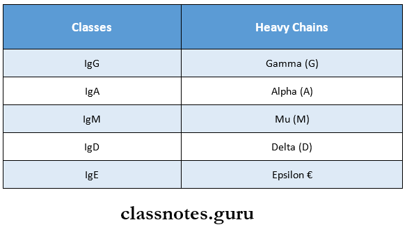
Types Of Immunoglobulins
Types of Immunoglobulins
Question 1. Define and classify antibodies and add a note on the structure and function of IgG.
Answer:
Antibodies Definition:
- Antibodies are substances which are formed in the serum and tissue fluids in response to an antigen and react with that antigen specifically and in some observable manner.
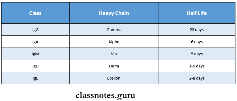
Antibodies Classification
- Antibodies are named as immunoglobulins.
- There are five groups of immunoglobulins (Ig]
Types of immunoglobulins
Read And Learn More: Microbiology Question and Answers
IgG
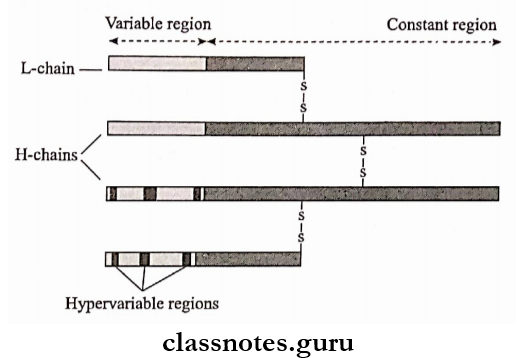
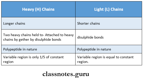
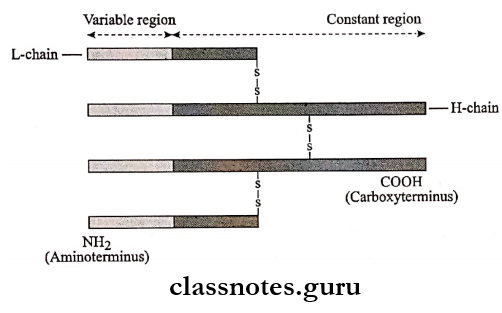
IgG Structure:
- IgG consists of
- Two Identical Heavy Chains (H) Chains
- They are gamma chains.
- Held together by disulphide bonds (S-S)
- They are longer chains.
- Consists of a smaller variable (V) region and a longer constant (C] region.
- Two Identical Light (L) Chains.
- Present in two forms. Kappa (K) and Lambada (L)
- Attached to heavy chains by disulphide bonds.
- They are shorter chains.
- The init variable and constant region are equal.
- Variable regions are present at the amino terminus
- The amino acid sequence here is highly variable.
- Constant region is present at the carboxy terminus.
- Two Identical Heavy Chains (H) Chains
IgG Functions
- Can be transported through the placenta
- Thus, provide natural passive immunity to the newborn.
- Participates in precipitation, complement fixation and neutralisation of toxins and viruses.
- Protects against active micro-organisms present in the blood and tissues.
Immunoglobulins
Question 1. Immunoglobulin
Answer:
Immunoglobulin
- Immunoglobulin is defined as a protein of animal origin endowed with known antibody activity.
Immunoglobulin Synthesis:
- They are synthesized by plasma cells and by lymphocytes.
Types of antibodies in immunology
Immunoglobulin Structures:
- Immunoglobulin consists of
- Two heavy chains
- Two light chains.
- The variable region is present at the amino terminus while the constant region is present at the carboxy terminus.
- Based on the heavy chains. Immunoglobulins are classified into 5 classes.
- Light chains in all the classes are Kappa (K) and lambda (L)
Immunoglobulin types and functions
Question 2. IgA/Secretory Immunoglobulin.
Answer:
IgA
IgA is the second major serum immunoglobulin
- IgA is the fast-moving alpha-globulin.
Serum Concentration:
- Normal serum concentration – 0.6 – 4.2 mg/ml.
- It is about 10 – 13% of total serum immunoglobulin.
- Half-life: 6 – 8 days
IgA Forms:
- IgA is present in two forms.
- Serum IgA
- Secretory IgA
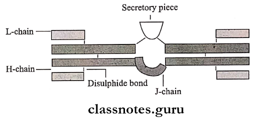
IgA Structure:
- IgA is a dinner consisting of two monomer units.
- It consists of
- Two Alpha-Heavy Chains
- Joined by disulphide bonds.
- Two Kappa (K) Or Lambda (L) Light Chains.
- Attached to heavy chains by disulphide bonds
- I Chains
- It is glycoprotein
- It joins the two monomer units of IgA
- S (Secretory) Piece.
- S (Secretory) Piece is glycine -rich polypeptide.
- S (Secretory) Piece is produced by mucosal or glandular epithelial cells.
- S (Secretory) Piece protects IgA from denaturation by bacterial proteases.
- Two Alpha-Heavy Chains
Classes of immunoglobulins
IgA Synthesis:
- Locally from plasma cells.
- In trace amount from serum.
IgA Functions:
- IgA, is present in secretions such as milk, saliva, tears, sweat, nasal fluids, colostrums and in secretions of respiratory, intestinal and genital systems.
- Hence called secretory immunoglobulin.
- Protects mucous membranes against micro-organisms.
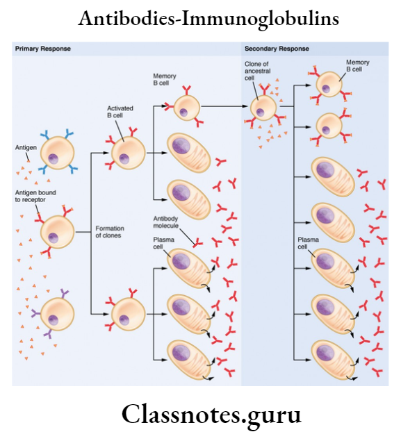
Antibodies Immunoglobulins Short Question And Answers
Question 1. Functions of antibodies.
Answer:
Functions Of Antibodies
Immunoglobulin classification
Question 2. Structure of IgM
Answer:
IgM Structure:
- IgM is a pentamer consisting of 5 immunoglobulin subunits and one molecule of J chain.
- J chain joins the Fc region of basic subunits.
- Each subunits consists of
- Two identical mu heavy (H) chains and
- Two identical Kappa (K) or Lambda (L) light chains.
- These chains are held together by disulphide bonds.
- Both chains have a variable (V) region and a constant (C) region.
Structure and types of immunoglobulins
Immunity Short And Long Essays Question And Answers
Types Of Immunity
- Innate immunity
- Acquired immunity
- Types
- Active immunity
- Natural active
- Artificial active
- Passive immunity
- Natural passive
- Artificial passive
- Active immunity
- Types
Immunity essay questions and answers
Read And Learn More: Microbiology Question and Answers
Antigens
- Heterophile Antigen
- These are the same or closely related antigens present in different tissues of more than one species
- Haptens
- These are substances unable to induce antibody formation on its own but can be immunogenic when linked to carrier proteins
- Haptens is a partial/incomplete antigen
Immunoglobulin
- These are substances which are formed in serum in response to an antigen
- Immunoglobulin consists of two heavy chains and two light chains held together by disulphide bonds
Immunoglobulin Classes
Types Of Antigen-Antibody Reactions
5. Complement
- Complement refers to a system of factors which occurs in normal serum and are activated by antigen-antibody reaction
- Complement is triggered by two parallel pathway
- Classical pathway
- Alternate pathway
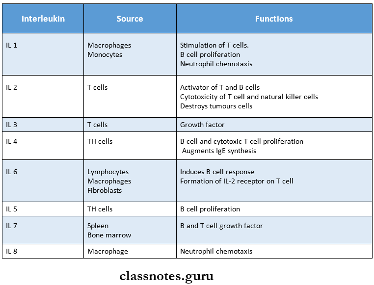
6. Interleukins (IL)
Immunity short and long answer questions
Hypersensitivity Reactions
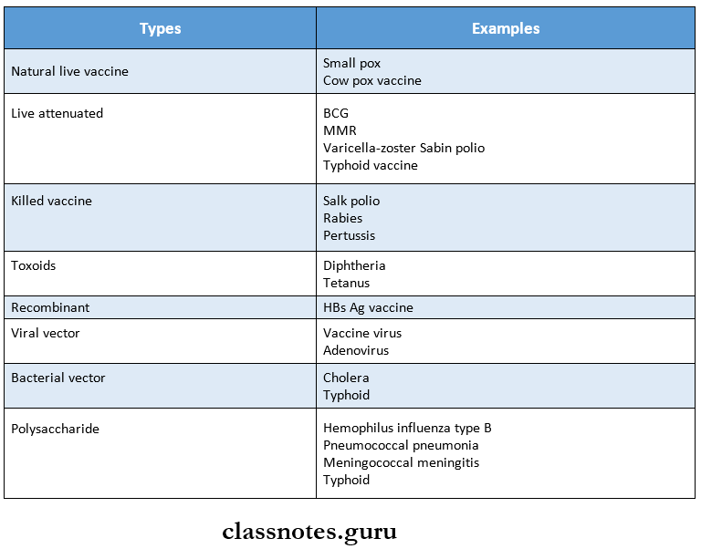
8. Vaccines
Types of immunity explained
Immunity
Question 1. Define and classify immunity. Discuss acquired immunity.
Answer:
Immunity Definition
1. Immunity:
- Immunity is defined as resistance exhibited by the host against any foreign antigen including micro-organisms.
Immunity Classification
- Immunity may be classified into different types as follows.
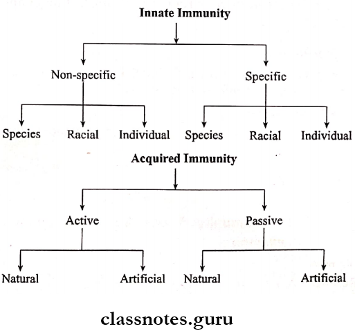
Acquired immunity Definition
- Acquired immunity is the resistance acquired by an individual during life.
Acquired immunity Types:
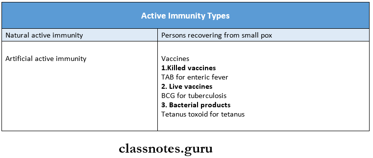
1. Active Immunity:
- Active Immunity is subdivided into two types.
- Natural Active Immunity.
- Acquired by natural subclinical or clinical infections.
- It is long-lasting.
- Example: A person recovering from smallpox develop natural active immunity.
- Artificial Active Immunity.
- Induced by vaccination.
- Natural Active Immunity.
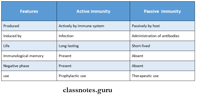
Active Immunity Mechanism
- Active immunity stimulates both humoral and cell-mediated immunity.
Innate and acquired immunity essay
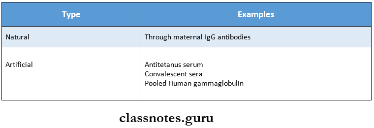
Passive Immunity
- Passive Immunity is subdivided into two types:
- Natural
- Immunity is transferred from mother to fetus transplacentally.
- Artificial
- Occurs through the parenteral administration of antibodies.
- Natural
Passive Immunity Mechanism
- Passive Immunity is induced in an individual by preformed vaccines against infective agents.
- Passive Immunity is short-lasting.
- Passive Immunity is used when immunity is required immediately.
Passive Immunity Uses
- To provide immediate short-term protection
- For suppression of active immunity.
- For treatment of serious infection
Innate immunity
Innate immunity Definition:
- Innate immunity is the resistance that individual possesses by birth.
Innate Immunity Types:
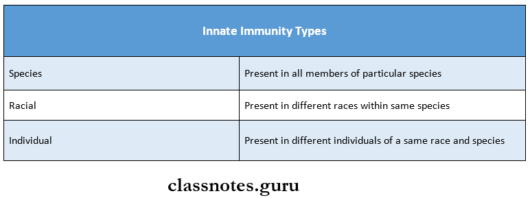
- Factors influencing it:
1. Age:
- Infants have immature immunity, while during old age there is gradual decreases in immunity.
- This increases susceptibility to infections.
2. Hormones:
- Hormonal disorders may lead to increased susceptibility to infections.
3. Nutrition:
- Malnutrition predisposes to infections.
Innate Immunity Mechanism
1. Epithelial Surfaces:
- Skin.
- Act as a mechanical barrier to micro-organisms
- Provides bacteriocidal secretions.
- The resident bacterial flora prevents colonization by pathogens.
- Respiratory Tract.
- Nasal passages arrest the inhaled particles.
- The mucous secretions of the respiratory tract act as a trapping mechanism.
- Cilia help to propel the particles toward the pharynx.
- Intestinal Tract.
- Mouth – inhibits micro-organisms.
- Acidic gastric pH – destroys bacteria.
- Intestine – prevents colonization of bacteria,
- Conjunctiva.
- Tears flushes away bacteria and dust particles.
- Lysozyme present in tears has bacteriocidal actions.
- Genitourinary Tract.
- Urine eliminates bacteria.
- Vaginal secretions destroy pathogens.
- Semen contains antibacterial substances.
Antibacterial Substances
- Antibacterial Substances include properdin, complement, lysozyme, beta-lysin, basic polypeptides, and interferons.
3. Cellular Factors:
- Phagocytic cells ingest pathogenic organisms and de¬stroy them.
4. Inflammation:
- Inflammation is a non-specific defense mechanism.
- Inflammation phagocytoses and destroys micro-organisms.
5. Fever:
- Stimulates production of interferon
6. Acute Phase Proteins:
- Activate alternate pathways of complement.
Immunity BSc nursing notes
Types Of Active Immunity With Examples
Difference Between Active And Passive Immunity
Difference between innate and adaptive immunity
Two Examples Of Passive Immunity
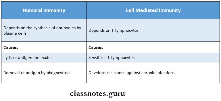
Humoral and cell-mediated immunity essay
Antigen Definition
- The antigen is a substance which, when introduced into a body evokes an immune response to produce a specific antibody with which it reacts in an observable manner.
Antigen Types
1. Complete Antigen.
- These can induce antibody formation by themselves.
2. Complete Antigen.
- These can induce antibody formation by themselves.
3. Incomplete Antigen/Haptens.
- These are unable to induce antibody formation on their own.
- But can become immunogenic when they are covalently linked to carrier proteins.