Infections Of Oral Cavity Question and Answers

Oral Medicine Infections Of Oral Cavity Important Notes

1. Theories of dental caries Theory

Oral Medicine Infections differences between recurrent aphthous and recurrent herpes ulcers

2. Zones of dental caries

  • Enamel caries
    • Zone 1 – Translucent zone
    • Zone 2 – Dark zone
    • Zone 3 – Body of lesion
    • Zone 4 – Surface zone
  • Dentinal caries
    • Zone 1 – Zone of fatty degeneration
    • Zone 2 – Zone of dental sclerosis
    • Zone 3 – Zone of decalcification of dentin
    • Zone 4 – Zone of bacterial invasion
    • Zone 5 – Zone of decomposed dentin

Read And Learn More: Oral Medicine Question and Answers

3. Ludwig’s angina

  • It is a severe form of cellulitis
  • Begins in submaxillary space and secondarily involves sublingual and submental spaces
  • Mandibular molars are source of infection
  • Manifests as rapidly developing broad-like swelling of the floor of the mouth
  • Results in elevation of the tongue with difficulty in swallowing and breathing
  • If the infection spreads to the neck it leads to edema of the glottis
  • Suffocation may lead to death
  • Emergency tracheostomy is done

Oral Medicine Infections Of Oral Cavity Short Essays

Question 1. Focal infection

Answer:

Focal infection

Focal infection is a localized or general infection caused by the dissemination of microorganisms or toxic products from a focus of infection

Focal infection Mechanism:

  • Spread of pathogenic micro-organisms from their primary site of infection to the distant part of the body via blood vessels or lymphatics
  • Spread of toxins liberated by the pathogenic microbes to distant organs either via blood vessels or lymphatics

Focal infection Significance:

  • It causes a great number of systemic diseases like
  • Arthritis
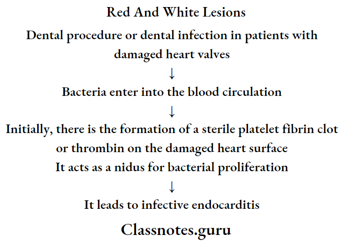
  • Valvular heart diseases
  • Gastro-intestinal diseases
  • Ocular diseases
  • Skin diseases
  • Renal diseases

Question 2. Dental fluorosis.

Answer:

Dental fluorosis

Dental fluorosis is caused by excessive intake of fluoride during tooth development

Dental fluorosis Clinical Features:

  • Lustreless, opaque white patches in the enamel may become mottled, striated, or pitted
  • Mottled areas may become stained yellow or brown
  • Hypoplastic areas may also be present to such an extent in severe cases that normal tooth form is lost
  • Enamel fluorosis is a developmental phenomenon due to excessive fluoride ingestion during amelogenesis
  • Once crowns are formed no further fluorosis occurs
  • The hypocalcified areas of the mottled enamel are less soluble in acids
  • They have a greater permeability to dyes
  • They emit fluorescence of higher intensity than normal enamel
  • Fluorosis occurs symmetrically within arches
  • The premolars are usually first affected followed by the second molar, maxillary incisor, canine, first molar, and mandibular incisors

Herpangina In Adults

Oral Medicine Infections Of Oral Cavity Short Answers

Question 1. Mucous patches

Answer:

Mucous patches

  • It is a feature of secondary syphilis
  • Mucous patches develop over the oral mucosa
  • They are painless, multiple, greyish-white plaques overlying the ulcerated surface

Question 2. Chemical burn

Answer:

Chemical burn

  • It is a burn caused due to caustic chemical agents
  • It produces coagulation necrosis of the epithelium

Chemical burn Causes:

  • Aspirin and aspirin-containing compounds
  • Hydrogen peroxide
  • Silver nitrate- Used in the treatment of aphthous ulcers
  • Toothache drop burns
  • Ethyl alcohol burns- Topical application of ethyl alcohol solution
  • Acid burns
  • Ingestion- Due to ingestion of caustic chemicals

Chemical burns Clinical Features:

  • Painful lesion
  • Appears as an irregularly shaped, white pseudomembrane-covered lesion
  • Gentle lateral pressure causes the white material to slide away exposing painful central red ulceration

Chemical Burn Management:

  • Topical application of dyclonine hydrochloride
  • A protective coat of emollient paste
  • Surgical debridement of necrosed tissue

Question 3. Causes for bleeding in the oral cavity

Answer:

Causes Of Bleeding From Mouth:

  • Bleeding diathesis
  • Carcinoma, squamous cell of head and neck – bleeding from the mouth
  • Crown placement
  • Dental caries
  • Dental cleaning
  • Dentures
  • Hemophilia
  • Idiopathic thrombocytopenic purpura
  • Leukemia
  • Pancytopenia
  • Periodontal disease
  • Plaque
  • Post tonsillectomy bleeding
  • Root canal
  • Stomatitis
  • Thrombocytopenia
  • Tonsillar abscess
  • Tooth extraction
  • Trauma
  • Viral Hemorrhagic Fevers

Question 4. Cellulitis

Answer:

Cellulitis Definition:

  • It is an acute, edematous, purulent inflammatory process that spreads diffusely through different tissue spaces

Cellulitis Sources Of Infections:

  • Periapical abscess
  • Pericoronitis
  • Periodontal abscess
  • Osteomyelitis
  • Infected post-extraction wound
  • Gunshot injuries
  • Oral soft tissue infections
  • Bloodborne infections

Cellulitis Clinical Features:

  • Large, diffuse, painful swelling over the face or neck
  • The overlying skin appears purplish
  • Fever, chills
  • Leukocytosis
  • Regional lymphadenopathy
  • Pus discharging sinuses

Cellulitis Complications:

  • Trismus
  • Dyspnoea
  • Dysphagia

Question 5. Causes of pigmentations

Answer:

Causes of pigmentations

  • Exogenous pigmentation
    • Occupational
      • Lead industry-lead
      • Match industry- Phosphorous
      • Fluorescent lamp industry- mercury
      • Photography- Silver
    • Habits
      • Tobacco
      • Pan
      • Foodstuffs
    • Therapeutic
      • Drugs like anti-malarial drugs
      • Metallic salts
    •  Others
      • Amalgam tattoo
      • Black hairy tongue
  • Causes of pigmentations Endogenous
    • Physiologic
      • Racial variation
      • Physiologic melanotic macule and papule
      • Fordyce’s granules
      • Pregnancy
    •  Pathological
      • Addison’s disease
      • Acromegaly
      • Peutz-Jeghers syndrome
      • Nevi
      • Malnutrition

Question 6. Caries vaccine

Answer:

Caries vaccine

  • It is a suspension of an attenuated or killed micro-organism administered for the prevention, amelioration, or treatment of infectious diseases

Caries vaccine Mechanism Of Action:

  • When the tooth erupts serum antibodies i.e. IgA stimulate opsonization and phagocytosis
  • These antibodies have an inhibitory effect on glucosyl transferase and acid production
  • It results in the inhibition of the metabolic activity of mutants in teeth

Caries vaccine Route Of Administration:

  • Oral route
  • Systemic route
  • Active gingivo-salivary route
  • Active immunization
  • Synthetic peptides
  • Coupling with cholera toxin subunits
  • Fusing with avirulent strains of salmonella
  • Liposomes- increases IgA antibodies
  • Passive immunization

Oral Medicine Infections Of Oral Cavity Viva Voce

  1. Denture sore mouth is rarely found under the mandibular denture
  2. Minocycline produced oral pigmentation
  3. Amalgam tattoo is the most common source of focal pigmentation
  4. Strawberry tongue is the result of an infection caused by streptococcus pyogenes
  5. The critical pH of dental caries is 5.2
  6. Initiation of dental caries is caused by streptococcus mutans
  7. Progression of dental caries is by lactobacillus
  8. Pioneer bacteria is seen in the earliest stage of caries

Infections Question and Answers

Oral Medicine Infections Important Notes

1. Burning mouth syndrome

  • Shows clinically healthy oral mucosa
  • A burning sensation of oral mucosa occurs
  • Pain starts in the morning and aggravates during the day

Oral Medicine Infections Long Essays

Question 1. Classify ulcers of the oral cavity. Describe the etiology, clinical features, and management of recurrent aphthous stomatitis.

Answer:

Ulcers Of Oral Cavity Classification:

1. Ulcers Of Oral Cavity Classification Acute multiple

  • Herpes virus infection
  • Primary herpes simplex virus infection
  • Coxsackie virus infection
  • Varicella-zoster virus infection
  • Erythema multiform

2. Ulcers Of Oral Cavity Classification Recurrent oral ulcers

  • Recurrent aphthous stomatitis
  • Behcet’s syndrome

3. Ulcers Of Oral Cavity Classification Chronic multiple ulcers

  • Pemphigus
  • Subeplthellal Bullous dermatoses

4. Ulcers Of Oral Cavity Classification Single ulcers

  • Histoplasmosis
  • Blastomycosis
  • Mucormycosis
  • It is a common disease characterized by the development of painful, recurrent, solitary ulceration of the oral mucosa

Read And Learn More: Oral Medicine Question and Answers

Ulcers Of Oral Cavity Classification Etiology:

  • Immunological abnormalities:
    • Due to T cell-mediated immunological abnormality
  • Genetic predisposition
    • Increased susceptibility to PAS
  • Microbial organism
    • Form L form of □hemolytic streptococci
  • Systemic factors
    • Nutritional deficiency
  • Cyclic neutropenia

Ulcers Of Oral Cavity Classification:

  • Minor aphthae: the ulcers are less than 1 cm in diameter
  • Major aphthae: they are over 1 cm in diameter
  • Herpetiform ulcers: they are small ulcers throughout the mucosa

Ulcers Of Oral Cavity Clinical Features:

  • Age and sex: common in women of the second and third decade of life
  • Site: it occurs most commonly on buccal and lingual mucosa, tongue, soft palate, pharynx, and gingiva
  • Prodromal symptoms:
    • It begins with burning, itching, or stinging for 24-48 hours
  • Next ulcer appears
  • The ulcer gradually enlarges over the next 48-72 hours
    • Later symptoms
    • The lesion is typically painful
  • It interferes with eating for several days
  • It begins as a single or multiple superficial erosion covered by the gray membrane is surrounded by localized areas of erythema
  • Lesions are round, symmetrical, and shallow
  • Multiple lesions are present
  • Minor aphthae:
    • Size: 0.3-1 cm
    • They heal without scar formation within 10-14 days
  • Major aphthae
    • Size: up to 5 cm in diameter
  • They interfere with speech and eating
  • The large portion may be covered with deep painful ulcers
  • The lesions heal slowly and leave a scar
  • Due to it, there is decreased mobility of the uvula and tongue
  • Herpetiform ulcers
    • Multiple small shallow ulcers often up to 100 in number
  • It begins as small pinhead-size erosions that gradually enlarge in size
  • Lesions are more painful
  • It is present continuously for one to three years
  • Patients get relief immediately with 2% tetracycline mouthwash

Ulcers Of Oral Cavity Classification Differential Diagnosis:

  • BMMP and pemphigus: absence of vesicles
  • Bedner’s aphthae: there is no aphthae
  • Erythema multiforme: aphthae ulcers are uniform in distribution

Ulcers Of Oral Cavity Classification Management:

  • Topical corticosteroid
    • Triamcinolone acetonide: 3-4 times in a day
  • Anesthetic cream
    • Orobase should be given
  • Tetracycline mouthwash:
    • 250 mg/ml use four times daily for 5-7 days
  • Nutritional supplements:
    • Replacement therapy with vitamin B12, ferritin, folate, and iron
  • Maintenance of oral hygiene
    • Chlorhexidine mouthwash is given

Oral Medicine Infections Short Essays

Question 1. Stomatitis medicamentosa

Answer:

Stomatitis medicamentosa

It is an allergic reaction associated with systemic drug intake.

Stomatitis medicamentosa Etiology:

  • Oral changes found with cutaneous reaction to drugs
  • Mucosal alterations may result from the following:
    • Myelosuppression
    • Direct cytotoxic effect
    • Xerostornic effects
    • Alterations of oral microbial flora

Stomatitis medicamentosa Clinical Features:

  • Painful, erythematous, erosive, or ulcerative lesions
  • Nonkeratinized locations are often affected initially
  • Fixed forms of drug-associated eruptions are relatively uncommon intraorally
  • The pseudomembranous necrotic surface may be noted

Stomatitis medicamentosa Differential Diagnosis:

  • Chemical or thermal burn
  • Erosive lichen planus
  • Pemphigus Vulgaris
  • Mucous membrane (cicatricial) pemphigoid
  • Erythema multiforme
  • Acute herpetic gingivostomatitis
  • Candidiasis

Stomatitis medicamentosa Treatment:

  • Identification and withdrawal of offending drug
  • Symptomatic management including topical preparations
  • Systemic corticosteroids if the mucosal reaction is not related to antineoplastic treatment

Question 2. Burning mouth syndrome.

Answer:

Burning mouth syndrome

Burning sensation in the structures in contact with the dentures without any visible change in the mucosa

Burning mouth syndrome Features:

  • Pain in the morning
  • Dry mouth
  • Persistent altered taste
  • Generalized symptoms

Burning mouth syndrome Etiology:

  • Irritation by ill-fitting dentures
  • Constant masticatory activity
  • Excessive friction on the mucosa
  • Candidal infection
  • Nutritional deficiency
  • Xerostomia
  • Medication

Burning mouth syndrome Management:

  • Counseling
  • Repair of ill-fitted dentures

Question 3. Allergic stomatitis.

Answer:

Allergic stomatitis

It occurs due to drug allergy

Allergic stomatitis Clinical Features:

  • Fever
  • Arthralgia
  • Inflammation
  • Ulceration
  • Vesicle formation
  • Lymph node enlargement
  • Erythematous skin

Allergic stomatitis Oral Manifestations:

  • Xerostomia
  • Taste alteration
  • Eating difficulty
  • Vesicle bullae found on the mucosa
  • The lesion has diffuse distribution
  • There may he ulceration and necrosis of the gingiva
  • Severe periodontal problems may occur
  • There may be erosion of teeth
  • Bathing of teeth with corrosive substances can spread to the pharynx.

Allergic stomatitis Differential Diagnosis:

  • Recurrent herpes simplex infection – it occurs in groups,
  • ANUG-punched out ulceration
  • Erythema multiforme- skin lesions are present

Allergic stomatitis Management:

  • Discontinuation of the drug: stop the allergic drug.
    • Use an alternative for it.
  • Antihistaminic drug: to relieve the acute sign.
  • Topical corticosteroid: to resolve localized reaction.
  • Adrenaline

Question 4. Treatment of aphthous stomatitis.

Answer:

Treatment of aphthous stomatitis

  • Topical corticosteroid
    • Triamcinolone acetonide: 3-4 times in a day
  • Anesthetic cream
    • Orobase should be given
  • Tetracycline mouthwash:
    • 250 mg/ml use four times daily for 5-7 days
  • Nutritional supplements:
    • Replacement therapy with vitamin B12, ferritin, folate, and iron
  • Maintenance of oral hygiene
    • Chlorhexidine mouthwash is given

Oral Medicine Infections Short Answers

Question 1. Major aphthous ulcers.

Answer:

Major Aphthae:

  • Size: up to 5 cm in diameter
  • They interfere with speech and eating
  • The large portion may be covered with deep painful ulcers
  • The lesions heal slowly and leave a scar
  • Due to it there is decreased mobility of the uvula and tongue

Question 2. Four differences between recurrent aphthous and recurrent herpes ulcers?

Answer:

Four differences between recurrent aphthous and recurrent herpes ulcers

Oral Medicine Infections differences between recurrent aphthous and recurrent herpes ulcers

Oral Medicine Infections Viva Voce

  1. Chronic osteomyelitis shows the moth-eaten appearance

Dermatological Diseases Short Question And Answer

Oral Medicine Dermatological Diseases Important Notes

1. Various findings of dermatological diseases

Oral medicine Dermatological Diseases Various finding of dermetological diseases

2. Ectodermal dysplasia

  • It is congenital dysplasia of ectodermal structures
  • Manifested as hypohidrosis, hypotrichosis and hypodontia

3. Pemphigus

  • Histological features:
    • There is a formation of vesicles or bullae intraepithelial just above the basal layer producing suprabasal split c
    • Intercellular bridges in suprabasal layers disappear due to edema resulting in acantholysis
    • Clumps of degenerating cells are found in vesicular areas called Tzanck cells

4. Scleroderma – features

  • Stiff and broad-like tongue
  • Lips become rigid
  • Microstomia
  • Dysphasia
  • Inability to open and close mouth
  • Extreme widening of PDL

Read And Learn More: Oral Medicine Question and Answers

5. Systemic lupus erythematosus

  • It is a multisystem inflammatory disorder

6. Steven Johnson syndrome

  • Severe bullous form of erythema multiforme involving the skin, eyes, oral cavity, and genitalia

Oral medicine Dermatological Diseases Systemic lupus erythematosis

7. Nikolsky’s sign

  • Loss of epithelium due to rubbing resulting in raw sensitive surface
  • Seen in
    • Pemphigus
    • Familial benign chronic pemphigus
    • Epidermolysis bullosa

8. Tzanck cells

  • They are multinucleated giant cells of epithelial origin
  • Seen in
    • Herpes
    • Pemphigus

9. Bulla are seen in

  • Intraepithelial bulla
    • Herpes simplex Herpes zoster Chicken pox Pemphigus
    • Familial benign pemphigus
    • Epidermolysis bullosa
    • Oral lesions of eiythema multiforme
  • Subepithelial bulla
    • Pemphigoid
    • Bullous pemphigoid
    • Bullous lichen planus
    • Dermatitis herpetiformis
    • Epidermolysis bullosa
    • Skin lesions of erythema multiforme.

Oral Medicine Dermatological Diseases.

Oral Medicine Dermatological Diseases Short Essays

Question 1. Lupus erythematosus.

Answer:

Lupus erythematosus

It is an autoimmune disorder characterized by the destruction of tissue due to the deposition of autoantibodies and immune complexes within it

Lupus erythematosus Types:

Oral medicine Dermatological Diseases Systemic And Discoid Lupus Erythematosus
Lupus erythematosus Generalized symptoms

  • Fever
  • Fatigue
  • Dysphagia
  • Depression
  • Splenomegaly
  • Lymphadenopathy
  • Leucopenia
  • Arthritis
  • Sjogren’s syndrome
  • Raynond’s phenomenon
  • Scleroderma
  • Pemphigoid
  • Pemphigus
  • Erythema multiforme

Lupus erythematosus Investigations:

  • Anti-nuclear antibodies are present
  • Anti-DNA antibodies are present
  • Polyclonal hyperactivity of the B lymphocytes
  • Decrease in the number of suppressor cells
  • Leucopenia
  • Thrombocytopenia
  • Hemolytic anemia
  • Hypergammaglobulinemia
  • Profuse proteinuria
  • Direct immunofluorescence
    • It reveals deposition of IgG, IgA, and IgM in the base¬ment membrane zone
  • Indirect immunofluorescence
    • It reveals circulating auto-antibodies

Lupus erythematosus Management:

  • Systemic steroids are given

Question 2. Nikolsky’s sign

Answer:

It is the diagnosis of pemphigus vulgaris

  • It is demonstrated by applying gentle pressure over the bullae
  • This results in the spreading of the lesion to the adjacent intact surface
  • Contacting an intact surface after pressing the lesion will result in the formation of a new lesion

Oral Medicine Dermatological Diseases Short Answers

Question 1. Koplik’s spots.

Answer:

Koplik’s spots

  • It is one of the important clinical features of measles
  • Site: buccal mucosa
  • Presentation
    • The mucosa becomes inflamed
    • Over it, there is the presence of white or white-yellow pinpoint papules

Question 2. Four differences between pemphigus vulgaris and benign mucous membrane pemphigoid

Answer:

Differences between pemphigus vulgaris and benign mucous membrane pemphigoid

Oral medicine Dermatological Diseases Differences between Pemphingus Vulgaris And Benign Mucous Membrane pemphigoid

Question 3. Mucous membrane pemphigoid.

Answer:

Mucous membrane pemphigoid

It is a relatively uncommon vesiculobullous lesion

Rare Vesiculobullous Lesion Clinical Features:

  • It usually produces mild erosion or desquamation of the gingival tissue
  • Vesicles or bullae arise from mucosal areas that have become erythematous earlier
  • In severe cases, large vesicles or bullae develop on the palate, cheek, alveolar mucosa, or tongue
  • They are quite large
  • They persist for several days
  • They are often tense and are relatively tough
  • Once the bullae rupture, they leave painful, eroded or ulcerated areas that heal slowly

Question 4. Pemphigus vegetans.

Answer:

Pemphigus vegetans

  • It is a common form of pemphigus lesion

Pemphigus vegetans Types:

  • Neumann type
  • Hallopean type

Pemphigus vegetans Clinical Features:

  • Flaccid bullae appear
  • They become eroded and form vegetation
  • It becomes covered by purulent exudates
  • It exhibits inflamed borders
  • It terminates in pemphigus Vulgaris

Pemphigus vegetans Oral Manifestations:

  • Granular/ cobblestone appearance
  • Gingival lesions are lace-like ulcers with purulent sur¬face on a red base or have a granular/ cobblestone appearance

Question 5. Define Vesicle and Pustule.

Answer:

Vesicle:

  • These are elevated blisters containing dear fluid that are under 1cm in diameter

Pustule:

  • It refers to loa raised lesion containing purulent material

Question 6. Target lesions

Answer:

Target lesions

  • Target lesions are a characteristic feature of erythema multiforme
  • They appear on extremities
  • They are concentric rings resulting from varying shades of erythema giving rise to target, iris, or Bullseye
  • They may be purpuric or paler in the center

Question 7. Auspitz sign.

Answer:

Auspitz sign

  • It is seen in psoriasis
  • If the deep scales on the surface of the lesion are re­moved, one or two tiny bleeding points are often dis­closed
  • This phenomenon is known as the “Auspitz sign”

Oral Medicine Dermatological Diseases Viva Voice

  1.  Nikolsky’s sign is a feature of Pemphigus
  2. Monro’s abscess is found in psoriasis
  3. Cicatricial pemphigoid primarily afr’ettc s
  4. Bull’s eye lesion is seen in erythema mar
  5. A butterfly rash is seen in systemic iu sese: (SLE)
  6. Antinuclear antibodies are a seer. in. SIS
  7. Kobner’s phenomenon is seen in Pen mar:
  8. Pemphigus vulgaris shows fish net pates immunofluorescence

Dermatological Diseases Question and Answers

Oral Medicine Dermatological Diseases Long Essays

Question 1. Enumerate vesiculobullous lesion. Describe in detail the erythema multiform.
(or)
Question 1. Classify vesiculobullous lesions of the oral cavity and Describe in detail erythema multiforme.

Answer:

Vesiculobullous Lesions Classification:

1. Hereditary:

  • Epidermolysis bullosa
  • Familial benign chronic pemphigus
  • Dyskeratosis cangenita

2. Viral:

  • Primary herpetic gingivastomatitis
  • Secondary herpetic gingivostomatitis
  • Chickenpox
  • Herpes zoster virus
  • Measles
  • Infectious mononucleosis
  • AIDS
  • Herpangina

3. Mucocutaneous:

  • Pemphigus Vulgaris
  • Pemphigus vegetans
  • Bullous mucous membrane pemphigoid
  • Lichen planus

4. Miscellaneous:

  • OSMF
  • Hyperacidity
  • Constipation
  • Impetigo
  • Erythema multiforme

Read And Learn More: Oral Medicine Question and Answers

Erythema Multiforme:

  • It is an acute inflammatory disease of the skin and mucous membrane that causes a variety of skin lesions

Erythema Multiforme Etiology:

  • Immune-mediated disease
  • Infections
    • Tuberculosis
    • Herpes simplex
    • Infectious mononucleosis
    • Histoplasmosis
  • Drug Hypersensitivity
    • Barbiturates
    • Sulfonamides
    • Phenylbutazone
    • Salicylates
    • Oral pills
  • Miscellaneous
    • Radiation therapy
    • Crohn’s disease
    • Vaccination

Erythema Multiforme Types:

  • Erythema multiform minor
  • Erythema multi forme major
  • Toxic epidermal necrolysis

Erythema Multiforme Clinical Features:

  • Age and sex: seen in children
  • Site: hands, feet, extensor surfaces of elbow and knees
  • Presentation
    • Rapidly developing erythematous macules, papules, vesicles, or bullae, often appear symmetrically over the hands and arms, legs and feet, and face and neck
    • The classic dermal lesions of it which often appear on the extremities are called “ target, iris, or bull’s eye”
    • These lesions consist of concentric rings separated by near near-normal color of the skin
    • Patients may complain of fever and malaise
    • It may be asymptomatic and in less than 24 hours, extensive lesions of oral mucosa may appear
    • It is characterized by a macule or papule, 0,5 to 2 cm in diameter
    • Typical skin lesions may be non-specific macules, papules, and vesicles
  • Toxic epidermal necrolysis
  • It occurs secondary to a drug reaction
  • It results in the sloughing of skin and mucosa in large sheets
  • It is common in females
  • It appears as if the patient is badly scalded
  • Oral manifestation
  • Site: lip, buccal mucosa, palate, tongue, and face
  • Presentation
  • It starts as bullae on the erythematous base and breaks into ulcers
  • The patient cannot eat or swallow and blood-stained saliva drools
  • The lesions are larger, irregular, deeper, and often bleed very freely
  • Healing occurs in 2 weeks

Erythema Multiforme Management:

  • Topical steroid
  • Removal of the causative agent
  • The patient should be rehydrated along with a soft diet intake
  • Topical anesthetic mouthwash to relieve pain
  • Systemic steroid: 30 mg/day prednisone for several days should be given
  • Acyclovir: if the lesion is associated with HSV
  • The patient is managed in burn centers

Question 2. Enumerate autoimmune diseases. Give clinical features and investigations of pemphigus vulgaris.

Answer:

Autoimmune Disorders:

  • Associated with mucocutaneous lesions
    • Recurrent aphthous ulcer
    • Behcet’s disease
    • Pemphigus
  • Salivary gland
    • Mikulicz’s disease
    • Sjogren’s syndrome
  • Blood disorder
    • Pernicious anemia
    • Purpura
  • Collagen disorder
    • Systemic lupus erythematous
    • Scleroderma
    • Rheumatic arthritis
  • Miscellaneous
    • Myasthenia gravis
    • Oral submucous fibrosis
  • Pemphigus

pemphigus vulgaris Clinical Features:

  • Age and sex: it is common In females 40-70 years of age
  • It represents rapidly developing vesicles or bullae on several areas of the skin and mucous membrane which contains clear fluid initially, but later on, there is the formation of pus
  • The vesicles or bullae rupture leaving painful ulcers
  • On oblique pressure, there is the stripping of normal mucosa
  • The disease involves the entire body
  • The patient’s condition becomes as serious as a severely burnt patient due to fluid loss and risk of secondary infections
  • Patients may even die due to septicemia
  • Skin lesions heal with scar formation while mucosal lesions without scar formation
  • Vesicles rupture resulting in ulcer
  • Ulcers are covered with blood-tinged exudates
  • They are painful
  • It may lead to excessive salivation and bleeding
  • The patient may have difficulty taking food
  • The patient may experience an extremely bad smell from the mouth.

Oral Medicine Dermatological Diseases

Pemphigus Vulgaris Investigations:

  • Tzanck smear:

Rub the lesion surrounding the area

Puncture the lesion

Absorb the secretion over cotton

Collect this overslide and stain it

Observe under microscope

Result: lesion shows acantholysis

Indirect immunofluorescent antibody test:

  • Antibodies against intercellular substances are seen

Direct test:

Antibody bind will) the inuminoglobln deposits hi the in
Tercellular substance

Shows fluorescence

Question 3. Discuss etiology, clinical features, differential diagnosis, Investigations, and management of pemphigus vulgaris.

Answer:

Pemphigus Vulgaris Etiology:

  • Pemphigus

Pemphigus Vulgaris Clinical Features:

  • Age and sex: it is common in females 40-70 years of age
  • It represents rapidly developing vesicles or bullae on several areas of the skin and mucous membrane which contains clear fluid initially, but later on, there is forma¬tion of pus
  • The vesicles or bullae rupture leaving painful ulcers
  • On oblique pressure, there is the stripping of normal mucosa
  • The disease Involves the entire body
  • The patient’s condition becomes as serious as a severely burnt patient due to fluid loss and risk of secondary infections
  • The patient may even die due to Septicemia
  • Skin lesions heal with scar formation while marocain lesions without tear formation
  • Vesldes rupture resulting in an ulcer
  • Ulcers are covered with blood-tinged exudates
  • They are painful
  • It may lead to excessive salivation and bleeding
  • The patient may have difficulty taking food
  • The patient may experience extreme;/ bed cm eh from ore mouth

Pemphigus Vulgaris Differential Diagnosis:

  • Dermatitis herpetiformis
  • Erythema multiform
  • EuJIouc lichen planus
  • Epidermolysis bullosa
  • Bullous pemphigoid
  • Cicatricial pemphigoid

Pemphigus Vulgaris Management:

  • Aim:
    • Decrease blister formation
    • Promote healing
    • Determine a minimal dose of medication
    • Control disease process
    • Drugs used
    • High dose of steroids
    • Use of immunosuppressive drugs
    • Antibiotics – to prevent secondary infection
    • Maintain fluid and electrolyte balance

Question 4.  Write clinical features, investigations, differential diagnosis, sis, and management of herpetic gingivostomatitis.

Answer:

Herpetic Gingivostomatitis Clinical Features:

  • Age – occurs during childhood
  • Headache
  • Fever
  • Nausea, anorexia
  • Lack of tactile and sensory sensation
  • Sore throat
  • Drooling of saliva
  • Bilateral cervical lymphadenopathy
  • Irritability
  • Myalgia
    • Site involved
    • Gingiva
    • Hard palate
    • Dorsum of tongue
    • Lips
    • Vermillion border
    • Perioral skin
    • Nasopharynx
  • Reddening of oral mucosa
  • Formation of numerous small, dome-shaped or pin¬head type vesicle
  • Size – 2-3 mm diameter
  • Vesicles contain clear fluid and rupture to form ulcers
  • Ulcers are multiple, small, circular, punctuate, shallow, and painful
  • Have red margins and yellowish or greyish floor
  • Small ulcers fuse to form diffuse, large, whitish ulcers
  • They are surrounded by a red halo
  • Gingival margins are red, swollen, and painful and have punched-out erosions
  • Difficulty in taking food
  • Difficulty in mastication
  • Difficulty in swallowing
  • Numerous vesicle formations over the tonsillar area and posterior pharynx

Herpetic Gingivostomatitis Investigations:

  • Patient history
  • Clinical findings
  • Direct smear
    • The material is obtained from the base of the lesion and smeared and stained
    • The finding of multinucleated cells with swelling, ballooning, and degeneration is adequate for the diagnosis
    • Inoculation of the virus from a suspected site to tis¬sue culture.
  • Serological studies

Herpetic Gingivostomatitis Differential Diagnosis:

  • Drug-induced pemphigus – related to drug
  • ANUG – has a bacterial origin

Herpetic Gingivostomatitis Management:

1. Local applications:

  • Using 8% zinc chloride, Talbot’s iodine, phenol, riboflavin, thiamine, etc.
  • Chlortetracycline used as a mouthwash

2. Palliative treatment:

  • Plaque, food debris, and superficial calculus are removed
  • Relief in pain is obtained with dyclonine hydrochlo¬ride

3. Supportive treatment:

  • Copious fluid intake and systemic antibiotic therapy i.e. aspirin is administered

Oral Medicine Question and Answers

Developmental Disorders Short Essays

Oral Medicine Developmental Disorders Short Essays

Question 1. Dysgeusia.

Answer:

Dysgeusia

  • It refers to a foul taste in the mouth
  • Causes:
    • Genetic factors
    • Smoking
    • Lower esophageal sphincter abnormalities
    • Defective gastric emptying
    • Increased abdominal pressure
    • Dietary behavior

Clinical Features:

  • It is more common in adults in the third decade of life
  • Water brash is a very common complaint
  • There is a sudden burst of salivation in the mouth

Management:

  • Patients should be encouraged to consume small frequent meals along with antacids instead of large meals
  • They should be educated not to go to bed immediately after a heavy meal
  • H2blockers: cimetidine 400 mg QID for 4 weeks
  • Proton pump inhibitors: omeprazole 20 mg once daily for 4 weeks
  • Half a teaspoon of baking soda is added to 250 ml of water, this solution is used as mouth rinse

Read And Learn More: Oral Medicine Question and Answers

Question 2. Regional odontodysplasia.

Answer:

Regional odontodysplasia

It is an uncommon but unique non-hereditary developmental disturbance of teeth characterized by defective formation of enamel and dentin in addition to abnormal pulp and follicle calcification

Etiology:

  • Local ischaemic changes in the tissue during odontogenesis

Clinical Features:

  • Both the dentition and are affected
  • There is no sex predilection
  • The maxilla is more affected than the mandible
  • It frequently occurs unilaterally
  • The centra! and lateral incisors arc rfTrrtrd
  • They have a soft leathery surface
  • They are yellowish-brown in color

Radiographic Features:

  • There is a marked decrease in radiodensity
  • The enamel and dentin are very thin
  • Pulp chambers are extremely large and open
  • They often contain pulp stones

Question 3. Enamel hypoplasia.

Answer

Enamel hypoplasia

It is an incomplete defective formation of organic enamel matrix

Classification:

  • Mild: There may be only a few small grooves, pits, and fissures on the enamel surface
  • Moderate: They exhibit rows of deep pits arranged horizontally across the surface
  • Severe: A considerable portion of enamel may be absent

Types:

  • Hypoplasia due to nutritional deficiency
    • Horizontal pitting occurs in rows on the teeth undergoing matrix formation at the time of dietary deficiency or during a febrile episode
    • Pitting picks up stains and discoloration occurs
  • Hypoplasia due to exanthematous disease
    • There is a temporary elevation of body temperature
    • Ameloblasts may be adversely affected
  • Syphilitic hypoplasia
    • It involves maxillary and mandibular permanent incisors and 1!’ molars
    • Upper incisors are screw-shaped with a central notch called” Hutchinson’s incisors”
  • Hypoplasia due to hypocalcemia
    • Tetany induced by decreased level of calcium in the blood
    • There is a defective formation of the enamel
    • It is usually a pitting type
  • Hypoplasia due to birth injury
    • Involves maxillary primary ln< Isom
    • It is due to T.i r rlisturbarx or mrtahollf dhordet
    • A wide band or line of enamel affects the primary truth of children associated with premature birth or low birth weight
    • It may affect the process of amelogenesis
  • Turner’s hypoplasia
    • It results in Hue to focal enamel hypoplasia
    • Tire trauma or the infection in the existing deciduous tooth may cause damage to the ameloblast cells forming the crown of the underlying permanent suc¬cessor
    • The tooth affected in this process is called “Turner’s tooth”

Types: Based On The Severity Of The Defect:

  • Slight pitting observed
  • Smooth surface with pitted areas
  • Grossly deformed with yellowish or brownish discoloration of the surface
  • Dental fluorosis:
    • It is due to disturbance in tootle formation caused by excessive intake of fluoride, during the formation period of dentition
  • Tetracycline hypoplasia
    • Tetracycline may be Inrorpocatcd in calcifying enamel matrix by the formation of tetracycline calcium orthophosphate complex
    • Varying degree of hypocakificatlon of teeth exists

Management:

  • Restoration: to confine the area of involvement
  • Crown: in severe hypoplasia
  • Bleaching with 30% H2O2
  • Calcium sucrose phosphate gel
  • Desensitizing paste

Question 4. Anodontla.

Answer:

Types:

  • True: It is a congenital absence of teeth
  • False: It is due to the extraction of teeth
  • Pseudo: It is due to multiple unerupted teeth in the jaw

Etiology:

  • Genetic causes: Hereditary syndrome
  • Radiation

Clinical Features:

  • Sex: it is common in women
  • Site: it may be unilateral or bilateral
  • Commonly missing teeth are 3rd molar, maxillary lateral incisor, maxillary or mandibular 2nd premolar

Features:

  • Microdontia
  • Reduced alveolar development
  • Increase freeway space
  • Retained primary teeth

Management:

  • Orthodontic treatment: to correct malocclusion
  • Prosthesis:
    • Traditional fixed prostheses and resin-bonded bridges are given

Question 5. Glossopyrosis.

Answer:

Glossopyrosis

It refers to a burning sensation in the tongue

Etiology:

  • Local factors:
    • Habits: excessive use of tobacco Dental causes- ill-fitted dentures
    • Referred pain from infected teeth Local tongue disorders Electrogalvanic discharge
    • Allergy to denture base materials
  • Systemic factors
    • Multiple myeloma
    • Amyloidosis
    • Pernicious anemia
    • Diabetes
    • Vitamin B deficiency
  • Neurological disorders
    • Trigeminal neuralgia
    • Damage to the lingual nerve

Management:

  • Removal of local cause
  • Muscle relaxants
  • Management of systemic causes
  • Topical analgesics 0.5% of Diphenhydramine
  • A mixture of 0.5% dyclonine or lidocaine with Diphenhydramine.

Oral Medicine Developmental Disorders

Question 6. Hairy tongue.

Answer:

Etiology:

  • Formation of excess keratin causes elongation of the filiform papillae on the dorsal tongue
  • May be infected with Candida albicans

Features:

  • Elongation of the filiform papillae
  • White to yellow
  • Located on the posterior dorsal tongue
  • Patients often have poor oral hygiene
  • Patients may complain of bad taste

Treatment:

  • Elimination of predisposing factors
  • Cleaning the dorsal tongue with a soft toothbrush
  • Treat Candidiasis if present

Question 7. Dentigerous imperfect.

Answer:

Dentigerous imperfect

  • Classification:
    • Shield type 1: It occurs with osteogenesis imperfecta
    • Shield type 2: It is not associated with osteogenesis imperfect
    • Shield type 3: It has got shell teeth appearance and multiple pulp exposure
  • Clinical Features:
    • Shield type 1:
      • Multiple bone fractures hyperextensible joints
      • Blue sclera Progressive deafness
      • Deciduous teeth are more affected
      • The color of the teeth varies from blue to brownish-violet to yellowish brown
      • Amber translucency of both the dentition
      • Rapid attrition of the teeth is seen
      • There is scalloping of DEJ
      • In the incisor region the crowns are more squarish The posteriors are flatter
    • Shield type 2:
      • Similar features but not associated with osteogenesis imperfect
    • Shield type 3:
      • Both the dentition are affected
      • The thickness of the enamel is normal
      • Dentin is very thin
      • Opalescent color, bell-shaped crown, and multiple pulp exposure

Question 8. Geographic tongue.

Answer:

Geographic tongue

It is defined as an irregularly shaped reddish area of depopulation and thinning of dorsal tongue epithelium which is surrounded by a narrow zone of regenerating papillae that are whiter than the surrounding tongue surface

Etiology:

  • Immunological reaction
  • Allergic reaction
  • Emotional factors
  • Hereditary factors
  • Infections
  • Nutritional deficiency

Classification:

  • Type 1- Lesions are confined to the tongue
  • Type 2- Lesions are also seen elsewhere in the mouth
  • Type 3- Lesions on the tongue that are not typical and that may be accompanied by lesions elsewhere in the mouth
  • Type 4- No tongue lesions are present but geographic areas are present in the mouth

Clinical Features:

  • Age-5-84 years
  • Sex- slight predilection to females
  • Site- dorsal surface and lateral margins of the tongue
  • Size- varies in diameter
  • Presentation
    • It is asymptomatic
    • The patient may complain of a burning sensation on spicy foods or intake of citrus fruits
    • It appears as an erythematous, non-indurated, atrophic lesion
    • Bordered by slightly elevated distinct rim
    • Multiple areas of desquamation of filiform papilla in an irregular fashion are seen
    • The central portion appears inflamed
    • Fungiform papilla persists as elevated red dots

Differential Diagnosis:

  • Psoriasis- skin lesions are present
  • Lichen planus

Management:

  • Topical application of anesthetic agents
  • Balanced diet
  • Elimination of irritants
  • Psychological reassurance
  • Topical corticosteroids

Question 9. Amelogenesis imperfecta.

Answer:

Amelogenesis imperfecta

  • Amelogenesis imperfecta is a developmental defect of the enamel with heterogenous etiology that affects the enamel of both the primary and permanent denti¬tion

Etiology:

  • Genetic mutation
  • It is an autosomal dominant trait

Clinical Features:

  • It has a wide range of clinical appearance
  • Enamel appears pitted with horizontal and vertical ridges
  • There is defective maturation of the crystal structure
  • Affected teeth are mottled, and opaque with white-brown yellowish discoloration
  • They have enlarged pulp chambers

Types:

  • Hypocalcified type
  • Hypomaturation type
  • Hypoplastic type

Treatment:

  • Veneering or capping of teeth

AIDS Question And Answers

Oral Medicine Aids Important Notes

1. Drugs used in the management of AIDS

  • Azidothymidine
  • Pentamidine
  • Didanosine

2. Hairy leukoplakia

  • It is HIV associated oral lesion
  • It is nonmalignant
  • The site involved: lateral border of the tongue
  • It appears as vertical white folds

Oral Medicine Aids Short Essays

Question 1. Oral manifestations of HIV.
Answer: Oral manifestations of HIV

  • Candidiasis
    • Erythematous
    • Hyperplastic
    • Pseudomembranous
    • Oesophageal
  • Herpes Simplex infection
  • Herpes Zoster infection
  • Hairy leukoplakia
  • Kaposi’s sarcoma
  • Angular cheilitis
  • HIV-gingivitis
  • HIV-periodontitis
  • Necrotizing ulcerative gingivitis
  • Necrotizing stomatitis
  • Major aphthae
  • Vesiculobullons lesion
  • Parotitis

Read And Learn More: Oral Medicine Question and Answers

  • Toxoplasmosis
  • Purpura
  • Osteomyelitis
  • Acute non-specific ulcers
  • Cytomegalo virus infection
  • Human papillomavirus infection
  • Squamous cell carcinoma
  • Lymphoma
  • Xerostomia
  • Facial palsy
  • Trigeminal neuropathy
  • Submandibular cellulitis
  • Delayed wound healing
  • Tuberculous ulcers
  • A typical oropharyngeal ulceration
  • Bacillary angiomatosis
  • Addisonian pigmentation
  • Unilateral or bilateral swelling of the salivary gland.

Oral Medicine Aids

Question 2. Laboratory diagnosis of HIV.
(or)
Two investigations into HIV infections

Answer:

1. Elisa (Enzyme-Linked Immunosorbent Assay):

  • It is a color reaction test

Method:

  • A serum containing antibodies is developed from the patient’s blood sample
  • It is added to the ELISA plate
  • Wash off the inactive antibodies
  • A second layer of antibodies, called a conjugate is added
  • Excess antibodies are again washed off
  • A substrate (chromogen) is added to it.

Result:

  • Color becomes a darker positive test
  • No color change – Negative test

2. Western Blot:

Method:

  • Viral proteins from the patient’s blood sample are passed through a gel
  • The separated proteins are then passed through an electric current
  • Human serum is added
  • A chromogen is added to it

Result:

  • A specific band of viral protein is detected.

Oral Medicine Aids Viva Voce

  1. An initial opportunistic infection in AIDS patients is pneumocystis carinii pneumonia
  2. AIDS patients become susceptible to infection when the T4 lymphocyte count is below 50 mm3
  3. A major target of HIV infection are the immune system and central nervous system
  4. Kaposi sarcoma is the most common neoplastic disease of AIDS
  5. Non-Hodgkin’s disease is the most rapidly increasing malignant disease of AIDS

Specific Systemic Disorder Question And Answers

Oral Medicine Specific Systemic Disorder Important Notes

1. Multinucleated giant cells are seen in

  • Cherubism
  • Hyperparathyroidism ‘
  • Aneurysmal bone cyst
  • Giant cell granuloma
  • Osteoclastoma
  • Osteosarcoma
  • Herpes
  • Leprosy
  • Eosinophilic granuloma

2. Specific Systemic  Russell’s bodies

  • They are immature plasma cells
  • Seen in
    • Chronic inflammatory disease
    • Multiple myeloma
    • Periapical granuloma

3. Specific Systemic  Infectious mononucleosis

  • Caused by Epstein Burr virus
  • Transmitted through oropharyngeal secretion through deep kissing
  • Oral excretion of the virus may continue for as long as 18 months following the onset of the disease

Oral Medicine Specific Systemic Disorder Short Essays

Question 1. Infective endocarditis.

Answer:

Infective endocarditis

  • It is a serious disorder which is the most common bacterial origin

Infective endocarditis Pathogenesis:

Specific Systemic Disorder

Infective endocarditis Predisposing Factors:

  • Rheumatic/congenital heart disease
  • Recent surgical correction of the congenital valvular defect within 6 months
  • Hypertrophic cardiomyopathy
  • Surgical trauma

Infective endocarditis Clinical Features:

  • Age – middle age group

Features:

  • Progressive weakness
  • Loss of weight
  • Dyspnea
  • Anorexia
  • Muscular and joint aches and pains
  • Low-grade fever
  • Petechiae hemorrhage in the conjunctiva and oral mucosa

Infective endocarditis Management:

  • 20,000,000 units of penicillin in combination with gentamicin for 2 weeks
  • Early removal of infected valve with a sterile replacement

Infective endocarditis Prevention:

  • Proper history should be taken from the patient
  • Administer the prophylactic antibiotic therapy before dental treatment
  • Consult the physician
  • Make the patient rinse with an antibacterial mouthwash
  • Use of a traumatic dental procedure

Question 2. Oral manifestations of renal diseases.

Answer:

Oral manifestations of renal diseases

  • Patients – may complaints of ammonic taste and smell due to a high concentration of urea in saliva
  • Xerostomia due to dehydration and mouth breathing
  • Oral mucosa is reddened and covered with thick exudates and a pseudomembrane
  • Stomatitis appears as frank ulceration with a red coat

Read And Learn More: Oral Medicine Question and Answers

  • Low caries index
  • Enamel hypoplasia
  • Pulpal narrowing and calcification
  • Severe tooth erosion
  • Loss of lamina dura

Question 3. Renal Osteodystrophy.

Answer:

Renal Osteodystrophy Clinical Features:

  • Muscle cramps are more common
  • Sensory neuropathy may cause paresthesia
  • Motor neuropathy may present as foot drop
  • Autonomic neuropathy may cause delayed gastric emptying, diarrhea, and postural hypertension
  • There may be hyperprolactinemia and hyperparathyroidism
  • There is a loss of libido and sexual functions
  • Growth retardation occurs
  • Bone fractures occur more frequently
  • CVS – Hypertension
    • Atherosclerosis
  • Skeletal – Gradual softening and bowing of bone

Radiographic Features:

  • Thinning of the bony cortex
  • Loss of lamina dura
  • The thickness of the mandibular cortex is reduced
  • Increase in medullary space

Renal Osteodystrophy Management:

  • Vitamin D supplement
  • A diet with high phosphate content is advised

Oral Medicine Specific Systemic Disorder Short Answers

Question 1. Bronchial Asthma.

Answer:

Bronchial Asthma

It is a spontaneously reversible spasmodic contraction of the smooth muscles of the bronchi resulting in bronchiolar narrowing

Bronchial Asthma Types:

  • Extrinsic asthma
  • Intrinsic asthma
  • Mixed asthma
  • Status asthmaticus

Bronchial Asthma Clinical Features:

  • Age and Sex – Common in young boys
  • Features:
    • The sensation of chest fullness
    • Increased heart rate
    • Dehydration
    • Wheezing, coughing, shortness of breath
    • Extreme fatigue
    • Severe hypoxia
    • Cyanosis

Bronchial Asthma Management:

  • Terbutaline-like drugs to prevent bronchial smooth muscle constriction
  • Xanthine derivatives like aminophylline
  • Corticosteroid like hydrocortisone
  • Emergency management
  • Inhalation of a solution containing 0.1 mg isoproterenol or 1:1000 epinephrine by nebulizer or
  • Injection of 0.1 ml of 1:1000 epinephrine

Question 2. Vasovagal Syncope.

Answer:

Vasovagal Syncope Features:

  • Nausea, vomiting
  • Rapid heart rate
  • Decreased blood pressure
  • Papillary dilation
  • Hyperpnea
  • Coldness in hand and feet
  • Loss of consciousness
  • Dizziness
  • Pallor
  • Weakness and sweating

Vasovagal Syncope Management:

  • The patient should be made to lie down in a supine position with legs raised
  • Loosen the tight clothing
  • 100% oxygen is administered
  • An ammonia ampule is crushed and held under the patient’s nose
  • After complete recovery, the patient should be slowly brought to semi reclined position.

Question 3. Brown’s tumor

Answer:

Brown’s tumor

It is an endocrine disorder occurring due to an excess of circulating parathyroid hormone

Brown’s Tumor Clinical Features:

  • Age and Sex – Common in middle-aged women
  • Classic triad:
    • Kidney stones
    • Bone resorption
    • Duodenal ulcers
  • Renal symptoms:
    • Renal calculi
    • Hematuria
    • Back pain
    • Psychological symptoms – Emotionally unstable
    • GIT symptoms – Anorexia, nausea, vomiting
  • Skeletal:
    • Bone pain a Pathologic fractures
    • Bone deformities
    • Hypercalcemia
  • Generalized symptoms:
  • Muscle weakness
  • Fatigue
  • Weight loss
  • Insomnia
  • Headache
  • Polydipsia and polyuria

Brown’s Tumor Oral Manifestations:

  • Brown tumor – Intraoral/Extraoral swelling appears

Brown’s tumor Teeth:

  • Gradual loosening
  • Drifting and loss of teeth
  • Malocclusion

Brown’s Tumor Management:

  • Surgical – Hyperplastic tissue is removed
  • Vitamin D – Oral administration of vitamin D
  • Parathyroidectomy
  • Restriction of dietary phosphate, phosphate binding agent, and aluminum salts

Oral Medicine Specific Systemic Disorder Viva Voce

  1. The palate is the most common oral site for Kaposi’s sarcoma
  2. Cafe au lait pigmentations are found in neurofibro¬matosis
  3. Amoxicillin is the drug of choice for rheumatic heart disease
  4. Epileptic seizures found in children are called as petit mal
  5. The tonic phase of epilepsy is associated with cyanosis
  6. Reiter’s disease is caused by chlamydia trachomatis

Herpangina In Adults Long Eassay

Viral Infections Important Notes

1. Herpangina

  • Caused by coxsackie virus
  • The prodromal phase consists of fever, chills, anorexia, sore throat, and dysphagia
  • Oral lesions start as punctuate macules and quickly evolve into papules and vesicles on the posterior pharynx, tonsils, faucial pillars, and soft palate

2. Herpes zoster

Common sites involved are the areas innervated by spinal cord segments D3 to L2 and the ophthalmic branch of the trigeminal nerve

3. Measles

  • It is an acute, contagious, hepatotropic viral infection
  • Characterized by fever, malaise, Koplik’s spots, cough, maculopapular rash
  • Koplik spots are prodromal and disappear after the onset of rash
  • They usually occur on the buccal mucosa as small, irregular bluish-white flecks surrounded by a red margin
  • Histologically it shows multinucleated giant cells

Viral Infections Long Essays

Question 1. Define vesicle. Write pathogenesis, clinical features, investigations, and management of primary herpetic infection.
Answer:

Vesicle:

These are elevated blisters containing clear fluid that are under 1 cm in diameter

Primary Herpetic Infection

  • Pathogenesis: Herpes simplex virus infects skin and neurons of the dorsal root ganglia

Primary Herpetic Infection Clinical Features:

Oral Medicine Viral Infections Primary herpetic infection clinical features

Primary Herpetic Infection  Oral Manifestations:

  • Site involved
  • Gingiva
  • Hard palate
  • Dorsum of tongue
  • Lips
  • Vermillion border
  • Perioral skin
  • Nasopharynx

Read And Learn More: Oral Medicine Question and Answers

  • Reddening of the oral mucosa
  • Formation of numerous small, dome-shaped, or pin-head type vesicle
  • Size-2-3 mm in diameter
  • Vesicles contains clear fluid and rupture to form ulcers
  • Ulcers are multiple, small, circular, punctuate, shallow and painful

Primary Herpetic Infection  Treatment:

  • Fluid administration
  • Acetaminophen- to reduce fever
  • Topical anesthesia- to decrease oral pain
  • Have red margins and yellowish or greyish floor
  • Small ulcers fuse to form diffuse, large, whitish ulcers
  • They are surrounded by a red halo
  • Gingival margins are red, swollen, and painful and have punched-out erosions
  • Difficulty in taking food
  • Difficulty in mastication
  • Difficulty in swallowing
  • Numerous vesicle formations over the tonsillar area and posterior pharynx

Primary Herpetic Infection  Investigations:

  • Lesions show features of epithelial hyperplasia with acanthosis and hyperkeratosis
  • Superficial epithelial cells show dense aggregates of nuclear chromatin
  • Minor atypical changes include basal cell hyperchromatic and an increased number of mitoses

Primary Herpetic Infection  Treatment:

  • Fluid administration
  • Acetaminophen- to reduce fever
  • Topical anesthesia- to reduce oral pain
  • Acyclovir- to reduce symptoms of infection

Viral Infections Short Essays

Question 1. Herpangina.
Answer:

Herpangina Clinical Features:

  • Age – Young children of the aged group 3 to 10 years
  • Incubation period – 2 – 10 days
  • Site – Commonly occurs over posterior pharynx, tonsil, faucial pillars, and soft palate

Herpangina Prodromal Symptoms:

  • Fever, chills
  • Headache
  • Anorexia, vomiting
  • Abdominal pain
  • Sore throat, dysphagia

Herpangina Ulceration:

  • The lesion initially appears as punctuate macule
  • This turns into papules and vesicles
  • Within 24 – 48 hours, vesicles rupture to form 1-2 mm ulcer
  • They show a grey base
  • They generally heal without treatment

Differential Diagnosis:

1. Primary herpes simplex infection: Herpangina occurs in epidemic

2. Herpes zoster: Segmental distribution of vesicles occurs

Treatment – Only palliative treatment is done

Question 2. Infectious Mononucleosis.
Answer:

Synonym – Glandular fever

  • It is a benign acute infectious disease caused due to the Epstein-Barr virus, a herpes virus that infects the B- lymphocytes

Infectious Mononucleosis Clinical Features:

  • Incubation period – 10 – 40 days
  • Age-Young age

Infectious Mononucleosis Features:

  • Sore throat
  • Fever -101oF – 103oF
  • Headache
  • Photophobia
  • Nausea, vomiting, diarrhea
  • Erythematous macular rash
  • Splenomegaly, hepatomegaly
  • Lymphadenopathy
  • Myalgia, arthralgia
  • Depression and cognitive defect

Infectious Mononucleosis Oral Manifestations: Site affected – soft palate, labial and buccal mucosa

Infectious Mononucleosis Features:

  • Petechiae over the soft palate
  • Ulcerative gingivitis, periodontitis
  • Stomatitis
  • Inflamed and enlarged tonsils
  • Tonsils are covered by pseudo-membrane
  • Sore throat
  • Dysphagia
  • Bleeding occurs from the oro-nasopharyngeal region and the gingiva

Herpangina In Adults

Infectious Mononucleosis Complications:

  • Airway obstruction.
  • Splenic rupture
  • Neurological involvement
  • Hemolytic anemia

Infectious Mononucleosis Diagnosis:

  • Positive Paul Bunnel test
  • Increase in WBC count

Infectious Mononucleosis Management:

  • For oral lesions
    • Topical anesthetic agent
    • Hydrogen peroxide rinses
  • For fever and pain
    • Antipyretics and Analgesics are prescribed
  • To control infection
    • Ganciclovir
    • Alfa interferon
  • To avoid complication
    • Corticosteroids are indicated

Oral Medicine Viral Infections Short Answers

Question 1. Herpes labialis.
Answer:

Herpes labialis

It occurs in patients with no prior infection with HSV-1 (Herpes Simplex Virus-1)

Herpes labialis Clinical Features:

  • Age:
    • It occurs in children and young adults
    • Incubation period – 5 – 7 days
    • Prodromal generalized symptoms
    • Fever, malaise
    • Headache
    • Nausea, vomiting
    • Painful mouth
    • Sore throat
    • Irritability
    • Excessive drooling of saliva
    • Lack of tactile sensation
    • Cervical lymphadenopathy
    • Later oral symptoms
    • Numerous vesicle formations over the keratinized mucosa
    • Vesicles are thin-walled
    • They contain clear fluid
    • They rupture leaving multiple, small, punctuate shallow painful ulcers of size 2-6 mm
    • Ulcers are surrounded by a red ring of inflammation
    • Ulcers may become secondarily infected
    • Healing starts in about 3 days and is completed within 7-14 days

Herpes labialis Treatment:

  • To prevent secondary infection – Antibiotics are given
  • To control fever – Antipyretics are given

Question 2. Post Herpetic Neuralgia.

Answer:

Post Herpetic Neuralgia

It is a complication of zoster infection

Post Herpetic Neuralgia Etiopathogenesis:

  • Nerve injury
    • Zoster virus attacks the peripheral nerve and leads to atrophy of dorsal horn cells in the spinal cord
  • Infection
    • Persistent infection of trigeminal ganglion

Post Herpetic Neuralgia Clinical Features:

  • Age and Sex – Common in older women
  • Presentation
    • Skin rashes appear
    • They are painful
    • Pain continues for weeks and months, more than 6 months
    • There may be paresthesia, hyperesthesia, and allodynia
    • There is also the presence of a sensory deficit

Post Herpetic Neuralgia Management:

  • Prevention: Use of live attenuated varicella-zoster vaccine
  • Topical therapy: Use of topical agents like lidocaine, capsaicin
  • Drug therapy: Use of Amitriptyline, Carbamazepine to minimize pain
  • Surgery: Carried out at the level of peripheral nerve or dorsal root
  • Steroid therapy: Steroid injections are given to reduce the pain of the patient

Question 3. Herpetic Whitlow.
Answer:

Herpetic Whitlow

  • It is caused by Herpes Simplex Virus
  • It is the infection of a finger by the virus through the break in the skin
  • A dentist may experience it through contact with lesions of the mouth or saliva of patients who are asymptomatic carriers of HSV
  • The lesions are usually preceded by prodromal symptoms of burning or tingling sensation

Oral Medicine Viral Infections Herpetic whitlow

Oral Medicine Viral Infections Viva Voce

  1. Herpes simplex virus causes oral ulcerations in immunocompromised patients
  2. Herpes virus shows prodromal symptoms preceding local lesions
  3. Acyclovir controls herpes infections by inhibiting DNA replication in HSV-infected cells
  4. Skin eruptions found in rheumatic fever are known as erythema marginatum
  5. Varicella Zoster virus is the most common viral infection in older patients
  6. Lipschutz bodies are seen in primary herpetic stomatitis
  7. Strawberry tongue is seen in scarlet fever
  8. Koplik’s spots are a characteristic feature of measles
  9. Rubella is teratogenic virus
  10. Herpetic whitlow occurs in fingers
  11. In herpes simplex there is ballooning degeneration of inclusion bodies called Lipschutz bodies
  12. Herpes simplex is caused by herpes simplex virus type 1
  13. Herpangina is caused by the coxsackie virus
  14. Measles is caused by the rubella virus, paramyxovirus

Antiviral Drugs Classification Question And Answers

Oral Medicine Drugs Short Essays

Question 1. Steroids in density. (or) steroids.

Answer:

Steroids in density

Oral Medicine Drugs Steroids in density

Question 2. Antiviral drugs, (or) Mention four anti-viral drugs.

Answer:

Antiviral drugs

Oral Medicine Drugs Antiviral drugs

Question 3. Antifungal drugs.

Answer:

Antifungal drugs

Oral Medicine Drugs Antifungal drugs

Question 4. Classification uses and adverse effects of Oral Penicillins.

Answer:

Oral Penicillins Classification:

  • Natural
    • Penicillin G
  • Semi-synthetic
    • Acid resistant: Penicillin V
    • Penicillinase resistant: Methicillin, Oxacillin
    • Aminopenicillin: Ampicillin, Bacampicillin
    • Antipsuedomonal penicillin:
    • Carboxypenicillin: carbenicillin
    • Ureidopenicillin: Azlocillin, Mazlocillin

Oral Penicillins Uses:

  • Orodental infection:
    • It is effective against a variety of aerobic and anaerobic infections
  • For pneumonia, meningitis, and osteomyelitis. Penicillin G is the drug of choice
  • Periodontal abscess response to Penicillin G
  • Penicillin G is the drug of choice for actinomycosis
  • Penicillin G is the drug of choice for anthrax, trench mouth
  • Benzathine penicillin is used as a prophylactic antibiotic

Oral Penicillins Adverse Effects:

  • Hypersensitivity reactions:
    • Manifestations range from skin rashes, urticaria, fever, bronchospasm, serum sickness and rarely exfoliative dermatitis, and anaphylaxis
  • Large doses of penicillin may produce confusion, muscle twitchings, convulsions, and coma
  • Suprainfections are rare due to their narrow spectrum of activity
  • Jarisch-Herxheimer reaction:
    • When penicillin is injected in a patient with syphilis, there is sudden destruction of spirochaetes and release of its lytic products
    • This triggers a reaction with fever, myalgia, shivering, exacerbation of syphilitic lesions, and vascular collapse.

Oral Medicine Drugs

Question 5. Indications and Contraindication of Corticosteroids.

Answer:

Corticosteroids Indications:

  • Rheumatoid arthritis: in progressive disease, steroids are given along with NSAIDs
  • Osteoarthritis: it is given intra, particularly with a minimum of 3 month intervals between two injections of steroids into the joints
  • Allergic diseases: steroids are given in cases with angioneurotic edema, hay fever
  • Bronchial asthma: acute exacerbations of asthma are treated with prednisolone
  • Collagen diseases: glucocorticoids are the first line of drugs
  • Eye diseases: allergic conjunctivitis is treated with steroid eye drops
  • Renal diseases: steroids are the first line of drug
  • Skin diseases: systemic steroids are life-saving in pemphigus
  • Liver diseases: steroids are useful in autoimmune chronic active hepatitis
  • Large doses of dexamethasone reduce cerebral edema
  • Steroids are useful in the treatment of acute lymphocytic leukemia

Corticosteroids Contraindications:

  • Peptic ulcer
  • Hypertension
  • Infections
  • Diabetes mellitus
  • Ocular infections
  • Osteoporosis

Read And Learn More: Oral Medicine Question and Answers

  • Psychoses
  • Epilepsy
  • CCF
  • Glaucoma
  • Renal failure

Oral Medicine Drugs Short Answers

Question 1. Acyclovir.

Answer:

Acyclovir

  • It is effective against herpes simplex virus, Varicella Zoster virus, and Epstein Barr virus

Acyclovir Mechanism:

  • Acyclovir is taken up by the various infected cells
  • It is converted to acyclovir triphosphate
  • This inhibits viral DNA synthesis by inhibiting viral; DNA polymerases and causing DNA chain termination

Acyclovir Adverse Effects:

  • Nausea
  • Vomiting
  • Headache
  • Rashes
  • Burning and itching
  • It may cause renal and neurotoxicity

Acyclovir Uses:

  • Herpes simplex virus infections
  • Diseases of the mouth, face, skin, esophagus, and brain
  • It is effective against primary and recurrent genital and labial herpes
  • Acyclovir eye drops are effective against HSV kerato-conjunctivitis
  • Herpes zoster
  • Acyclovir shortens the duration of illness
  • Chickenpox
  • Acyclovir reduces the duration and severity of illness

Question 2. Diclofenac sodium.

Answer:

Diclofenac sodium

  • It is an analgesic, antipyretic, and anti-inflammatory agent
  • Its tissue penetrability is good
  • It attains good concentration in synovial fluid
  • Adverse effects are mild

Diclofenac sodium Dose:

  • 50 minds/ tds
  • The gel is available for topical application

Diclofenac sodium Uses:

  • Treatment of chronic inflammatory conditions like rheumatoid arthritis and osteoarthritis
  • Acute musculoskeletal pain, painful dental lesions
  • Postoperatively for relief of pain and inflammation

Question 3. NSAIDs

Answer:

NSAIDs

Nonsteroidal anti-inflammatory drugs are aspirin-type or non-opioid analgesics

NSAIDs Classification:

1. Nonselective COX inhibitors

  • Salicylic acid derivatives
    • Aspirin, sodium salicylate, diflunisal
  • Para-aminophenol derivatives
    • Paracetamol
  • Pyrazolone derivative
    • Phenylbutazone, azapropazone
  • Indole acetic acid derivative
    • Indomethacin. etodolac
  • Aryl acetic acid derivative
    • Diclofenac, aciclofenac, ketorolac
  • Propionic acid derivative
    • Ibuprofen, carprofen, naproxen, ketoprofen
  • Anthranilic acids
    • Plufenamic acid, mefanamic acid
  • Oxicams
    • Piroxicam tenoxicam
  • Alkanones
    • Nabumetone

2. Selective COX-2 inhibitors

  • Nimesulide, celecoxib, rofecoxib

Mechanism Of Action:

  • NSAIDs inhibit the prostaglandin synthesis by inhibiting the enzyme cyclo-oxygenase

Question 4. Anti-oxidants.

Answer:

Anti-oxidants

  • Antioxidants a molecules capable of inhibiting the oxidation of other molecules

Anti-oxidants Uses:

  • Inhibit oxidation reactions
  • Used as an ingredient in dietary supplements
  • Prevents cancer, coronary heart disease
  • Industrial use as preservatives in food and cosmetics
  • Prevents degradation of rubber and gasoline

Anti-oxidants Agents:

  • Thiols
  • Ascorbic acid
  • Polyphenols
  • Glutathione
  • Superoxide dismutase

Question 5. Analgesics for pulpal pain

Answer:

Analgesics for pulpal pain

  • Analgesics used for pulpal pain are

1. Opioids

  • They have short half-lives
  • Require repeated dosing
  • Side effects are dose-dependent
  • Have high abuse potential
  • Ex: Morphine

2. NSAIDs- Like celecoxib, diclofenac, ibuprufen, naproxen

  • Act by inhibiting cyclo-oxygenase enzyme responsible for the formation of prostaglandin that promotes pain and inflammation
  • It is used along with acetaminophen- Ibuprufen 600 mg plus acetaminophen 1000 mg administered every 6 hours for 24 hours is effective

Question 6. Clindamycin

Answer:

Clindamycin

  • It is a congener of lincomycin
  • Bonds to 50S ribosomal subunit

Clindamycin Actions:

  • Suppresses protein synthesis
  • Inhibits streptococci, staphylococci, and pneumococci, and anaerobes

Clindamycin Uses:

  • Anaerobic infections
  • Streptococcal and staphylococcal infections
  • P.jiroveci infection
  • T.gondii
  • Prophylaxis in valvular heart disease patients

Clindamycin Adverse effects:

  • Diarrhea
  • Skin rashes
  • Neuromuscular blockade
  • Intravenous administration causes thrombophlebitis

Oral Medicine Drugs Viva Voce

  1. The required adult dose of acyclovir in severe herpes zoster is 800 mg 5 times daily
  2. The use of corticosteroids is contraindicated in primary herpes
  3. Atropine used in peptic ulcer may lead to Xerostomia
  4. Isoniazid hydrochloride may induce hepatitis
  5. Azathioprine can produce stomatitis and Xerostomia
  6. Pilocarpine and cevemeline are contraindicated in patients with pulmonary disease
  7. The required adult dose of acyclovir in severe herpes zoster is 800 mg 5 times daily
  8. The use of corticosteroids is contraindicated in primary herpes
  9. Atropine used in peptic ulcer may lead to Xerostomia
  10. Isoniazid hydrochloride may induce hepatitis
  11. Azathioprine can produce stomatitis and Xerostomia
  12. Pilocarpine and cevemeline are contraindicated in patients with pulmonary disease