Gluteal Region Lower Limb Question And Answers

Gluteal Region Question And Answers

Question 1. Give a description of the gluteal region. Mention about the superficial and deep fascia.
Answer:

Gluteal Region

It is a large area covering the side and back of the pelvis. It extends from the iliac crest to the gluteal fold. The muscles, nerves, and vessels in the pelvis are covered by the gluteus maximus muscle and buttock.

  • Superficial Fascia
    • It contains:
      • A thick layer of fat, especially in females.
      • Cutaneous nerves.
  • Deep Fascia
    • Thck over the gluteus medius muscle.
    • But it is thin over the gluteus maximus muscle and encloses the muscle on splitting.

Read And Learn More: Anatomy Question And Answers 

Question 2. List the muscles of the gluteal region.
Answer:

Muscles Of Gluteal Region They are:

  • Intrinsic muscles are:
    • Gluteus maximus
    • Gluteus medius
    • Gluteus minimus
    • Pyriform
    • Superior Gemelli
    • Inferior Gemelli
    • Obturator internus
    • Obturator externus
    • Quadratus femoris.
  • Extrinsic Muscle
    • Tensor fascia lata.

Question 3. Write a note on the gluteus maximus muscle.
Answer:

Gluteus Maximus Muscle

Gluteal Region Attachments Of The Gluteus Maximus

Gluteus Maximus Origin

  • Outer slope of a dorsal segment of the iliac crest
  • Posterior gluteal line
  • Posterior part of gluteal surface of ilium behind the posterior gluteal line
  • Aponeurosis of erector spinae
  • Dorsal surface of lower part of the sacrum
  • Side of coccyx
  • Sacrotuberous ligament
  • Fascia covering gluteus medius.

Gluteus Maximus Insertion

  • Deep fiers of lower part of the muscle are inserted into the gluteal tuberosity
  • The greater part of muscle is inserted into the iliotibial tract.

Gluteus Maximus Nerve Supply

  • Inferior gluteal nerve.
  • Gluteus Maximus Actions
  • Chief extensor of the hip joint
  • Assist in standing up from the sitting position.

Question 4. What are the structures undercover gluteus maximus?
Answer:

The Structures Undercover Gluteus Maximus

Gluteal Region Structures Under Cover Of Gluteus Maximus

Structures Undercover Gluteus Maximus

  1. Muscles
    • Gluteus medius
    • Gluteus minimus
    • Reflcted head of rectus femoris
    • Pyriformis
    • Obturator internus with 2 gemelli
    • Quadratus femoris
    • Obturator externus
    • Origin of 4 hamstrings
    • Insertion of upper fiers of adductor magnus
  2. Vessels
    • Superior gluteal vessels
    • Inferior gluteal vessels
    • Inferior pudendal vessels
    • Ascending branch of medial circumflex femoral artery
    • Trochanteric anastomosis
    • Cruciate anastomosis
    • First perforating artery
  3. Nerves
    • Superior gluteal nerve
    • Inferior gluteal nerve
    • Sciatic nerve
    • The posterior cutaneous nerve of thigh
    • Nerve to quadratus femoris
    • Pudendal nerve
    • Nerve to obturator internus
    • Perforating cutaneous nerves
  4. Bones And Joints
    • Ilium
    • Ischium with ischial tuberosity
    • Upper end of the femur with the greater trochanter
    • Sacrum
    • Coccyx
    • Hip joint
    • Sacroiliac joint
  5. Ligaments
    • Sacrotuberous
    • Sacrospinous
    • Ischiofemoral
  6. Bursae
    • Trochanteric bursa of gluteus maximus
    • Bursa of the ischial tuberosity
    • Bursa in between the gluteus maximus and vastus lateralis

Question 5. Write about the origin, insertion, nerve supply, and actions of gluteus medius and minimus.
Answer:

Gluteal Region Gluteus Medius And Minimus

Question 6. Write about the origin, insertion, nerve supply, and actions of the remaining muscles of the gluteal region.
Answer:

Gluteal Region Muscles Of Gluteal Region

Muscles Of Gluteal Region Clinical Anatomy

  • In the normal posture, the weight is equally distributed on both the lower limbs and the center of gravity falls between the two limbs.
  • If the right limb is lifted of the ground, this normal balancing mechanism is disturbed. Naturally, there will be a tendency for the right pelvis to sink from the normal horizontal plane.
  • This is prevented by the pull of the left gluteus medius and minimus, and the right pelvis is raised from the normal horizontal plane.
  • This mechanism helps to balance the body, when one tends to stand on one leg or even running.
  • But when paralysis of these abductors of one side occurs, the opposite side pelvis sinks. This is a positive Trendelenburg sign, the resulting gait is called lurching gait.

Gluteal Region The Action Of Normal And Paralyzed Gluteus Medius And Minimus Muscles

(1) Dotted arrow shows the normal action of the limb;
(2) Solid arrow shows the paralyzed muscle and
(3) Hollow arrow shows the dipping of the normal limb

  • Intramuscular Injections are given in the anterosuperior quadrant of the gluteal region over the gluteus medius and minimus as it is the safest site.
  • When the gluteus maximus is paralyzed, patient cannot stand up from a sitting position without support.
  • Bursae associated with gluteus maximus is prone to bursitis. If it is over ischial tuberosity, it is called ‘never’s bottom’.

Gluteal Region Safe Quadrant And Site For Intramuscular Injection In Gluteal Region

Question 7. Briefly explain gluteal ligaments.
Answer:

  • Sacrotuberous Ligaments
    • Ligament which extends from side to side of the sacrum and coccyx to the medial of the ischial tuberosity.
  • Sacrospinous Ligament
    • Ligament which extends from the ischial spine to the side of the sacrum and coccyx.
  • Sacrotuberous and sacrospinous ligaments converts the greater and lesser sciatic notch to greater and lesser sciatic foramen respectively.

Gluteal Region Multiple Choice Questions

Question 1. Th structure coming through the lesser sciatic foramen is:

  1. Tendon of obturator internus
  2. Pudendal nerve
  3. Internal pudendal artery
  4. Nerve to obturator internus

Answer: 1. Tendon of obturator internus

Question 2. For surgical access to the femoral neck, the greater trochanter is separated. All the muscles will be removed with greater trochanter, except:

  1. Piriformis
  2. Gluteus medius
  3. Gluteus minimus
  4. Quadrats femoris

Answer: 4. Quadrats femoris

Question 3. Which of the following has the same root value as that of tibial nerve?

  1. Sciatic nerve
  2. Common peroneal nerve
  3. Obturator nerve
  4. Accessory obturator nerve

Answer: 1. Sciatic nerve

Question 4. Which of the following muscle is not supplied by obturator nerve?

  1. Part of adductor magnus inserted into linea aspera
  2. Obturator internus
  3. Obturator externus
  4. Gracilis

Answer: 2. Obturator internus

Question 5. Trendelenburg sign is positive in defective:

  1. Gluteus maximus muscle
  2. Gluteus medius
  3. Gluteus minimus
  4. Both b and c

Answer: 4. Both b and c


Anatomy Back Of Thigh Lower Limb Question And Answers

Back Of Thigh Question And Answers

Question 1. Give a description of back of the thigh. What are its contents?
Answer:

Back Of The Thigh

The back of the thigh extends from the gluteal fold above to the back of the knee joint below. It is completely separated from the anterior compartment by the lateral intermuscular septum but incompletely separated by an ill-defined posterior intermuscular septum with the medial compartment.

Back Of Thigh Contents

  • Muscles
    • Hamstring muscles
    • Short head of biceps femoris.
  • Nerve
    • Sciatic nerve.
  • Arteries
    • Arterial anastomosis in back of the thigh
    • Perforating branches of the profundal femoris artery.

Read And Learn More: Anatomy Question And Answers 

Question 2. Write a note on the hamstring muscles. Explain their origin, insertion, nerve supply, and actions.
Answer:

Hamstring Muscles

  1. Semitendinosus
  2. Semimembranosus
  3. Biceps femoris (only long head)
  4. Ischial head of adductor magnus.
  • Characteristic features of hamstring muscles are All of them:
    • Arises from the ischial tuberosity
    • Inserted into either one bone of leg
    • Supplied by the tibial part of the sciatic nerve
    • Are floors of knee and extensors of the hip joint.
  • The tibial collateral ligament of the knee joint morphologically represents the degenerated tendon of the adductor magnus muscle which is attached below to the tibia, so it is considered as hamstring muscle.
  • The short head of biceps femoris is not considered as hamstring muscle.

Hamstring Muscles Biceps Femoris

Back Of Thing Hamstring Muscles Biceps Femoris

Back Of Thing Attachments Of The Semimembranosus Muscle

Back Of Thing Attachments Of The Semitendinosus Muscle

Back Of Thing Attachments Of The Biceps Femoris Muscle

Rest of Muscles of Posterior Compartment of Thigh

Back Of Thing Rest Of Muscles Of posterior Compartment Of Thigh

Hamstring Muscles Clinical Anatomy

  • If hamstring muscles are paralyzed gluteus maximus muscle alone is not capable of maintaining erect poster, person cannot stand.
  • In some persons hamstrings are short and they cannot touch the toe when extending the knee and they are not fit for gymnastics.

Back Of Thing Cross-section At The Level Of Midthigh To Depict Contents Of Osteofascial Compartments

Question 3. Draw the cross-section at the level of mid-thigh.
Answer:

The Cross-Section At The Level Of Mid-Thigh

Back Of Thing Transverse Section Through The Middle Of Left Thigh

Question 4. Write a note on the popliteal fossa with its boundaries and contents. How popliteal artery, tibial nerve, and popliteal vein are arranged in the popliteal fossa?
Answer:

Popliteal Fossa With Its Boundaries

Back Of Thing Popliteal Fossa

The popliteal fossa is a diamond-shaped depression, lying behind of knee joint, the lower part of the femur, and upper part of the tibia, best felt at the back of the semi-fixed knee joint.

Popliteal Fossa Boundaries

  1. Superomedially
    • Semitendinosus and semimembranosus
  2. Inferomedially
    • The medial head of the gastrocnemius
  3. Inferolaterally
    • The lateral head of the gastrocnemius and plantaris muscle
  4. Floor/anterior wall
    • From above downwards:
      • The popliteal surface of the femur
      • The capsule of knee joint and oblique popliteal ligament
      • Popliteal fascia covering popliteus muscle
    • The popliteal surface of the tibia
  5. Roof/posterior wall
    • Deep fascial popliteal fascia
    • Superficial fascia containing
      • Small saphenous vein
      • Cutaneous nerves
        • The terminal part of posterior cutaneous nerve of thigh
        • The posterior division of medial cutaneous nerve of the thigh
        • Sural communicating nerve

Popliteal Fossa Contents

  • Popliteal artery and branches
  • Popliteal vein and branches
  • Tibial nerve and branches
  • Common peroneal nerve and branches
  • Popliteal pad of fat
  • The terminal part of posterior cutaneous nerve of thigh
  • Descending genicular branch of obturator nerve
  • Terminal part of the short saphenous vein.

Arrangements Of The Popliteal Artery, Tibial Nerve And Popliteal Vein In Popliteal Fossa

  • In the upper part of the popliteal fossa from medial to lateral side: Artery, vein, nerve (AVN)
  • In the middle part of the popliteal fossa from superficial to deep: Nerve, vein, artery (NVA)
  • In the lower part of the fossa from medial to lateral
  • side: Nerve, vein, artery (NVA).


Lower Limb Sole Of Foot Question And Answers

Sole Of Foot Question And Answers

Question 1. Give a description of the sole of foot with the peculiarities of skin, superficial fascia, and deep fascia.
Answer:

The Sole Of Foot

The region in the upper limb corresponding to the sole of the foot is hand. However, due to functional difference, sole and hand differ in their anatomical arrangements.

Sole Of Foot Skin of Sole

  • It is specialized because:
    • It is thick and hairless
    • It is creased for grip
    • Firmly adherent to the underlying deep fascia
    • It has numerous sweat glands.

Sole Of Foot Superficial Fascia

  • It is thick and dense over weight-bearing points. It contains:
    • Subcutaneous fat
    • Cutaneous nerves.

Read And Learn More: Anatomy Question And Answers 

Sole Of Foot Deep Fascia

  • It is modifid to form:
    • Plantar Aponeurosis
      • It is the thickened part of the fascia covering the sole
      • It represents the distal part of plantaris muscle
      • It is triangular in shape
      • The apex is proximal and the base is distal
      • At the base, it is divided into 5 processes
      • Each process splits into the superficial and deep slip
      • Superficial slip is attached to the skin
      • Deep slip again splits and embraces the flexor retinaculum.
      • Functions:
        • It fies the skin to the sole
        • It protects deeper structure in the sole
        • Helps to maintain the longitudinal arch of the foot
        • It gives origin to the first layer of the sole.
    • Deep transverse metatarsal ligaments.
    • Fibrous flexor sheaths.

Sole Of Foot Plantar Aponeurosis

Sole Of Foot Clinical Anatomy

Plantar fasciitis can occur due to stretching of palmar aponeurosis in policemen.

Question 2. List the muscles of the sole of the foot. Give their nerve supply and actions.
Answer:

The Muscles Of Sole Of Foot

  • There are 18 intrinsic muscles and 4 extrinsic tendons in the sole.
  • Intrinsic muscles are arranged in 4 layers.
  • They are chiefly concerned about maintaining the arches of the foot.

Muscles of First Layer of Sole of Foot

Sole Of Foot Muscles Of First Layer Of Sole Of Foot

Muscles Of The Second Layer Of The Sole Of The Foot

Sole Of Foot Muscles Of Second Layer Of Sole Of Foot

Muscles of Third Layer of Sole of Foot

Sole Of Foot Muscles Of Third Layer Of Sole Of Foot

Muscles of Fourth Layer of Sole of Foot

Sole Of Foot Muscles Of Fourth Layer Of Sole Of Foot

Question 3. Explain in detail about the arches of the foot by classifying and comparing them.
Answer:

Arches Of Foot

  • The human foot is uniquely architectured to perform complex functions.
  • Arches of the foot help in weight bearing, fast walking, running, and jumping.
  • These arches are maintained by intrinsic and extrinsic muscles of the sole in addition to ligaments, aponeurosis, and the shape of bones.
  • Arches of the foot are classified into:
    • Longitudinal:
      • Medial
      • Lateral
    • Transverse:
      • Anterior
      • Posterior

Sole Of Foot Height Of Arches Of Foot From The Ground (1)Medial View Of Medial Longitudinal Arch (2)Lateral View Of Lateral Longitudinal Arch

Sole Of Foot Medial Longitudinal Arch And Lateral Longitudinal Arch

Sole Of Foot Anterior Transverse Arch And Posterior Transverse Arch

Arches Of Foot Functions

  • It distributes body weight to the weight-bearing parts of the sole.
  • They act as springs and help in walking and running.
  • They also act as shock absorbers while stepping and jumping.
  • The soft tissue of the sole is protected due to the concavity created by the arches.

Arches Of Foot Clinical Anatomy

  • Flat Foot (Pes Planus): Occurs due to medial longitudinal arch deformity.
  • High-Arched Foot (Pes Cavus): Occurs due to exaggeration of the longitudinal arch of the foot.
  • Club Foot/Talipes: Combined defect of ankle and foot resulting in an inability to walk normally. For example, talipes equinus, talipes varus, talipes valgus, etc.

Mnemonics

  • Tarsal bones of ankle ‘Tiger Cubs Need MILC’:
    • Superior, then clockwise on right foot:
    • Talus
    • Calcaneus
    • Navicular
    • Medial cuneiform
    • Intermediate cuneiform
    • Lateral cuneiform
    • Cuboid

Sole Of Foot Multiple Choice Question

Question 1. Which is the main joint of the medial longitudinal arch?

  1. Calcaneocuboid
  2. Subtalar
  3. Talocalcaneonavicular
  4. Ankle

Answer: 3. Talocalcaneonavicular

Joints Of Lower Limb Anatomy Question And Answers

Joints Of Lower Limb Question And Answers

Question 1. Explain in detail about hip joint under the headings—type, articular surfaces, ligaments, factors providing stability to the joint, relations, bursae around the joint, blood supply, nerve supply, movements, and muscles involved.
Answer:

Hip Joint

  • It is homologous with the shoulder joint of upper limb.
  • But it is more stable at the cost of mobility compared to shoulder joint which is more mobile at the cost of stability.

Hip Joint Type

  • Ball and socket type of synovial joint.
  • It is simple and multiaxial joint.

Hip Joint Articular Surfaces

  • Head of the femur articulates with the acetabulum of the hip bone.
  • Head of femur is covered with hyaline cartilage except at the fovea capitis.
  • Acetabulum have a horse shoe-shaped lunate articular surface with an acetabular notch and an acetabular fossa.
  • The articular surface of the acetabulum is also covered with cartilage.

Read And Learn More: Anatomy Question And Answers 

Hip Joint Ligaments

  • Capsular Ligament
    • Unlike in the shoulder joint, joint capsule is strong and dense limiting the wide range of movements.
    • Attachments:
    • Medially on the hip bone, on the outer aspect of the acetabular labrum, and transverse acetabular ligament.
    • Laterally on the femur, anteriorly to the intertrochanteric line, and posteriorly about 1 cm in front of the intertrochanteric crest.
      • The joint capsule is thick and strong anterosuperior where maximum stress occurs especially when standing and running.
        • It is thin and loose posteroinferiorly through which dislocation of hip joint occurs commonly.
        • The capsule is made up of two layers of fibers. The inner circular and outer longitudinal fibers.
        • Joint capsule is lined inside by a synovial membrane.
  • Iliofemoral Ligament/Ligament of Bigelow
    • It is an inverted y­shaped ligament located anterior aspect of the joint and it is blended with the joint capsule.
    • Its apex is attached to the lower half of the anterior inferior iliac spine and the base to the intertrochanteric line.
    • It is one of the strongest ligaments of the body preventing falling body backwards in a standing posture
  • Pubofemoral Ligament
    • It is a triangular-shaped ligament located anteromedially to the joint.
    • Its base is directed upwards and attached to the iliopubic eminence, superior pubic ramus, and obturator crest.
    • Its apex is directed downwards and blends with the anteroinferior part of the capsule and medial band of iliofemoral ligament.
  • Ischiofemoral Ligament
    • It is a relatively weak ligament located posteriorly.
    • It is attached superiorly to the ischium and inferiorly to the greater trochanter deep to the iliofemoral ligament.
  • Ligament of Head of Femur/Round Ligament/ligament
    • Terms of Head of Femur
      • It is a flt triangular ligament.
      • Its apex is attached to the fovea of head of femur and the base to the transverse acetabular ligament.
      • It transmits arterial branches from the obturator and medial circumflex femoral arteries to the head of femur.
    • Acetabular Labrum
      • It is a fibrocartilaginous rim attached to the acetabular margin.
      • It deepens the acetabulum and grasps the head of femur in its position.
    • Transverse Acetabular Ligament
      • It bridges the acetabular notch making it into the acetabular foramen.
      • The acetabular foramen transmits the vessels and nerves to the hip joint.

Joints Of Lower Limb Ligaments Of Hip Joint

Hip Joint Factors Providing Stability To The Hip Joint

  • Acetabulum is deep and the depth is increased by the presence of acetabular labrum
  • Strong ligaments.
  • Strength of the surrounding muscles.
  • Length and obliquity of the neck of femur.

Joints Of Lower Limb Relations Of Hip Joint As Seen In Cross-section Of The Thigh Passing The Hip Point

Hip Joint Relations

  • Anteriorly: Tendon of iliopsoas separated from the joint capsule by the bursa, femoral vessels, and nerve.
  • Posteriorly: From below upwards:
    • Tendon of obturator externus covered by Quadratus femoris
    • Obturator internus and gemelli
    • Piriformis
    • Sciatic nerve
    • Gluteus maximus
  • Superiorly: Reflcted head of rectus femoris covered by gluteus minimus, gluteus medius and gluteus maximus.
  • Inferiorly: Lateral fiers of pectineus and obturator externus, gracilis, adductor longus, brevis, Magnus, and hamstring muscles.

Bursae Around Hip Joint

  • Subgluteal bursa
  • Subpsoas bursa.

Hip Joint Arterial Supply

  • Medial circumflex femoral artery
  • Lateral circumflex femoral artery
  • Obturator artery
  • Superior gluteal artery
  • Inferior gluteal artery.

Joints Of Lower Limb Arterial Supply Of Head And Neck Of Femur

Hip Joint Nerve Supply

  • Femoral nerve via nerve to rectus femoris
  • The anterior division of obturator nerve
  • Nerve to quadratus femoris
  • Superior gluteal nerve.

Movements And Muscles Involved

Joints Of Lower Limb Hip Joint Movements And Muscles Involved

Mnemonics

  • Hip: Lateral rotators “Play Golf Or Go On Quaaludes”:
  • From top to bottom:
    • Piriformis
    • Gemellus superior
    • Obturator internus
    • Gemellus inferior
    • Obturator externus
    • Quadratus femoris

Hip Joint Clinical Anatomy

  • Dislocation Of Hip Joint
    • Hip joint can dislocate congenitally or accidentally.
    • Congenital dislocation of the hip joint should be identified soon after birth and treated.
    • Accidental dislocation of the hip occurs most commonly posteriorly because the joint capsule and ligaments on the posterior aspect of the joint is weak.
    • This type of dislocation can injure the sciatic nerve.
    • Anterior dislocations of the hip joint are very rare because the joint capsule and the ligaments of the anterior aspect of the joint are very strong.
  • Fracture Of Neck Of Femur
    • Fractures of the neck or femur can occur in individuals, especially females after the age of 60 due to osteoporosis. The limb is shortened and laterally rotated because of the pull of the muscles of the back. It leads to avascular necrosis of the neck of the femur.
    • A time gap is given for the fracture segment to unite if it is young individuals. Hip replacement surgeries are preferred in elderly patients.
    • Shenton’s line is a continuous curve formed by the upper border of the obturator foramen and lower margin of the neck of the femur seen in a normal X­ray pelvis. But following posterior dislocation and fracture of the neck of the femur, this line is disrupted.
    • Shoemaker’s line is a line extending from the greater trochanter, advancing through the anterior superior iliac spine, to pass through the umbilicus. But in fracture of neck of the femur or in posterior dislocations, this line passes below the umbilicus.

Question 2. Explain in detail about the knee joint under the headings—type, articular surfaces, ligaments, bursae around the joint, blood supply, nerve supply, movements and muscles involved, and locking and unlocking of the knee joint.
Answer:

Knee Joint

The Knee Joint is the largest joint in the body. It is also more complex than other joints due to the presence of three articulations.

Knee Joint Type

  • Knee Joint is a modified hinge type of synovial joint.
  • Knee Joint is a compound joint. It incorporates two condylar joints between the condyles of femur and tibia and one saddle joint between the femur and patella.

Joints Of Lower Limb Attachments Of the Capsule Of knee Joint To Femur

Knee Joint Ligaments

  • Fibrous articular capsule
  • Knee Joint is very thin and deficient anteriorly where it is replaced by quadriceps femoris, patella and ligamentum patellae.

Knee Joint Articular Surfaces

  • The lateral and medial condyles of the femur articulate with the lateral and medial condyles of the tibia respectively. It is of condylar type.
  • The patella articulates with the femur. It is of saddle type.
  • Attached on
    • Femur
      • ½ to 1 cm beyond the articular margins except:
        • Anteriorly where it is replaced by the suprapatellar bursa in the middle
        • Posteriorly where it is attached to the intercondylar line
        • Laterally where it encloses the origin of the popliteus muscle.
    • Tibia
      • ½ to 1 cm beyond the articular margins except:
        • Posteriorly where it encloses the tendon of the popliteus muscle.
        • It is lined inside by a synovial membrane.

Knee Joint Ligamentum Patellae

  • Knee Joint Ligamentum is the central part of the common tendon of insertion of quadriceps femoris (the remaining portion of tendon forms lateral and medial patellar retinacula)
  • Knee Joint Ligamentum is 7.5 cm in length and 2.5 cm in width.
  • Superiorly, it is attached to the apex of the patella and inferiorly to smooth upper part of the tibial tuberosity.

Joints Of Lower Limb Ligamentum Patellae

Tibial Collateral Ligament/Medial Ligament

  • Tibial Collateral Ligament is a long and strong fibrous band located at the medial side of the knee joint.
  • Tibial Collateral Ligament has two parts—superficial and deep.
  • The superficial part is longer, attached above to the medial epicondyle of femur just below the adductor tubercle and below to the medial border and posterior part of the medial surface of the shaft of the tibia.
  • The Tibial Collateral Ligament covers the inferior medial genicular nerves and vessels, and the anterior part of the tendon of the semimembranosus.
  • The Tibial Collateral Ligament is crossed superficially by the tendon of the sartorius, gracilis, and semitendinosus.
  • The deep part is shorter, attached above the medial epicondyle of the femur just below the adductor tubercle and below to the medial condyle of tibia above the groove for the tendon of the semimembranosus.
  • The Tibial Collateral Ligament blends with the fibrous capsule and peripheral margin of the medial meniscus in the middle.

Fibular Collateral Ligament/Lateral Ligament

  • The fibular Collateral Ligament is a short, strong, and cord-like ligament located on the lateral side of the knee joint.
  • The fibular Collateral Ligament is attached to the lateral epicondyle of the femur just above the popliteal groove and below, it is attached to the head of the fibula in front of its apex.
  • In the lower part, the ligament is crossed by the tendon of the biceps femoris.
  • Unlike the medial ligament, the lateral ligament does not blend with the lateral meniscus due to the presence of the tendon of the popliteus.
  • The fibular Collateral Ligament is also free from the capsule separated by inferior lateral genicular nerves and vessels.

Joints Of Lower Limb Tibial Collateral Ligament And Medial Meniscus In MRI Of Knee In Coronal Plane

Joints Of Lower Limb MRI Of Coronal View Of Knee Showing Fibular Collateral Ligament

Oblique Popliteal Ligament

  • The Oblique Popliteal Ligament is an expansion from the tendon of the semimembranosus muscle located posteriorly.
  • The Oblique Popliteal Ligament runs upwards and laterally blends with the capsule of the knee joint.
  • The Oblique Popliteal Ligament is attached to an intercondylar line of the femur.
  • The Oblique Popliteal Ligament strengthens the capsule posteriorly and is in close relation with the knee joint.
  • The Oblique Popliteal Ligament is pierced by middle genicular vessels and nerves, the posterior division of the obturator nerve.

Arcuate Popliteal Ligament

  • It is a posterior expansion from the short part of the fibular collateral ligament.
    • Anterior and Posterior Cruciate Ligaments
      • They are two thick and strong intracapsular fibrous bands crossing each other like ‘X’, hence the name
        cruciate.
      • They are named anterior and posterior according to their site of attachment in the tibia.
      • They are considered to be the direct bonds of union between the tibia and femur.
      • They maintain anteroposterior stability of the knee joint.

An Anterior Cruciate Ligament (ACL)

  • ACL is attached below to the anterior part of the intercondylar area of the tibia and runs upwards, backward, and laterally to get attached on the posterior part of the medial surface of the lateral condyle of femur.
  • ACL prevents the anterior displacement of tibia.

Posterior Cruciate Ligament (PCL)

  • PCL is attached below the posterior part of the intercondylar area of tibia and runs upward forward and medially to the anterior part of the lateral surface of the medial condyle of femur.
  • PCL prevents the posterior displacement of tibia.

Joints Of Lower Limb Positive Anterior Drawer Sign In Rupture Of Anterior Cruciate Ligament

Joints Of Lower Limb Positive Posterior Drawer Sign In Rupture Of Posterior Cruciate Ligament

Menisci/Semilunar Cartilages Of Knee Joint

  • They are crescent or semilunar-shaped intra­articular discs made up of fibrocartilage.
  • They divide the joint cavity into two. The upper compartment (meniscofemoral) and the lower compartment (meniscotibial).
  • Each meniscus has:
    • Two ends: Anterior and posterior attached to the tibia
    • Two borders: Thck, convex, fixed outer border and thin, concave, free inner border
    • Two surfaces: Concave upper surface for the articulation with the femur. The flat lower surface which rests on the peripheral 2/3rd of the tibial condyle
    • The peripheral part is vascular and the inner part is avascular nourished by the synovial fluid.

Medial Meniscus

  • Almost semicircular.
  • Has anterior and posterior horns and both are attached to the intercondylar area of the tibia.
  • It is attached to the deep part of the tibial collateral ligament.

Lateral Meniscus

  • Almost circular.
  • It also has anterior and posterior horns and both are attached to the intercondylar area of the tibia.
  • The posterior horn of the lateral meniscus is attached to the medial condyle of the femur by anterior and posterior meniscofemoral ligaments.
  • The medial part of the tendon of the popliteus muscle is attached to the lateral meniscus.
  • This, the movements of the lateral meniscus is controlled by the popliteus muscle and anterior and posterior meniscofemoral ligaments.

Mnemonics

Menisci attachments in the knee ‘Each meniscus has something attached to it:

  • The medial meniscus has the medial collateral ligament.
  • The lateral meniscus is attached to the popliteal muscle.

Functions of Menisci

  • Menisci increase the concavity of the condylar surface of the tibia ensuring the best congruence with the condylar surface of the femur.
  • They act as shock absorbers.
  • They help to lubricate the joint cavity.
  • Flexion and extension of the knee joint take place in the upper compartment made by the menisci where as medial and lateral rotation take places in the lower compartment.
  • Since they are innervated by sensory nerves, they have a proprioceptive role too.

Joints Of Lower Limb Menisci Of Knee Joint

Transverse Ligament:

  • It connects the anterior end of the lateral and medial menisci.
    • Bursae Around Knee Joint
      • Around 13 bursae are found around the knee.
    • Anteriorly
      • Subcutaneous prepatellar bursa
      • Subcutaneous infrapatellar bursa
      • Deep infrapatellar bursa
      • Suprapatellar bursa.
    • Laterally
      • Bursa deep to the lateral head of the gastrocnemius
      • Bursa in between the fibular collateral ligament and biceps femoris
      • Bursa in between the fibular collateral ligament and tendon of popliteus
      • Bursa in between tendon of popliteus and lateral condyle of tibia.
    • Medially
      • Bursa deep to the medial head of gastrocnemius
      • Anserine bursa
      • Bursa deep to the tibial collateral ligament
      • Bursa deep to the semimembranosus.

Joints Of Lower Limb Transverse Section Of Right Knee Joint To Show Its Relations At Risk Structures Are tibial Nerve And Popliteal Vessels

Relations of Knee Joint

  • Anteriorly
    • Anterior bursae
    • Ligamentum patellae
    • Patellar plexus of nerves
  • Posteriorly
    • At the middle
      • Popliteal vessels
      • Tibial nerve
    • Posterolaterally
      • The lateral head of the gastrocnemius
      • Plantaris
      • Common peroneal nerve
    • Posteromedially
      • The medial head of the gastrocnemius
      • Semitendinosus
      • Semimembranosus
      • Gracilis
      • Popliteus at its insertion
  • Medially
    • Sartorius
    • Gracilis
    • Semitendinosus
    • Great saphenous vein
    • Saphenous nerve
    • Semimembranosus
  • Laterally
    • Biceps femoris
    • Tendon of origin of popliteus

Relations of Knee Joint Blood Supply

  • Five genicular branches of the popliteal artery
  • Descending genicular branch of femoral artery
  • Descending branch of lateral circumflex femoral artery
  • Two recurrent branches of the anterior tibial artery
  • Circumflex fibular branch of the posterior tibial artery.

Relations of Knee Joint Nerve Supply

  • Sciatic nerve through its genicular branches of tibial and common peroneal nerve
  • Femoral nerve
  • Obturator nerve through its posterior division.

Movements And Muscles Involved

Joints Of Lower Limb Knee Joint Movements And Muscles Involved

  • Locking and Unlocking of the Knee Joint
    • The locking mechanism helps the knee to remain in a standing position without much muscular effort.
    • This is possible due to the fact that the anteroposterior diameter of the medial condyle of the femur is larger than that of the lateral condyle.
    • As a result, towards the end of extension movement, the lateral condyle of the femur is fully occupied whereas a portion of the medial condyle is still unused by the tibia.
    • So the lateral condyle acts as an axis around which the femur rotates medially on the tibia to engage the medial condyle completely.
    • This is done by the extensors of the knee joint, especially vastus medialis.
    • Now the knee joint is locked and the muscles around the joint can relax at the expense of taut ligaments.
    • This mechanism is a blessing when one stands.
    • Unlocking is done by the popliteus muscle by laterally rotating the femur on the tibia.
    • Now the flexors can act and flex the knee joint.
  • Clinical Anatomy
    • The knee joint is:
      • Largest joint in the body
      • Compound joint
      • Weight-bearing joint.
  • Due to these specialties, it is more prone to injuries and damages.
  • Injury to the anterior cruciate ligament causes anterior dislocation of the tibia and the anterior drawers sign will be positive. Injury to the posterior cruciate ligament causes posterior dislocation of the tibia, and the posterior drawers sign will be positive.
  • The medial meniscus is more prone to injury than the lateral meniscus. this is due to the fact that the medial meniscus is fied to the capsule and tibial collateral ligament whereas the lateral meniscus is protected by the pull of the popliteus muscle with which it is attached.
  • As age advances, the knee joint is more prone to osteoarthritis.
  • If fluid is collected in the joint cavity, the patellar tap shows a floating patella, and fluid is aspirated from either side of the ligamentum patellae.

Question 3. Write a short note on the deltoid ligament.
Answer:

Deltoid Ligament

  • The Deltoid Ligament is a very strong triangular ligament seen on the medial side of the ankle joint
  • Deltoid Ligament has superficial and deep parts.

Joints Of Lower Limb Attachments Of Deltoid Ligament of Ankle Joint

The Deltoid Ligament Superficial Part

  • It has 3 sets of filters and all of them have a common superior attachment to the apex and margins of medial malleolus.
  • But they have different attachments in the inferior part.
  • Anterior fibers or talonavicular fiers are attached inferiorly to the tuberosity of the navicular bone and to the medial margin of the spring ligament.
  • Middle fibers or talocalcaneal fiers are attached inferiorly to the whole length of sustentaculum tali.
  • Posterior fibers or posterior tibiotalar fibers are attached to the medial tubercle and to the adjoining part of the medial surface of the talus.

The Deltoid Ligament Deep Part

  • It has the same superior attachment as that of the superficial part.
  • Inferiorly it is attached to the anterior part of the medial surface of the talus.

Question 4. Explain about inversion and eversion of the foot by comparing them.
Answer:

Inversion And Eversion Of The Foot By Comparing Them

Joints Of Lower Limb Eversion And Inversion Of Foot

Inversion And Eversion Of The Foot Joints Taking Part

  • Subtalar joint
  • Talocalcaneonavicular joint
  • Transverse tarsal/midtarsal joint.

Inversion And Eversion Of The Foot Axis of Movement

  • Around the oblique axis

Joints Of Lower Limb Deltoid Ligament Axis Of Movement

Inversion And Eversion Of The Foot Functional Significance

  • Helps to walk in uneven and sloppy ground.
  • When the foot is in the ground, this movement occurs in its modified form called supination and pronation.

Mnemonic

Inversion vs Eversion Muscles in Leg

  • Second letter rule for inversion/eversion:
    • Eversion muscles:
      • pEroneus longus
      • pEroneus brevis
      • pEroneus tertius
    • Inversion muscles:
      • tIbialis anterior
      • tIbialis posterior.

Joints Of Lower Limb Multiple Choice Questions

Question 1. The number of ossification centers in the lower limb (on one side) in full term newborns are_____:

  1. Six
  2. Five
  3. Two
  4. One

Answer: 2. Five

Question 2. In Clergyman’s knee, which bursa is affected?

  1. Prepatellar
  2. Suprapatellar
  3. Subcutaneous infrapatellar
  4. Semimembranosus

Answer: 3. Subcutaneous infrapatellar

Question 3. What is the deformity if patient walks on the toes?

  1. Talipes varus
  2. Talipes valgus
  3. Talipes equinus
  4. Talipes calcaneus

Answer: 3. Talipes equinus

Question 4. Which of the following is maximally taut while walking downhill?

  1. Medial collateral ligament
  2. Posterior cruciate ligament
  3. Lateral collateral ligament
  4. Anterior cruciate ligament

Answer: 2. Posterior cruciate ligament

Question 5. What is true about lateral meniscus?

  1. Attached to fibular collateral ligament
  2. Attached to popliteus
  3. Bucket hand tear is common
  4. Semicircular in shape

Answer: 2. Attached to the popliteus

Nerve Supply Of Lower Limb Question And Answers

Nerve Supply Of Lower Limb Question And Answers

Question 1. Give an outline of the nerve supply of the lower limb.
Answer:

Nerve Supply Of The Lower Limb

  • The nerves of lower limb originate from:
    • Lumbar plexus (L1-L4)
    • Sacral plexus (L4-S4)
  • Three main nerves originate from the plexus and innervate the lower limb. They are:
    • Femoral nerve: Nerve of the anterior compartment of the thigh.
    • Obturator nerve: Nerve of the medial compartment of the thigh.
    • Sciatic nerve: Nerve of the posterior compartment of thigh.

Mnemonics

  • Thgh: Innervation by compartment ‘MAP OF Sciatic’:
    • Medial compartment: Obturator
    • Anterior compartment: Femoral
    • Posterior compartment: Sciatic

So all the thigh muscles in that compartment get innervated by that nerve.

  • The sciatic nerve divides into the tibial nerve and the common peroneal/common fibular nerve, a little above the popliteal fossa.
    • The tibial nerve divides into:
      • Medial plantar nerve
      • Lateral plantar nerve.
    • Common peroneal/common fibular nerve divides into:
      • Superficial peroneal nerve
      • The deep peroneal nerve.

Read And Learn More: Anatomy Question And Answers 

Question 2. Explain in detail about the femoral nerve under the headings—origin, root value, beginning, course, termination, branches, and innervation.
Answer:

Femoral Nerve

It is the nerve of the anterior compartment of the thigh.

Femoral Nerve Origin

  • Largest branch of lumbar plexus.

Femoral Nerve Root Value

  • L2, L3, L4

Femoral Nerve Beginning

  • Emerges at the lateral border of psoas major in the abdomen.

Femoral Nerve Course

  • It enters the thigh posterior to the inguinal ligament just lateral to the femoral sheath.

Nerve Supply Of Lower Limb Femoral Nerve

Femoral Nerve Termination

  • Below the level of the inguinal ligament, it divides into anterior and posterior divisions separated by the lateral circumflex femoral artery.

Femoral Nerve Branches and Innervation

Nerve Supply Of Lower Limb Femoral Nerve Branches And Innervation

Femoral Nerve Clinical Anatomy

  • Rarely in stab wounds in the groin region, the femoral nerve can get injured and paralysis of the quadriceps femoris with or without loss of sensation on the anterior and medial aspects of the thigh can occur.

Question 3. Explain in detail about the obturator nerve under the headings—origin, root value, beginning, course, termination, branches, and innervations.
Answer:

Obturator Nerve It is the chief nerve of the medial compartment of the thigh.

Obturator Nerve Origin

  • From lumbar plexus.

Obturator Nerve Root Value

  • L2, L3, L4

Obturator Nerve Beginning

  • Emerges from the medial border of the psoas major muscle within the abdomen.
  • It crosses the pelvic brim and runs downwards through the obturator foramen.

Obturator Nerve Termination

  • The obturator nerve terminates after exiting through the obturator canal by dividing into anterior and posterior divisions.

Obturator Nerve Branches and Innervations

Nerve Supply Of Lower Limb Obturator Nerve Branches And Innervation

Obturator Nerve Clinical Anatomy

  • In disease to the knee joint, pain may get referred to hip joint also because the obturator nerve supplies both joints.

Nerve Supply Of Lower Limb Obturator Nerve

Question 4. Explain in detail about the sciatic nerve under the headings—origin, root value, beginning, course, termination, relations, branches, and innervations.
Answer:

Sciatic Nerve

  • Sciatic Nerve is the thickest nerve in the body.
  • Sciatic Nerve is the nerve of the posterior compartment of the thigh.

Sciatic Nerve Origin

  • Arises from sacral plexus.

Sciatic Nerve Root Value

  • L4, L5, S1, S2, S3.

Sciatic Nerve Beginning

  • From pelvis.

Sciatic Nerve Course

  • The sciatic nerve leaves the pelvis by passing through the greater sciatic foramen below the piriformis muscle and enters the gluteal region.
  • From the gluteal region, it runs downwards with slight lateral convexity between the ischial tuberosity and greater trochanter.
  • At the lower border of the gluteal maximus, it enters the back of the thigh and runs vertically downwards up to the superior angle of the popliteal fossa.

Sciatic Nerve Termination

  • The sciatic nerve splits to the tibial and common peroneal nerve at the level of the upper 2/3rd and lower 1/3rd of the thigh.

Sciatic Nerve Relations

  • Deep relations: From above downwards:
    • Body of ischium
    • Tendon of obturator internus
    • Quadratus femoris
    • Adductor magnus.
  • Superficial relations: From above downwards:
    • Gluteal Maximus
    • Long head of biceps femoris.

Nerve Supply Of Lower Limb Sciatic Nerve

Sciatic Nerve Branches and Innervations

Nerve Supply Of Lower Limb Sciatic Nerve Branches And Innervation

Sciatic Nerve Clinical Anatomy

  • When a person sits on a hard surface for a longer time, compression of the sciatic nerve leads to paresthesia of the lower limb known as sleeping foot
  • When compression of the nerve root of the sciatic nerve occurs, the patient complains of shooting pain along the cutaneous distribution of the sciatic nerve. The pain starts in the gluteal region, then back to the thigh, then the lateral side of the leg, and to the dorsum of the foot.

Question 5. Explain in detail about the common peroneal nerve under the headings—origin, root value, beginning, course, termination, branches, and innervations.
Answer:

Common Peroneal Nerve Origin

  • Common Peroneal Nerve is a smaller terminal branch of the sciatic nerve.

Common Peroneal Nerve Root Value

  • L4, L5, S1, S2.

Common Peroneal Nerve Beginning

  • Begins as a smaller terminal branch of the sciatic nerve in the back of the thigh.

Common Peroneal Nerve Course

  • Common Peroneal Nerve descends downwards along the medial side of the tendon of the biceps femoris up to the lateral angle of the popliteal fossa.
  • Common Peroneal Nerve further descends downwards to turn around the lateral surface of fibula.
  • The rest of the nerve lies in the substance of the peroneus longus muscle.

Common Peroneal Nerve Termination

  • Terminates as superficial and deep peroneal nerves.

Common Peroneal Nerve Branches and Innervations

Nerve Supply Of Lower Limb Peroneal Nerve Branches And Innervation

Nerve Supply Of Lower Limb Common Peroneal Nerve Branches And Innervation

Common Peroneal Nerve Clinical Anatomy

  • Injury to the common peroneal nerve causes ‘foot drop’ and the person has a stepping gait.

Question 6. Explain in detail about the tibial nerve under the headings—origin, root value, beginning, course, termination, branches, and innervations.
Answer:

Tibial Nerve Origin

  • Tibial Nerve is a large terminal branch of the sciatic nerve.

Tibial Nerve Root Value

  • L4, L5, S1, S2, S3.

Tibial Nerve Beginning

  • Larger subdivision of the sciatic nerve in the back of the thigh.

Tibial Nerve Course

  • Tibial Nerve runs vertically downwards from the superior angle to the inferior angle of the popliteal fossa.
  • In the popliteal fossa, it crosses the popliteal artery from the lateral to the medial side, superficial to it.
  • The Tibial Nerve descends downwards to the back of the leg as a neurovascular bundle along with the posterior tibial artery and passes deep to the flexor retinaculum.

Tibial Nerve Termination

  • Tibial Nerve terminates as medial and lateral plantar nerves as it lies deep in the flexor retinaculum.

Tibial Nerve Branches and Innervations

Nerve Supply Of Lower Limb Tibial Nerve Branches And Innervation

Tibial Nerve Clinical Anatomy

  • Tarsal tunnel syndrome is caused by compression of the tibial nerve in the tarsal tunnel.

Question 7. Explain in detail about the cutaneous innervations of the lower limb.
Answer:

The Cutaneous Innervations Of The Lower Limb

Cutaneous supply of the lower limb originating from the lumbar and sacral plexus. In addition, T12, and L1 spinal segments also provide nerve fibers for the cutaneous supply.

Nerve Supply Of Lower Limb Cutaneous Supply Of Lower Limb

Nerve Supply Of Lower Limb Cutaneous Innervations

Anterior Aspect of Thigh

  • Seven cutaneous nerves supply skin of the front of thigh.
  • They are emerging from the lumbar plexus.

Nerve Supply Of Lower Limb Cutaneous Innervations Anterior Aspect Of Thigh

Medial Aspect of Thigh

  • Cutaneous branch of the obturator nerve.
  • The medial cutaneous nerve of the thigh—a branch of the femoral nerve.

Posterior Aspect of Thigh

  • The Posterior Nerve Of The Thigh: Direct branch of sacral plexus.

Patellar Plexus

  • The skin over the patella, ligamentum patellae, and upper end of the tibia are supplied by a plexus of nerves around them.
  • These are formed from branches from:
    • The lateral cutaneous nerve of the thigh
    • The intermediate cutaneous nerve of the thigh
    • The medial cutaneous nerve of thigh
    • Infrapatellar branch of the saphenous nerve.

Front, Dorsum, Lateral, and Medial Side of Leg

Nerve Supply Of Lower Limb Cutaneous Innervations Front, Dosum, Lateral And Medial Side Of Leg

Back Of Leg

Nerve Supply Of Lower Limb Cutaneous Innervations Back Of Leg

Sole Of Foot

Nerve Supply Of Lower Limb Cutaneous Innervations Sole Of Foot

Clinical Anatomy

Sometimes in obese or elderly people, the lateral cutaneous nerve of the thigh may get compressed by the inguinal ligament in its course and may present with pain and numbness over the anterolateral aspect of the thigh called meralgia paresthetica.

Question 8. Draw the dermatomes of the lower limb
Answer:

The Dermatomes Of The Lower Limb

Nerve Supply Of Lower Limb Dermatomes Of Lower Limb

Nerve Supply Of Lower Limb Multiple Choice Questions

Question 1. Which is not a branch of the common perineal nerve?

  1. Lateral inferior genicular
  2. Recurrent genicular
  3. Sural
  4. Sural communicating

Answer: 3. Sural

Question 2. Which dermatome is located over the medial border of the foot?

  1. S1
  2. S2
  3. L4
  4. L5

Answer: 3. L4

Question 3. Which nerve is tested if a physician pinches the skin of a patient between big toe and the second toe?

  1. Superfiial perineal
  2. Deep perineal
  3. Sural
  4. Saphenous

Answer: 2. Deep perineal

Question 4. The lateral cutaneous branch of the following nerves enters the gluteal region:

  1. Subcostal and iliohypogastric
  2. Subcostal, ilioinguinal and iliohypogastric
  3. Subcostal and ilioinguinal
  4. Iliohypogastric and ilioinguinal

Answer: 1. Subcostal and iliohypogastric

Question 5. Foot drop can be caused due to the injury of:

  1. Obturator nerve
  2. Femoral nerve
  3. Sciatic nerve
  4. Anterior tibial nerve

Answer: 3. Sciatic nerve


Thoracic Cage Anatomy

Wall Of Thorax And Thoracic Cavity Question And Answers

Question 1. What is the thoracic cage?
Answer:

It is an osseocartilaginous skeletal framework.

Wall Of Thorax And Thoracic Cavity Thoracic Cage

  • Formed By
    • Anteriorly: All 3 parts of the sternum (manubrium, body, and xiphoid)
    • Posteriorly: All 12 thoracic vertebrae and intervertebral discs between them
    • Laterally: 12 ribs and the costal cartilages to which the ribs are attached.

Read And Learn More: Thorax Anatomy

  • The first 7 ribs (true ribs) articulate with the sternum through bicoastal cartilage.
  • The 8th–10th ribs (false ribs) end by getting attached to the next higher cartilage.
  • The anterior ends of the 11th and 12th ribs are free, so-called floating ribs.
  • Ribs are attached posteriorly to the corresponding thoracic vertebrae.
  1. Atypical ribs: 1, 2, 10, 11, 12
  2. Typical ribs : 3, 4, 5, 6, 7, 8
  3. Costal cartilage : Hyaline cartilage.

Ossification Centers Of A Typical Rib

  1. Primary Center: Forms at the completion of the second month of fetal life
  2. Secondary Center: Forms at puberty.

Thoracic Cage Applied Anatomy

  • Accessory ribs may sometimes be present
    • Rib Attached To 7th Cervical Vertebrae: Cervical rib
    • Rib Attached To Fist Lumbar Vertebrae: Lumbar rib
  • Costal cartilages make the thorax more elastic, in old age costal cartilage calcifies and loses elasticity.

Thoracic Cage Joints

  • First Cartilage With Manubrium Sternum: Synchondrosis
  • 2nd-7th Cartilage With Sternum: Synovial joint
  • 8th, 9th, 10th Cartilage With Next Higher Cartilage: Synovial joint

Question 2. What is a thoracic inlet?
Answer:

Wall Of Thorax And Thoracic Cavity Thoracic Inlet

Thoracic Inlet

  • The Thoracic Inlet is the upper end of the thorax
  • Also known as superior thoracic aperture
  • Thoracic Inlet is a kidney-shaped, narrow opening
  • Its size corresponds to the diameter of the neck

Thoracic Inlet Boundaries

  1. Anteriorly: Upper border of manubrium sterni
  2. Posteriorly: Upper end of the body of T1 vertebrae
  3. Laterally: Inner border of the first rib and its cartilage
  • The thoracic inlet is divided into two halves, each half is formed by a subpleural membrane (it is fused with conical pleura inferiorly)
  • There is a central cleft in between both halves.

Important Structures Passing Through The Inlet

  • Trachea, Esophagus, lung apices
  • Muscles: Sternohyoid, sternothyroid
  • Blood vessels
    • Brachiocephalic artery
    • Left common carotid and left subclavian artery
    • Internal thoracic artery
    • Brachiocephalic veins
  • Nerves: Phrenic nerves, vagus nerves, sympathetic trunk.

Question 3. What is a thoracic outlet?
Answer:

Thoracic Outlet

  • The Thoracic Outlet is the inferior aperture of the thorax.
  • The Thoracic Outlet is separated from the abdominal cavity by the diaphragm.
  • Its size corresponds to the diameter of the upper part of the abdomen.

Thoracic Outlet Boundaries

  1. Anteriorly: 7th–10th costal cartilages
  2. Posteriorly: Lower border of 12th thoracic vertebrae
  3. Laterally: 11th and 12th pairs of ribs.

Since the inferior aperture is closed by the diaphragm, structures from the thorax to the abdomen have to pass through large and small openings in the diaphragm.

Bones of thoracic cage

Question 4. Write briefly about the boundaries and contents of the scalene triangle.
Answer:

Scalene Triangle

  • Narrow triangular space
  • Boundaries
    • Anteriorly: Scalenus anterior
    • Posteriorly: Scalenus medius
    • Inferiorly: Upper surface of first rib
  • Contents
    • Subclavian artery
    • Lower trunk of brachial plexus.

Scalene Triangle Applied Anatomy

  • Cervical rib syndrome: Due to the presence of a cervical rib or a congenitally hypertrophied scalenus anterior muscle.
  • The scalene triangle gets narrowed and results in compression of the lower trunk of the brachial plexus and subclavian artery.

Question 5. Describe in detail about the origin and insertion of the diaphragm. What is meant by the dome of the diaphragm?
Answer:

Wall Of Thorax And Thoracic Cavity Dime Of Diaphragm

It is a large muscle that closes the thoracic outlet

Diaphragm Formation: It has 3 points of origin

  1. Sternal Part: Arise from the back of the xiphoid process as two slips, right and left
  2. Costal Part: Consisting of broad slips arising from the inner surface of 7th–12th ribs and their costal cartilage
  3. Lumbar Part: Consists of right and left crura, which arises from
  • The anterolateral surface of a body of lumbar vertebrae
  • Medial and lateral arcuate ligaments
  1. Medial arcuate ligament: Fascia covering psoas major
  2. Lateral arcuate ligament: Fascia covering quadratus lumborum
  • Medial margins of two crura are joined to each other to form a median (not medial) arcuate ligament at the level of the lower border of T-12 vertebrae

Diaphragm Insertion

All muscular fibers of the diaphragm run upwards and converge to get inserted into the flat central tendon (located below the pericardium)

Dome Of Diaphragm

  • It is the upper convex part of the diaphragm
  • The central part of the dome is made of central tendon
  • And peripheral part by muscular fibers.

Question 6. What are the openings of the diaphragm? What are the structures passing through it?
Answer:

Wall Of Thorax And Thoracic Cavity Openings Of Diaphragm And Its Structure Passes

Mneumonic—Voice Of America (VOA)

Structures Passing Through Small Openings

  • Superior epigastric artery
  • Musculophrenic artery
  • 8th–11th intercostals nerves and vessels
  • Subcostal nerves and vessels
  • Sympathetic trunk
  • Splanchnic nerves.

Wall Of Thorax And Thoracic Cavity Openings Of Diaphragm

Abbreviation: IVC = Inferior vena cava; NV = Neurovascular

Question 7. What is the nerve supply of the diaphragm?
Answer:

Nerve Supply of Diaphragm

  1. Sensory supply: Lower six intercostals nerves, right and left phrenic nerves
  2. Motor supply: Right and left phrenic nerves.

Question 8. Write a note on the development of the diaphragm.
Answer:

Development Of Diaphragm

  1. Central tendon from septum transversum
  2. A dorsal-paired portion of the diaphragm from the pleuroperitoneal membrane. A circumferential portion from the lateral thoracic wall
  3. A dorsal unpaired portion from the dorsal mesentery of the esophagus.

Question 9. Write a note on intercostal space–muscles, nerves, blood supply, and lymphatic drainage.
Answer:

Wall Of Thorax And Thoracic Cavity Intercostal Space

Intercostal Space

  • The space between two adjacent ribs and their costal cartilages is called intercostal space
  • There are 11 intercostal spaces between 12 ribs on both sides:
    • Intercostal space contains
      • Intercostal muscles
      • Intercostal nerves, vessels, and lymphatics
  • 3rd–6th intercostal spaces formed between the typical ribs are traversed by vessels and nerves which are confined only to the thorax. As a result, these intercostal spaces are called typical intercostal spaces.

Intercostal Muscles They are arranged in three layers:

  1. External intercostal muscles
  2. Internal intercostal muscles
  3. Innermost muscles: Sternocostalis, intercostalis intimi, subcostalis.

1. External Intercostal Muscles: Takes origin from the lower margins of ribs 1 to 11, and is inserted on the upper margin of the rib just below it.

  • External Intercostal Muscles Parts It has 2 parts, namely:
    • Fleshy interosseous part: Located between the ribs
    • Membranous interchondral part: Located between the adjacent costal cartilages.

2. Internal Intercostal Muscles: Takes origin from the lower margins of ribs and costal cartilage and to the floor of the costal groove, and are inserted on the upper margin of the rib and costal cartilage just below it

  • Internal Intercostal Muscles Parts: It also has 2 parts, namely:
    • Fleshy Part: Long part, located between the anterior end of intercostal space to the angle of the rib
    • Membranous Part: Short part, located beyond the angle of the rib.
  • Internal Intercostal Muscles Extent
    • External intercostal muscle is replaced by an external intercostal membrane, between the costochondral junction and sternum but posteriorly it is continuous with the posterior fibers of the superior costotransverse ligament
    • The internal intercostal muscle is continuous posteriorly as an internal intercostal membrane.

Wall Of Thorax And Thoracic Cavity Internal Intercostal Muscles

3. Innermost Intercostal Muscles

  • Innermost Intercostal Muscles Sternocostalis:
    • The anterior part of the innermost muscles
    • Lies behind the sternum and costal cartilage
  • Innermost Intercostal Muscles Intercostalis intimi
    • Present on the middle two fourth of intercostal space
  • Innermost Intercostal Muscles Subcostalis
    • Present over the posterior part of intercostal space.

Diaphragm Nerve Supply All intercostal muscles are supplied by intercostal nerves of the corresponding space.

Intercostal Nerves

  • They are derived from the anterior primary rami of T1 to T11 spinal nerves
  • They are eleven in number
  • Anterior primary rami of T 12 spinal nerve gives rise to the subcostal nerve (It runs in the abdominal wall below the 12th rib)

Classification Of Intercostal nerves are classified into:

  1. Typical Intercostal Nerves
    • They are confined to their own intercostal spaces in the thoracic wall
    • 3rd, 4th, 5th and 6th intercostal nerves.
  2. Atypical Intercostal Nerves
    • They extend beyond the thoracic wall and partly or completely supply other areas
    • 1st, 2nd, 7th, 8th, 9th, 10th and 11th intercostal nerves.

The Course Of A Typical Intercostal Nerve

Wall Of Thorax And Thoracic Cavity Course Of A Typical Intercostal Nerve

Branches of Typical Intercostal Nerves

Wall Of Thorax And Thoracic Cavity Branches Of Typical Intercostal Nerves

Intercostal Arteries

Wall Of Thorax And Thoracic Cavity Intercostal Arteries

The Upper Nine Intercostal Spaces Contain Three Arteries:

  1. Single large posterior intercostal artery
  2. Two small anterior intercostal arteries
  3. The 10th and 11th spaces only contain a single posterior intercostal artery.

Posterior Intercostal Arteries

In the upper two intercostal spaces, the posterior intercostal arteries are branches of the superior intercostal artery.

Drainage Origin

  • In the lower nine intercostal spaces, the posterior intercostal arteries arise from the descending thoracic aorta
  • Right intercostal arteries in the lower nine intercostal spaces are longer than left since the descending thoracic aorta lies near the left of the vertebral column.

Wall Of Thorax And Thoracic Cavity Course Of A Typical Intercostal Nerve

Drainage Branches

  • Muscular Branches: Supply intercostal, pectoral, and serratus anterior muscle.
  • Dorsal Branches: Supply vertebrae, spinal cord, muscles, and skin of the back.

Anterior Intercostal Arteries: There are two anterior intercostal arteries each in the upper nine intercostal spaces.

Anterior Intercostal Arteries Origin

  • Upper six spaces: Internal thoracic artery.
  • 7th–9th spaces: Musculophrenic artery.

Anterior Intercostal Arteries Termination

Wall Of Thorax And Thoracic Cavity Termination

Lymphatic Drainage of Intercostal Space

Wall Of Thorax And Thoracic Cavity Intercostal Veins

Lymphatics From The Anterior Part Of Intercostal Space: Anterior Intercostal Nodes.

Lymphatics from the anterior part of intercostal space: Posterior intercostal nodes.

Structure of thoracic cage

Question 10. Describe the origin, course, and branches of the internal thoracic artery. Write a brief on the internal thoracic vein.
Answer:

Wall Of Thorax And Thoracic Cavity Internal Thoracic Vein

The internal thoracic artery is also known as the internal mammary artery.

Internal Thoracic Artery Origin

Two cm above the sternal end of the clavicle, from the inferior part of the first part of the Subclavian artery.

Internal Thoracic Artery Course

  • Runs medially and downwards behind the sternal end of the clavicle and fist costal cartilage
  • The runs downwards behind the fist to sixth costal cartilage (about 1 cm lateral to sternal margin). And terminates in the 6th intercostal space by dividing into two arteries.
    1. Musculophrenic artery
    2. Superior epigastric artery
  • In its course, the internal thoracic artery also has several other branches, namely:

Anterior Intercostal Arteries

  • Two anterior intercostal arteries are given off to each upper six intercostal spaces.
  • In each intercostal space, two anterior intercostal arteries anastomoses with posterior intercostal artery.

Pericardiophrenic Branches

  • Pericardiophrenic is given near the upper end of the internal thoracic artery.
  • Pericardiophrenic runs downwards along with the phrenic nerve and reaches the diaphragm, supplying the pericardium and pleura.

Perforating Branches

  • Given in the upper six intercostal spaces.
  • Supplies Pectoralis major muscle and Breast.
  • From 2nd to 4th intercostal spaces, these branches are found to be larger in females (to supply breasts).

1. Musculophrenic Artery

  • The lateral terminal branch of the internal thoracic artery.
  • Course: From the origin, it runs downward and laterally deep into the coastal margin.
  • Reaches the abdominal wall by passing through a small opening in the diaphragm.
  • Branches: Anterior intercostal branches to the 7th, 8th, and 9th intercostal spaces.

2. Superior Epigastric Artery

  • The medial terminal branch of the internal thoracic artery
  • Course: Runs downwards through the gap between coastal and xiphoid origin of the diaphragm into the abdomen
  • It supplies the anterior mediastinum, pericardium, and diaphragm.

3. Applied Anatomy

The internal thoracic artery is the commonly used coronary graft since it is less prone to develop atherosclerosis due to its histological peculiarity (its wall contains elastic tissue only).

Internal Thoracic Vein

  • The internal thoracic artery is accompanied by a number of small veins.
  • These veins eventually join each other forming one large vessel, the internal thoracic vein.
    • Course: Runs upwards through the medial aspect of the internal thoracic artery.
    • Termination: Drain into Brachiocephalic vein.
    • Tributaries: Corresponds to branches of the internal thoracic artery.

Question 11. Write a note on the azygos vein.
Answer:

Azygos Vein

  • It is present only on the right side (azygos means unpaired)
  • Arises opposite to L1 or L2 vertebrae

Azygos Vein Formation: Formed by the union of the lumbar azygos vein, right subcostal vein, and right ascending lumbar vein.

Wall Of Thorax And Thoracic Cavity Veins Of Thoracic Wall

  • Lumbar Azygos Vein:
    • It lies right of the lumbar vertebrae
    • Its lower end communicates with the inferior vena cava
    • Its upper end joins with other veins of the azygos system to form the azygos vein
  • Right Subcostal Vein:
    • It accompanies the corresponding artery
  • Right Ascending Lumbar Vein:
    • It is a vertical channel connecting lumbar veins (Sometimes, the lumbar azygos vein may be absent, then the azygos vein is formed by the subcostal vein and ascending lumbar vein)

Wall Of Thorax And Thoracic Cavity Lumbar Azygos Vein Flowchart

Thoracic skeleton

  • Applied Anatomy
    • Since the azygos vein forms a channel between the superior vena cava and inferior vena cava, in the case of the superior vena cava obstruction acts as the main channel through which blood from the upper half of the body is transmitted to the inferior vena cava.

Question 12. Write a note on the hemiazygos vein.
Answer:

Hemiazygos Vein

  • Also known as the inferior azygos vein, since it is the mirror image of the lower part of the azygos vein.
  • Present only on the left side.
  • Formation: By union of left ascending lumbar vein and left subcostal vein.

Wall Of Thorax And Thoracic Cavity Hemiazygos Vein

Question 13. Write a note on the accessory hemiazygos vein.
Answer:

  • Also known as the superior azygos vein, since it is the mirror image of the upper part of the azygos vein.
  • It is also connected superiorly to the left superior intercostal vein.
  • Sometimes it joins the hemiazygos vein or has a connection to both the azygos and hemiazygos vein.

Tributaries:

4th to 8th left posterior intercostal veins.(Sometimes left bronchial veins also join with it).

Question 14. What is thoracic sympathetic trunk and what are its branches?
Answer:

Wall Of Thorax And Thoracic Cavity Branches Of The Thoracic Part Of Sympathetic Trunk

Thoracic Sympathetic Trunk

  • The sympathetic trunk is a long nerve cord extending from the base of the skull to the coccyx.
  • The thracic part of the sympathetic trunk is situated on both sides of the thoracic vertebral column (one each per side), in front of the heads of ribs, and is a ganglionated chain.
  • It is considered a component of the posterior mediastinum since they pass through it.
  • There are 12 thoracic ganglia, but the fist thoracic ganglion is fused with the inferior cervical ganglion, to form a cervicothoracic ganglion
  • So the lower eleven (T2-T12) thoracic ganglia are considered as true thoracic ganglia.

Wall Of Thorax And Thoracic Cavity Thoracic Sympathetic Trunck

Arteries of Thorax

Wall Of Thorax And Thoracic Cavity Arteries Of Thorax

Thoracic Sympathetic Truck Branches

  • Laterally: Sympathetic ganglia is connected to adjacent
  • Thoracic spinal nerves by white and grey rami communicans.

Medially: It supplies the viscera.

Medial branches arising from T1-T5 ganglia are postganglionic, they are connected to the nerve plexus supplying viscera

Wall Of Thorax And Thoracic Cavity Thoracic Sympathetic Trunk Branch

Medial branches from T6-T12 are preganglionic, they join together and form three nerves, they are:

Wall Of Thorax And Thoracic Cavity Thoracic Sympathetic Trunk Branches

Wall Of Thorax And Thoracic Cavity Multiple Choice Questions

Question 1. Which is considered a ‘false’ rib?

  1. 1
  2. 2
  3. 3
  4. 11

Answer: 4. 11

Question 2. All the following structures pass through the superior aperture of thorax except:

  1. Left common carotid artery
  2. Left sympathetic trunk
  3. Right recurrent laryngeal nerve
  4. Thoracic duct

Answer: 3. Right recurrent laryngeal nerve

Question 3. Which rib usually articulates with more than one vertebral body?

  1. Rib 1
  2. Rib 2
  3. Rib 11
  4. Rib 12

Answer: 2. Rib 2

Rib cage anatomy

Question 4. Azygos vein:

  1. Formed by the arch of the aorta
  2. Arches the groove of the left lung
  3. Opens in to inferior vena cava
  4. Ascends through the aortic opening in the diaphragm

Answer: 4. Ascends through the aortic opening in the diaphragm

Question 5. The external intercostal membrane is a continuation of the:

  1. Internal intercostal muscle
  2. Posterior intercostal muscle
  3. External intercostal muscle
  4. None of the above

Answer: 3. External intercostal muscle


Superior Mediastinum Anatomy

Mediastinum Question And Answers

Question 1. What are the boundaries of mediastinum and how is it divided?
Answer:

Boundaries of Mediastinum

  • It lies between two lungs
  • Boundaries
    • Anteriorly: Sternum
    • Posteriorly: Thoracic vertebral column (T1–T12)
    • Superiorly: Thoracic inlet
    • Inferiorly: Diaphragm
    • Laterally: Mediastinal pleura
  • Mediastinum is divided into:
    • Superior mediastinum
    • Inferior mediastinum

Read And Learn More: Thorax Anatomy

By a transverse plane passing through the sternal angle and the intervertebral disc between T4 and T5.

Mediastinum Subdivisions Of Mediastinum

Superior mediastinum anatomy

Question 2. Write a note on superior mediastinum and what are its contents.
Answer:

Superior Mediastinum Boundaries:

  • Anteriorly: Manubrium part of sternum
  • Posteriorly: T1-T4 vertebrae and intervertebral discs between them
  • Superiorly: Thoracic inlet
  • Inferiorly: Transverse plane passing through the sternal angle and the intervertebral disc between T4 and T5

Mediastinum Transverse Section Through The Superior Madiastinum Just Above The Summit Of The Ach Of The Arota To Show Some Relations Of The Trachea

Superior Mediastinum Contents

  • Phrenic nerve
  • Vagus nerve
  • Thoracic duct
  • Left recurrent laryngeal nerve
  • Brachiocephalic veins
  • Aortic arch and its 3 branches (Brachiocephalic artery, left common carotid artery, left Subclavian artery)
  • Mnemonic: PVT Left BAttle
  • Muscles: Sternothyroid, sternohyoid
  • Other veins left superior intercostal vein, superior vena cava (upper part)
  • Trachea and esophagus
  • Thmus.

Question 3. Write a note on inferior mediastinum and what are its divisions and contents.
Answer:

Inferior Mediastinum Boundaries:

  • Anteriorly: Body of sternum
  • Posteriorly: T5-T12 vertebrae and intervertebral discs between them
  • Superiorly: Transverse plane passing through the sternal angle and the intervertebral disc between T4 and T5
  • Inferiorly: Diaphragm (superior surface)
  • Laterally (on both sides): Mediastinal pleura.

The inferior mediastinum is further subdivided into:

  • Anterior mediastinum
  • Middle mediastinum
  • Posterior mediastinum.
  • By the Pericardial sac.

Anterior Mediastinum Boundaries:

  • Anteriorly: Body of sternum
  • Posteriorly: Pericardium
  • Laterally (on both sides) : Mediastinal pleura
  • Superiorly: Transverse plane passing through the sternal angle and the intervertebral disc between T4 and T5
  • Inferiorly: Diaphragm (superior surface).

Anterior Mediastinum Contents:

  • Mediastinal branches of internal thoracic artery
  • Sternopericardial ligaments
  • Lymphatics and lymph nodes
  • Thus.

Mediastinum Lateral View Of Thoracic Cavity Showing Few Contents

Middle Mediastinum Boundaries:

  • Anteriorly: Sternopericardial ligaments
  • Posteriorly: Azygos vein, descending aorta,
  • Esophagus
  • Laterally (on both sides): Mediastinal pleura
  • Superiorly: Transverse plane passing through the sternal angle and the intervertebral disc between T4 and T5
  • Inferiorly: Diaphragm (superior surface).

Middle Mediastinum Contents

  • Pericardium and the heart
  • Arteries: Ascending aorta, pulmonary trunk and pulmonary arteries
  • Veins: Superior vena cava, azygos vein, pulmonary veins
  • Nerves: Deep cardiac plexus and phrenic nerve
  • Tracheobronchial lymph nodes
  • Bifurcation of trachea and principal bronchi.

Posterior Mediastinum Boundaries:

  • Anteriorly
  • Pericardium
  • Pulmonary vessels
  • Bifurcation of trachea
  • Posteriorly: T5–T12 vertebrae and the intervertebral discs between them
  • Laterally (on both sides): Mediastinal pleura
  • Superiorly: Transverse plane passing through the sternal angle and the intervertebral disc between T4 and T5
  • Inferiorly: Diaphragm (superior surface).

Posterior Mediastinum Contents:

  • Arteries: Descending thoracic aorta and its branches
  • Veins: Azygos, hemiazygos, and accessory hemiazygos veins
  • Nerves: Vagus nerve, splanchnic nerve
  • Posterior mediastinal lymph nodes
  • Esophagus
  • Thoracic duct.

Mnemonic: Posterior mediastinum structures. There are four birds:

  1. The esophagus (esophagus)
  2. The vaGOOSE nerve
  3. The azyGOOSE vein
  4. The thoracic DUCK (duct).

Anatomy of superior mediastinum

Mediastinum Multiple Choice Question

Question 1. The contents of the anterior mediastinum in an adult are all except:

  1. Thymus gland
  2. Phrenic nerve
  3. Branches of internal thoracic artery
  4. Areolar tissue

Answer: 1. Thymus gland

Question 2. The posterior mediastinum has the following except:

  1. Esophagus
  2. Thoracic duct
  3. Inferior vena cava
  4. Right descending thoracic aorta

Answer: 4. Right descending thoracic aorta

Structures in superior mediastinum

Question 3. Which of the following structures is/are located entirely within the mediastinum?

  1. Thoracic duct
  2. Phrenic nerves
  3. Pericardial sac
  4. Vagus nerves

Answer: 3. Pericardial sac

Question 4. A tumor growing in the posterior mediastinum could directly affect which of the following structures?

  1. Azygos vein
  2. Superior vena cava
  3. Aortic arch
  4. Right atrium of the heart

Answer: 1. Azygos vein

Question 5. The thymus is located in:

  1. Posterior mediastinum
  2. Anterior mediastinum
  3. Middle mediastinum
  4. Superior mediastinum

Answer: 2. Anterior mediastinum


Pleurae Anatomy

Pleurae Question And Answers

Question 1. Write a short note on pleural cavities and the types of pleura.
Answer:

Pleural Cavities

  • Pleural cavities are present on both sides of the mediastinum
  • They envelop the lungs.

Pleurae Pleural Cavity And Costomediastinal Recess In Cross-section

Pleural Cavities Extent:

  • Superiorly: Above the fist rib (4 cm above fist costal cartilage)
  • Inferiorly: Up to a level, just above the costal margin
  • Medially: Related to the mediastinum.

Read And Learn More: Thorax Anatomy

Pleura

  • Pleura are thin serous membranes
  • Pleura is formed by:
    • Mesothelium (single layer of fat cells)
    • Connective tissue
  • Pleura is divided into two, based on the location
    • Parietal Pleura: Outer layer of pleural cavity
    • Visceral Pleura (Pulmonary Pleura): Firmly attached to lung surfaces and fissures
  • The pleural cavity is actually the space between the parietal and visceral pleura
  • It is filled with a thin layer of serous fluid, this helps to reduce friction between the visceral and parietal pleura, so that the visceral pleura can freely slide over the parietal pleura.

Parietal Pleura

  • Due to its close relation with the thoracic wall
  • It is divided into four parts, based on the part of the thoracic wall to which it is related:
  1. Costal Pleura:
    • Costal Pleura lines the inner aspect of ribs and intercostal spaces and also an inner aspect of the sternum
    • The Costal Pleura is loosely attached to the corresponding surfaces through the endo thoracic fascia.
  2. Diaphragmatic Pleura:
    • The diaphragmatic Pleura covers the upper surface of the diaphragm.
  3. Mediastinal Pleura
    • Mediastinal Pleura is part of the pleura covering the mediastinum
    • Between T 5 and T 7 levels, the mediastinal pleura extends as a sleeve-like tube over the structures passing between the mediastinum and the lung (pulmonary vessels, bronchus)
    • The tubular covering of the mediastinal pleura along with the structures passing through it forms the root of the lung
    • Mediastinal pleura becomes continuous with the visceral pleura at the hilum of the lung.
  4. Cervical Pleura
    • Also known as the Dome of Pleura
    • Cervical Pleura is a dome-shaped layer
    • Cervical Pleura extends 5 cm above the first costal cartilage and 2.5 cm above the medial 1/3rd of the clavicle
    • The cervical pleura is covered superiorly by the Suprapleural membrane

Parietal Pleura Relations:

  • Anteriorly
    • Scalenus anterior muscle
    • Subclavian artery
  • Posteriorly
    • First rib
    • Cervicothoracic ganglion
    • Superior intercostal artery

Nerve Supply Of Parietal Pleura:

They are supplied by somatic afferent fibers through intercostal nerves and phrenic nerves

Costal Pleura: Intercostal nerve

Diaphragmatic pleura and mediastinal pleura: Phrenic nerve.

Parietal Pleura Applied Anatomy

  • Since the costal pleura is innervated by intercostal nerves, its pain is felt to the thoracic wall
  • Since diaphragmatic and mediastinal pleura are innervated by phrenic nerves, pain is felt on the supraclavicular region and lateral surface of the neck.

Visceral Pleura

  • Also known as the pulmonary pleura
  • It is firmly attached to the surfaces and fissures of the lung
  • It is continuous with the parietal pleura at the hilum
  • Nerve supply: By visceral afferent nerves, so, as a result, its pain is insensitive.

Anatomy of pleura

Question 2. What is the pulmonary ligament?
Answer:

Pulmonary Ligament

  • The parietal pleura runs downwards as a thin fold from the root of the lung and extends beyond the hilum up to the mediastinum, known as the pulmonary ligament
  • It contains loose areolar tissue and few lymphatics
  • It stabilizes the position of the inferior lobe and also acts as a space for pulmonary veins to expand during increased venous return.

Question 3. Write a note on pleural recess.
Answer:

Pleural Recess

  • In normal quiet respiration, the lungs do not fully occupy the anterior and inferior regions of the pleural cavity
  • These regions/spaces are called a pleural recess
  • They are spaces provided for the lungs to expand during deep inspiration

Pleurae Pleural Cavity And Costomediastinal Recess

  1. Costomediastinal Recess
    • Anterior recess, formed by costal and mediastinal pleura
    • Lies behind sternum and costal margin
    • The left costomediastinal recess is larger than the right
    • In normal quiet respiration, the right costomediastinal recess is filed, but a part of the left costomediastinal recess is free due to the presence of cardiac notch.
  2. Costodiaphragmatic Recess
    • Largest recess
    • Recess used for clinical purposes
    • Formed between costal and diaphragmatic pleura
    • Lies inferiorly
    • Extent: From 8th to 10th ribs in the mid-axillary line
    • Vertically it is about 5 cm long
    • Only gets filed during deep inspiration.
  • Pleural Recess Applied Anatomy
    • Pneumothorax: The pleural cavity sometimes gets filled with air
    • Traumatic Pneumothorax: It can be due to injury to the thoracic wall or lung, leading to leakage of air into the pleural cavity
    • Spontaneous pneumothorax: Occurs by leakage of air from the lung, resulting from rupture of cysts or lesions, it is associated with pulmonary tuberculosis.

Question 4. Write a brief on the blood supply and lymphatic drainage of the pleura.
Answer:

Parietal Pleura

1. Arterial Supply:

  • Intercostal arteries
  • Internal thoracic artery
  • Musculophrenic artery.

2. Venous Drainage: Azygos vein and internal thoracic vein.

Visceral/Pulmonary Pleura

  • Arterial supply: Bronchial arteries
  • Venous drainage: Bronchial veins.

Lymphatic Drainage Of Pleura

1. Parietal Pleura

  • Intercostal lymph nodes
  • Internal mammary lymph nodes
  • Posterior mediastinal lymph nodes
  • Diaphragmatic lymph nodes.

2. Visceral/Pulmonary Pleura:

Bronchopulmonary Lymph Nodes

Pleural cavity anatomy

Pleurae Multiple Choice Questions

Question 1. ‘Pulmonary cavity’ and ‘pleural cavity’ are different names for the same thing:

  1. True
  2. False

Answer: 2. False

Question 2. The lowest extent of the pleural cavity into which tissue does not extend is known as:

  1. Costodiaphragmatic recess
  2. Costomediastinal recess
  3. Cupola
  4. Inferior mediastinum

Answer: 1. Costodiaphragmatic recess

Question 3. At which location is the parietal pleura continuous with visceral pleura?

  1. On the surface of the mediastinum
  2. Throughout the entire pulmonary cavity
  3. At the hilum of the lungs
  4. On the diaphragm

Answer: 3. At the hilum of the lungs

Pleura histology

Question 4. The portion of the parietal pleura that extends above the first rib is called the:

  1. Costodiaphragmatic recess
  2. Costomediastinal recess
  3. Costocervical recess
  4. Cupola

Answer: 4. Cupola

Question 5. The pleural cavity near the cardiac notch is known as the:

  1. Costodiaphragmatic recess
  2. Costomediastinal recess
  3. Cupola
  4. Hilum
  5. Pulmonary ligament

Answer: 2. Costomediastinal recess

Lung Anatomy

Lungs Questions and Answers

Question 1. Describe the gross features of the right and left lungs. Mention the relations of mediastinal surface of both lungs.
Answer:

Right And Left Lungs

  • Paired organs of respiration
  • Lie on either side of the mediastinum
  • They are covered by pleura

Read And Learn More: Thorax Anatomy

Lungs Lobes And Fissures Of Lungs Viewed From Anterior Aspect

  • Lungs receive air through the bronchi, which are the continuations of the trachea
  • The right lung is larger than the left (since the heart bulges more to the left)
  • At birth it is grey in color, later it becomes black due to the deposition of carbon particles through inhalation
  • Weight: Right lung – 700 g, left lung 600–650 g
  • Shape: Cone-shaped
  • It has:
    • Apex: Upper end, 2.5 cm above the medial 1/3rd of the clavicle
    • Base (Broader inferior surface): Sits on the diaphragm
    • Two surfaces:
  1. Costal Surface
    • Rounded lateral surface
    • It is in relation to ribs and intercostal spaces
  2. Mediastinal Surface
    • Medial surface
    • Mediastinal Surface Relations:
      • Anteriorly: Mediastinum
      • Posteriorly: Thoracic vertebral column
      • Three borders
        1. Anterior Border: Sharp border
          • Lies anteriorly
          • It separates costal and mediastinal surfaces
        2. Posterior Border: Smooth rounded border
          • It separates the vertebral and costal surfaces
        3. Inferior Border: Sharp border
          • It separates the base from the other two surfaces.
  • All the surfaces of the lung are covered by the visceral pleura except at an area in the mediastinal surface, called as the hilum
  • The pulmonary artery and principal bronchus enter the lung and the pulmonary vein leaves the lung through the hilum.

Lungs Relationship Of Structures At Hilum Of Right Lung And left Lung

Lung anatomy diagram

Lungs Right And Left Lungs

Lungs Relatiojs Of Mediastinal Surface Of Lung

Lungs Relations Of Medial Surface Of Right Lung

Lungs Relations Of Medial Surface Of Left Lung

Lung structure and function

Right and Left Lungs Blood Supply of Lung

  • Arterial Supply
    • Right Side: One bronchial artery (Branch of 3rd posterior intercostal artery)
    • Left Side: Two bronchial arteries (Branch of descending thoracic aorta).
  • Venous Drainage
    • Right Side
      • Superior And Middle Lobes: Upper pulmonary vein
      • Inferior Lobe: Lower pulmonary vein
    • Left Side
      • Superior Lobe: Upper pulmonary vein
      • Inferior Lobe: Lower pulmonary vein

Right And Left Lungs Nerve Supply

  • Receives autonomic nerves from the anterior and posterior pulmonary plexus.
    • Sympathetic Nerves: Bronchial dilatation
    • Parasympathetic Nerves: Bronchial constriction.

Lymphatic Drainage: Drained by two sets of vessels.

  1. Superficial
  2. Deep.

Right And Left Lungs Applied Anatomy

  • Bronchitis: Inflmmation of the bronchi
  • Pneumonia: Inflammation of the lung parenchyma
  • Obstruction of the bronchus due to any cause can lead to the collapse of the part supplied by it.

Question 2. Write a note on the development of the lungs.
Answer:

Development Of Lungs

  • Lungs develop from lung buds which develop at the caudal end of the laryngotracheal tube
  • It divides into two knob-like bronchial buds, which grow into celomic ducts, the primordia of pleural cavities
  • They are surrounded by splanchnic mesenchyme
  • Each bronchial bud enlarges to form primary bronchus
  • Primary bronchus on the right side forms superior and inferior secondary bronchi, and on the left side, forms two secondary bronchi for superior and inferior lobes
  • The secondary bronchus forms the tertiary bronchus. Ten on the right and nine on the left side each And surrounding mesenchyme gives rise to a bronchopulmonary segment
  • Cartilage, smooth muscles, connective tissue, and capillaries are derived from surrounding splanchnic mesenchyme
  • Visceral pleura from splanchnic mesoderm and parietal pleura from somatopleuric mesoderm.

Question 3. What is the root of the lung?
Answer:

Root Of Lung

  • The root Of the Lung is a short pedicle
  • The root Of the Lung attaches the medial surface of the lungs to the mediastinum
  • Root Of the Lung is formed by structures passing between the lungs and the mediastinum
  • The root Of the Lung is covered by the mediastinal pleura, which is continuous with the visceral pleura at the hilum.

Structures Located At The Root

  • Pulmonary artery
  • Superior and inferior pulmonary veins
  • Main bronchus
  • Bronchial vessels: Arteries and veins (Arteries – one on the right side and two on the left side)
  • Nerves
  • Lymphatics
  • Bronchopulmonary lymph nodes.

Arrangement Of Structures In The Root

Lungs Arrangement Of Structures In The Root

Abbreviation: A = Artery; B = Bronchus; V = Vein

Question 4. Write a short note on bronchopulmonary segments.
Answer:

Bronchopulmonary Segments

  • Bronchopulmonary segments are areas of the lung supplied by a single segmental bronchus.
  • Each segmental bronchus is accompanied by a branch of the pulmonary artery.
  • The artery lies in the dorsolateral aspect of the bronchus

Lungs Terms Used To Describe The Terminal Ramifications Of The Bronchial Tree

  • The bronchopulmonary segment is functionally independent and is a well-defined anatomical and surgical segment of the lung
  • That is, it is the smallest area of the lung that can be removed surgically without affecting adjacent areas of the lung
  • There are 10 bronchopulmonary segments for each lung
  • Each segment has the shape of an irregular cone
  • It has:
    • Apex: Present at the origin of segmental bronchus
    • Base: Directed towards the surface of the lung
  • Bronchopulmonary Segments Contents:
    • Segmental bronchus
    • Segmental artery
    • Autonomic nerves
    • Lymphatics
  • Bronchopulmonary segments are drained by a segmental vein, which lies in the connective tissue between adjacent bronchopulmonary segments
  • Pulmonary veins run between two segmental bronchus and thus drain more than one segment.

Lungs Bronchopulmonary Segments Of Right And Left Lungs

Human lung anatomy PDF

Bronchopulmonary Segments Applied Anatomy

Normally infection of bronchopulmonary segments remains confined to it, but tuberculosis can spread from one segment to another.

Lungs Bronchopulmonary Segments

Lungs Multiple Choice Questions

Question 1. Above the hilum of the right lung, we find all except:

  1. Pulmonary artery and bronchial artery
  2. Bronchus
  3. Pulmonary vein
  4. Arch of aorta

Answer: 4. Arch of the aorta

Question 2. All the following options are true about bronchopulmonary segments except:

  1. They are surgically resectable
  2. Veins are present in between each segment
  3. Pyramidal in shape
  4. Secondary bronchus open into them

Answer: 4. Secondary bronchus open into them

Question 3. The right hilum of the lung consists of all the structures except:

  1. Pulmonary vein
  2. Pulmonary artery
  3. Bronchus
  4. Inferior vena cava

Answer: 4. Inferior vena cava

Lung anatomy for medical students

Question 4. Which one of the following structures passes posterior to the root of the right lung?

  1. hemiazygos vein
  2. Right vagus nerve
  3. Right phrenic nerve
  4. Thoracic aorta
  5. Right recurrent laryngeal nerve

Answer: 2. Right vagus nerve

Question 5. The lung is supplied by:

  1. Pulmonary artery
  2. Pulmonary vein
  3. Bronchial artery
  4. All the above

Answer: 3. Bronchial artery


Diseases Of The Oral Cavity Question And Answers

Diseases Of The Oral Cavity Important Notes

  1. Stomatitis
    • Stomatitis describes inflammation of the lining of the mouth
    • Stomatitis TypesDiseases Of The Oral Cavity Stomatitis Types

Diseases of oral cavity questions and answers

  1. Cancrumoris
    • Progression Of Lesion
      • Area of edema and induration on the lip
      • The area becomes ischaemic and necrotic
      • Spreads over larger areas of lips, cheeks, and jaws and destroys them
  2. Ulcers Of TongueDiseases Of The Oral Cavity Ulcers Of Tongue
  3. Carcinoma Of Tongue
    • Sites
      • Anierior or 2/ 3rd – 50 %
      • Posterior 1 /3rd – 20 %
      • Dorsum -10 %
      • Tip – 10%
      • Undersurface- 10%
    • Predisposing Factor
      • Pipe smoking
      • Syphilis
      • Chronic superficial glossitis
      • Alcohol
      • Chronic irritation
    • Carcinoma Of Tongue Types
      • An ulcer
      • A warty growth
      • An indurated plaque or mass
      • A fissure
    • Carcinoma Of Tongue Feature
      • Presents as painless irregular ulcer
        • Edges – raised and everted
        • Floor – covered by yellowish-grey slough
        • Base – indurated
      • Repealed spitting
      • Excessive salivation
      • Fetor oris
      • Ankyloglossia
      • Hoarseness of voice and dysphagia
      • Regional lymphadenopathy
      • Later blood stained saliva occurs
      • Pain occurs due to the involvement of the lingual nerve
    • Carcinoma Of Tongue Treatment
      • Surgery
      • Radiotherapy
      • Block dissection in case of cervical Jymphadenopathy
  4. Carcinoma Of Lip
    • In 90% of cases, it involves the lower lip
    • Related to exposure to sunlight
    • Males are commonly affected
    • Present as a flat nodule or indurated crack at skin-vermillion junction
  5. Etiology Of Carcinoma Of The Cheek
    • Sepsis
    • Smoking
    • Spirit
    • Sharp tooth
    • Syphilis
    • Spices
  6. Causes Of Macroglossia
    • Lymphangioma
    • Haemangioma
    • Neurofibroma
    • Muscular macroglossia
    • Primary mesodermal amyloidosis
    • Infiltrating carcinoma

Common oral diseases MCQs with answers

Read And Learn More: General Surgery Question and Answers

Diseases Of The Oral Cavity Long Essays

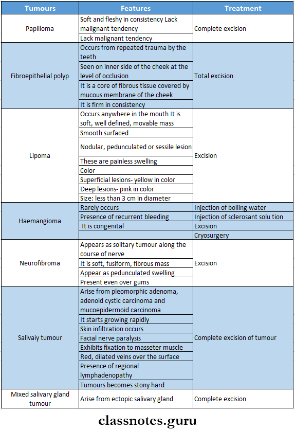
Question 1. Enumerate and write tumors of the cheek and floor of the mouth
Answer:

Tumors Of The Cheek And Floor Of The Mouth

Diseases Of The Oral Cavity Tumours Of Cheek And Floor Of The Mouth

Question 2. Discuss the etiology, clinical features, diagnosis, and management of carcinoma of the tongue.
Answer:

Carcinoma Of Tongue: Carcinoma of the tongue is a common lesion

Carcinoma Of Tongue Etiology:

  • Premalignant conditions like
    • Leukoplakia
    • Erythroplakia
  • Six
    • Smoking
    • Syphilis
    • Spices
    • Spirit
    • Sepsis.
    • Sharp tooth

Carcinoma of Tongue Clinical Features

  • Age- 50 years of age
  • Sex- males are commonly affected
  • Site- common in anterior 2/3rd of tongue and edges
  • Excessive salivation
  • A painless lump or ulcer develops on the surface of the tongue
  • Foetor oris
  • Ankyloglossia
  • Pain due to involvement of nerves
  • Hoarseness of voice and dysphagia- if carcinoma involves posterior 1/3rd of tongue
  • Enlarged cervical lymph nodes

Dental diseases questions and answers

Carcinoma of Tongue Diagnosis:

  • Edge biopsy- done under general anesthesia
  • FNAC- of lymph nodes
  • Indirect and direct laryngoscopy- to examine posterior third growth
  • CT scan to detect extension of carcinoma
  • Chest radiograph- to detect bronchopneumonia

Carcinoma of Tongue Management

  • Surgery
    • Wide incision
      • Indicated if the growth is less than 1 cm in diameter
      • A wide incision with a 1 cm margin and depth of 1 cm is used
    • Partial Glossectomy
      • Indicated when the lesion is less than 2 cm and confined to the lateral border of the tongue
      • About one-third of the tongue is removed
    • Hemiglossectomy
      • Indicated when radiotherapy fails
      • Removal of half of the anterior 2/3rd of the tongue is done
    • Total glossectomy
      • Indicated in pervasive growth involving the entire tongue
      • Combined with radiotherapy
    • Commando Operation
      • Indicated when carcinoma of the tongue is fixed to the mandible
      • It comprises of
        • Hemiglossectomy
        • Hemimandibulectomy
        • Removal of the floor of the mouth
        • Radical neck dissection
  • Radiotherapy
    • Interstitial radiotherapy
      • Indicated as preliminary treatment when the growth is more than 1 cm in diameter in the anterior 2/3rd of the tongue
    • Teletherapy
      • Useful in carcinoma of posterior l/3rd of the tongue when the lesion is larger than 2 cm in diameter
      • Cobalt 60 unit is used
    • When there is a large tumor, both the primary site and neck are irradiated to 4500 rads
  • Chemotherapy
    • Amethoprin- 50 mg/ day for 5 days can be used to reduce the size of the tumor
  • Treatment of lymph node
    • When lymph nodes are not enlarged- prophylactic block dissection of the neck is done
    • When lymph nodes are enlarged- Commando’s operation is carried out
    • When enlarged lymph nodes are fixed- deep radiotherapy is done

Oral cavity disorders short questions

Question 3. Describe the etiology, clinical features, diagnosis, and treatment of carcinoma of the cheek.
(or)
Describe the etiology, and pathology of carcinoma of the cheek and how you will manage it if it involves the mandible
Answer:

Carcinoma Of Cheek: Squamous cell carcinoma is a common carcinoma of the cheek

Carcinoma Of Cheek Etiology:

  • Six Ss
    • Smoking
    • Spirit
    • Spices
    • Sharp tooth
    • Sepsis
    • Syphilis
  • Premalignant Conditions
    • Leukoplakia
    • Erythroplakia
    • Hyperplastic candidiasis
    • Submucosal fibrosis
  • Betel Nut Chewing

Diseases Of The Oral Cavity Carcinoma of Cheek Pathology

Carcinoma of Cheek Clinical Features:

  • Development of exophytic growth- cauliflower-like growth
  • Nonhealing ulcer develops
  • Edges are everted
  • Ulcer bleeds on touch
  • Pain occurs when it is infected
  • Fixity to underlying structures
  • Trismus
  • Dysphagia
  • Halitosis
  • Enlargement of submandibular and upper deep cervical lymph nodes

Carcinoma of Cheek Diagnosis:

  • Wedge biopsy
  • OPG- to rule out an extension
  • FNAC- from lymph nodes
  • CT scan- to detect extension

Carcinoma of Cheek Treatment

  • Surgery
    • Wide excision followed by split skin graft for small superficial ulcer
    • Wide excision followed by flap reconstruction for infiltrative ulcer
  • Radiotherapy
    • External radiotherapy
      • Large total doses of 6000-8000 cGy units are given
    • Interstitial radiotherapy
      • Indicated in infiltrative small lesions
  • Advanced Carcinoma Of The Cheek
    • T3 and T4 lesions require surgery followed by postoperative radiotherapy
    • Repaired by myocutaneous flap
    • Reconstruction after surgery can be done by
      • Split skin graft
      • Deltopectoral cutaneous flap
      • Forehead flap
      • Pectoralis major myocutaneous flap
      • Cortical bone graft
      • Free flaps
  • Chemotherapy
    • Drugs used are
      • Methotrexate
      • Vincristine
      • Bleomycin
      • Adriamycin

Oral diseases viva questions

Question 4. Discuss the etiology, pathology, and clinical features of oral carcinoma and the management of gingival carcinoma

Answer:

Oral Carcinoma It is the common malignant neoplasm in the head and neck region

Oral Carcinoma Etiology:

  • Tobacco smoking
  • Use of smokeless tobacco
  • Alcohol consumption
  • Malnutrition
  • 3 fitted dentures
  • Radiations
  • Viral Infections
  • Immunosuppression
  • Chronic infections
  • Occupational hazards
  • Genetic factors
  • Pre-existing oral diseases

Oral Carcinoma Pathology: It is characterized by malignant cells

  • These cells show variable degrees of differentiation
  • Cells Invade through the basement membrane into the dermis

Oral Carcinoma Arrangement:

  • Cellfi arc arranged in concentric layers called epithelial pearls
  • They contain keratin material in the center of the coll masses
  • Cells are separated by lymphocytes

Oral Carcinoma Clinical Features:

  • Associated with oral leukoplakia and erythroplakia
  • Initial Symptoms are
    • Asymptomatic
    • White or rod, variegated patch
    • Nodule or fissure over oral mucosa
    • Painless
  • Later Symptoms
    • Past enlarging
    • Exophytic or invasive ulcer
  • Persistent induration around the periphery
  • Presence of superadded candidal infections
  • Painful lesions due to secondary infections
  • Fixation to the underlying tissues
  • Trismus
  • Invasion of the alveolar bone
  • Enlarged regional lymph nodes
  • Pathological fractures of the jaw bone

Gingival Carcinoma: Occurs due to tobacco and betel nut chewing

Gingival Carcinoma Management:

  • Surgical excision
  • Radiotherapy
  • Chemotherapy

BDS oral pathology questions and answers

Question 5. Discuss differential diagnosis, clinical features, and management of gum swelling.
Answer:

Gum Swelling Differential Diagnosis:

  • Chronic gingivitis
  • Gingival abscess
  • Periodontitis

Gum Swelling Clinical Features:

  • Mandibular gingiva is more affected than maxilla
  • The attached gingiva Is more affected than the free gingiva
  • The initial lesion appears as verrucous leukoplakia or as a small ulceration with an indurated margin
  • bony invasion occurs
  • Mobility and premature loss of teeth
  • Nonhealing extraction socket
  • Extends to the neighboring structures

Gum Swelling Management:

  • Surgical excision
  • Radiotherapy
  • Chemotherapy

Question 6. Discuss the etiology of oral cancer and how to detect nearly oral cancer and describe the steps to prevent it.
Answer:

Oral Cancer It is the common malignant neoplasm in the head and neck region

Oral Cancer Etiology:

  • Tobacco Smoking
    • Cigarettes
    • Bidis
    • Pipes
    • Cigars
  • Use Of Smokeless Tobacco
    • Snuff dipping
    • Gutkha
    • Tobacco chewing
  • Alcohol Consumption
  • Malnutrition
    • Vitamin deficiency
  • 3 fitted Dentures
    • Broken prosthesis
    • Chronic irritation
  • Radiations
    • Actinic radiation
    • X-ray radiation
  • Viral Infections
    • Herpes simplex virus
    • Human papilloma virus
    • Human immunodeficiency virus
    • Epstein burr virus
  • Immunosuppression
    • AIDS
    • Organ transplants
  • Chronic Infections
    • Candidiasis
    • Syphilis
  • Occupational hazards
  • Genetic factors
  • Pre-existing oral diseases

Oral Cancer Investigation

  • Histopathological Examination
    • Exhibit excessive proliferation of malignant cells
    • Cellular pleomorphism
    • Xuclear hyperchromatic
    • Breakdown of basement membrane
    • Intense inflammatory cell infiltration
  • Radiographic Examination
    • Exhibits bone destruction
    • Shows ill-defined radiolucent areas
    • Expansion and destruction of cortical plates
    • Pathological fractures of bone
  • Exfoliative Cytology
    • Used to detect the neoplastic cells as these cells tend to exfoliate or shed to the surface
  • Toluidine Blue Test
    • Detects dysplastic changes
  • Acridine Binding Method
    • Detects dysplastic cells
  • DNA Probe
    • Help to determine the DNA content of tumor cells
  • Tumor Markers
    • Carcinoembryonic antigen and alpha-fetoprotein are tumor markers that detect tumor

Oral Cancer Prevention:

  • Primary Prevention
    • Avoid exposure to tobacco and other deleterious habits
    • Primary Prevention Methods
      • Reducing tobacco habits by making tax hikes for tobacco-related products
      • Making changes in the manufacturing process of tobacco items
  • Secondary Prevention
    • It Includes
      • Early detection and treatment of already-developed cancer
      • Prompt management of potentially risky precancerous lesions and conditions
      • Treatment of early cancer

Oral lesions questions and answers

Question 7. Define oral ulcer. Classify ulcers. Discuss the differential diagnosis of ulcers of the oral cavity and management of ulcers of the palate.
Answer:

Oral Ulcer Ulcer is a break in the continuity of the covering epithelium

Oral Ulcer Classification:

  • Clinically
    • Spreading ulcer
    • Healing ulcer
    • Callous ulcer
  • Pathological
    • Nonspecific Ulcer
      • Traumatic ulcer
      • Arterial ulcer
      • Venous ulcer
      • Neurogenic user
      • Infective ulcer
    • Specific Ulcer
      • Syphilitic ulcer
      • Tubercle ulcer
    • Malignant Ulcer
      • Epithelioma
      • Rodent ulcer

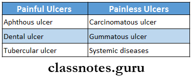
Oral Ulcer Differential Diagnosis:

  • Aphthous Ulcer
    • It is a small painful ulcer seen on the tip, undersurface, and sides of the tongue in its anterior part
    • It is small and superficial
    • Surrounded by a hyperaemic zone
    • It is quite painful
    • Has yellowish border
    • It is a painful ulcer
  • Dental Ulcer
    • Caused by mechanical irritation
    • Occurs at the periphery or undersurface of the tongue
    • It is elongated
    • Presents a slough at the base and is surrounded by a zone of erythema and induration
    • It is painful
  • Syphilitic Ulcer
    • Called snail track ulcer
    • Occurs rarely on the tongue
  • Tuberculous Ulcer
    • Young adults are affected
    • Ulcers are shallow, multiple, and greyish-yellow in color
    • Seen at the tip, margin, and dorsum of the tongue
  • Post Pertussis Ulcer
    • Occurs in children with whopping cough
    • Seen at the upper part of frenum linguae and undersurface of the tip
  • Chronic Nonspecific Ulcer
    • Occurs in the anterior 2/3rd of the tongue
    • It is moderately indurated
    • Painless
  • Carcinomatous Ulcer
    • Occurs in elder persons
    • The site involves- the anterior 2/3rd of the tongue
    • It may be a single or multiple
  • Herpetic Ulcer
    • Common in children and young adults
    • Associated with acute neuralgic pain
    • Vesicles appears
  • Ulcers Due To Glossitis
    • Ulcers are superficial and multiple with hyperemia
    • Pain occurs during meals

Management of Ulcer of Palate:

  • Removal of the causative agent
  • The membrane is swabbed away with pledgets of cotton soaked with hydrogen peroxide
  • Mouth should be kept clean by repeated use of mouthwash
  • Antibiotics used if required

Question 8. Surgical anatomy of the maxillary sinus, clinical features and management of maxillary sinusitis
Answer:

Maxillary Sinus: It is pyramidal with a base forming the lateral nasal wall and an apex at the root of the zygoma.

  • Capacity: 10-15 ml
  • Size: Height 3.5 cm; Width 2.5 cm; Anteroposterior depth 3.2 cm
  • Roof: Formed by the floor of the orbit
  • Floor: Alveolar process of maxilla
  • Anterior Wall: Facial surface of maxilla
  • Posterior Wall: Sphenomaxillary wall

Medial Wall: Lateral wall of nasal cavity

Vascular And Nerve Supply:

  • Blood Supply: Facial artery
    • Infraorbital artery
    • Greater palatine artery
  • Nerve Supply:
    • Infraorbital nerve
    • Anterior, middle, and posterior superior alveolar nerves
  • Lymphatic Drainage: Submandibular lymph nodes

Maxillary Sinusitis Clinical Features:

    • Pain on lowering your head
    • Tenderness in canine fossa
    • Redness of the area
    • Nasal discharge
    • Nose block
    • Change in voice
    • Dry cough
    • Fever
    • Malaise
    • Headache

Maxillary Sinusitis Management:

  • Antibiotics:
  • Decongestants:
  • Analgesics
  • Antihistamines
  • Steam inhalation
  • Local heat application
  • Antral lavage
  • Irrigation of sinus through lukewarm water

Oral cavity diseases long and short questions

Question 9. What are the causes of oral malignancy? How do you manage carcinoma of the cheek?
Answer:

Causes of Oral Malignancy

  • Tobacco Smoking
    • Cigarettes
    • Bidis
    • Pipes
    • Cigars
  • Use Of Smokeless Tobacco
    • Snuff dipping
    • Guikha
    • Tobacco chewing
  • Alcohol Consumption
  • Malnutrition
    • Vitamin deficiency
  • 3 fitted Dentures
    • Broken prosthesis
    • Chronic irritation
  • Radiations
    • Actinic radiation
    • X-ray radiation
  • Viral Infections
    • Herpes simplex virus
    • Human papilloma virus
    • HIV
    • Epstein Burr virus
  • Immunosuppression
    • AIDS
    • Organ transplants
  • Chronic Infection
    • Candidiasis
    • Syphilis
  • Occupational Hazards
  • Genetic Factors
  • Pre-Existing Oral Diseases

Diseases Of The Oral Cavity Short Essays

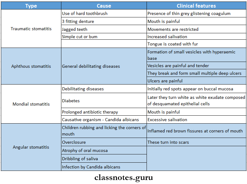
Question 1. Stomatitis

Answer:

Stomatitis

  • Stomatitis used to describe any kind of inflammation of the lining of the mouth
  • It may affect the surface of the tongue

Stomatitis Causes:

  • Trauma
  • Mechanical
  • Chemical agents
  • Thermal injury
  • Radiotherapy
  • Idiopathic
  • Malnutrition

Stomatitis Types:

  • Traumatic Stomatitis
    • It is covered by a thin grey glistening coagulum
    • It heals on its own
  • Aphthous Stomatitis
    • May be solitary or multiple
    • Small vesicles with hyperaemic base appear
    • These are painful and tender
    • These vesicles break and form painful ulcers
  • Monilial Stomatitis
    • Occurs due to Candida albicans
      • Starts as a spot on the buccal mucosa
      • Excessive salivation occurs
      • Swallowing is painful
  • Recurrent Aphthous Ulceration
    • Seen in adults
    • Seen on the inner sides of the lips and cheek and the undersurface of the tongue
    • These recur in different parts of the mouth
    • Ulcers are very painful
    • There is excessive salivation
    • They heal on their own

Question 2. Erythroplakia
Answer:

Erythroplakia

  • Erythroplakia is a red patch or plaque which cannot be characterized clinically or pathologically as any other condition and which has no apparent cause
  • Erythroplakia has clearly defined margins

Erythroplakia Types:

  • Homogenous Erythroplakia
    • The lesion appears bright red, velvety soft areas on the oral mucosa
    • Has irregular but well-defined margins
  • Erythroplakia Interspersed With Patches Of Leukoplakia
    • There is the presence of multiple, irregular erythematous areas
    • Few white leukoplakic patches occur
  • Speckled Erythroplakia
    • There is the presence of soft, irregular, raised, erythematous areas in the epithelium with a granular surface
    • There are some tiny, focal white plaques distributed all over red surfaces

Erythroplakia Differential Diagnosis

  • Erosive lichen planus
  • Early squamous cell carcinoma
  • Atrophic candidiasis
  • Kaposi’s sarcoma
  • Contact allergy

Erythroplakia Treatment:

  • Deep and wide surgical excision of the lesion
  • Regular follow up

Oral mucosal diseases questions

Question 3. Carcinoma of lip
Answer:

Carcinoma Of Lip Clinical Features:

  • Age/sex: elderly males
  • Non-healing ulcer
  • Edge is everted
  • Induration present
  • The floor is covered with slough
  • Bleeding spots present
  • Fix to underlying subcutaneous tissue
  • Cervical Lymphadenopathy

Carcinoma Of Lip Differential Diagnosis:

  • Keratoacanthoma
  • Ectopic salivary gland tumour
  • Pyogenic granuloma
  • Leukoplakia

Carcinoma Of Lip Treatment:

  • Surgery:
    • Abbe flap
    • Estlander flap
  • Radiotherapy
    • Dose: 4000-6000 cGv units

Question 4. Etiology, clinical features, and management of malignant sinusitis
Answer:

Malignant Sinusitis Etiology

  • Genetic mutation
  • Smoking
  • Long-term exposure to chemicals and irritants
  • Infections like human papillomavirus

Malignant Sinusitis Clinical features

  • Difficulty breathing through the nose
  • Loss of sense of smell
  • Nose bleed
  • Discharge from nose
  • Facial pain and swelling
  • Watery eyes
  • A sore or lesion on the roof of the mouth
  • Vision problems
  • Lump in neck
  • Difficulty in the opening of the mouth

Questions on dental caries and oral infections

Question 5. Write about Leukoplakia
Answer:

Leukoplakia

Leukoplakia is a whitish patch or plaque that cannot be characterized, clinically or pathologically, as any other disease and which is not associated with any other physical or chemical causative agent except the use of tobacco.

Leukoplakia Etiology:

  • Smoking
  • Spices
  • Spirits
  • Sharp tooth
  • Sunlight
  • Syphilis

Leukoplakia Stages

  1. Keratosis
  2. Acanthosis
  3. Dyskeratosis
  4. Speckled leukoplakia
  5. Carcinoma in situ

Leukoplakia Treatment

  • Stoppage of all habits
  • Surgical excision
  • Cryosurgery
  • Administration of vitamin A

Diseases Of The Oral Cavity Short Answers

Question 1. Angular stomatitis
Answer:

Angular Stomatitis

Angular Stomatitis is a superficial ulceration at the corner of the mouth

Angular Stomatitis Causes:

  • Licking the corners of the mouth
  • Over closure
  • Dribbling of saliva
  • Atrophic oral mucosa

Angular Stomatitis Clinical Features:

  • Inflamed red-brown fissures at the comer of the mouth
  • Results in scar formation

Angular Stomatitis Treatment:

  • Improve general health
  • Application of mercurochrome or Gentian violet B Vitamin supplement
  • Iron supplements

Question 2. Tongue tie
(or)
Ankyloglossia
Answer:

Tongue Tie

Tongue Tie is a congenital developmental condition characterized by fixation of the tongue to the floor of the mouth

Tongue Tie Clinical Features:

  • Males are commonly affected
  • Defective speech
  • Dental deformities
  • Difficulty in sucking
  • Difficulty in swallowing
  • Tension in anterior lingual gingiva

Tongue Tie Treatment:

  1. Frenulectomy- in severe cases of ankyloglossia

Fungal infections of oral cavity questions

Question 3. Cancrum Oris
Answer:

Cancrum Oris

Cancrum Oris is an extensive ulcerative disease of cheek mucosa occurring in malnourished children

Cancrum Oris Precipitating Factors:

  • Malnutrition
  • Major diseases like diphtheria
  • Vincent’s organism

Cancrum Oris Treatment:

  • Ryle’s tube-feeding
  • Improve the nutrition
  • Antibiotics: metronidazole-400 mg TID for 7-10 days
  • Reconstructive surgery

Cancrum Oris Complications:

  • Fibrosis
  • Septicaemia
  • Restricted jaw movement
  • Death

Question 4. Lingual thyroid
Answer:

Lingual Thyroid

  • Lingual thyroid is a round red swelling seen at the back of the tongue at the foramen caecum
  • The lingual thyroid contains thyroid tissue at the foramen caecum

Lingual Thyroid Complications:

  • Hemorrhage
  • Respiratory obstruction
  • Dysphagia
  • Speech impairment

Lingual Thyroid Treatment: L-thyroxin replacement therapy- to reduce the size of the swelling

Salivary gland disorders questions

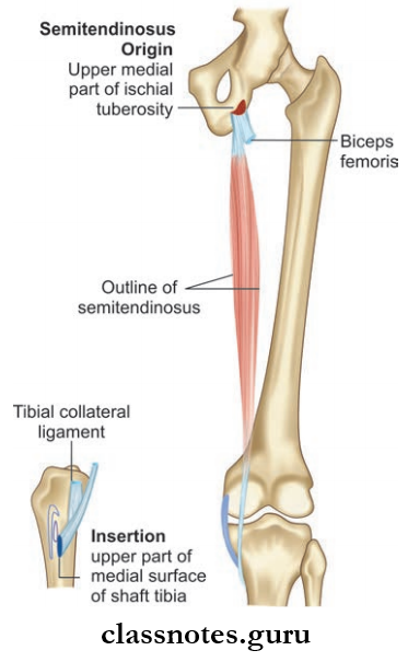
Question 5. Ulcers Of Tongue
Answer:

Ulcers Of Tongue

  • Aphthous Ulcer
    • It is a small painful ulcer seen on the tip, undersurface, and sides of the tongue in its anterior part
    • It is small and superficial
    • Surrounded by a hyperaemic zone
  • Dental Ulcer
    • Caused by mechanical irritation
    • Occurs at the periphery or undersurface of the tongue
  • Syphilitic Ulcer
    • Called snail track ulcer
    • Occurs rarely on the tongue
  • Tuberculous Ulcer
    • Ulcers are shallow, multiple, and greyish-yellow in color
    • Seen at the tip, margin, and dorsum of the tongue
  • Post pertussis Ulcer
    • Occurs in children with whopping cough
    • Seen at the upper part of frenum linguae and undersurface of the tip
  • Chronic Nonspecific Ulcer
    • Occurs in the anterior 2/3rd of the tongue
    • It is moderately indurated
  • Carcinomatous Ulcer
    • Occurs in elder persons
    • The site involves- the anterior 2/3rd of the tongue
    • It may be a single or multiple
  • Herpetic Ulcer
    • Common in children and young adults
    • Associated with acute neuralgic pain
    • Vesicles appear
  • Ulcers Due To Glossitis
  • Ulcers Are Superficial And Multiple With Hyperemia
  • Pain Occurs During Meals

Diseases Of The Oral Cavity Ulcers Of Tongue Painful And Painless Ulcers

Question 6. Haemangioma of tongue
Answer:

Haemangioma Of Tongue

  • Cavernous haemangioma occurs on tongue
  • Arises from veins
  • It consists of multiple dilated venous channels
  • It is a spongy swelling

Haemangioma Of Tongue Management:

  • Conservative Treatment
    • Injection of sclerosing agent
    • Cautery
  • Surgery
    • Ligation of feeding vessels
    • Excision of the lesion
    • Diathermy to control hemorrhage

Oral manifestations of systemic diseases Q&A

Question 7. Predisposing factors- carcinoma of the tongue
Answer:

Predisposing Factors- Carcinoma Of TheTongue

  • Predisposing factors of carcinoma of the tongue are
    • Pipe smoking
    • Syphilis
    • Chronic superficial glossitis
    • Alcohol
    • Chronic irritation
    • Betel nut

Question 8. Glossitis
Answer:

Glossitis

Involvement of the tongue due to any cause with or without inflammation is called glossitis

Glossitis Causes:

  • Local Causes
    • Mechanical trauma
    • Mechanical irritation
    • Allergic reaction
    • Dry mouth
  • Systemic Causes
    • Herpes simplex virus
    • Iron deficiency anemia
    • Aphthous ulcer
    • Oral lichen planus
    • Pemphigus Vulgaris

Glossitis Clinical Features:

  • Pain and tenderness in the tongue
  • Swelling in the tongue
  • Color changes to beefy dark red or pale
  • Halitosis
  • Difficulty in speech, eating, and swallowing
  • Loss of papillae on the tongue

Question 9. Sinusitis
Answer:

Sinusitis Etiology:

  • Nasal infections
  • Dental infections
  • Trauma

Sinusitis Causative Organisms:

  • Streptococcus
  • Pneumococci
  • Staphylococci
  • Clinical Features:
  • Pain on lowering your head
  • Tenderness in canine fossa
  • Redness of the area
  • Nasal discharge
  • Nose block
  • Change in voice
  • Dry cough
  • Fever
  • Malaise
  • Headache

Question 10. Anaplasia
Answer:

Anaplasia Definition:

  • Anaplasia is a lack of differentiation and is a characteristic feature of most malignant tumors.
  • Depending upon the degree of differentiation, the extent of anaplasia is also variable i.e., poorly differentiated malignant tumors have a high degree of anaplasia.

Result of Anaplasia:

  1. Loss of polarity
  2. Pleomorphism
  3. N: C ratio changes from 1:5 to 1:1
  4. Anisonucleosis.
  5. Hyperchromatism.
  6. Prominent nucleolus
  7. Tumorgaint cells
  8. Chromosomal abnormalities.

Oral cancer signs symptoms questions

Question 11. Metaplasia
Answer:

Metaplasia Definition: Metaplasia is defined as a reversible change of one type of epithelial or mesenchymal adult cells to another type of adult epithelial or mesenchymal cells, usually in response to abnormal stimuli.

Metaplasia Types: Metaplasia is divided into the following two types.

  1. Epithelial Metaplasia
    1. Squamous metaplasia
    2. Columnar metaplasia
  2. Mesenchyme Metaplasia
    1. Osseous metaplasia
    2. Cartilaginous metaplasia

Question 12. Oral thrush
Answer:

Oral Thrush Features:

  • The lesions appear soft, white, and slightly elevated plaques
  • The Sites Involved Are:
  1. Buccal mucosa
  2. Tongue
  3. Gingiva
  4. Palate
  5. The floor of the mouth
  6. Entire oral cavity’ is involved in severe cases

Oral Thrush Person Affected Arc

  1. HIV patients
  2. Cancer patients undergoing chemotherapy or radiotherapy
  3. Neonates and infants
  4. Debilitated and chronically ill patients

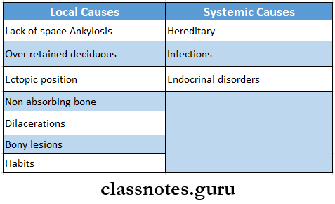
Question 13. Impacted teeth
Answer:

Impacted Teeth Definition: It is the cessation of the eruption of a tooth caused by a physical barrier or ectopic positioning of a tooth

Impacted Teeth Causes:

Diseases Of The Oral Cavity Impacted Teeth Causes

Question 14. Micrognathia
Answer:

Micrognathia

Micrognathia is an orofacial anomaly characterized by the development of jaws smaller than normal

Micrognathia Causes:

  • Pierre- Robin syndrome
  • Hallerman- Steriff syndrome
  • Trisomy 13
  • Trisomy 18
  • Turner syndrome
  • Marfan syndrome
  • Progeria

Micrognathia Types:

  • Pseudo micrognathia
  • True micrognathia

Micrognathia Clinical Features:

  • Defective alignment of teeth
  • Crowding
  • Malocclusion
  • Retruded chin
  • Difficulty in feeding
  • Difficulty in speech

Question 15. Macroglossia
Answer:

Macroglossia

Macroglossia is characterized by an abnormally large tongue in the oral cavity

Macroglossia Types:

  • Congenital
  • Acquired

Macroglossia Clinical Features:

  • Causes displacement of teeth
  • Develops tongue-thrusting habits
  • Results in obstructive sleep apnea
  • Spacing in teeth
  • Distortion of the mandibular arch
  • The lateral margin of the tongue exhibits scalloping indentation
  • Defective speech
  • Unesthetic appearance

Macroglossia Treatment:

  • Removal of the causative agent
  • Surgical reduction or trimming

Multiple choice questions on precancerous oral lesions

Question 16. Giant cell epulis
Answer:

Giant Cell Epulis

  • Peripheral giant cell granuloma is also called giant cell epulis
  • It appears in the mouth as an overgrowth of tissue due to irritation or trauma
  • They frequently appear on gingiva Color ranges from red to bluish-purple
  • It can be pedunculated or sessile
  • Common in females
  • More often found over the mandible rather than the maxilla
  • The underlying alveolar bone can be destroyed resulting in cauterization
  • Treatment involves surgical removal of bone

Clinical case-based questions on oral infections

Question 17. Causes of Macroglossia
Answer:

Causes of Macroglossia

Macroglossia is an abnormally large tongue

Question 18. Subacute osteomyelitis
Answer:

Subacute Osteomyelitis

  • It is a chronic low-grade infection of bone characterized by lack of systemic manifestation
  • The causative organism is Staphylococcus
  • The onset is insidious
  • Pain is a common symptom
  • Swelling and tenderness over the involved area are seen
  • Subacute Osteomyelitis Treatment
    • Surgical debridement
    • Antibiotics

Diseases Of The Oral Cavity Viva voce

1. Cancrum Oris is associated with leukemia

2. Carcinoma of the lip commonly occurs over the lower lip