
Pleurae Question And Answers
Question 1. Write a short note on pleural cavities and the types of pleura.
Answer:
Pleural Cavities
- Pleural cavities are present on both sides of the mediastinum
- They envelop the lungs.
Pleural Cavities Extent:
- Superiorly: Above the fist rib (4 cm above fist costal cartilage)
- Inferiorly: Up to a level, just above the costal margin
- Medially: Related to the mediastinum.
Read And Learn More: Thorax Anatomy
Pleura
- Pleura are thin serous membranes
- Pleura is formed by:
- Mesothelium (single layer of fat cells)
- Connective tissue
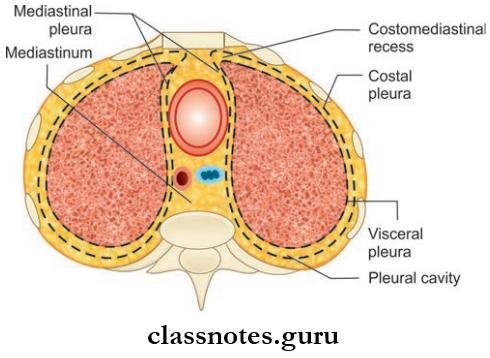
- Pleura is divided into two, based on the location
- Parietal Pleura: Outer layer of pleural cavity
- Visceral Pleura (Pulmonary Pleura): Firmly attached to lung surfaces and fissures
- The pleural cavity is actually the space between the parietal and visceral pleura
- It is filled with a thin layer of serous fluid, this helps to reduce friction between the visceral and parietal pleura, so that the visceral pleura can freely slide over the parietal pleura.
Parietal Pleura
- Due to its close relation with the thoracic wall
- It is divided into four parts, based on the part of the thoracic wall to which it is related:
- Costal Pleura:
- Costal Pleura lines the inner aspect of ribs and intercostal spaces and also an inner aspect of the sternum
- The Costal Pleura is loosely attached to the corresponding surfaces through the endo thoracic fascia.
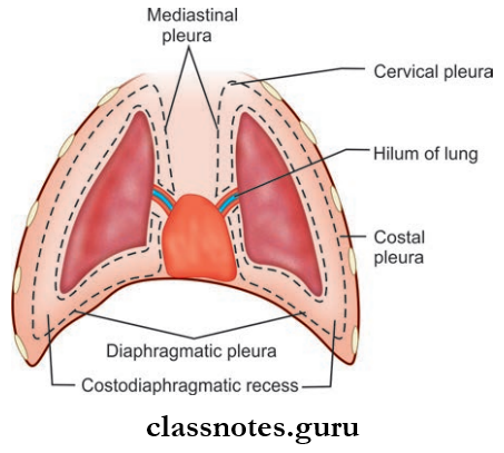
- Diaphragmatic Pleura:
- The diaphragmatic Pleura covers the upper surface of the diaphragm.
- Mediastinal Pleura
- Mediastinal Pleura is part of the pleura covering the mediastinum
- Between T 5 and T 7 levels, the mediastinal pleura extends as a sleeve-like tube over the structures passing between the mediastinum and the lung (pulmonary vessels, bronchus)
- The tubular covering of the mediastinal pleura along with the structures passing through it forms the root of the lung
- Mediastinal pleura becomes continuous with the visceral pleura at the hilum of the lung.
- Cervical Pleura
- Also known as the Dome of Pleura
- Cervical Pleura is a dome-shaped layer
- Cervical Pleura extends 5 cm above the first costal cartilage and 2.5 cm above the medial 1/3rd of the clavicle
- The cervical pleura is covered superiorly by the Suprapleural membrane
Parietal Pleura Relations:
- Anteriorly
- Scalenus anterior muscle
- Subclavian artery
- Posteriorly
- First rib
- Cervicothoracic ganglion
- Superior intercostal artery
Nerve Supply Of Parietal Pleura:
They are supplied by somatic afferent fibers through intercostal nerves and phrenic nerves
Costal Pleura: Intercostal nerve
Diaphragmatic pleura and mediastinal pleura: Phrenic nerve.
Parietal Pleura Applied Anatomy
- Since the costal pleura is innervated by intercostal nerves, its pain is felt to the thoracic wall
- Since diaphragmatic and mediastinal pleura are innervated by phrenic nerves, pain is felt on the supraclavicular region and lateral surface of the neck.
Visceral Pleura
- Also known as the pulmonary pleura
- It is firmly attached to the surfaces and fissures of the lung
- It is continuous with the parietal pleura at the hilum
- Nerve supply: By visceral afferent nerves, so, as a result, its pain is insensitive.
Anatomy of pleura
Question 2. What is the pulmonary ligament?
Answer:
Pulmonary Ligament
- The parietal pleura runs downwards as a thin fold from the root of the lung and extends beyond the hilum up to the mediastinum, known as the pulmonary ligament
- It contains loose areolar tissue and few lymphatics
- It stabilizes the position of the inferior lobe and also acts as a space for pulmonary veins to expand during increased venous return.
Question 3. Write a note on pleural recess.
Answer:
Pleural Recess
- In normal quiet respiration, the lungs do not fully occupy the anterior and inferior regions of the pleural cavity
- These regions/spaces are called a pleural recess
- They are spaces provided for the lungs to expand during deep inspiration
- Costomediastinal Recess
- Anterior recess, formed by costal and mediastinal pleura
- Lies behind sternum and costal margin
- The left costomediastinal recess is larger than the right
- In normal quiet respiration, the right costomediastinal recess is filed, but a part of the left costomediastinal recess is free due to the presence of cardiac notch.
- Costodiaphragmatic Recess
- Largest recess
- Recess used for clinical purposes
- Formed between costal and diaphragmatic pleura
- Lies inferiorly
- Extent: From 8th to 10th ribs in the mid-axillary line
- Vertically it is about 5 cm long
- Only gets filed during deep inspiration.
- Pleural Recess Applied Anatomy
- Pneumothorax: The pleural cavity sometimes gets filled with air
- Traumatic Pneumothorax: It can be due to injury to the thoracic wall or lung, leading to leakage of air into the pleural cavity
- Spontaneous pneumothorax: Occurs by leakage of air from the lung, resulting from rupture of cysts or lesions, it is associated with pulmonary tuberculosis.
Question 4. Write a brief on the blood supply and lymphatic drainage of the pleura.
Answer:
Parietal Pleura
1. Arterial Supply:
- Intercostal arteries
- Internal thoracic artery
- Musculophrenic artery.
2. Venous Drainage: Azygos vein and internal thoracic vein.
Visceral/Pulmonary Pleura
- Arterial supply: Bronchial arteries
- Venous drainage: Bronchial veins.
Lymphatic Drainage Of Pleura
1. Parietal Pleura
- Intercostal lymph nodes
- Internal mammary lymph nodes
- Posterior mediastinal lymph nodes
- Diaphragmatic lymph nodes.
2. Visceral/Pulmonary Pleura:
Bronchopulmonary Lymph Nodes
Pleural cavity anatomy
Pleurae Multiple Choice Questions
Question 1. ‘Pulmonary cavity’ and ‘pleural cavity’ are different names for the same thing:
- True
- False
Answer: 2. False
Question 2. The lowest extent of the pleural cavity into which tissue does not extend is known as:
- Costodiaphragmatic recess
- Costomediastinal recess
- Cupola
- Inferior mediastinum
Answer: 1. Costodiaphragmatic recess
Question 3. At which location is the parietal pleura continuous with visceral pleura?
- On the surface of the mediastinum
- Throughout the entire pulmonary cavity
- At the hilum of the lungs
- On the diaphragm
Answer: 3. At the hilum of the lungs
Pleura histology
Question 4. The portion of the parietal pleura that extends above the first rib is called the:
- Costodiaphragmatic recess
- Costomediastinal recess
- Costocervical recess
- Cupola
Answer: 4. Cupola
Question 5. The pleural cavity near the cardiac notch is known as the:
- Costodiaphragmatic recess
- Costomediastinal recess
- Cupola
- Hilum
- Pulmonary ligament
Answer: 2. Costomediastinal recess