
Trachea Esophagus And Thoracic Duct Question And Answers
Question 1. Describe the features and relations of the esophagus.
Answer:
Esophagus Anatomy
Esophagus Structure
- Esophagus is a long muscular tube
- Esophagus Length: 25 cm
- Esophagus Width: 2 cm
- Esophagus Function: Transport of food from pharynx to the stomach
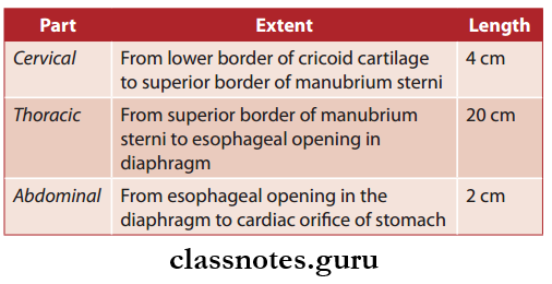
- Parts of esophagus.
Read And Learn More: Thorax Anatomy
Esophagus Gross Features
- Esophagus Extent: Pharynx to cardiac orifice of stomach
- Esophagus Lumen:
- It is flattened anteroposteriorly
- Normally it is in a collapsed state but dilates during the passage of food
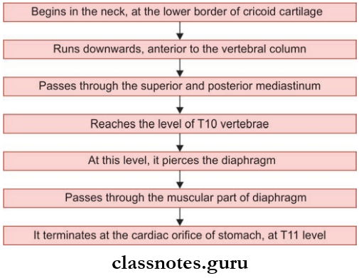
Esophagus Course
Esophagus Anatomy
Esophagus Curvatures: The Esophagus during its course shows few curves
- Two anteroposterior curvatures
- Two side-to-side curvatures.
Esophagus Anatomy
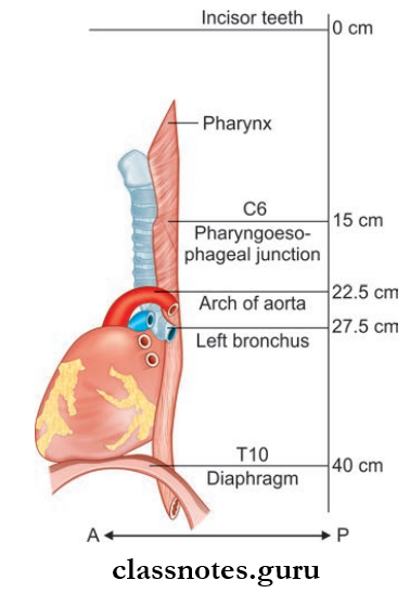
Esophagus Constrictions There are four sites of constrictions present in the esophagus
- First Constriction: It is at a distance of 15 cm, at the pharyngoesophageal junction
- Second Constriction: It is at a distance of 22.5 cm at the place where it is crossed by the arch of the aorta
- Third Constriction: It is at a distance of 27.5 cm, at a place where it is crossed by the left principal bronchus
- Fourth Constriction: It is at a distance of 40 cm, at a place where it pierces the diaphragm.
Thoracic duct anatomy
Esophagus Applied Anatomy: Constrictions of the esophagus act as potential sites at which the swallowed foreign bodies can get stuck.
Esophagus Relations:
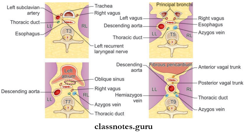
1. Esophagus Relations Cervical part
- Anteriorly
- Trachea
- Recurrent laryngeal nerve
- Posteriorly
- Prevertebral fascia
- Vertebral column
- Laterally
- Common carotid artery
- Lateral lobe of the thyroid
- Left Side
- Thoracic duct
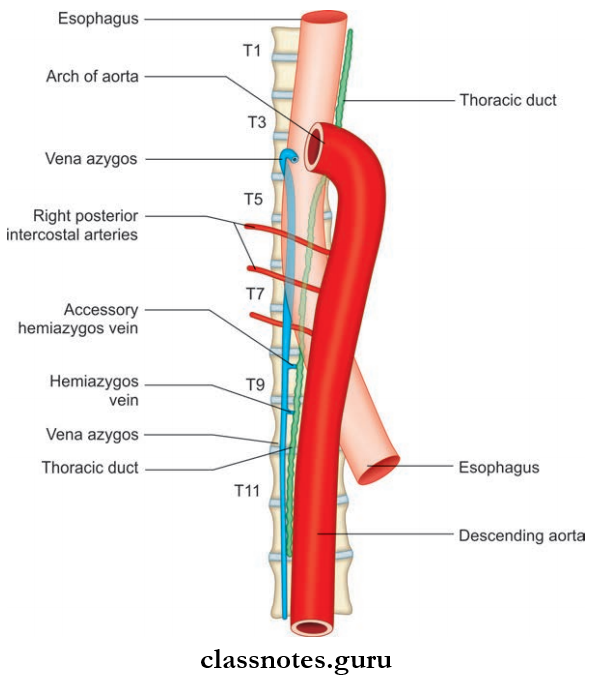
2. Esophagus Relations Thoracic Part
- Anteriorly
- Trachea
- Arch of aorta
- Right principal bronchus
- Fibrous pericardium
- Oblique sinus
- Diaphragm
- Posteriorly
- Vertebral column
- Right posterior intercostal arteries
- Thoracic duct
- Azygos and hemiazygos veins
- Descending thoracic aorta
- Right
- Pleura
- Right lung
- Right vagus
- Azygos
- Left
- Arch of aorta
- Left subclavian artery
- Thoracic duct
- Pleura
- Left lung
Thoracic duct function
3. Esophagus Relations Abdominal Part
- Anteriorly
- The posterior surface of the left lobe of the liver
- Left gastric nerve
- Posteriorly
- Left crus of the diaphragm
- Right gastric nerve
Question 2. Write a note on blood supply, lymphatic drainage, and nerve supply of the esophagus.
Answer:
Esophagus Arterial Supply
- Cervical Part: Inferior thyroid arteries
- Thoracic Part: Descending thoracic aorta and bronchial arteries
- Abdominal Part: Left gastric artery and left phrenic artery
Esophagus Venous Drainage
- Cervical Part: Inferior thyroid
- Thoracic Part: Azygos and hemiazygos veins
- Abdominal Part: Hemiazygos and left gastric vein
Esophagus Lymphatic Drainage
- Cervical Part: Deep cervical lymph nodes
- Thoracic Part: Posterior mediastinal nodes
- Abdominal part: Left gastric nodes
Esophagus Nerve Supply
- Sympathetic Supply:
- These nerves reach it through splanchnic branches of the sympathetic trunk.
- Esophagus Parasympathetic Supply:
- The Upper Half Of The Esophagus: Recurrent laryngeal nerve
- Lower Half Of Esophagus: Esophageal plexus
Question 3. Write a brief on the development of the esophagus.
Answer:
- Developed from posteriormost part of the foregut
- It is short in the beginning but it lengthens quickly due to the descent of the heart and lungs
- Musculature is derived from the splanchnic mesenchyme surrounding the foregut.
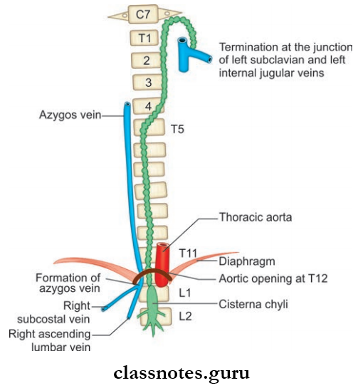
Question 4. Write a note on the thoracic duct.
Answer:
Esophagus Disease
Thoracic Duct Features
- Largest lymph vessel in the body
- It is 40–45 cm long
- Extent: From the upper part of the abdomen to the lower part of the neck.
Thoracic Duct Gross Features
Thoracic Duct has a characteristic beaded appearance due to the presence of numerous valves.
Thoracic Duct Course
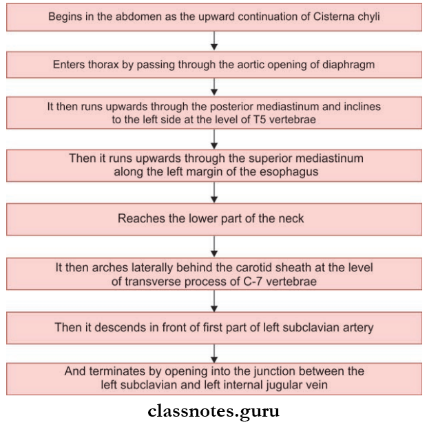
Mnemonic: Thoracic duct: Relation to azygos vein and esophagus.
‘The duck between two geese: Thoracic duct (duck) is between two geese, azygos and esophagus
Esophagus Disease
Question 5. Write a note on the trachea.
Answer:
Trachea Structure
- Wide tube
- Lying more or less in the midline, in the lower part of the neck
- Length: 10–15 cm
- External Diameter:
- Males – 2 cm
- Females – 1.5 cm
- Internal diameter – 12 mm
- The upper end of the trachea is the continuation of the larynx
- The laryngotracheal junction lies at the level of the 6th cervical vertebrae
- The trachea enters the thorax through the superior thoracic inlet and occupies the superior mediastinum
- Lower end of trachea end by bifurcating into right and left principal bronchi
- The level of bifurcation corresponds to the lower border of the T4 vertebrae or the lower border of the manubrium sterni.
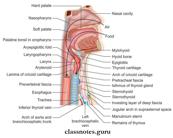
Trachea Relations:
1. Trachea Relations Anteriorly
- Manubrium sterni
- Sternothyroid muscle
- Left brachiocephalic veins
- Arch of aorta
- Brachiocephalic trunk
2. Trachea Relations Posteriorly
- Esophagus
- T1–T4 vertebrae
3. Trachea Relations Right side
- Right lung
- Right vagus
- Azygos vein
4. Trachea Relations Left side
- Arch of aorta
- Left subclavian artery
- Left common carotid artery
- Left recurrent laryngeal nerve
- Arterial Supply: Inferior thyroid arteries
- Venous Drainage: Left brachiocephalic vein
- Lymphatic Drainage: Pretracheal and paratracheal nodes
Trachea Nerve Supply:
- Sympathetic Supply: From middle cervical ganglion
- Parasympathetic Supply: From vagus and recurrent laryngeal nerves.
Thoracic duct drainage
Trachea Esophagus And Thoracic Duct Multiple Choice Questions And Answers
Question 1. Trachea bifurcates at the level of:
- Lower border of T4 vertebrae
- The lower border of T5 vertebrae
- The upper border of T3 vertebrae
- The upper border of T4 vertebrae
Answer: 1. Lower border of T4 vertebrae
Question 2. One should watch for _____________ while passing a Ryle’s tube for gastric analysis:
- Structures anterior to the esophagus
- Constrictions of the esophagus
- Pericardium
- Trachea
- None of the above
Answer: 2. Constrictions of the esophagus
Question 3. The length of the esophagus in an adult is:
- 15 cm
- 20 cm
- 25 cm
- 30 cm
Answer: 3. 25 cm
Thoracic duct location
Question 4. What is true of the anatomy of the trachea?
- Starts at the level of C4
- Drains to axillary lymph nodes
- Is supplied by the glossopharyngeal nerve
- Is marked at its lower end by the sternal angle
Answer: 4. Is marked at its lower end by the sternal angle
Question 5. The esophagus is narrowest at:
- Level of cricopharyngeus
- C6
- At cardiac orifice
- C4
Answer: 1. Level of cricopharyngeus