
Superior Vena Cava And Aorta Question And Answers
Question 1. Describe briefly on the formation, course, relations, and tributaries of superior vena cava.
Answer:
Superior Vena Cava
- Large venous channel
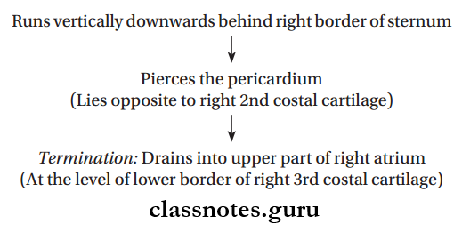
- Receives blood from upper half of body and drain it into the upper end of right atrium
- 7 cm long, 1.25 cm in diameter
- Valve less
Read And Learn More: Thorax Anatomy
- Location:
- Extrapericardial part: Superior mediastinum
- Intrapericardial part: Middle mediastinum.
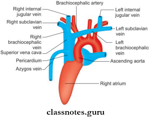
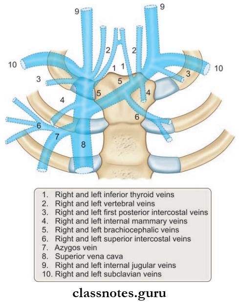
Superior Vena Cava Formation
- Formed by the union of right and left brachiocephalic veins
- The point of formation lies at the lower border of the sternal end of the fist costal cartilage.
Superior Vena Cava Course
Superior Vena Cava Relations:
- Anterior
- Chest wall
- Right internal thoracic vessels
- Anterior margin of right lung and pleura
- Medial
- Ascending aorta
- Brachiocephalic artery
- Posterior
- Right pulmonary artery
- Right bronchus
- Trachea
- Right vagus
- Lateral
- Right pleura
- Right lung
- Right phrenic nerve
- Pericardiophrenic vessels
Superior Vena Cava Tributaries
- Right and left brachiocephalic veins
- Azygos vein: At the lower end of extra pericardial part
- Mediastinal veins
- Pericardial veins.
Vena cava anatomy
Question 2. What are the parts of aorta?
Answer:
Parts of Aorta
- Largest artery
- Receives oxygenated blood from left ventricle
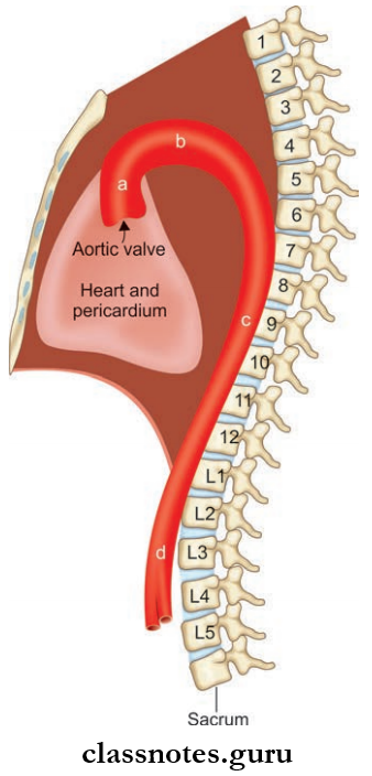
- Aorta can be divided into four parts:
- Ascending aorta
- Arch of aorta
- Descending thoracic aorta
- Abdominal aorta.
Question 3. Write a note on ascending aorta.
Answer:
Structure ascending aorta is 5 cm long, 3 cm in diameter, enclosed in a pericardium.
Ascending Aorta Origin
- From the upper end of left ventricle
- At the lower border of 3rd costal cartilage behind the left half of the sternum
Ascending Aorta Course: Runs upwards, forward, and to the right
Ascending Aorta Termination: At the level of sternal angle, it continues as the arch of the aorta
Ascending Aorta Branches: Right coronary artery and left coronary artery.
Question 4. What are aortic sinuses?
Answer:
- They are dilatations of the vessel wall at the roots of the aorta, just above the cusps of the aorta
- They Are Three In Number:
- Anterior: Gives rise to the right coronary artery
- Right Posterior And Left Posterior: Gives rise to the left coronary artery.
Aortic Sinuses Relations:
- Anteriorly
- Sternum
- Pericardium
- Pulmonary trunk
- Infundibulum of right ventricle.
- Posteriorly
- Right principal bronchus
- Right pulmonary artery
- Left atrium
- Transverse sinus.
Vena cava function
Question 5. Write a note on the arch of the aorta.
Answer:
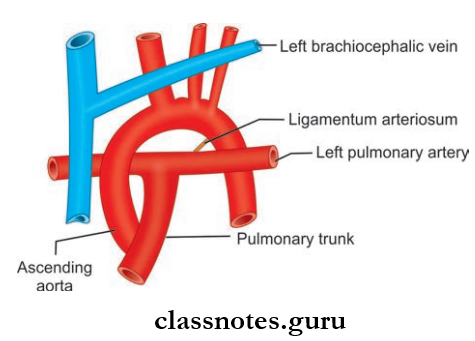
Arch Of Aorta Introduction
- Location: Superior mediastinum
- Level: Lower half of manubrium sterni
- It is the continuation of the ascending aorta at the sternal angle and also ends at the level of sternal angle (Both origin and termination at the same level)
- Derived from the ventral part of the aortic sac, its left horn and left arch artery
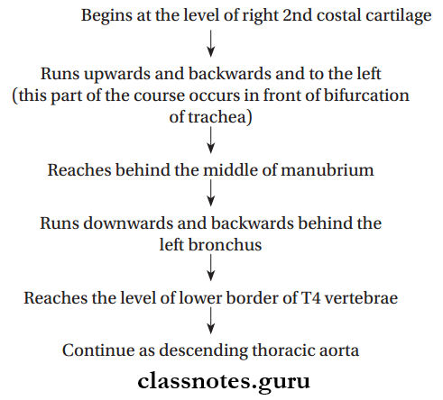
Arch Of Aorta Course:
Arch Of Aorta Relations:
- Anterior And To The Left
- Left pleura and lung
- Left superior intercostal vein
- Nerves:
- Left phrenic nerve
- Left vagus nerve
- Left cardiac nerves
- Posterior And To The Left
- Trachea
- Esophagus
- Left recurrent laryngeal nerve
- Thoracic duct
- Vertebral column.
- Superior
- Brachiocephalic trunk
- Left common carotid artery
- Left subclavian artery
- Left brachiocephalic vein
- Thymus.
- Inferior
- Bifurcation of the pulmonary trunk
- Left branches
- Ligamentum arteriosum
- Superfiial cardiac plexus
- Left recurrent laryngeal nerve.
Arch Of Aorta Branches
- Brachiocephalic artery
- Left common carotid artery
- Left subclavian artery.
Vena cava circulation
Superior Vena Cava And Aorta Multiple Choice Questions
Question 1. Which of the following statements concerning the relations of the arch of the aorta is incorrect?
- The ascending aorta arches backward to reach the body of the fourth thoracic vertebra
- The arch is crossed on its left side by the phrenic and vagus nerves
- The left recurrent laryngeal nerve passes upwards on the left side of the arch of the aorta
- Ends by becoming the thoracic aorta posterior to the 2nd left sternocostal joint
Answer: 3. The left recurrent laryngeal nerve passes upwards on the left side of the arch of the aorta
Question 2. The arch of the aorta is related to:
- Jugular notch
- Angle of Louis
- The midline of the manubrium sterni
- Second costal cartilage
Answer: 3. Midline of manubrium sterni
Question 3. Which of the following is false about the arch of the aorta?
- It starts from the 2nd right sternocostal joint
- It arches over the anterior surface of the trachea while ascending diagonally posteriorly
- It is a content of superior and posterior mediastinum
- It is crossed anteriorly by the left phrenic nerve and left vagus nerve
Answer: 3. It is a content of superior and posterior mediastinum