
The scapular region consists of the muscles, intermuscular spaces, nerves, vessels, and anastomosis around the scapula.
Scapular Region Question And Answers
Question 1. Enumerate the muscles around the scapula.
Answer:
- Scapulae originate from the scapula and are inserted into the humerus, hence called scapulohumeral muscles.
- Scapulae are also called intrinsic shoulder muscles.
- The scapula acts on the glenohumeral joint.
- Scapulae are:
- Deltoid
- Supraspinatus
- Infraspinatus
- Teres major
- Teres minor
- Subscapularis.
Read And Learn More: Upper Limb
Question 2. Write a note on the deltoid muscle.
Answer:
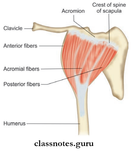
Deltoid Muscle
- The Deltoid Muscle is a thick, powerful, and curved triangular muscle covering the shoulder joint contributing to its rounded contour.
- The Deltoid Muscle resembles the inverted Greek letter delta, hence the name
- Structurally, it is divided into 3 parts:
- Clavicular part (unipennate)
- Acromion part (multipennate)
- Spinous part (unipennate)
Scapula Anatomy Notes PDF
Deltoid Muscle Origin
- Clavicular Part (Unipennate): Anterior part of lateral 1/3rd of clavicle
- Acromion Part (Multipennate): Lateral border of acromion
- Spinous part (Unipennate): Lower lip spine of the scapula
Deltoid Muscle Insertion
- Deltoid tuberosity of the humerus
- Nerve Supply
- Accessory nerve
Deltoid Muscle Actions
- Anterior Fibers: Flexion and medial rotation
- Middle Fibers: Abduction of the arm
- Posterior Fibers: Extension and medial rotation of the arm
Deltoid Muscle Clinical Anatomy
- Intramuscular injections are given commonly in the lower half of the muscle to avoid injury to the axillary nerve which winds around the neck of the humerus under the muscle.
- In the shoulder region, injury to the supraspinatus tendon is common, and the patient feels difficulty in the initiation of abduction of the shoulder joint.
- The tendon of the supraspinatus may undergo degeneration and subsequent calcification as advances and results to rupture of the tendon.
Question 3. Write about the origin, insertion, nerve supply, and actions of the muscles around the scapula.
Answer:
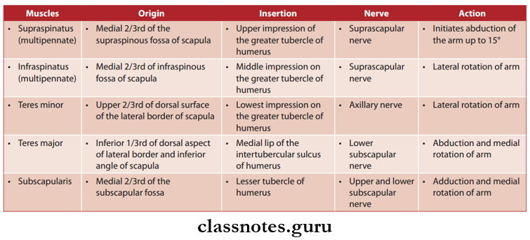
Question 4. What is the rotator cuff or musculocutaneous cuff of the shoulder joint? Write about its formation and functions.
Answer:
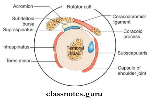
- It is a fibrous sheath formed by the flattened tendons of four scapulohumeral muscles.
- They are:
- Supraspinatus fusing superiorly
- Infraspinatus fusing posteriorly
- There is minor fusion posteriorly
- Subscapularis fusing anteriorly
- It is blended with the capsule of the shoulder joint.
Scapula Bones – Medical Students’ Guide
Rotator Cuff Or Musculocutaneous Cuff Functions
- It gives strength to the shoulder joint.
- It grasps and holds the relatively larger head of the humerus
- against smaller and shallower glenoid cavities.
Rotator Cuff Or Musculocutaneous Cuff Clinical Anatomy
- The cuff is deficient inferiorly, through which inferior dislocation of the humerus from the joint can take place more easily.
Mnemonic: Rotator cuff muscles
- The SITS muscles:
- Clockwise from top:
- Supraspinatus
- Infraspinatus
- Teres minor
- Subscapularis
- A pro baseball pitcher has injured his rotator cuff muscles.
- As a result, he SITS out for the rest of the game and then gets sent to the minor leagues.
Scapula Bone Viva Questions and Answers
Question 5. Write a short note on the subacromial bursa.
Answer:
- Suba cromial Bursa is the largest bursa of the body.
- The Subacromial Bursa is situated below the coracoacromial arch and the deltoid muscle.
- Under the bursa, there are:
- Tendon of supraspinatus
- Greater trochanter of the humerus.
Subacromial Bursa Functions
- Subacromial Bursa protects the supraspinatus tendons against friction with the acromion process.
- Subacromial Bursa facilitates the movements of the greater tubercle of the humerus under the acromion during overhead abduction.
Subacromial bursa Clinical Anatomy
- Subacromial bursitis commonly appears after inflammation of the supraspinatus tendon. It causes pain when pressure is applied just below the acromion.
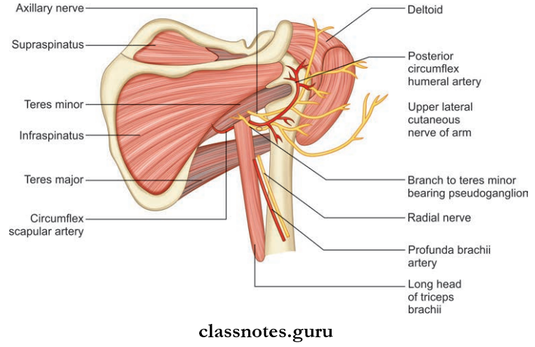
Question 6. List the intermuscular spaces with their boundaries and contents.
Answer:
- Quadrangular Spaces are two triangular and one quadrangular spaces formed by the muscles in the scapular region.
- Quadrangular Spaces are seen clearly from behind after reflecting the posterior part of the deltoid.
- They are:
Quadrangular Space Boundaries:
- Superior:
- Teres minor posteriorly
- Subscapularis anteriorly
- The capsule of the shoulder joint between the above two muscles
- Inferior: Teres major
- Medial: Long head of triceps
- Lateral: Surgical neck of the humerus.
Structures Passing Through Quadrangular Space:
- Axillary nerve
- Posterior circumflex humeral artery and vein.
Upper Triangular Space Boundaries
- Superior: Teres minor
- Lateral: Long head of triceps
- Inferior: Teres major
Structures Passing Through Upper Triangular Space:
- Circumflx scapular artery.
Lower Triangular Space Boundaries:
- Medial: Long head of triceps
- Lateral: Shaft of the humerus
- Superior: Teres major.
Scapula Structure and Features Essay
Structures PassingThrough Lower Triangular Space:
- Radial nerve
- Profunda brachii artery and vein.
Scapular Region
Question 1. The following part of the scapula forms the lateral most palpable landmark on the shoulder:
- Superior angle
- Glenoid cavity
- Coracoid process
- Acromion
Answer: 4. Acromion
Question 2. Subacromial bursa separates coracoacromial arch from the tendon of:
- Subscapularis
- Teres minor
- Supraspinatus
- Infraspinatus
Answer: 3. Supraspinatus
Question 3. Which of the following has actions similar to that of teres minor?
- Supraspinatus
- Infraspinatus
- Subscapularis
- Teres major
Answer: 2. Infraspinatus
Upper Limb Anatomy – Scapula Explained
Question 4. Which muscle does NOT substantially contribute to the stability of the shoulder joint?
- Subscapularis
- Supraspinatus
- Infraspinatus
- Teres minor
Answer: 1. Subscapularis
Question 5. Which is NOT a boundary of the quadrangular space?
- Teres major
- Teres minor
- The long head of the triceps
- Latissimus dorsi
Answer: 4. Latissimus dorsi