Diseases Of Bone And Joints Oral Pathology Essay Question And Answers

Diseases Of Bone And Joints Important Notes

  1. Fibrous Dysplasia Definition: It is an idiopathic condition, in which an area of normal bone is gradually replaced by abnormal fibrous connective tissue, which then again undergoes osseous metaplasia, and eventually the bone is transformed into a dense lamellar bone.
    • Fibrous Dysplasia Classification:
      • Monostotic – Only one bone is involved
      • Polyostotic – More than one bone is involved
        • Jaffe’s type -Polyostotic along with cafe-au-lait-skin pigmentation
        • Albright syndrome – characterized by polyostotic fibrous dysplasia, cafe-au-lait skin pigmentation, and endocrine disturbances
    • Diseases Of Bone And Joint Features
      • Cafe au lait pigmentation of skin
      • Unilateral swelling of the jaw
      • Precocious puberty
      • Egg crackling of the cortex of the bone is present
      • Later ground glass appearance is seen
      • Maxillary lesions causes obliteration of maxillary sinus
      • Spindle-shaped fibroblasts are arranged in a whorled pattern
  2. Paget’s disease
    • It is characterized by excessive and abnormal remodeling of bone
    • Affects the adult skeleton
    • Patients suffer from deafness, blindness, and facial paralysis
    • There is a progressive enlargement of the skull and maxilla because of which the patient has to change the hats and dentures frequently
  3. Cherubism
    • Manifests by the age of 3-4 years
    • Painless symmetric swelling of the mandible or maxilla occurs
    • Results in chubby face appearance
    • The deciduous teeth shed prematurely and numerous teeth are absent
    • X-ray shows numerous unerupted teeth floating in cyst-like spaces
  4. Cleidocranial dysplasia
    • it is characterized by abnormalities of the skull, shoulder girdle, jaws, and teeth
    • Skull – delayed closure of sutures and wormian bones
    • Shoulder – partial or complete absence of clavicles
    • Teeth – prolonged retention of deciduous and delayed eruption of permanent
    • Numerous supernumerary teeth are found in the mandibular premolar and incisor areas
  5. Blue sclera Is seen In
    • Osteogenesis imperfecta
    • Marfan syndrome
    • Cherubism
    • Ehlers Danlos syndrome
    • Osteopetrosis
    • Fetal rickets
    • Normal infants
  6. Marfan’s syndrome
    • Long thin extremities
    • Hyperextensibility of joints
    • Spidery fingers
    • Arachnodactyly
    • Bifid uvula
    • CVS complications
  7. Albright’s syndrome
    • Precocious puberty
    • Polyostotic fibrous dysplasia
    • Cafe-au- lait pigmentation
  8. Down syndrome
    • It occurs due to trisomy 21
    • Features
      • Hypermobility
      • Macroglossia
      • Flat face
      • Large anterior fontanelle
      • Sexual underdevelopment
      • Cardiac abnormalities
  9. Cotton wool appearance is seen in
    • Paget’s disease
    • Chronic sclerosing diffuse osteomyelitis
    • Fibrous dysplasia
    • Cemento-osseous dysplasia
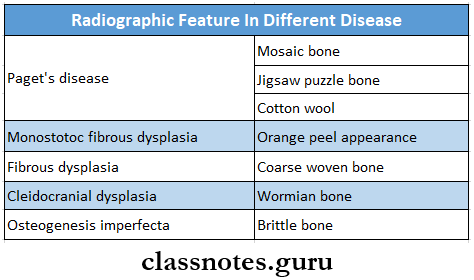
  10. Radiographic features in different disease
    Diseases Of Bone And Joints Radiographic Feature In Different Disease
    Serum affine phosphatase is elevated In  

    • Malignancy
    • Abscess of Ihrer
    • Amyloidosis
    • Leukemia
    • Sarcoidosis
  11. Pierre Robin syndrome
    • Features
      • Micrognathia
      • Geffc palate
      • Glossoprosis
  12. Diseases with cafe-Au lait spots are
    • Albright syndrome
    • Yon Recklinghausen neurofibromatosis
    • Bloome’s syndrome
    • Fanconi’s syndrome
    • Cowden’s syndrome
    • Tuberculosis sclerosis
    • Watson’s syndrome
    • Ataxia telangiectasia

Diseases Of Bone And Joints Short Question And Answer

Question 1. Classify the diseases of TMJ. Write etiology and clinical features of ankylosis
Answer:

Classification of Diseases of Temporomandibular Joint:

  1. Disorders due to extrinsic factors
    • Masticatory muscle disorders
      • Myofunctional pain dysfunction syndrome
      • Myositis
    • Problems due to trauma
      • Traumatic arthritis
      • Fracture
      • Internal disc derangement
      • Tendonitis
  2. Disorders due to intrinsic factors
    • Trauma
      • Dislocation
      • Fracture
    • Internal disc displacement
      • Anterior disc displacement with reduction
      • Anterior disc displacement without reduction
    • Arthritis
      • Osteoarthritis
      • Rheumatoid arthritis
      • Juvenile arthritis
      • Infantile arthritis
    • Developmental defects
      • Agenesis
      • Hypoplasia
      • Hyperplasia
    • Ankylosis
    • Neoplasm
      • Benign
      • Malignant

Read And Learn More: Oral Pathology Questions and Answers

Ankylosis: Ankylosis means stiff joint

Ankylosis Etiology:

  • Trauma
  • Congenital
  • Infections- osteomyelitis
  • Inflammation- Osteoarthritis
  • Systemic diseases-typhoid
  • Measles
  • Prolonged trismus

Ankylosis Types:

  • False or true ankylosis
  • Extra articular or intra articular
  • Fibrous or bony
  • Unilateral or bilateral
  • Partial or complete

Ankylosis Clinical Features:

  1. Unilateral ankylosis
    • Deviation of the chin on the affected side
    • The fullness of the face on the affected side
    • Flatness on the unaffected side
    • Crossbite
    • Angle’s class 2 malocclusion
    • Condylar movements absent on the affected side
  2. Bilateral ankylosis
    • Inability to open mouth
    • Neck chin angle reduced
    • Class 2 malocclusion
    • Protrusive upper incisors
    • Multiple carious teeth

Question 2. Enumerate bone disorders affecting the jaws. Describe the pathogenesis, clinical features, radiographic appearance, and histopathology of fibrous dysplasia.
Answer:

Bone Disorders Affecting the Jaws:

  • Osteogenesis imperfecta
  • Osteopetrosis
  • Fibrous dysplasia
  • Cheruhism
  • Mandibulofaci dysostosis
  • Pierre Robin malformation
  • Achondroplasia
  • Chondroectodermal dysplasia
  • Cleidocranial dysplasia
  • Down’s syndrome
  • Marfan syndrome
  • Infantile cortical hyperostosis

Fibrous Dysplasia:

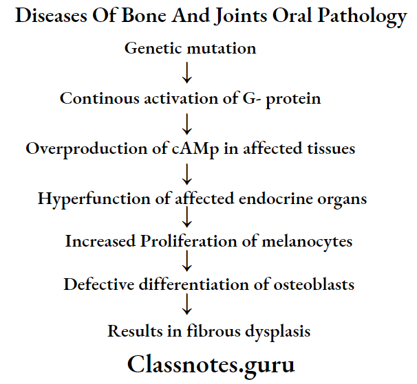
  • Fibrous dysplasia is a skeletal developmental anomaly of the bone-forming mesenchyme that manifests as a defect in osteoblastic differentiation and maturation

Fibrous Dysplasia Pathogenesis:

Diseases Of Bone And Joints Oral Pathology

Fibrous Dysplasia Types:

  • Monostotic form
  • Polyostotic form
  • Jaffe’s type
  • Albright syndrome

Fibrous Dysplasia Clinical Features:

  • Age- Occurs in the first and second decade of life
  • Sex- common in females
  • Site involved
    • Skull
    • Facial bones
    • Clavicles
    • Pelvic bones
    • Long bones-femur, tibia, humerus
  • Skeletal lesions
    • Unilateral distribution of lesions
    • Swelling on the affected side
    • Recurrent bone pain
    • Cessation of growth
    • Pathological fractures
  • Skin lesions
    • Cafe-au- Jail pigmentations
    • It consists of irregularly, pigmented, light brown, flat, melanotic spots
  • Oral manifestations
    • Slow enlarging, painless, unilateral swelling of the jaw
    • Facial deformity
    • Expansion and distortion of cortical plates,
    • Displacement of regional teeth
    • Disturbances in teeth eruption
    • Severe malocclusion
    • Maxillary lesions lead to Exophthalmos, proptosis, and nasal obstruction
    • Mandibular protuberance
  • Precocious puberty
    • Premature vaginal bleeding
    • Breast development
    • Presence of axillary and pubic hairs at the age of 2-3 years

Fibrous Dysplasia Radiographic Features:

  • Initially, it produces unilocular or multilocular radiolucent areas in bone
  • Expansion and distortion of cortical plates occurs
  • Displacement of teeth
  • The egg-cell crackling of the cortex of the bone is present
  • Later a classical ground glass or orange peel appearance of bone is seen
  • The margin of the lesion blends with the surrounding normal bone
  • Mandibular lesions cause bulging of the US inferior border
  • Narrowing of periodontal ligament
  • Thinning of lamina dura
  • Maxillary lesions causes obliteration of maxillary sinus

Fibrous Dysplasia Histopathology:

  1. Monostatic fibrous dysplasia
    • Consists of proliferating fibroblasts in the stroma of interlacing collagen fibers
    • Trabeculae of bone are multiple, coarse, irregular, and immature
    • This produces a Chinese letter pattern
    • Spheroidal areas of calcification are seen
    • Presence of giant cells
    • At the margin, the lesion blends with the surrounding bone
    • Gradually the amount of cellularity decreases and the amount of bone tissue increases
    • There is remodeling of woven bone
  2. Polyostotic fibrous dysplasia
    • Areas of fibrous metaplasia within flat and tubular bones
    • Well defined lesions
    • Rich in spindle-shaped fibroblasts arranged in a whorled pattern
    • Presence of giant cells
    • Collagen fiber bundles lack orientation

Question 3. Enumerate the osteodystrophies. Write in detail about Paget’s disease of bone.
Answer:

Osteodystrophies: Osteodystrophies are disorders of bone other than neoplastic and inflammatory conditions

Osteodystrophies Classification:

  1. Fibro-osseous lesions
    • Fibrous dysplasia
    • Periapical cementitious dysplasia
    • Focal cementitious dysplasia
  2. Giant cell lesions
    • Cheru be
    • Central giant cell granuloma
    • Peripheral giant cell granuloma
  3. Developmental disorders of bone
  4. Metabolic disorders of bone
    • Brown’s tumor
  5. Miscellaneous
    • Rickets
    • Osteomalacia

Paget’s Disease: It is a bone disorder characterized by excessive, tin- coordinated phases of bone resorption and subsequent deposition of new bone in the same area

Paget’s Disease Clinical Features:

  • Age- fifth, sixth, seventh decade of life
  • Sex- common in males
  • Sites involved
    • Weight-bearing areas- vertebral column, femur
    • Skull
    • Pelvis
    • Sternum
    • Common in maxilla than mandible
  • Present as deep and aching bone pain
  • Bilateral swelling of the involved bone
  • Bowing deformity of weight-bearing areas
  • Results in monkey-like stance
  • Waddling gait
  • Involvement of facial bones is referred to as dementia- sis ossa
  • Headache
  • Deafness, blindness
  • Facial paralysis
  • Enlargement of skull
  • Bowing of legs
  • The increased localized temperature of the skin

Paget’s Disease Histopathology:

  • The initial stage shows osteoclastic bone resorption
  • Bone is replaced by highly vascularised cellular connective tissue
  • Osteoclasts are larger and multinucleated
  • The later stage shows the deposition of new lamellar bone by osteoblast cells
  • Fatty bone marrow is replaced by fibrous stroma
  • Bone resorption and deposition produce prominent reversal and resting lines
  • The irregular pattern of such lines produces a jigsaw- puzzle or mosaic pattern
  • The affected bone is thick, sclerotic
  • Obliteration of the medullary cavity occurs
  • Chronic inflammatory cells and dilated blood capillaries are present

Paget’s Disease Radiographic Features:

  • Initially, there is the presence of radiolucent areas in the affected bone
  • In the next stage, involved bone shows haphazardly arranged newly formed bone in radiolucent areas
  • This produces the cotton wool appearance
  • The radiopacity of lesions increases due to increased osteosclerosis
  • Prognathic and pagetoid mandible
  • Obliteration of maxillary sinus
  • Hypercementosis of tooth
  • Loss of lamina dura
  • Obliteration of periodontal ligament space
  • Root resorption

Question 4. Clinical features of monostotic fibrous dysplasia
Answer:

Monostotic Fibrous Dysplasia: It is a form of fibrous dysplasia that involves single-bone

Monostotic Fibrous Dysplasia Clinical Features:

  • Common in children and young adults
  • Painless swelling of the jaw
  • Common in mandible
  • The protuberance of its inferior border
  • Misalignment or displacement of regional teeth
  • The overlying mucosa is intact
  • Maxillary lesions involve the maxillary sinus, the floor of the orbit, and the zygomatic process
  • There is a bulging of canine fossa

Question 5. Cleidocranial dysplasia
Answer:

Cleidocranial dysplasia

It is a hereditary disorder characterized by abnormal growth of the bones in the face, skull, and clavicles with a tendency for the failure of tooth eruption

Cleidocranial dysplasia Clinical Features:

  • Absence or hypoplasia of one/ both clavicles
  • Hypermobility of shoulder joints
  • Elongated frontal and occipital skull plates
  • Underdeveloped entire mid-face
  • Delayed closure of fontanelles
  • High and narrow arched palate
  • Underdeveloped paranasal sinuses
  • Photophobia
  • Multiple unerupted and impacted teeth

Cleidocranial dysplasia Radiographic Features:

  • Open sutures
  • Open fontanelles
  • Partial/complete loss of clavicles
  • Multiple impacted teeth
  • Thin roots of teeth

Question 6. Etiopathogenesis and Histopathology of cherubism
Answer:

Cherubism: It is a rare benign hereditary condition characterized by bilaterally symmetrica] enlargement of the mandible

Cherubism Etiopathogenesis:

  • It results due to
    • Anomalous development of bone
    • Latent hyperparathyroidism
    • Hormone dependent neoplasm
    • Trauma
    • Disturbance in the development of bone-forming mesenchyme

Cherubism Histopathology:

  • The presence of numerous multinucleated giant cells
  • Stroma consists of a large number of spindle-shaped fibroblasts
  • Numerous small vessels and capillaries are present
  • They are lined by endothelial cells and perivascular cuffing
  • Advanced lesions show
  • Increase in fibrous tissue
  • Decrease in giant cells
  • Formation of new bone

Question 7. MPDS
Answer:

MPDS

  • It is a disorder characterized by facial pain limited to mandibular function, muscle tenderness, joint sounds, absence of significant organic and pathologic changes in TMJ
  • It may be due to functional derangement of dental articulation, psychological state of mind, or physiological state of the joint
  • Coined by Laskin

MPDS Etiology:

  1. Extrinsic factors
    • Occlusal disharmony
    • Trauma
    • Environmental influences
    • Habits
  2. Intrinsic factors
    • Internal derangement of TMI
    • Anterior locking of disc
    • Trauma

MPDS Features:

  • Unilateral preauricular pain
  • Dull constant sound
  • Muscle tenderness
  • Clicking noise
  • Altered jaw function
  • Absence of radiographic changes
  • Absence of tenderness in ext. auditory meatus

MPDS Management:

  1. Reassurance
  2. Soft diet
  3. Occlusal correction: 7 ‘R’s
    • Remove-extract the tooth
    • Reshape grind the occlusal surface
    • Reposition orthodontically treated
    • Restore conservative treatment
    • Replace by prosthesis
    • Reconstruct TMJ surgery
    • Regulate control habits
  4. Isometric exercises
    • Opening and closing of mouth 10 times a day
  5. Medicaments
    • Aspirin: 0.3-0.6 gm/ 4 hourly
    • NSAIDS: for 14-21 days
    • Pentazocine: 50 mg/ 2-3 times a day
  6. Heat application
    • It increases circulation
  7. Diathermy
    • Causes heat transmission to deeper tissues
  8. LA injections
    • 2% lignocaine into trigger points
  9. Steroid injection
    • As anti-inflammatory
  10. Anti-anxiety drugs
    • Diazepam-2-5 mg * 10 days
  11. TENS
  12. Acupuncture

Question 8. Cherubism
Answer:

Cherubism

It was described by Jones in 1933

Cherubism Classification:

  • Based on the severity and location of the lesion
    • Grade 1- Affects Minus of the mandible
    • Grade 2- Affects ramous and body of the mandible and maxillary tuberosity
    • Grade 3 – after maxilla ami mandible entirely

Cherubism etiology:

  • Autosomal dominant trail latent hyperparathyroidism
  • Trauma
  • Disturbance in bone-forming mesenchymal

Cherubism Clinical Features:

  • Age and sex- 2-3 years males are affected
  • Site-angle of mandible bilaterally
  • Bilateral, painless, symmetrical swelling giving a chubby appearance
  • Swelling is firm to hard in consistency
  • Maxillary swelling causes pressure over the floor of the orbit
  • Due to this, pupils turn upwards giving a “heavenward look”
  • Difficulty in speech, deglutition, mastication, and respiration
  • Limited jaw movements
  • Expansion and widening of alveolar ridge
  • Flattening of palatal vault
  • Chronic lymphadenopathy
  • Malocclusion

Question 9. Osteogenesis Imperfecta
Answer:

Osteogenesis Imperfecta

It is a genetically transmitted disease of bone characterized by defective matrix formation and lack of mineralization

Osteogenesis Imperfecta Clinical Features:

Diseases Of Bone And Joints Osteogenesis Imperfecta Clinical Features

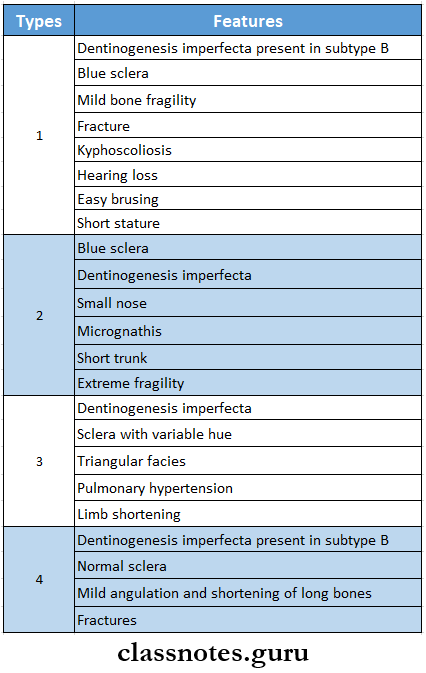
Osteogenesis Imperfecta Oral Manifestations:

  • Large head
  • Frontal bossing
  • Maxillary hypoplasia
  • Bulbous crowns of teeth
  • Class 3 malocclusion
  • Severe attrition of deciduous teeth
  • Multiple impacted permanent teeth
  • Increased incidence of osteomyelitis

Question 10. Osteopetrosis
Answer:

Osteopetrosis

  • It is also known as marble disease
  • It is a rare bone disorder characterized by increased bone density

Osteopetrosis Clinical Features:

  • Decreased bone marrow activity leading to anemia, leukopenia, and pancytopenia
  • Hepatosplenomegaly
  • Deafness, blindness, and facial paralysis due to narrowing of cranial foramina
  • Defective enamel formation
  • Short roofs
  • Pathological fractures
  • Increased incidence of osteomyelitis

Question 11. Blue sclera
Answer:

Blue sclera

  • Blue sclera is due to unusually transparent or thin sclera which causes increased visibility of choroids
  • It is seen in
    • Osteogenesis imperfecta
    • Marfan syndrome
    • Cherubism
    • Ehlers-Danlos syndrome
    • Osteopetrosis
    • Fetal rickets
    • Normal infants

Question 12. Leontiasis ossa
Answer:

Leontiasis ossa

The involvement of facial bones in Paget’s disease is known as leontiasis ossa

Leontiasis ossia Features:

  • Progressive enlargement of the maxilla
  • Widening of alveolar ridges
  • Loosening of teeth
  • Flattening of palate
  • Mouth remains open
  • In edentulous patients, there is difficulty in wearing dentures

Question 13. Albright’s syndrome
Answer:

Albright’s syndrome Features:

  • Common in females
  • It is a severe form of fibrous dysplasia involving nearly all the bones in the body
  • It is accompanied by pigmentations of the skin and endocrine disorders
  • Endocrine disorders
    • Precocious puberty
    • Goitre
    • Hyperthyroidism
    • Hyperparathyroidism
    • Cushing’s syndrome
    • Acromegaly
  • Skin lesions
    • These are coffee with milk color spots
    • There is an irregular flat area of increased skin pigmentation
  • Vaginal bleeding occurs
  • Long bones are frequently affected

Question 14. Marfan’s syndrome
Answer:

Marfan’s syndrome

It is a hereditary syndrome

Marfan’s Syndrome Clinical Features:

  • Long, thin extremities resembling spider fingers
  • Hyperextensibility of joints
  • Habitual dislocations
  • Kyphosis
  • Aortic regurgitation
  • Cardiac aneurysm
  • Mitral valve prolapse
  • Myopia, cataract
  • Retinal detachment
  • Psychological trauma

Marfan’s Syndrome Oral Manifestations:

  • Long and narrow face
  • High arched palate
  • Bifid uvula
  • Presence of multiple odontogenic cysts
  • Malocclusion
  • Temporomandibular joint dysarthrosis

Question 15. Mandibulofacial dysostosis
Answer:

Mandibulofacial dysostosis

It is a hereditary- disease characterized by defects in structures derived from 1st and 2nd branchial arches

Mandibulofacial dysostosis Clinical Features:

  • Malformation of the external ear- the absence of an external auditor canal, deformity in the middle and internal ear
  • Antimongoloid palpebral fissures
  • Coloboma of the outer portion of lower eyelids
  • Hypoplasia of the mandibular body and zygoma
  • Narrow face and depressed cheek
  • Results in bird-face appearance
  • Crowding and malocclusion of teeth
  • High arched palate
  • Atypical hair growth
  • Parotid hypoplasia
  • Narrowing of larynx and trachea
  • Difficulty in speech and respiration

Question 16. Serum alkaline phosphatase
Answer:

Serum alkaline phosphatase

  • Alkaline phosphatase occurs in many tissues of the body, especially in osteoblasts
  • It is elevated in
    • Malignancy
    • Abscess of liver
    • Amyloidosis
    • Leukemia
    • Sarcoidosis

Question 17. Pierre Robin syndrome
Answer:

Pierre Robin syndrome

It is a hereditary disease

Pierre Robin syndrome Features:

  • Mandibular micrognathia giving bird face appearance
  • Downward and backward placement of tongue
  • Difficulty in breathing, airway maintenance, feed- ind and speech
  • Malocclusion of teeth
  • Presence of multiple missing teeth or supernumerary teeth
  • Absence of TMJ
  • Mongolism
  • Congenital heart defects
  • Hydrocephaly, microcephaly
  • Mental retardation
  • Psychological trauma

Question 18. Cotton wool appearance
Answer:

Cotton wool appearance

  • Cotton wool appearance is a radiographic feature of Paget’s disease
  • In the later stage of the disease, new bone is formed in the present radiolucent areas
  • It results from thickened, disorganized trabeculae which lead to areas of sclerosis in previously lucent areas of bone
  • These areas are poorly calcified

Question 19. Peaud orange radiographic appearance
Answer:

Peaud orange radiographic appearance

  • It is seen in the later stage of fibrous dysplasia
  • Initially, there is the presence of unilocular or multi-locular radiolucent areas
  • Later quite opaque areas develop due to delicate trabeculae
  • This results in a proud orange or orange peel appearance
  • It is not well-circumscribed
  • Its margins blend with the surrounding bone

Question 20. Down syndrome
(or)
Trisomy 21
Answer:

Down syndrome

  • Down’s syndrome/trisomy 21/mongolism affects approximately 1 in 1000 births.
  • It is the most common chromosomal disorder and is the commonest cause of mental retardation.

Down syndrome or Trisomy 21 Etiology:

  • Late maternal age
  • Nondisjunction of chromosome 21 during an early stage of embryogenesis.

Down Syndrome or Trisomy 21 Clinical Features:

  • Epicanthal folds and flat facial profile,
  • Slanting eyes produce a mangoloid appearance.
  • Hands are short with a transverse single palmar crease.
  • Abnormalities of ears, trunk, pelvis, and phalanges
  • Cardiac malformations
  • Congenital malformations are common and quite disabling
  • Risk of developing acute leukemia, especially megakaryocytic leukemia.

Down syndrome or Trisomy 21 Oral Manifestation:

  • Deficient maxilla- class 3 relation,
  • Open mouth,
  • Large tongue,
  • Caries free teeth due to excess salivation.

Question 21. Brown tumor
Answer:

Brown tumor

  • The brown tumor is also known as hyperparathyroidism
  • It is an endocrine disorder occurring due to an excess of circulating parathyroid hormone

Brown tumor Types:

  • Primary hyperparathyroidism
  • Occurs due to tumour of glands
  • Secondary hyperparathyroidism
  • Occurs in response to hypocalcemia
  • Tertiary hyperparathyroidism
  • Occurs after long-standing secondary hyperparathyroidism

Brown Tumour Clinical Features: Age and sex- common in middle-aged women

  1. Classic triad
    • Kidney stones
    • Bone resorption
    • Duodenal ulcers
  2. Renal symptoms
    • Renal calculi
    • Hematuria
    • Back pain
  3. Psychological symptoms
    • Emotionally unstable
  4. GIT symptoms
    • Anorexia
    • Nausea, vomiting
  5. Skeletal
    • Bone pain
    • Pathologic fractures
    • Bone deformities
    • Hypercalcaemia
  6. Generalised symptoms
    • Muscle weakness
    • Fatigue
    • Weight loss
    • Insomnia
    • Headache
    • Polydipsia and polyuria
  7. Oral manifestations
    • Intraoral and extraoral swelling
    • Gradual loosening of teeth
    • Drifting and loss of teeth
    • Malocclusion

Question 22. Philadelphia chromosome
Answer:

Philadelphia chromosome

  • Philadelphia chromosome is the translocation of chromosomal material from chromosome 22 to chromosome 9
  • It is seen in leukemic patients

Question 23. Cafe au lait spots
Answer:

Cafe au lait spots

  • Cafe-au-lait spots are pigmented macules
  • They are arranged in linear or segmental patterns near the midline of the body

Diseases with Cafe-Au-Lait Spots are:

  • Albright syndrome
  • Von Recklinghausen’s neurofibromatosis
  • Bloome’s syndrome
  • Fanconi’s anaemia
  • Cowden’s syndrome
  • Tuberculosis sclerosis
  • Watson’s syndrome
  • Ataxia telangiectasia


Diseases Of Bone And Joints Viva Voce

  1. Pathognomic feature of osteogenesis imperfecta is blue sclera
  2. Ankylosis means stiff joint
  3. Cotton wool appearance is seen in Paget’s disease
  4. Ground glass appearance is seen in monostotic fibrous dysplasia
  5. Mosaic bone and jigsaw puzzle appearance is seen in Paget’s disease
  6. Chinese letter appearance is seen in Monostotic fibrous dysplasia
  7. The brown tumor occurs due to an excess of circulating parathyroid hormone
  8. Philadelphia chromosome is the translocation of chromosomal material from chromosome 22 to chromosome 9


Oral Pathology Question and Answers

Developmental Disturbances Of Oral And Paraoral Structures Short Question And Answers

Developmental Disturbances Of Oral And Paraoral Structures Short Question And Answers

Question 1. Gardener’s syndrome
Answer:

Gardener’s syndrome

It is a hereditary disorder characterized by colorectal polyps in association with various other lesions involving skin, eyes, teeth, and skeletal system

Gardener’s Syndrome Clinical Features:

  • Multiple intestinal polyps
  • Multiple osteomas of the skin, paranasal sinuses, and jaw
  • Facial deformity
  • Difficulty in mouth opening
  • Multiple supernumerary teeth, impacted teeth, and odontomas
  • Desmoid tumors of soft tissue and dermoid cysts of skin are present
  • Pigmented lesion in ocular fundus

Gardener’s Syndrome Treatment:

  • Prophylactic colectomy
  • Surgical removal of osteomas and dermoid cyst

Question 2. Ramsay Hunt syndrome
Answer:

Ramsay Hunt syndrome

It is a zoster infection of geniculate ganglion with involvement of external ear and oral mucosa

Read And Learn More: Oral Pathology Questions and Answers

Ramsay Hunt syndrome Clinical Features:

  • Facial paralysis
  • The pain of external auditory meatus
  • Pinna of the ear
  • Vesicular eruption in the oral cavity and oropharynx
  • Hoarseness of voice
  • Tinnitus
  • vertigo

Question 3. Melkersson-Rosenthal syndrome
Answer:

Melkersson-Rosenthal syndrome

  • The melkersson-Rosenthal syndrome consists of
    • Recurrent attacks of facial paralysis identical to Bell’s palsy
    • Nonpitting, non-inflammatory painless edema of the face
    • Chelitis granulomatosa
    • Scrotal tongue
    • Persistent unilateral edema of orbit and eyelid

Developmental Disturbances Of Oral And Paraoral Structures.

Question 4. Hairy tongue
(or)
Black hairy tongue
Answer:

Black hairy tongue Etiology:

  • Formation of excess keratin
  • Infections- like candidiasis

Black hairy tongue Clinical Features:

  • Elongation of filiform papillae
  • Color- white to yellow
  • Located on the posterior dorsal surface of the tongue
  • Poor oral hygiene
  • Bad taste in the mouth

Black hairy tongue Treatment:

  • Elimination of predisposing factors
  • Cleaning of the dorsal surface of the tongue with a soft toothbrush
  • Treat candidiasis

Question 5. PeutJeghers syndrome
Answer:

PeutzJeghers syndrome Features:

  • Recurrent abdominal pain due to familial intestinal polyps
  • Cutaneous pigmentation in the perioral region
  • Precocious puberty
  • Gastrointestinal bleeding
  • Pigmentation of buccal mucosa

Question 6. Xerostomia
Answer:

Xerostomia

It refers to the subjective sensation of dry mouth associated with salivary hypofunction

Xerostomia Etiology:

  • Developmental- salivary aplasia
  • Water or metabolic imbalance
  • Iatrogenic causes
  • Medications- antihistamines, decongestants, antidepressants, antihypertensives
  • Radiation

Xerostomia Clinical Features:

  • Reduction in salivary secretion
  • Residual saliva is foamy or thick
  • Fissured dorsum of the tongue
  • Atrophy of filiform papilla
  • Difficulty in mastication and swallowing
  • Susceptibility to infection
  • Dry mouth
  • More prone to dental caries

Question 7. Turner’s hypoplasia
Answer:

Turner’s hypoplasia

  • Turner’s hypoplasia is enamel hypoplasia occurring due to trauma or infection to the deciduous dentition
  • Commonly affects incisors or premolars
  • Periapical infection of deciduous teeth affects the ameloblastic layer of underlying permanent teeth
  • As a result, permanent teeth get discolored or pitted

Question 8. Dilaceration
Answer:

Dilaceration

It refers to an angulation or sharp bend or curve anywhere along the root portion of the tooth

Dilaceration Clinical Features:

  • Involves both dentition
  • Seen at the coronal portion of the teeth
  • The tooth looks like hook-shaped due to bending in the root
    Treatment:
  • Extraction of involved teeth

Question 9. Talons cusp
Answer:

Talons cusp

It is an anomalous projection from the lingual aspect of the maxillary and mandibular permanent incisors

Talons cusp Clinical Features:

  • It arises from the cingulum area of the tooth which extends to the incisal edge as a prominent T-shaped projection
    • Asymptomatic
    • Cosmetic problems
    • Susceptible to caries
    • Consist of normal-appearing enamel, dentin, and vital pulp tissue

Associated Syndrome:

Rubinstein Taybi syndrome

Talons cusp Treatment:

Restorative measures- to prevent caries

Question 10. Taurodontism
Answer:

Taurodontism

Taurodontism is a peculiar developmental condition in which the crown of the tooth is enlarged at the expense of its roots

Taurodontism Pathgenesis:

  • It occurs due to failure of the Hertwig’s epithelial root sheath to invaginate at the proper horizontal level

Taurodontism Clinical Features:

  • It involves both the sex
  • It commonly affects multi-rooted permanent molar teeth and sometimes premolar
  • It rarely occurs in primary dentition
  • Common in Neanderthal men
  • The affected tooth exhibits an elongated pulp chamber with rudimentary roots
  • Teeth are usually rectangular with minimum constriction at the cervical area
  • The furcation area of the teeth is more apically placed
  • Teeth often have a greater apical-occlusal height

Question 11. Unerupted teeth
Answer:

Unerupted teeth

  • It is an uncommon condition
  • Causes delayed eruption of permanent teeth

Unerupted teeth Causes:

  • Retained deciduous teeth
  • Failure of eruption of permanent teeth
  • Lack of eruptive force
  • Cleidocranial dysplasia

Question 12. Amelogenesis imperfecta
Answer:

Amelogenesis imperfecta

It is a developmental anomaly characterized by defective enamel formation

Amelogenesis imperfecta Clinical Features:

  • Affects both dentition
  • Color- chalky white to yellow
  • Prone to disintegration
  • Open contact points due to loss of enamel
  • Abraded occlusal surfaces and incisal edges
  • Abrasion of dentin
  • Cheesy consistency of enamel
  • Alteration in the eruption process
  • Anterior open bite
  • Presence of grooves and wrinkles on enamel surfaces
  • The presence of some white opaque flecks at incisal margins gives Snow-capped teeth appearance

Question 13. Shell teeth
Answer:

Shell teeth

  • In type 3 dentinogenesis imperfect the dentin appears very thin and pulp chambers and root canals are extremely large
  • Because of this, the teeth appear thin shells of enamel and dentin
  • Thus it is described as shell teeth
  • Seen in association with enamel aplasia

Question 14. Ghost Teeth
Answer:

Ghost Teeth

  • It is a radiographic feature of regional odontodysplasia
  • It involves both dentition
  • Permanent teeth show delayed eruption and defective mineralization
  • There is a marked decreased radiodensity
  • Enamel and dentin are very thin
  • Pulp chambers are extremely large and open
  • This results in the ghostly appearance of the involved teeth

Question 15. Tooth ankylosis
Answer:

Tooth ankylosis

Fusion between the tooth and bone is called ankylosis

Tooth ankylosis Clinical Features:

  • Asymptomatic
  • Produces dull, muffled sound on percussion
  • Loss of periodontal ligament
  • Mild sclerosis of the bone
  • Blending of bone with tooth root
  • This leads to difficulty in the extraction

Question 16. Submerged teeth
Answer:

Submerged teeth

Submerged teeth are ankylosed deciduous teeth

Submerged teeth Causes:

  • Trauma
  • Infection
  • Disturbed local metabolism
  • Genetic factor

Submerged teeth Clinical Features:

  • Commonly affects mandibular second molars
  • It prevents the exfoliation of deciduous teeth and the eruption of their successor
  • It is located below the occlusal level of other teeth
  • There is a lack of physiological mobility in the teeth
  • It imparts solid sound on percussion

Radiographic Features:

  • Absence of periodontal ligament
  • Blending of tooth root and bone

Question 17. Enameloma

Answer:

Enameloma

  • The ectopic formation of enamel in the form of globule on the root surface is referred to as Enameloma
  • It is usually located in the cementoenamel junction or the cervical third of the root surface

Enameloma Clinical features

  • It appears as yellowish white, spherical, or globular structure adherent to the furcation areas of the root surface
  • Diameter ranges from 1-3 mm
  • More common in the roots of maxillary molars
  • May produce localized periodontal destruction in molars
  • Radiological features
  • Appears as hemispherical dense radio parities projecting from the root surface.

Diseases Of Nerves And Muscles Essay Question And Answers

Diseases Of Nerves And Muscles Important Notes

1. Disease and involved nerve

Diseases Of Nerves And Muscles Disease And Involved Nerve

2. Disease and affected areas

Diseases Of Nerves And Muscles Disease And Affects Areas

3. Causes of burning mouth syndrome

Diseases Of Nerves And Muscles Causes Of Burning Mouth Syndrome

4. Features of Eagle’s Syndrome

  • Elongation of the styloid process
  • Ossification of the stylohyoid ligament
  • Dysphagia
  • Sore throat
  • Otalgia
  • Glossodynia
  • Headache
  • Vague orofacial pain
  • Pain along the distribution of internal and external carotid artery

5. Horner’s syndrome is characterized by

  • Miosis – contraction of the pupil
  • Ptosis – drooping of the eyelid
  • Anhidrosis and vasodilatation over face.

Diseases Of Nerves And Muscles

Diseases Of Nerves And Muscles Short Question And Answers

Question 1. Mention the different types of neuralgias. Explain in detail about trigeminal neuralgia
Answer:

Types of Neuralgias:

  • Trigeminal neuralgia
  • Paratrigeminal neuralgia
  • Atypical neuralgia
  • Geniculate neuralgia
  • Glossopharyngeal neuralgia
  • Migrainous neuralgia
  • Occipital neuralgia
  • Postherpetic facial neuralgia
  • Sphenopalatine ganglion neuralgia
  • Superior laryngeal neuralgia
  • Tympanic plexus neuralgia

Trigeminal Neuralgia: It refers to the pain along the distribution of any branch of the trigeminal nerve

Trigeminal Neuralgia Etiology:

  • Idiopathic
  • Traumatic compression of the nerve
  • Biochemical change in the nerve cells
  • Abnormal blood vessels

Read And Learn More: Oral Pathology Questions and Answers

Trigeminal Neuralgia Clinical Features:

  • Commonly affects older adults
  • Females are more commonly affected
  • Causes severe, unilateral, and lancinating types of pain
  • Pain lasts for only a few seconds or minutes and then disappears
  • Pain occurs on stimulation of trigger zones
  • Trigger zones are:
    • Vermillion border of lips
    • Around eyes
    • Ala of nose
  • Stimulation of these zones occurs by
    • Shaving
    • Washing face
    • Applying lotion, cosmetics
    • Chewing
    • Brushing
    • Touching
    • Strong breeze
  • Pain produces spasmodic contractions of facial muscles
  • So this is called Tic doulorreux
  • This leads to a poor lifestyle

Trigeminal Neuralgia Treatment:

  • Peripheral neurectomy
  • Injection of alcohol or boiling water into gasserian ganglion
  • Injection of steroid or anesthetic agent into the ganglion
  • Electrocoagulation
  • Administration of carbamazep pine and phenytoin
  • Microsurgical decompression of the trigeminal root

Trigeminal Neuralgia Differential Diagnosis:

  • Migraine
  • Sinusitis
  • Tumors of the nasopharynx
  • Trotter’s syndrome
  • Postherpetic neuralgia
  • TMJ disorder
  • Intracranial hemorrhage
  • Acute pulpitis

Question 2. Bell’s palsy
Answer:

Bell’s palsy

Idiopathic paralysis of the facial nerve of sudden onset

Bell’s palsy Etiology: 5 Hypothesis:

  • Rheumatic
  • Cold
  • Ischaemia
  • Immunological
  • Viral

Bell’s Palsy Clinical Features:

  • Pain in post auricular region
  • Sudden onset
  • Unilateral loss of function
  • Loss of facial expression
  • Absence of wrinkles on the forehead
  • Inability to close the eye- an effort to do so causes rolling of the eyeball upwards
  • Watering of eye
  • Inability to blow the cheek
  • Nasolabial fold disappears
  • The tip of the nose deviates
  • Loss of taste sensation
  • Hyperacusis
  • Slurring of speech

Bell’s Palsy Management:

  • Physiotherapy
  • Facial exercises
  • Massaging
  • Electrical stimulation
  • Protection to eye
  • Covering of eye with a bandage
    • Medical management
      • Prednisolone 60-80 mg per day
      • 3 tablets for 1st 4 days
      • 2 tablets for 2nd 4 days
      • 1 tablet for 3rd 4 days
    • Surgical treatment
      • Nerve decompression
      • Nerve grafting

Question 3. Myasthenia gravis
Answer:

Myasthenia gravis

Myasthenia gravis: It is an acquired autoimmune disorder characterized clinically by the weakness of skeletal muscles and fatigability on exertion

Myasthenia gravis Etiology:

  • Idiopathic
  • Autoimmune- antibodies are produced against acetylcholine receptors of the muscles

Myasthenia gravis Clinical Features:

  • Mainly involves middle-aged women
  • Weakness of voluntary muscles
  • Muscles of mastication and facial expression are involved
  • Difficulty in mastication and deglutition
  • Dropping of jaw
  • Slow and slurred speech
  • Taste alteration
  • Diplopia and ptosis
  • Weakness of neck muscles
  • Loss of weight
  • Dry mouth
  • Atypical facial pain
  • Candidiasis
  • Hyperplasia of the thyroid gland
  • Death due to respiratory failure

Myasthenia gravis Treatment: Intramuscular administration of physostigmine

Question 4. Sphenopalatine neuralgia
Answer:

Sphenopalatine neuralgia

It is a pain syndrome referable to nasal ganglion

Sphenopalatine neuralgia Etiology:

  • Irritation of nasal ganglion
  • Irritation to vidian nerve

Sphenopalatine neuralgia Clinical Features:

  • Males below 40 years of age are commonly affected
  • Unilateral intense pain in the region of eyes, maxilla, ear, and mastoid, the base of the nose, beneath the zygoma
  • Pain is rapid in onset
  • Persists for 15 minutes to several hours
  • Absence of trigger zones
  • Pain occurs at exactly at same time every day so it is called an alarm clock headache
  • Sneezing
  • Swelling of the nasal mucosa
  • Severe nasal discharge
  • Epiphora
  • Watering of eyes
  • Paraesthesia of skin over the lower half of the face
  • Sphenopalatine neuralgia Treatment:
  • Alcohol injection into sphenopalatine ganglion
  • Use of ergotamine or methysergide
  • Surgical correction of septal defects

Question 5. Eagle’s syndrome
Answer:

Eagle’s Synonym: DISH syndrome

Eagle’s Syndrome Types:

  1. Classic type- occurs after tonsillectomy
  2. Carotid artery syndrome- Results from calcification of stylohyoid ligament
  3. Traumatic Eagle’s syndrome- develops after a fracture of the stylohyoid ligament

Eagle’s Syndrome Clinical Features:

  • Age- common in adults
  • Elongated styloid process
  • Pain In the lateral pterygoid area and side of the lower face and neck
  • Difficulty In swallowing
  • Sore throat
  • Glossodynia
  • Headache
  • Dull lo severe hemiacial pain
  • Blurred vision
  • Vertigo

Eagle’s Syndrome Radiographic Features:

  • Elongation of the styloid process is seen
  • Eagle’s Syndrome Management:
  • Topical anaesthesia
  • Surgical resection or segmentation of elongated styloid process
  • Corticosteroid injection

Question 6. Frey’s syndrome
Answer:

Frey’s syndrome

This is auriculotemporal nerve syndrome

Frey’s syndrome Causes: Iatrogenic causes followed by parotidectomy

Frey’s Syndrome Features:

  • Pain in auriculotemporal nerve distribution
  • Gustatory sweating
  • Flushing on the affected side
  • Frey’s syndrome Diagnosis:
  • Positive starch iodine test

Frey’s Syndrome Treatment:

  • Topical application of anticholinergic
  • Radiation therapy
  • Surgical procedures
  • Skin excision
  • Nerve section
  • Tympanic neurectomy

Question 7. Myofunctional pain dysfunction syndrome
Answer:

Myofunctional pain dysfunction syndrome

  • It is a disorder characterized by facial pain limited to mandibular function, muscle tenderness, joint sounds, absence of significant organic and pathologic changes in TMJ
  • It may be due to functional derangement of dental articulation, psychological state of mind, or physiological state of joint

Myofunctional pain dysfunction syndrome Etiology:

  1. Extrinsic factors
    • Occlusal disharmony
    • Trauma
    • Environmental factors
    • Habits
  2. Intrinsic factors
    • Internal derangement of TMJ
    • Anterior locking of disc
    • Trauma

Myofunctional pain dysfunction syndrome Clinical Features:

  • Unilateral preauricular pain
  • Dull constant
  • Muscle tenderness
  • Clicking noise
  • Altered jaw function
  • Absence of radiographic changes
  • Absence of tenderness in the external auditory meatus

Myofunctional pain dysfunction syndrome Management:

  • Reassurance
  • Soft diet
  • Occlusal correction
  • Isometric exercises
  • Heat application
  • Diathermy
  • Anaesthetic injections
  • Steroids
  • Drugs
  • Aspirin- 0.3-0.6 gm/4 hourly
  • Pentazocine- 50 mg- 2-3 times a day
  • Diazepam- 2-5 mg for 10 days
  • Acupuncture

Question 8. Trigeminal neuralgia (or) Tie douloureux (or) Fotherglll’s disease
Answer:

Trigeminal neuralgia Synonyms:

  • Tic douloureux
  • Trifacial neuralgia
  • Fothergill’s disease

Trigeminal neuralgia Clinical Features:

  • Commonly affects older adults
  • Females are more commonly affected
  • Causes severe, unilateral, and lancinating types of pain
  • Pain lasts for only a few seconds or minutes and then disappears
  • Pain occurs on stimulation of trigger zones
  • Trigger zones are:
    • Vermillion border of lips
    • Around eyes
    • Ala of nose
  • Stimulation of these zones occurs by
    • Shaving
    • Washing face
    • Applying lotion, cosmetics
    • Chewing
    • Brushing
    • Touching
    • Strong breeze
  • Pain produces spasmodic contractions of facial muscles
  • So this is called Tic doulorreux
  • This leads to a poor lifestyle

Question 9. Trigger zones
Answer:

Trigger zones

  • Trigger zones are cutaneous zones located along the distribution of division of nerve
  • These are:
    • Vermillion border of lips
    • Around eyes
    • Ala of nose
  • Stimulation of these zones occurs by
    • Shaving
    • Washing face
    • Applying lotion, cosmetics
    • Chewing
    • Brushing
    • Touching
    • Strong breeze

Trigger zones Results:

  • Pain
  • Poor lifestyle

Question 10. Glossopharyngeal neuralgia
Answer:

Glossopharyngeal neuralgia

It refers to the pain occurring through the distribution of the glossopharyngeal nerve

Glossopharyngeal neuralgia Etiology:

  • Abnormal blood vessels pressing on the glossopharyngeal nerve
  • Growth at the base of the skull
  • Tumor or infection in the mouth

Glossopharyngeal neuralgia Clinical Features:

  • Pain occurs unilaterally on stimulation of trigger zones
  • Pain occurs in the ear, pharynx, tonsillar area, and posterior part of the tongue
  • Pain lasts for a few seconds to a few minutes

Glossopharyngeal neuralgia Trigger Zones:

  • Posterior oropharynx
  • Tonsillar fossa

Glossopharyngeal neuralgia Triggering Factors:

  • Chewing
  • Coughing
  • Talking
  • Swallowing
  • Laughing

Question 11. Facial causalgia (or) Atypical facial pain
Answer:

Facial causalgia

It constitutes a group of conditions in which there is a vague, deep, poorly localized pain in the regions supplied by the 5th and 9th cranial nerves and 2nd and 3rd cervical nerves

Facial causalgia Causes:

  • Injury to any peripheral or proximal branch of the trigeminal nerve
  • Facial trauma
  • Nasal skull fracture
  • Following extraction of multi-rooted teeth

Facial causalgia Treatment: Use of tricyclic antidepressants

Question 12. Gustatory sweating
Answer:

Gustatory sweating

  • It is sweating occurring on the forehead, face, and neck soon after ingesting food
  • Occurs as a result of nerve damage as in Frey’s syndrome

Diseases Of Nerves And Muscles Viva Voce

  1. Glossodynia – painful tongue
  2. Glossopyrosis – burning tongue
  3. Neuritis is inflammation of the nerve
  4. Causalgia is used to describe severe pain arising after injury or sectioning of peripheral sensory nerve
  5. Atypical facial pain lacks a trigger zone
  6. Atypical odontalgia is pain localized only to teeth
  7. Multiple sclerosis is an idiopathic inflamed demo lining eating disease of CNS
  8. Myotonia is the failure of muscle relaxation over the face after cessation of voluntary contraction
  9. Myasthenia gravis is an acquired autoimmune disorder characterized by weakness of skeletal muscle and fatigability of striated muscle on exertion
  10. Myositis is inflammation of muscle tissue.

Oral Pathology Miscellaneous Essay Question And Answers

Oral Pathology Miscellaneous Short Question And Answers


Oral Pathology

Question 1. Autoclave
Answer:

Autoclave

Autoclave is the process of sterilization by saturated steam under high pressure above 100 degrees C temperature

Sterilization Conditions:

Oral Pathology Miscellaneous Autoclave Sterilization Condition

Autoclave Uses:

  • Articles sterilised in autoclave are:
    • Culture media
    • Rubber articles like tube, gloves, etc
    • Syringes and surgical instruments
    • OT gowns, dressing materials
    • Endodontic instruments
    • Hand instruments

Question 2. Odontalgia
Answer:

Odontalgia

  • Odontalgia refers to chronic tooth pain
  • It is throbbing and constant toothache
  • Intensity of pain varies from very mild to very severe
  • It occurs without any cause
  • Pain may be associated with a dental procedure
  • It is not relieved by any dental procedure
  • It is not aggravated by hot or cold food or drinks or chewing or biting
  • It is diagnosed by clinical history and radiographic examination

Question 3. Gram staining
Answer:

Gram staining

Gram staining is an essential procedure used in the identification of bacteria and is frequently the only method required to study their morphology

Gram staining Method:

  • Primary staining with pararosanilinc dye for 1 minute
  • Application of Gram’s iodine over slide for 1 minute
  • Decolonization with organic solvent for 10-30 seconds
  • Counterstaining with dye of contrasting colour for 30 seconds

Question 4. Histopathology of dentin
Answer:

Histopathology of dentin

Histopathology of dentin consists of:

  1. Dentinal tubules
    • They extend through the entire thickness of the dentin from dentin enamel junction to the pulp
    • They follow an S-shaped path
  2. Peritubular dentin
    • It is the dentin that immediately surrounds the dentinal tubules
    • It is hyper mineralized structure
  3. Intertubular dentin
    • It is dentin located between dentinal tubules
    • It forms main body of dentin
  4. Presenting
    • It is first formed dentin
    • It lines pulpal portion of tooth
    • It consist of collagen and non-collagenous components
  5. Odontoblast process
    • They are cytoplasmic extensions of the odontoblasts
    • It extends into dentinal tubules

Question 5. Common and special stains used
(or)
Histopathological diagnosis of the lesions
Answer:

Common and special stains used

Oral Pathology Miscellaneous Histopathological Diagnosis Of The Lesions

Question 6. Haemotoxylin and eosin staining
Answer:

Haemotoxylin and eosin staining

  • Haemotoxylin and eosin is routine staining methods
  • Haemotoxylin is basic dye with an affinity for nucleic acid
  • It stains either regressive or progressive
  • With regressive stain, the slides are left in the solution for a set period of time
  • With progressive stain, the slide is dipped in solution until the desired intensity of staining is achieved
  • Eosin is acidic dye for cytoplasmic components of cell

Question 7. Barodontalgia or Aerodontalgia
Answer:

Barodontalgia or Aerodontalgia

It is an unusual type of dental pain that occurs as an effect of change in the pressure

Barodontalgia or Aerodontalgia Clinical Features:

  • It affects some persons who experience pain in the tooth during high altitude flight or during deep sea diving
  • At ground levels, the tooth is completely asymptomatic
  • Pain occurs few hours or days later

Read And Learn More: Oral Pathology Question And Answers

  • It occurs in an endodontically treated tooth with improper obturation of canals
  • The entrapped air in the improperly obturated root canals may expand during flight or during diving which creates pressure in the periapical nerve budles and produces pain

Question 8. Dead tracts of Fish
Answer:

Dead tracts of Fish

  • Dentinal tubules are emptied by complete retraction of the odontoblast process from the tubule or through the death of the odontoblast
  • The dentinal tubules become sealed off so that in-ground section air-filled tubules appear by transmitted light as black dead tracts
  • They are most often seen in coronal dentin
  • Frequently bound by bands of sclerotic dentin
  • These areas demonstrate decreased sensitivity
  • They are the initial step in the formation of sclerotic dentin

Question 9. The isomorphic phenomenon at Kolnwr
Answer:

The isomorphic phenomenon at Kolnwr

  • If the feature of lichen planus
  • It refers to the development of skin lesions of lichen planus
  • It extends along the areas of Injury or irritation
  • It sometimes exhibits periods of regression and recurrence

Question 10. Culture media for Candida albicans and Tubercle bacilli
Answer:

Culture Media For Candida Albicans:

  • Sabouraud’s dextrose agar media
    • Media is inoculated and incubated at 25-37 degrees C for 24 hours
    • Cream-colored smooth pasty colonies appear
    • On grain staining of it shows gram-positive budding yeast cells

Culture Media For Tubercle Bacilli:

  1. Lowenstein-jensen media
    • Results in dry, rough, buff-coloured colonies which gets raised with a wrinkled surface
  2. Liquid media
    • Bacilli grow over it as surface pellicle

Question 11. Fixatives
Answer:

Fixatives

Oral Pathology Miscellaneous Histopathological Diagnosis Of The Lesions

Question 12. ELISA
Answer:

ELISA

It Is color reaction test

ELISA Method:

  • A serum containing antibodies Is developed from the patient’s blood sample
  • it is added to die ELISA plate
  • Wash off the inactive antibodies
  • A second layer of antibodies, called a conjugate is added
  • Excess antibodies are again washed off
  • A substrate (chromogen) is added to it.

ELISA Result:

  • Color becomes darker positive test
  • No color change – Negative lest

Question 13. Microtome
Answer:

Microtome

  • It is a tool used to cut sections for study under microscope
  • Microtome Types
    • Sledge
    • Rotary
    • Cryomicrotome
    • Ultramicrotome
    • Vibrating microtome
    • Laser microtome
    • Saw
  • Microtome Knives used with it are
    • Steel
    • Glass
    • Diamond
  • Microtome Uses
    • Histological examination
    • Frozen section
    • Electron microscopy
    • Spectroscopy

Question 14. Foam cells
Answer:

Foam cells

  • They are a type of cell containing cholesterol
  • Contains low-density lipoprotein
  • Present in
    • Chlamydia
    • Toxoplasma
    • Tuberculosis
  • Foam cells are formed when circulating monocyte-derived cells are migrated to the atherosclerotic site or fat deposits in blood vessel walls
  • It can lead to atherosclerosis

Question 15. Reiter’s syndrome
Answer:

Reiter’s syndrome

  • There is a tetrad of manifestations
    • Urethritis – urethral discharge associated with burning sensation and itching
    • Arthritis – bilateral, symmetrical, and polyarticular
    • Conjunctivitis – mild
    • Mucocutaneous lesions – red or yellow keratotic macules or papules

Question 16. Decalcifying Agents
Answer:

Decalcifying Agents

  • They are acidic substances that combine with lime forming water-soluble compounds that easily can be removed
  • Decalcifying Agents Types
    • Chelating agents
      • Take up the calcium ions
      • Example: EDTA
    • Acids
      • Help to produce of solution of calcium ions
      • Example:
        • Weak acid – acetic acid, formic acid
        • Strong acid – nitric acid, hydrochloric acid

Question 17. Technique of exfoliative cytology
Answer:

Technique of exfoliative cytology

Oral Pathology Miscellaneous Technique Of Exfoliative Cytology

The technique of exfoliative cytology Results:

  1. Class 1: Normal
  2. Class 2: atypical
  3. Class 3: Intermediate
  4. Class 4: Suggestive of cancer
  5. Class 5: Positive for cancer

Diseases Of The Skin Oral Pathology Essay Question And Answers

Diseases Of The Skin Important Notes

  1. Features of hereditary ectodermal dysplasia
    • Hyperhidrosis
    • Hypotrichosis
    • Hypodontia
  2. Lichen planus
    • It is a relatively common dermatological disorder occurring on skin and oral mucous membranes and refers to lace-like patterns produced by symbolic algae and fungal colonies on the surface of rocks in nature
    • Lichen Planus Histopathology
      • Hyperortho or hyper para keratinization
      • Thickening of granular cell layer
      • Acanthosis
      • Intercellular edema of the Spinus cell layer
      • The sawtooth appearance of recipes
      • Presence of Civatte bodies
      • Apoptotic keratinocytes
  3. Wickham’s striae
    • It is tiny white elevated dots present at the intersection of white lines
    • Seen in lichen planus
  4. Psoriasis
    • Small, sharply delineated dry papules each covered by delicate silvery scale
    • Auspitz’s sign is present
    • Extensor surfaces of extremities are affected
    • Psoriasis Histopathology
      • Absence of stratum granulosum
      • Elongation and clubbing of rete pegs
      • Intraepithelial microabscess formation called Monro abscess
  5. Histopathology o0f pemphigus
    • Formation of vesicles or bullae within the epithelium
    • Supra basilar split
    • Loss of intercellular bridges
    • Acanlholysls
    • Disruption of prickle cells
    • Presence of Tzanck cells
  6. Auspltz’s sign
    • Seen in psoriasis
    • If the deep scales are removed, one or more tiny bleeding points are disclosed
  7. Tzanck test – used for
    • Pemphigus
    • Herpes simplex
  8. Ehler Donlos syndrome
    • Ehler-Donlos Syndrome Features
      • Hyperelasl icity of skin
      • Hyperextension joints
      • Excessive bruising
      • The patient is known as Rubber Man
      • Scarred area of skin appears as crumpled cigarette papers – Gorlin’s sign
      • Hypermobility of joints
      • Retarded wound healing
  9. Crest syndrome
    • Associated with scleroderma
    • It consists of:
      • C – Calcinosis cutis
      • R – Raynaud’s phenomenon
      • E – Esophageal dysfunction
      • S – Scleroductyly
      • T – Scleroductyly

Diseases Of The Skin Long Essays

Question 1. Classify vesiculobullous lesions. Write In detail about the types, clinical features, pathogenesis, and histopathology of pemphigoid.
Answer:

Vesiculobullous lesions Classification:

  1. Hereditary
    • Epidermolysis bullosa
    • Familial benign chronic pemphigus
    • Dyskeratosis congenita
  2. Viral
    • Primary herpetic gingivostomatitis
    • Secondary herpetic gingivostomatitis
    • Chickenpox
    • Herpes zoster virus
    • Measles
    • Infectious mononucleosis
    • AIDS
    • Herpangina
  3. Mucocutaneous
    • Pemphigus Vulgaris
    • Pemphigus vegetans
    • Bullous mucous membrane pemphigoid
    • Lichen planus
  4. Miscellaneous
    • Oral submucous fibrosis
    • Hyperacidity
    • Constipation
    • Impetigo
    • Erythema multiforme

Read And Learn More: Oral Pathology Questions and Answers

Pemphigoid: Pemphigoid is a group of relatively uncommon autoimmune vesiculobullous lesions characterized histologically by the subepithelial bullae formation in the basement membrane zone of the skin and epithelium

Vesiculobullous lesions Types:

  • Cicatricial pemphigoid
  • Bullous pemphigoid

Vesiculobullous lesions Clinical Features:

1. Cicatricial pemphigoid/ Benign mucous membrane pemphigoid

  • Common in females
  • Age group-40-50 years
  • Sites involved
    • Conjunctiva
    • The skin around the genitalia and near body orifices
    • Mucosal surfaces of
    • Nose
    • Larynx
    • Pharynx
    • Esophagus
    • Vulva
    • Vagina
    • Penis
    • Anus
  • Causes obliteration of palpebral fissure

Vesiculobullous lesions Oral Manifestations:

  • Mild erosion or desquamation of the gingival tissue
  • Thick-walled vesiculobullous lesions appear
  • They rupture leaving a raw, eroded, bleeding surface
  • Desquamative gingivitis
  • Pain
  • Positive Nikolsky’s sign
  • Irritation from denture, calculus, and plaque
  • Secondary infection
  • Sore throat
  • Dysphagia

2. Bullous pemphigoid

  • Affects elderly people
  • Skin lesions occur over the trunk and limbs
  • Lesions persist for several weeks to several months
  • Vesicles and bullae are thick-walled
  • It leaves raw, eroded areas that heal rapidly

Vesiculobullous lesions Pathogenesis:

Diseases Of The Skin Pemphigoid Pathogenesis

Vesiculobullous lesions Histopathology:

  • Extracellular edema and vacuolation occur in the basement membrane zone
  • There is the formation of subepithelial vesicles or bullae
  • The subepithelial bullae cause separation of the full-thickness epithelium from the underlying lamina propria
  • The epithelium forms the roof of intact bullae
  • Absence of acantholysis and epithelial degeneration
  • Presence of polymorphonuclear neutrophils within the vesicular fluid
  • Subepithelial connective tissue shows inflammatory cell infiltration by lymphocytes, macrophages, and eosinophils
  • Dilatation of blood vessels

Question 2. Describe the pemphigus vulgaris.
Answer:

Pemphigus Vulgaris:

  • Pemphigus is a group of vesiculobullous lesions of the skin and mucous membrane which is characterised by the formation of intraepithelial vesicles or bullae causing separation of the epithelium above the basal cell layer
  • Pemphigus vulgaris is the most common type of pemphigus

Pemphigus Vulgaris Clinical Features:

  • Age- 50-60 years of age
  • Sex- Both sexes are equally affected
  • Vesicles and bullae develop
  • They vary in diameter from millimeter to centimeter
  • These lesions cover a large surface over the skin
  • They contain thin, watery fluid
  • When bullae rupture, they leave a raw eroded surface
  • Ruptured vesicles leave extremely painful, superficial, erythematous ulcers with ragged borders
  • Ulcers bleed profusely
  • Gentle traction or oblique pressure on the unaffected areas around the lesion causes denudation or stripping of the normal skin or mucous membrane
  • This is known as Nikolsky’s skin
  • Skin lesions appear over the scalp, trunk, and umbilical area

Pemphigus Vulgaris Oral Manifestations:

  • 3-defined, irregularly shaped, gingival, buccal, or palatal erosions develop
  • Such lesions are painful and slow to heal
  • Hoarseness of voice
  • Excessive salivation
  • Difficulty In feeding
  • Extremely foul smell in the mouth

Pemphigus Vulgaris Histopathology:

  • Formation of the vesicle or bullae within the epithelium
  • Results in suprapatellar split or separation
  • The basal cell layer remains attached to the lamina propria
  • Loss of intercellular bridges and collection of edem fluid results in acantholysis
  • Disrupts prickle cells
  • Clumps of large hyperchromatic epithelial cells desquamate and lie free within the vesicular fluid
  • These cells are rounded and smooth- called Tzanck cells
  • A small number of neutrophils and lymphocytes are present

Pemphigus Vulgaris Differential Diagnosis:

  • Dermatitis herpetiformis
  • Erythema multiforme callosum
  • Bullous lichen planus
  • Epidermolysis bullosa
  • Pemphigoid
  • Aphthous ulcers

Pemphigus Vulgaris Treatment:

  • Aim of treatment
    • Decrease blister formation
    • Promote healing
    • Determine a minimal dose of medication
    • Control disease process
  • Drugs used
    • High dose of steroids
    • Use of immunosuppressive drugs
    • Antibiotics to prevent secondary infections
    • Maintain fluid and electrolyte balance

Question 3. List out precancerous lesions, Describe the etiology, clinical features, and histopathology of lichen planus.
Answer:

Precancerous Lesions:

  • It is defined as morphologically altered tissue in which cancer is more likely to occur than its normal counterparts
  • They are
    • Leukoplakia
    • Erythroplakia
    • Mucosal changes associated with smoking habits
    • Carcinoma in situ
    • Bowen’s disease
    • Actinic keratosis

Lichen Planus: It is a relatively common dermatological disorder occurring on skin and oral mucous membranes and refers to lace-like patterns

Lichen Planus Etiology:

  • Cell-mediated immune response
  • Autoimmunity
  • Immunodeficiency
  • Genetic factor
  • Psychogenic factor
  • Infections
  • Habits- tobacco chewing, betelnut chewing
  • Vitamin deficiency
  • Patients with secondary syphilis

Lichen Planus Clinical Features:

  • Sites involved
    • Buccal mucosa
    • Tongue
    • Lips
    • Gingiva
    • The floor of the mouth
    • Palate
  • Initially, there is a burning sensation in the oral mucosa
  • It appears as radiating white and grey velvety thread-like papules in a linear, angular, or reform arrangement forming typical lacy, reticular patterns, rings, and streaks
  • Wickham’s striae- tiny white elevated dots are present at the intersection of white lines
  • It may be superimposed on candidal infections

Diseases Of The Skin Lichen Planus Clinical Features

Lichen Planus Management:

  • Removal of the causative agent
  • Steroid therapy
  • Cryosurgery and cauterization
  • Psychotherapy
  • Papsone therapy
  • PUVA therapy

Lichen Planus Histopathology

  • The overlying surface epithelium exhibits hyper ortho keratinization or hyper para keratinization
  • Thickening of the granular cell layer
  • Acanthosis
  • Intercellular edema in the spouse cell layer
  • Shortened and pointed rete pegs producing saw tooth appearance
  • Necrosis and liquefaction degeneration of the basal cell layer of the epithelium
  • The presence of a few round or ovoid, amorphous, eosinophilic bodies within epithelium called Civatte bodies
  • They represent apoptotic keratinocytes which are transported to lire connective tissue for phagocytosis
  • A thick band-like infiltration of chronic inflammatory cells is present

Diseases Of The Skin Short Essays

Question 1. Erythema multiforme
Answer:

Erythema multiforme

Erythema multiforme is an acute self-limiting dermatitis characterized by a distinctive clinical eruption

Erythema multiforme Precipitating Factors:

  1. Infections
    • Tuberculosis
    • Herpes simplex
    • Mycoplasma pneumonia
    • Infectious mononucleosis
  2. Drug hypersensitivity
    • Barbiturates
    • Sulfonamides
    • Salicylates
  3. Hyperimmune reactions
  4. Miscellaneous
    • Radiation therapy
    • Crohn’s disease
    • Vaccinations

Erythema multiforme Clinical Features:

  • Age- 2nd-4th decade of life
  • Sex- males are frequently affected
  • Initially, it is asymptomatic
  • Later erythematous discrete macules, papules or vesicles, and bullae distributed in symmetrically
  • Site involved
    • Hands and arms
      • Feet and legs
      • Face
      • Neck
  • Size- A few centimeters or less in diameter
  • Appearance- a concentric ring-like appearance of lesions resulting from varying shades of erythema
  • This gives rise to ‘target’, ‘iris’, or ‘bull’s eye

Erythema multiforme Oral Manifestations:

  • Site involved
    • Tongue
    • Palate
    • Buccal mucosa
    • Gingiva
  • Hyperaemic macules, papules, or vesicles appear
  • They become eroded or ulcerated
  • Ulcers are diffuse, extremely painful, have irregular borders, and are covered by slough
  • They bleed profusely
  • Gets easily secondarily foul smell foulsmell in my mouth
  • Difficulty in eating and swallowing
  • Weakness
  • Dehydration
  • Tracheobronchial ulceration
  • Pneumonia

Erythema multiforme Histopathology:

  • Intracellular edema of spinous layer of epithelium and edema of superficial connective tissue
  • A zone of severe liquefaction degeneration in the upper layers of epithelium
  • Intraepithelial vesicle formation
  • Thinning of basement membrane
  • Dilatation of superficial capillaries and lymphatic vessels
  • Inflammatory cell infiltration present in connective tissue

Erythema multiforme Treatment:

  • Treat the cause
  • Analgesics- for pain relief
  • Antibiotics- to treat infections
  • Anti-histamines
  • Topical steroids
  • Antacids- for oral ulcers
  • Use of 0.05% chlorhexidine mouthwash

Question 2. Hereditary ectodermal dysplasia
Answer:

Hereditary ectodermal dysplasia

Hereditary ectodermal dysplasia is a large, heterogeneous group of inherited X-linked recessive disorders characterized by the defective formation of ectodermal structures of the body

Hereditary Ectodermal Dysplasia Clinical Features:

  • Occurs commonly in males
  • Hereditary Ectodermal Dysplasia  Hypotrichosis
    • Absence of hair
    • Reduction in hair follicles varies from sparse scalp hair to complete absence of hair
    • Hair bulbs may be distorted, bifid, and small
  • Hereditary Ectodermal Dysplasia  Hyperhidrosis
    • Teeth show abnormal morphogenesis or are absent
    • Affects both perdition
    • The teeth that are present are small in size and abnormal in shape
  • Skin is soft, dry, and smooth
  • Heat intolerance
  • Fine wrinkling and hyperpigmentation over periocular skin
  • Nails are often brittle and dun or show abnormal ringing
  • Lack of breast development
  • Deficient hearing or vision
  • Cleft lip or palate
  • Missing fingers or toes
  • Xerostomia
  • Rhinitis, sinusitis, pharyngitis
  • Dysphagia
  • Hoarseness of voice
  • Depressed nasal bridge
  • Frontal bossing
  • Protuberant lips

Hereditary ectodermal dysplasia Treatment:

  • Early dental evaluation
  • Construction of artificial dentures and regular change of it
  • Artificial saliva is given

Question 3. Pemphigus
Answer:

Pemphigus

Pemphigus is a group of vesiculobullous lesions of the skin and mucous membrane which is characterized by the formation of intraepithelial vesicles or bullae causing separation of the epithelium above the basal cell layer

Pemphigus Types:

  • Pemphigus Vulgaris
  • Pemphigus vegetans
  • Pemphigus foliaceus
  • Pemphigus erythematosus
  • Brazilian pemphigus

Pemphigus Etiopathogenesis

Diseases Of The Skin Pemphigus Etiopathogenesis

Pemphigus Histopathology:

  • Formation of the vesicle or bullae within the epithelium
  • Results in supra-basilar split or separation
  • The basal cell layer remains attached to the lamina propria
  • Loss of intercellular bridges and collection of edem fluid results in acantholysis
  • Disrupts prickle cells
  • Clumps of large hyperchromatic epithelial cell desquamate lie free within the vesicular fluid
  • These cells are rounded and smooth- called Tzanck cells
  • A small number of neutrophils and lymphocytes are present

Question 4. Lupus erythematosus
Answer:

Lupus erythematosus

  1. It is characterized by the destruction of tissue due to the deposition of autoantibody and immune complexes within it
  2. It is an autoimmune disorder
  3. Lupus Erythematosus Forms
    • Systemic – includes
      • Skin lesions
      • Oral lesions
      • Hepatosplenomegaly
      • Pneumonia
      • Cardiac problems
      • Renal problems
      • Neural problems
      • Hematological problems
      • Joint problems
      • Ocular problems
      • GIT disturbances
      • Discoid

Features Systemic form Skin lesions

  • Fixed, erythematous rashes with butterfly configuration over the malar region and across the bridge of the nose
  • Produces itching or burning sensation
  • Aggravates on exposure to sunlight
  • Hyperpigmentation of skin
  • Extensive loss of hair over the scalp

Features Systemic form Oral lesions

  • White, hyperkeratotic plaque-like areas
  • Oral and nasopharyngeal ulceration
  • Presence of erythematous lesions
  • Formation of hemorrhagic macules that become ulcerated
  • Severe burning sensation

Discoid forms Skin lesions

  • Butterfly configuration
  • Elevated, red or purple macules covered by yellow or greyscale
  • Forceful removal of covering causes numerous carpet-track extensions
  • Loss of hair over the scalp

Discoid from Oral lesions

  • Multiple white plaques with central atrophy
  • Shallow ulcers – small slit-like
  • Pain and burning sensation in the mouth
  • Pemphigus Histopathology
  • The epithelium is atrophic, hyper ortho ortho, or para-keratinized
  • Keratin plugging and acanthosis present
  • Liquefactive degeneration of the basal cell layer
  • Inflammatory cell infiltration occurs Deposition of antigen-antibody complexes
  • Short

Diseases Of The Skin Short Question And Answers

Question 1. Nikolsky’s sign
Answer:

Nikolsky’s sign

  • Nikolsky’ssign is a characteristic feature of pemphigus
  • Gentle traction or oblique pressure on an affected area around the lesion causes stripping of the normal skin or mucous membrane occurs
  • This is known as Nikolsky’s sign
  • It is caused due to the presence of perivascular edema that disrupts the dermal-epidermal junction

Question 2.Monro’s abscess
Answer:

Monro’s abscess

  • Monro’s abscess are histologic feature of psoriasis
  • Intraepithelial microabscesses in the upper part of the Malpighian layer is known as Monro’s abscess
  • They are collections of neutrophils within the parakeratin layer of epithelium

Question 3. Grinspan syndrome
Answer:

Grinspan syndrome

  • It was described by Grinspan
  • It refers to the triad of
    • Diabetes mellitus
    • Lichen planus
    • Vascular hypertension

Question 4. Oral lichen planus
Answer:

Oral lichen planus

It is a relatively common dermatological disorder occurring on skin and oral mucous membranes and refers to lace-like patterns

Oral lichen planus Etiology:

  • Cell-mediated immune response
  • Autoimmunity
  • Immunodeficiency
  • Genetic factor
  • Psychogenic factor
  • Infections
  • Habits- tobacco chewing, betelnut chewing
  • Vitamin deficiency
  • Patients with secondary Syphilis

Oral lichen planus Clinical Features:

  • Site involved
    • Buccal mucosa
    • Tongue
    • Lips
    • Gingiva
    • The floor of the mouth
    • Palate
  • initially, there is a burning sensation of oval mucosa
  • It appears as radiating white and grey velvety thread-like papules in a linear, angular or region arrangement forming typical lacy, reticular pah terns, rings, and streaks
  • Wickham’s striae- liny white elevated dots are present at the intersection of white lines
  • It may be superimposed on candidal infections

Question 5. Erythema multiforme
Answer:

Erythema multiforme

Erythema multiforme is an acute self-limiting dermatitis characterized by a distinctive clinical eruption

Erythema multiforme Clinical Features:

  • Age- 2nd-4th decade of life
  • Sex- males are frequently affected
  • Initially, it is asymptomatic
  • Later erythematous discrete macules, papules or vesicles, and bullae distributed in symmetrically
  • Site involved
    • Hands and arms
    • Feet and legs
    • Face
    • Neck
  • Size- A few centimeters or less in diameter
  • Appearance- a concentric ring-like appearance of lesions resulting from varying shades of erythema
  • This gives rise to ‘target, ‘iris’, or ‘bull’s eye

Erythema multiforme Oral Manifestations:

  • Site involved
    • Tongue
    • Palate
    • Buccal mucosa
    • Gingiva
  • Hyporaemic macules, papules, or vesicles appear
  • They become eroded or ulcerated
  • Ulcers are tlliluuo, extremely painful, have Irregular borders, and are covered by slough
  • They bleed profusely
  • Gel and easily secondary Infected
  • Font smell In the mouth
  • Difficulty In eating and swallowing
  • Weakness
  • Dehydration
  • Tracheobronchial ulceration
  • Pneumonia

Question 6. Steven-Johnson syndrome
Answer:

Steven-Johnson syndrome

It is a severe form of erythema multiforme with widespread Involvement typically Involving skin, oral cavity, eyes, and genitalia

Steven-Johnson syndrome Clinical Features:

  • Fever, malaise
  • Photophobia
  • Eruptions on oral mucosa, genital mucosa, and skin

Steven-Johnson Syndrome Skin Lesions:

  • Hemorrhagic
  • Vesicles and bullae are present
  • Eye Lesions:
  • Photophobia
  • Conjunctivitis
  • Corneal ulceration
  • Keratoconjunctivitis

Steven-Johnson syndrome Genital Lesions:

  • Non-specific urethritis
  • Balanitis
  • Vaginal ulcers

Steven-Johnson Syndrome Oral Manifestations:

  • Oral mucosa may be extremely painful
  • Difficulty in mastication
  • Presence of mucosal vesicles or bullae
  • They rupture and leave a surtax coveted with thicks white or Yellow exudate

Question 7. Lupus erythematous cells
Answer:

Lupus erythematous cells

  • Lupus erythematous or LE cells ute characteristic feature of acute systemic form of lupus erythema ptosis
  • These are neutrophil leukocytes, which have phagocytosed other leukocytes
  • They are large, circular, basophilic inclusions within a neutrophil

Question 8. Tzanck cells
Answer:

Tzanck cells

  • They are characteristic of pemphigus
  • They are acantholytic multinucleated epithelial cells
  • Present freely within the vesicular space
  • They have large nuclei and condensation of chromatin along the cell wall

Question 9. Paul-Bunnel test
Answer:

Paul-Bunnel test

It is a diagnostic test for infectious mononucleosis

Paul-Bunnel test  Procedure:

  • Collect sheep’s RBCs and human’s RBCs
  • Agglunate both

Paul-Bunnel test Result:

  • Agglutination is observed
  • Normal titre-1:8
  • In infectious mononucleosis titer becomes-1:4096

Question 10. White sponge nevus
Answer:

White sponge nevus

White sponge nevus is a hereditary skin disease characterized by the occurrence of white, thickened, corrugated mucosal lesions in the oral cavity

White sponge nevus Clinical Features:

  • Occurs in childhood
  • It is congenital
  • Lesions involve the cheeks, palate, gingiva, the floor of the mouth, and a portion of the tongue
  • Mucosa appears thickened and folded Of corrugated with a soft and spongy texture
  • Color- White
  • Ragged white areas may appear which can be rt moved by gentle rubbing without any bleeding
  • They are asymptomatic

Question 11. Corps, rounds, and grains
Answer:

Corps, rounds, and grains

  • Corps, rounds, and grains are histologic font notes of keratosis follicular
    • Corps and rounds
      • Slightly larger than normal squamous cells
      • Present in the granular layer and superficial spinous layer
      • The nucleus is round, homogenous, and basophilic
      • It has a distinct cell membrane
    • Grains
      • They are small, elongated pnrakoratotic cells
      • Present in the keratin layer

Diseases Of The Skin Oral Pathology

Question 12. Clinical types of lichen planus
Answer:

Clinical types of lichen planus

  • Linear
  • Popular
  • Confluent
  • Reticular
  • Annular
  • Pigmented
  • Vesicular or bullous
  • Erosive or atrophic
  • Hypertrophic

Question 13. Suprabasilar split/cleft
Answer:

Suprabasilar split/cleft

  • It is a histological feature of pemphigus
  • There is the formation of a vesicle or bulla entirely intraepithelial just above the basal layer
  • It results in a suprabasal split
  • Following this, the basal cell layer remains attached to the lamina propria
  • This gives row-of-tomb stones appearance

Question 14. Wickham’s striae
Answer:

Wickham’s striae

  • Wickham’s striae are characteristic features of lichen planus
  • The surface of the skin papules is covered by a characteristic very fine lace-like network of greyish-white lines known as Wickham’s striae
  • They can occur anywhere on the skin surface but are commonly seen over
    • Flexor surfaces of wrist and forearms
    • The inner aspect of the knees and thighs

Question 15. Civatte bodies
Answer:

Civatte bodies

  • Civatte bodies are characteristic features of lichen planus
  • They are round or oval, amorphous, eosinophilic bodies present within the epithelium
  • They represent apoptotic keratinocytes or other necrotic epithelial components which are transported to the connective tissue for phagocytosis
  • They are also known as colloid, hyaline, or cytoid bodies

Question 16. Ehlers Danlos syndrome
Answer:

Ehlers Danlos syndrome

Ehlers-Danlos syndrome is a group of hereditary disorders characterized by defective or abnormal collagen synthesis in various body organs

Ehlers-Danlos Syndrome Clinical Features:

  • It affects the skin, and joints, and is characterized by hyperelasticity of skin, hyperextend- sive joints, and excessive bruising
  • The patient is known as Rubber Man
  • Scarred areas of skin appear as “crumpled cigarette papers”
  • Gorlin’s sign- Patient can touch the tip of the nose with their tongue
  • Mobility of teeth and marked periodontal weakness
  • Retarded wound healing
  • Enamel hypoplasia
  • Large pulp stones are present
  • Formation of irregular dentin
  • Hypermability and subluxation of the temporo- mandibular joint
  • Loss of normal scalloping of dentin enamel junction

Question 17. CREST syndrome
Answer:

CREST syndrome

CREST syndrome is associated with scleroderma

Diseases Of The Skin CREST Syndrome

Question 18. Target lesions
Answer:

Target lesions

  • Target lesions are characteristic features of erythema multiforme
  • They appear on extremities
  • They are concentric rings resulting from varying shades of erythema giving rise to target, iris, or Bullseye
  • They may be purpuric or paler in the center

Question 19. Tzanck test
Answer:

Tzanck test

Tzanck smear is prepared

Diseases Of The Skin Tzanck Test

Tzanck test Result: Lesion shows acantholysis

Question 20. Auspit sign
Answer:

Auspit sign

  • It is seen in psoriasis
  • If the deep scales on the surface of the lesion are removed, one or two bleeding points are often disclosed
  • This phenomenon is known as the Auspilz sign

Diseases Of The Skin Viva Voce

  1. Butterfly-shaped cutaneous lesions of the face are seen in lupus erythema l os is
  2. Carpet track extensions are seen in discoid lupus erythema ptosis
  3. Antinuclear antibodies are seen in systemic lupus erythematosus
  4. Target, iris, or bull’s eye are seen in erythema multiforme
  5. Steven Johnson’s syndrome is a very severe bullous form of erythema multiforme
  6. Saw tooth retepegs are seen in lichen planus
  7. Monro’s abscess is seen in psoriasis
  8. Corps, rounds, and grains are histologic features of keratosis follicularis
  9. Ehler-Danlos syndrome is a group of hereditary disorders characterized by defective or abnormal collagen synthesis in various body organs
  10. Civatte bodies are characteristic features of lichen planus
  11. Kobner’s phenomenon is seen in psoriasis

Cysts And Tumours Of Odontogenic Origin Essay Question And Answers

Cysts And Tumours Of Odontogenic Origin Important Notes

  1. Cyst
    • Cyst Definition
      • It is a soft fluctuant swelling containing fluid in a sac lined by epithelium and endothelium
    • Cyst Classification
      • Congenital cysts
        • Sequestration dermoid
        • Tubulodermoid or tubuloembryonic cyst
        • Cysts of embryonic remnants
      • Acquired cyst
        • Retention cyst
        • Distension cyst
        • Exudation cyst
        • Degeneration cyst
        • Cystic tumours
        • Implantation dermoid
        • Traumatic cyst
        • Parasitic cysts
  2. Radiological types of dentigerous cyst
    • Central
      • Covers crown of unerupted teeth
    • Circumferential
      • Covers the crown from all sides t. Lateral
    • Covers crown from side
      • Seen in partially erupted and mesioangular impaction of mandibular molar
  3. Complication of dentigerous cyst
    • Ameloblastoma
    • Epidermoid carcinoma
    • Mucoepidermoid carcinoma
  4. Phases of aneurysmal bone cyst
    • Osteolytic initial phase
    • Active growth phase
    • Mature stage
    • Healing phase
  5. Odontogenic keratocyst
    • Arises from
      • Dental lamina
      • The primordium of developing tooth germ
      • The basal layer of oral epithelium
    • Characteristic histological feature
      • Cystic lumen containing a large amount of desquamated cells
      • Formation of multiple small microcysts called satellite cysts
      • The fibrous capsule is devoid of inflammatory cells
      • Hyalinization may occur
  6. Phases of development of radicular cyst
    • Phase of proliferation
    • Phase of mystification
    • Phase of enlargement
  7. Gorlin’s cyst
    • It is an odontogenic cyst of the jawbone
    • Causes bony hard swelling of the jaw
    • Ghost cells are characteristic features of it
  8. Classification of odontogenic tumors
    • Benign tumors
      • Epithelial origin
        • Ameloblastoma
        • Calcifying epithelial odontogenic tumor
    • Mesenchymal origin
      • Ameloblastic fibroma
      • Calcifying epithelial odontogenic cyst
      • Odontoma
    • With epithelial and mesenchymal origin
      • Odontogenic fibroma
    • Malignant tumour
      • Odontogenic carcinoma
        • Malignant Ameloblastoma
        • Primary intraosseous carcinoma
    • Odontogenic sarcoma
      • Ameloblastic fibrosarcoma
      • Ameloblastic fibrodentinosarcoma
    • Odontogenic carciosarcoma
  9. Pindborg tumour
    • Arises from
      • Cells of stratum intermedium of darnel organs
      • Reduced enamel epithelium
      • Remnants of dental lamina
    • Consists of sheets or islands of closely packed cells arranged in a cribriform pattern
    • Amyloid gets deposited between tumor cells
  10. Ameloblastoma
    • It is a true neoplasm of enamel organ type tissue which does not undergo different to the point enamel formation
    • Derived from
      • Cell rests of Serres or epithelium rests of Malassez
      • Epithelium of odontogenic cyst
      • Disturbances of developing epithelium of jaws
      • Heterotropic epithelium in other parts of the body
  11. AOT arises from
    • Enamel organ
    • Epithelial lining of dentigerous cyst
    • Epithelial rests of Malassez
    • Remmants of dental laminma
  12. Odontomes
    • They are common hamartomatous odontogenic lesions with limited growth potential
    • Odontomes Types
      • Complex odontoma
      • Compound odontoma
    • Radiograph appearance
      • Complex odontoma – sunburst appearance
      • Compound odontoma – a big teeth appearance

Cysts And Tumours Of Odontogenic Origin

Cysts And Tumours Of Odontogenic Origin Long Essays

Question 1. Classify benign and malignant tumors of the oral cavity. Write in detail about the clinical features histological features and radiographical features of ameloblastoma.
Answer:

Benign and malignant tumors of the oral cavity Classification:

  1. Benign tumors
    • Epithelial origin
      • Ameloblastoma
      • Calcifying epithelial odontogenic tumor
    • With mesenchymal origin
      • Ameloblastic fibroma
      • Calcifying epithelial odontogenic cyst
      • Odontoma
    • With epithelial and mesenchymal origin
      • Odontogenic fibroma
  2. Malignant tumors
    • Odontogenic carcinoma
      • Malignant ameloblastoma
      • Primary intraosseous carcinoma
    • Odontogenic sarcoma
      • Ameloblastic fibrosarcoma
      • Ameloblastic fibrodentinosarcoma
    • Odontogenic carcinosarcoma

Ameloblastoma: Ameloblastoma is a benign locally aggressive neoplasm arising from odontogenic epithelium

Ameloblastoma Clinical Features:

  • Age- 1st-7th decade of life
  • Sex- both sexes are equally affected
  • Pain and swelling occur in the involved area
  • Swelling is slow enlarging, painless, ovoid, bony hard
  • expansion and distortion of cortical plates
  • Pain and paraesthesia
  • Inflammation

Read And Learn More: Oral Pathology Questions and Answers

  • Dental trauma
  • Ulceration of mucosa
  • Loosening of teeth
  • Epitaxis
  • Nasal obstruction
  • Larger lesions cause fluctuations in the affected area
  • Thin shell of bone near lesion cracks under digital pressure called eggshell crackling
  • Pathological fractures
  • Extraosseous ameloblastoma produces small, nodular growth in the gingiva

Ameloblastoma Types:

  1. Solid or multicystic ameloblastoma
    • Slow-growing locally invasive ameloblastoma
    • High recurrence rate
  2. Unicystic type
    • The lesion can be enucleated
    • Rarely seen in maxilla
    • Recurrence rate is low
  3. Peripheral ameloblastoma
    • Does not invade bone
    • Treated in early stages of development

Ameloblastomal Histopathology:

  1. Plexiform ameloblastoma
    • Neoplastic epithelial cells proliferate in the form of long continuous anastomosing strands or cords
    • Form fishnet pattern
    • Peripheral cells are tall columnar resembling ameloblasts
    • The center portion of strands consists of triangular-shaped cells resembling stellate reticulum cells
    • Cells between columnar and stellate reticulum cells resemble stratum intermedium
    • Connective tissue is loose and vascular
  2. Follicular ameloblastoma
    • Epithelial cells proliferate in the form of multiple, discrete follicles or islands
    • It resembles an enamel organ
    • Peripheral cells are tall columnar resembling ameloblasts with reverse polarization
    • The center of the follicles consists of loosely arranged, polyhedral or triangular
    • Cells between a peripheral and central group of cells appear stratum intermedium
    • Microcyst formation is seen within connective tissue stroma
    • Connective tissue consists of collagen bundles, fibroblasts, and blood vessels

Ameloblastoma Radiological features

  • Presents as well well-defined multilocular radiolucent area
  • If locules are large it gives a soap bubble appearance
  • If locules are small present as a honeycomb
  • Causes expansion and distortion of cortical plates
  • Margins are irregular and scalloped
  • Root resorption is seen
  • Causes expansion of the lower border of the mandible

Cysts And Tumours Of Odontogenic Origin Histologic Features Of Follicular Ameloblastoma

Cysts And Tumours Of Odontogenic Origin Histologic Features Of Plexiform Ameloblastoma

Question 2. Define cyst. Classify cysts of odontogenic origin. Write in detail about the dentigerous cyst.
Answer:

Cyst: It is a pathological cavity containing fluid, semi-fluid, or gas which is usually lined by epithelium and is not formed by the accumulation of pus

Cysts of odontogenic origin Classification:

  1. Epithelial cyst
    • Odontogenic cysts
      • Developmental cysts
        • Primordial cyst
        • Gingival cysts of infants
        • Dentigerous cyst
        • Calcifying epithelial odontogenic cyst
      • Inflammatory cyst
        • Radicular cyst
        • Residual cyst
        • Paradental cyst
    • Nonodontogenic cysts
      • Nasopalatine cyst
      • Globulamaxillary cyst
      • Nasolabial cyst
  2. Non-epithelial cyst
    • Simple bone cyst
    • Traumatic bone cyst
    • Solitary bone cyst
    • Aneury bone cyst

Dentigerous Cyst: It is an odontogenic cyst covering the crown of impacted teeth

Dentigerous Cyst Clinical Features:

  • An age-1st-3rd decade of life
  • Sex- common in males
  • Site- Mandibular 3rd molar, maxillary canines, maxillary 3rd molar
  • Causes expansion of bone
  • Palpation of the affected area of bone gives the crepitus-like sensation
  • Smaller lesions are asymptomatic
  • Pain occurs when it gets secondarily infected
  • Facial asymmetry
  • Displacement of adjacent teeth
  • Resorption of roots
  • Paraesthesia and anesthesia on the affected area
  • Pathological fractures may occur

Dentigerous Cyst Radiological Features:

  • Unilocular, well-defined radiolucency
  • Has sclerotic margin
  • Displacement of the tooth is seen
  • Large cyst look multiloeular
  • Expansion and distortion of cortical plates
  • Resorption of roots

Dentigerous Cyst Types:

  1. Central
    • Covers the crown of an unerupted tooth
    • Pushes involved tooth away from its direction eruption
  2. Circumferential
    • Covers the crown from all sides
  3. Lateral type
    • Covers the crown from the side
    • Seen in partially erupted and mesioangular impaction of mandibular molar

Dentigerous Cyst Histopathology:

  • The cystic cavity is lined by a thin Liver of non-keratinized, odontogenic epithelium
  • Cells are flat or cuboidal in shape
  • They may undergo mucous metaplasia
  • Nest islands and strands of odontogenic epithelium are set within the capsule
  • Localized areas of the bud-like proliferation of cystic epithelial cells known as mua proliferation
  • Discontinuity of epithelium is seen when the cyst gets secondarily infected
  • The epithelium is- supported by loosely arranged connective tissue
  • Connective tissue stroma consists of young fibroblast cells separated by mucopolysaccharides and collagen bundles

Question 3. Describe the pathogenesis and histological features of odontogenic keratocyst and radiographical features.
(or)
Give clinical features and histological features of the primordial cyst.
Answer:

Odontogenic Keratocyst: Odontogenic keratocyst is a common cystic lesion of the jaw that arises from remnants of tire dental lamina

Odontogenic Keratocyst Pathogenesis: Odontogenic keratocyst arises from

  1. Dental lamina
  2. The primordium of developing tooth germ
  3. Basal lover of oral epithelium
  • Develops due to cystic degeneration of cells of stellate reticulum in developing tooth germ

Odontogenic Keratocyst Clinical features:

  • Age- 2nd-3rd decade of life
  • Sex- common in males
  • Site- common in the mandible
  • It is an asymptomatic condition
  • If gets secondarily infected, causes the expansion of cortical plates
  • Mobility of teeth
  • Pain and tenderness of the involved area
  • Extradsseous lesions may develop over gingiva
  • Multiple lesions develop in association with nevoid basal cell carcinoma syndrome
  • Paraesthesia of lower lip and teeth
  • Discharge of pus
  • Maxillary lesions cause displacement or destruction of the floor of the orbit and protrusion of the eyeball

Odontogenic Keratocyst Histopathology:

  • The cystic cavity is lined by uniform keratinized stratified odontogenic epithelium
  • Epithelium exhibits para-keratinization
  • The basal layer consists of tall columnar or cuboidal cells
  • Cells have basophilic nuclei with reverse polarity
  • Howell-defined granular layer
  • Cells in the suprabasal layer are polyhedral in shape
  • Mitotic activity is high
  • Cystic lumen contains large amounts of desquamated keratin
  • Formation of multiple small micro cysts called daughter cysts or satellite cysts
  • A fibrous capsule is usually thin and devoid of inflammatory cells
  • Syndrome-associated cysts consist of thick epithelium lining and nests of basaloibudding offending-off from cystic lining
  • Hyalinization may occur in the cyst capsule

Odontogenic Keratocyst Radiological features

  • Unilocular or multilocular radiolucency
  • Margins: well-defined sclerotic margins
  • Scalloping of border
  • In the multilocular variant, the central cavity with satellite cysts is observed
  • Expansion of cortical plates
  • Proximity of cortical plates
  • Soap bubble appearance

Odontogenic Keratocyst  Classification: 

Cysts And Tumours Of Odontogenic Origin Classification

Question 4. Write the etiology and histological features of Gorlin’s cyst.
Answer:

Calcifying Epithelial Odontogenic Cyst/Gorlin’s Cyst: It is a rare odontogenic cystic lesion of the jawbone

Gorlin’s Cyst Clinical Features:

  • Age- 2nd decade of life
  • Sex- both sexes are equally affected
  • Site- mandibular premolars, anterior of maxilla
  • Size- 2-3 cm in diameter
  • Causes bony bard swelling of the jaw
  • Expansion and distortion of cortical plates
  • Involved teeth are vital
  • Extraosseous lesions are circumscribed, sessile, or pedunculated gingival swelling
  • Perforation of cortex

Gorlin’s Cyst Histopathology:

  • The cystic cavity is lined by keratinized epithelium
  • Some cells resemble stellate reticulum
  • Basal cells are columnar or cuboidal
  • The presence of many ghost cells occurs
  • They are swollen, eosinophilic, abnormally keratinized, devoid of nuclei
  • They become paler leaving a faint outline
  • Ghost cells fuse to form a larger cellular mass which fills up the cystic lumen
  • Ghost cells undergo calcification
  • Cystic lumen consists of multiple, small, basophilic calcified bodies
  • Melanin pigmentation occurs in cysts
  • Connective tissue capsule contains satellite micro cysts and multiple multi-nucleated giant cells

Question 5. Describe pathogenesis, clinical features, histopathology, radiological features, and differential diagnosis of calcifying epithelial odontogenic tumor or Pindborg tumor
Answer:

Calcifying Epithelial Odontogenic Tumour/ Pindborg Tumour:

It is a locally aggressive neoplasm

Pindborg Tumour Pathogenesis:

  • It arises from
  • Cells of stratum intermedium of enamel organ
  • Reduced enamel epithelium
  • Remnants of dental lamina

Pindborg Tumour Clinical Features:

  • Age- Middle age
  • Sex- both sexes are equally affected
  • Site- mandible over gingiva
  • Present as slow enlarging bony hard swelling
  • Causes expansion of cortical plates
  • Displacement of teeth
  • Pain
  • Paresthesia
  • Maxillary lesions lead to nasal airway obstruction
  • Nodular swelling over the gingiva occurs

Pindborg Tumour Histopathology:

  • Tumour consists of sheets or islands of closely packed, polyhedral epithelial cells
  • Neoplastic cells have a cribriform arrangement
  • Cells contain oval-shaped nuclei, prominent nucleoli, eosinophilic cytoplasm
  • The presence of prominent intercellular bridges
  • Amyloids get deposited between tumor cells
  • Several calcified bodies or masses occur within the lesion
  • These masses are arranged as concentrically laminated rings
  • Such masses fuse to form large complex masses within tissue
  • A large number of clear cells are found
  • It is a non-capsulated lesion

Cysts And Tumours Of Odontogenic Origin Histologic Features Of CEOT

Pindborg Tumour Radiological Features:

  • Presents as a well-defined, multilocular radiolucent area
  • Calcification within the tumor appears as multiple, small, radiopaque foci
  • This produces a driven snow appearance
  • The border of the lesion is scalloping
  • Expansion and destruction of cortical plates
  • Perforation of cortical plates

Pindborg Tumour Pathological fractures

  • Larger lesions produce mixed features of radiolucency and radiopacity
  • Peripheral lesions cause superficial cupped-out erosion of the cortical bone

Differential Diagnosis:

  • Calcifying epithelial odontogenic cyst
  • Adenomatoid odontogenic tumor
  • Poorly differentiated carcinoma
  • Ameloblastoma
  • Dentigerous cyst
  • Central ossifying fibroma

Question 6. Classify odontogenic tumor. Describe in detail odontomas
Answer:

Odontomes: Common hamartomatous odontogenic lesions with limited growth potential

Pindborg Tumour Types:

  • Complex odontoma: Consists of a mass of haphazardly arranged enamel, dentin, and cementum
  • Compound odontoma: Consists of collections of numerous small, discrete, tooth-like structures

Pindborg Tumour Clinical Features:

  • Age: children and young adults
  • Sex: both
  • Site: compound- in the maxilla
    • Complex- mandible
  • Small asymptomatic lesion
  • Size- varies from small to 6 cm in diameter
  • Expansion of cortical plates
  • Displacement of regional teeth
  • Impacted or retained deciduous teeth
  • Pain, inflammation
  • Ulceration
  • Fistula formation

Pindborg Tumour Radiographic Features:

  • Appears as well-defined radiolucencies with well-corticated borders
  • Surrounded by a thin radiolucent zone representing a capsule
    • Compound- A bag of teeth appearance
      • Appear as numerous, small miniature teeth or tooth-like structures projecting from a single focus
      • Present between roots of erupted permanent teeth or above the crown of impacted teeth
    • Complex- sunburst appearance
      • Radiopaque mass within jawbone is present

Pindborg Tumour Histopathology:

  • Small islands of epithelial host cells are seen
  • Odontomes contain soft tissue consisting of odontogenic epithelium, secretory ameloblasts, developing enamel organs, reduced enamel epithelium, odontoblasts, and cementoblasts
    • Compound odontoma
      • There is the presence of multiple separate denticles embedded in fibrous tissue stroma
      • Consist of enamel, dentin, cementum, and- pulp tissues
      • Number of denticles- varies from 2-3 to 20-30
    • Complex odontoma
      • Consists of irregularly arranged enamel, dentin, cementum, and pulp
      • Enamel is fully calcified and appears as small clefts or circular empty spaces
      • Dentin forms the bulk of tissue
      • It lies in direct contact with connective tissue
      • Cementum is present as a thin layer at the periphery of the tumor

Pindborg Tumour Treatment: Surgical enucleation

Question 7. Describe the clinical and histological features of adenoameloblastoma.
(or)
Describe in detail about adenomatoid odontogenic tumour
Answer:

AdenomatoidOdontogenic Tumour:

  • Also known as adenoameloblastoma or ameloblastic-adenomatoid tumor
  • It is a well-circumscribed tumor characterized by the formation of multiple duct-like structures by neoplastic epithelial cells
  • Origin: Reduced enamel epithelium

AdenomatoidOdontogenic Tumour Clinical Features:

  • Age: Young age
  • Sex: Common in Female
  • Site- Maxillary anterior region
  • Present as slow enlarging, small, bony hard swelling
  • Elevation of the upper lip
  • Displacement of teeth
  • Expansion of cortical plates
  • Associated with unerupted teeth
  • Nodular swelling occurs over gingiva

AdenomatoidOdontogenic Tumour Radiographic Features:

  • Well-defined, unilocular, radiolucent area enclosing tooth or tooth-like structure
  • Multiple interior small radiopaque foci are seen
  • This is known as snow-flake calcifications
  • Unilocular lesions are present between the roots of erupted teeth
  • Expansion and distortion of cortical plates are seen
  • There is displacement of roots
  • The border of the lesion is not well-corticated

AdenomatoidOdontogenic Tumour Histopathology:

  • It shows spindle-shaped, neoplastic odontogenic epithelial cells proliferating multiple duct-like patterns
  • Each such structure is lined by a single layer of tall columnar cells resembling ameloblasts
  • The nuclei of these cells are polarized away from central space
  • Lumen of it is filled with homogenous eosinophilic coagulum
  • Small foci of calcification are scattered all over the lesion
  • Neoplastic cells are arranged in solid nests, sheets, or rosette-like patterns in some cases
  • Connective tissue is loose and thin
  • Neoplasm is well-capsulated

Cysts And Tumours Of Odontogenic Origin Histologic Features Of Adenoameloblastoma

Cysts And Tumours Of Odontogenic Origin Short Essays

Question 1. Lateral periodontal cyst
Answer:

Lateral periodontal cyst

A lateral periodontal cyst develops in association with the lateral root surface of the erupted vital tooth

Lateral periodontal cyst Clinical Features:

  • Occurs in adult males
  • Commonly seen in maxillary and mandibular anterior region
  • It is usually asymptomatic
  • It appears as small, painless soft tissue swelling anterior to interdental papillae
  • The overlying mucosa is normal
  • Color- may be bluish
  • Associated teeth are vital
  • Size- less than 1 cm in diameter

Lateral periodontal cyst Histopathology:

  • Present as a small cystic cavity lined by non-keratinized stratified squamous epithelium
  • Epithelial cells appear flattened resembling reduced enamel epithelium
  • Some papillary infolding is seen
  • The epithelium contains a cluster of glycogen-rich, clear cells with vacuolated cytoplasm
  • Connective tissue is non-inflamed

Question 2. Radicular cyst/ Periapical cyst
Answer:

Radicular cyst

A radicular cyst is an odontogenic cystic lesion associated with the apex of a non-vital tooth

Periapical cyst Pathogenesis:

  1. Phase of initiation
    • Bacterial infection leads to stimulation of cell rest of Malassez
  2. Phase of proliferation
    • Excessive and exuberant proliferation of cell rests
  3. Phase of mystification
    • Deprivement of nutrition of central cells results in necrosis
    • Cyst formation occurs
  4. Phase of enlargement
    • Enlargement of the cavity occurs due to
      • Higher osmotic tension of the cystic fluid
      • Release of bone resorting factors

Periapical cyst Clinical Features:

  • Age- young age
  • Sex- common in males
  • Site- common in maxillary anterior
  • Involved teeth are nonvital
  • Smaller cysts are asymptomatic
  • Larger lesions produce slow enlarging, bony hard swelling
  • Expansion and distortion of cortical plates
  • Severe bone destruction
  • The springiness of jaw bones
  • Pain occurs if a secondary infection is present
  • Intraoral and extraoral pus discharge
  • Pathological fractures
  • Formation of an abscess called “cyst abscess”

Periapical cyst Histopathology:

  • The cystic cavity is lined by nonkeratinized stratified squamous epithelium
  • This epithelium is encircled by all the sides by a connective tissue capsule in an arcading pattern
  • This capsule is made up of chronic inflammatory cell infiltration

The structures seen are:

  1. Goblet cells
    • Present in the lining of the cyst
    • They are mucous-secreting cells
  2. Cholesterol clefts
    • They are multiple small, ribbon-shaped or needle-shaped, cleft-like spaces
    • Present in the cystic lumen or connective tissue capsule
  3. Multiple laminated crescent-shaped or hairpin-shaped hyaline structures
  4. Rushton bodies
    • Present within cystic lining or in connective tissue
  5. Russell bodies
    • These are plasma cells surrounded by immunoglobulin
  6. Round or irregularly shaped squamous epithelial islands within the capsule

Cysts And Tumours Of Odontogenic Origin Histologic Features Of Radicular Cyst

Question 3. Aneurysmal bone cyst
Answer:

Aneurysmal bone cyst

An aneurysmal bone cyst is a cystic lesion involving bone anywhere in the body

Aneurysmal bone cyst Clinical Features:

  • Age- second decade of life
  • Sex- common in females
  • Site-Mandibular molar-ramus area and maxillary posterior area
  • Present as rapidly enlarging, diffuse, firm swelling of the jaw
  • Causes facial asymmetry
  • Swelling is painful
  • Expansion and thinning of bone results in egg-shell crackling
  • Perforation of cortical bone
  • Pathological fracture of affected jawbone
  • Paraesthesia of regional teeth
  • Difficulty in mouth opening due to impingement on the capsule of TMJ
  • Maxillary lesions cause nasal bleeding, pressure sensation in the eye and nasal obstruction

Aneurysmal bone cyst Radiological Features:

  • Reveals multilocular radiolucent area resembling the honey-comb appearance
  • The border is well-demarcated or diffuse
  • There is a ballooning expansion of cortical plates
  • Displacement of teeth and resorption of roots are seen
  • Blow-out bulging of the lower border of the mandible is seen

Aneurysmal bone cyst Histopathology:

  • Consists of multiple blood-filled spaces lined by spindle-shaped or flat endothelial cells
  • Spaces are separated by loose connective tissue
  • There is the presence of multiple multinucleated giant cells, scattered osteoids, areas of hemorrhage, and hemosiderin pigmentation
  • The Cyst wall consists of a lace-like pattern of calcification

Question 4. Ameloblastic fibroma?
Answer:

Ameloblastic fibroma

Ameloblastic fibroma is a benign odontogenic tumor containing both epithelial and mesenchymal components

Ameloblastic fibroma Clinical Features:

  • Age- below 20 years of age
  • Sex- common in females
  • The site commonly involved are mandibular posterior region
  • Present as slow-growing, painless, bony hard swelling of the jaw
  • It is asymptomatic
  • Causes mobility of regional teeth and facial asymmetry
  • It is associated with impacted or unerupted molar teeth

Ameloblastic fibroma Histopathology: Ameloblastic fibroma consists of epithelial and mesenchymal components

  1. Epithelial component
    • Consist of multiple sharply defined strands, islands, or narrow cords
    • Cells at the periphery of the cord are tall columnar resembling ameloblasts
    • Center cells resemble stellate reticulum cells
    • The epithelium component is bordered by a narrow cell-free zone of hyaline connective tissue
  2. Mesenchymal component
    • Consist of plump stellate or ovoid cells and loose fibroblastic stroma
    • It resembles dental papilla

Cysts And Tumours Of Odontogenic Origin Histologic Features Of Amelobastic Fibroma

Question 5. Malignant potential of dentigerous cyst
Answer:

Malignant potential of dentigerous cyst

  • A dentigerous cyst leads to
    • Epidermoid carcinoma
    • Mucoepidermoid carcinoma of salivary gland
  • Features of dentigerous cyst that leads to malignancy are
    • Hyperchromatism of basal cell nuclei
    • Polarization of basal cells
    • Cytoplasmic vacuolization
    • Presence of budding and protruding epithelial islands from lining epithelium
    • Mucous secreting celts present leads to malignancy

Question 6. Potential complications of dentigerous cyst
Answer:

Potential complications of dentigerous cyst

  1. Ameloblastoma
    • Arises from lining epithelium or nests of odontogenic epithelium
  2. Scellous celt carcinoma
  3. Mucoepidermoid carcinoma
    • Arises from lining epithelium of event

Question 7. Epidermoid cyst
Answer:

Epidermoid cyst

Epidermal cysts are the result of the implantation of epidermal elements and their subsequent cystic transformation

Epidermoid cyst Etiology:

  • Sequestration and implantation of epidermal rest during the embryonal period
  • Iatrogenic or surgical implantation of the epithelium into jaw mesenchyme

Epidermoid cyst Clinical Features:

  • Age- third to fourth decade of life
  • Sex- common in males
  • Associated with Gardner syndrome
  • Asymptomatic
  • Pain and tenderness occur when the cyst becomes inflamed or infected
  • Discharge of foul-smelling cheese-like material
  • Difficulty in swallowing and feeding
  • Difficulty in speech
  • Appear as firm, round, mobile subcutaneous nodules
  • Color- flesh-colored, yellow or white

Question 8. Gingival cyst of adult
Answer:

Gingival cyst of adult

Gingival cysts in adults are small developmental odontogenic cysts of gingival soft tissue

Gingival cyst of adult Clinical Features:

  • Age- fifth and sixth decade of life
  • Sex- common in females
  • Site- common in the mandible
  • Present over gingiva as firm, fluid-filled, dome-like swelling over gingiva
  • Present over attached gingiva or interdental papil- lain canine-premolar area
  • Size- less than 1 cm in diameter
  • It is well-circumscribed
  • Surface- smooth
  • Color- normal or bluish
  • Adjacent teeth are vital

Gingival cyst of adult Histopathology:

  • The cyst cavity is lined by thin epithelial lining made up of lat or cuboidal cells
  • Cells contain pyknotic nuclei with perinuclear cytoplasmic vacuoles
  • The cystic lumen contains layers of keratin
  • Epithelial cells are arranged in a whorled pattern
  • It may contain clear cells

Question 9. Hemorrhagic cyst
Answer:

Hemorrhagic cyst

A hemorrhagic cyst is characterized by a cavity in bone lined by fibrous tissue

Hemorrhagic cyst Clinical Features:

  • Age- in young peoples
  • Sex- common in females
  • Site involved
  • Mandibuar body, symphysis or ramus
  • Maxillary anterior region
  • It is asymptomatic
  • It produces pain when secondarily infected
  • Paraesthesia of lip
  • Expansion of cortical plates
  • Displacement of regional teeth

Hemorrhagic cyst Pathogenesis:

Cysts And Tumours Of Odontogenic Origin Gingival Cyst Of Adult

Hemorrhagic cyst Radiographic Features:

  • Unilocular radiolucent area
  • The cystic margin is well-demarcated
  • It appears scalloping between the roots of the teeth
  • Root resorption of adjacent teeth

Question 10. Cholesterol crystals
Answer:

Cholesterol crystals

  • Cholesterol crystals appear as clear needle-like spaces or cleft
  • They get dissolved on histological examination
  • Associated with multinucleated giant cells
  • Seen in radicular cyst and odontogenic keratocyst
  • Source
    • Disintegration of RBC
    • Degeneration and disintegration of lymphocytes and plasma cells
    • Giant cells
    • Circulating plasma lipids
  • Complications
    • Fistula formation
    • Osteomyelitis
    • Cellulitis
    • Squamous cell carcinoma

Question 11. Globulomaxillary cyst
Answer:

Globulomaxillary cyst

It is a developmental or fissural cyst arising from bony suture between the maxilla and pre-maxilla

Globulomaxillary cyst Clinical Features:

  • Present as small swelling between the upper lateral incisor and canine
  • Asymptomatic
  • Pain occurs only when it is secondarily infected
  • Elevation of the upper lip
  • Associated teeth are vital

Globulomaxillary cyst Radiographic Features:

  • Shows the inverted pear-shaped radiolucent area between the roots of the upper lateral incisor and canine
  • Causes divergence of roots of teeth

Globulomaxillary cyst Histopathology:

  • The cystic cavity is lined by stratified or pseudostratified ciliated columnar epithelium
  • Connective tissue capsule consists of chronic inflammatory cell infiltration

Question 12. Rushton bodies
Answer:

Rushton bodies

  • Rushton bodies or hyaline bodies are linear or curved bodies
  • They are brittle and fracture immediately
  • Present within the lining epithelium of cyst-like a dentigerous cyst
  • Source
    • Keratinized secondary enamel cuticle
    • Odontogenic epithelium
    • Hematogenous origin from thrombi

Question 13. Enamel pearl
Answer:

Enamel pearl

  • Enamel pearl is also called ectopic enamel
  • It is hemispheric or dome-shaped white calcified projections of enamel
  • It is a localized bulging of the odontoblastic layer
  • Appears radiopaque area
  • Seen in
    • Roots of maxillary molars
    • Furcation areas
  • Composed of normal enamel

Question 14. Ghost cells
Answer:

Ghost cells

  • It occurs in a variety of odontogenic and nonodontogenic lesions
  • Ghost cells contain nuclear remnants of cytoplasmic organelles and numerous tonofilaments
  • They are large, vacuolated cells
  • Their presence indicates proliferative odontogenic epithelium
  • Reason for its formation
    • Intracellular edema
    • Presence of dilated degenerated membranous organelles
  • Seen in
    • Odontomes
    • Ameloblastoma
    • Ameloblastic fibro-odon to mas
    • Ameloblastic odontomas

Question 15. Golln Goltz syndrome
Answer:

Golln Goltz syndrome

It is transmitted as an autosomal dominant trait

Golln Goltz syndrome Clinical features: It is composed of

  • Cutaneous anomalies
    • Basal cell carcinoma
    • Benign dermal cysts and tumors
    • Palmar pitting
    • Palmar and plantar keratosis
    • Dermal Calcinosis
  • Dental anomalies
    • OKC
    • Mild mandibular prognathism
    • Rib anomalies
    • Vertebral anomalies
    • Brachymetacarpalism
    • Cleft lip and palate
  • Ophthalmologic abnormality
    • Hypertelorism with wide nasal bridge
    • Dystopia canthorum, internal strabismus
    • Congenital blindness
  • Neurologic anomalies
    • Mental retardation
    • Dural calcification
    • Agenesis of corpus callosum
    • Congenital hydrocephalus
    • Medulloblastoma
  • Sexual abnormality
    • Hypogonadism in males
    • Ovarian tumours

Question 16. Liesegang rings
Answer:

Liesegang rings

  • Seen in CEOT or Pindborg tumour
  • They are calcified masses or bodies found within the lesion
  • They are hematoxyphilic around degenerating tumor cells
  • Some of these structures are fused to form large complex masses


Cysts And Tumours Of Odontogenic Origin Viva Voce

  1. Odontogenic cysts are derived from epithelium associated with the development of dental apparatus
  2. Odontogenic keratocyst is derived from the rest of the dental lamina
  3. Odontogenic keratocyst (OKC) represents a central cavity having a satellite cyst
  4. A dentigerous cyst surrounds the crown of Impacted teeth
  5. Epstein’s pearls are found along Midpalatine raphe
  6. Bohn’s nodules are scattered over the palate mostly along the junction of the hard and soft palate
  7. A hyaline body called Rushton body is found in great numbers in the epithelium of residual cysts
  8. A globulomaxillary cyst is found within the bone between the maxillary lateral incisor and canine teeth
  9. The nasolabial cyst arises at the junction of the globular process, lateral nasal process, and maxillary process
  10. Liesegang rings are a form of calcification seen in the Pindborg tumor
  11. Hyaline rings found in AOT are duct-like structures lined by Eosinophilic rims of varying thickness
  12. Ghost cells are a characteristic feature of odontomas
  13. Adenoameloblastoma is commonly associated with missing teeth
  14. Snowflake calcifications are seen in adenoameloblastoma

Tumours Of Salivary Glands Essay Question And Answers

Tumours Of Salivary Glands Long Essays

Question 1. Classify salivary gland tumors. Write about histogenesis and clinical features of pleomorphic adenoma
(or)
Enumerate benign tumors of salivary glands. Describe clinical and histopathological features of pleomorphic adenoma
Answer:

Salivary gland tumors Classification:

  1. Based on the spread of tumours
    • Benign tumors
      • Pleomorphic adenoma
      • Cystadenoma
      • Myoepithelioma
      • Canalicular adenoma
      • Oxyphilic adenoma
    • Malignant tumors
      • Malignant pleomorphic adenoma
      • Mucoepidermoid carcinoma
      • Adenoid cystic carcinoma
      • Acinar cell adenocarcinoma
      • Epidermoid carcinoma
  2. Histological classification
    • Adenoma
      • Pleomorphic adenoma
      • Myoepithelioma
      • Basal cell adenoma
      • Warthin’s tumour
      • Canalicular adenoma
      • Cystadenoma
    • Carcinoma
      • Acinic cell carcinoma
      • Mucoepidermoid carcinoma
      • Adenoid cystic carcinoma
      • Adenocarcinoma
      • Squamous cell carcinoma
    • Nonepithelial tumours
    • Malignant lymphomas
    • Secondary tumours
    • Unclassified tumours
    • Tumor like lesions
    • Sialoadenesis
      • Oncocytosis
      • Necrotizing sialometaplasia
      • Salivary gland cyst

Pleomorphic Adenoma: It is a benign mixed tumor of the salivary gland

Pleomorphic Adenoma Clinical Features:

  • An age-5th-6th decade of life
  • Sex- common in females
  • Site- common in parotid gland
  • Appears as slow growing, exophytic, solitary lesion
  • Swelling of gland
  • The smooth surface of the lesion
  • Painless lesion
  • Superficial lesion- located near the angle of the mandible
  • Deeper lesions- present over the lateral wall of the oropharynx
  • Minor gland neoplasm exhibits a firm and nodular swelling
  • Palatal lesions cause surface ulceration
  • In buccal mucosa presents as a small, planless nodular lesion

Pleomorphic Adenoma Histopathological Features:

  • It has a pleomorphic nature of epithelial and mesenchymal tissue
    • Epithelial component
      • Proliferation of glandular, basophilic epithelial cells in the form of diffuse sheets or clusters
      • Such cells are polygonal, spindle, or stellate-shaped
      • They form duct-like structure
      • Arranged in clumps or interlacing strands
      • Each duct-like structure exhibits an inner row of cuboidal or columnar cells and an outer row of spindle-shaped myoepithelial cells
      • The center of it contains clear eosinophilic material
      • Myoepithelial cells are cuboidal, flattened, or spindle-shaped surrounded by connective tissue stroma
      • Epithelial cells proliferate around the salivary gland duct
      • They exhibit squamous metaplasia
    • Connective tissue stroma
      • Exhibits metaplastic changes
      • Presence of hyaline, elastic, or myxochon-droid elements
      • Consist of a delicate network of collagen bundles
      • The fibrous area consists of dense collagen bundles
      • In myxoid areas- strands of epithelial cells are surrounded by mucoid material
      • The fibromyxoid area contains abundant elastic tissues
      • The chondroid area consists of isolated, rounded epithelial cells lying within lacunae within mucoid material
      • Mucoid material is composed of glycosaminoglycans and chondroitin sulfate

Tumours Of Salivary Glands

Read And Learn More: Oral Pathology Questions and Answers

Question 2. Discuss in detail mucoepidermoid carcinoma
Answer:

Mucoepidermoid Carcinoma: It is a malignant tumor of the salivary gland

Mucoepidermoid Carcinoma Clinical features:

  • Age 30-50 years of age
  • Sex- common in females
  • Site involved
    • Parotid gland
    • Minor salivary glands of palate, lips, buccal mucosa, tongue, and retromolar area
  • Characterised by slow-growing, painless swelling
  • Hemorrhage, ulceration, and paraesthesia may occur
  • Jaw involvement causes bony expansion
  • Parotid lesions are present as a well-defined, focal, movable nodular swelling
  • Size- varies between 1-4 cm in diameter
  • Facial nerve paralysis
  • Low-grade tumors are fluctuant, non-ulcerated with a slight bluish color of growth
  • High-grade tumours are firm

Mucoepidermoid Carcinoma Histopathology:

  • Mucoepidermoid carcinoma consists of three types of cells
    • Large pale mucous-secreting cells
    • Epidermoid cells
    • Intermediate type of cells
  • According to the distribution of cells, the tumor is divided into 2 grades

Tumours Of Salivary Glands Mucoepidermoid Carcinoma

Mucoepidermoid Carcinoma Treatment:

  • Surgical excision
  • Radiotherapy

Question 3. Discuss in detail adenoid cystic carcinoma
Answer:

Adenoid Cystic Carcinoma: It is a malignant neoplasm arising from the glandular epithelium of the salivary gland

Adenoid Cystic Carcinoma Clinical Features:

  • Age-50-70 years of age
  • Sex- common in females
  • Site involved
    • Commonly in the submandibular gland
    • Minor glands over the tongue and palate
  • Characterized by slow enlarging growth with surface ulceration
  • Parotid tumours are asymptomatic
  • They are located surrounding the nerve trunk
  • Anesthesia, paraesthesia, or palsy
  • Fixation and induration of tumor to underlying structures
  • Submandibular tumors are large
  • Palatal lesions cause toothache, loosening of teeth, and delayed healing of socket

Adenoid Cystic Carcinoma Histopathology:

  • Characterized by the presence of numerous small, polygonal, or cuboidal cells
  • They have hyperchromatic nuclei and minimum mitotic activity
  • Double layer of tumor cells are arranged in a duct-like pattern
  • Gives Swiss cheese appearance
  • Connective tissue stroma surrounds the tumour cells forming cylinders
  • Tumour cells spread through perineural or intraneural spaces

Adenoid Cystic Carcinoma Subtypes:

  1. Cribiform pattern
    • Consist of small, uniform, polygonal cells with basophilic cytoplasm
    • These cells are penetrated by numerous cylindrical spaces
  2. Solid pattern
    • Tumor cells proliferate to form solid masses with central necrosis
    • Tubular pattern
    • Tumor cells proliferate as small tubular units with a single central lumen

Adenoid Cystic Carcinoma Treatment: Surgical excision

Question 4. Discuss in detail about clinical features and histological features of Sjogren’s syndrome
Answer:

Sjogren’s Syndrome:

  • It is a chronic autoimmune disease
  • Characterized by oral and ocular dryness, exocrine dysfunction, and lymphocytic infiltration

Sjogren’s Syndrome Clinical Features:

  • Decreased salivary function
  • Dry mouth
  • Difficulty in chewing, swallowing, and speech
  • Increased risk of caries
  • Altered taste
  • Dry cracked lips
  • Angular cheilitis
  • Mucosa reveals fissuring and lobulation on the surface resulting in the cobble-stone appearance
  • Mucosa appears red, dry, tender, and smooth
  • It is called the parchment-like appearance of the mucosa
  • Minimal salivary pooling
  • The tooth is smooth and painful
  • Erosion of enamel
  • Susceptible to infection
  • Increased risk of developing malignant lymphoma
  • Difficulty in wearing dentures
  • Diffuse, firm, painless enlargement of major salivary glands
  • High risk of bacterial sialadenitis
  • Keratoconjunctivitis
  • Burning sensation in the eye
  • Blurred vision and itching sensation in the eye

Sjogren’s Syndrome Histopathological Features:

  • Infiltration of lymphocytes in the intralobular ducts of involved salivary gland replacing entire lobule
  • Atrophy of salivary gland acini and proliferation of ductal epithelial cells
  • Ductal epithelial hyperplasia obliterates the ductal lumen
  • Formation of discrete islands of epithelial tissue called myoepithelial islands

Question 5. Write about clinical features and histopathology of Warthin’stumour.
Answer:

Warthin’stumour Clinical features

  • Age: 50-70 years
  • Sex: common in males
    • Site: common in the parotid gland especially in the lower part overlying angle of the mandible
    • Characterized by slow enlarging, well-circumscribed soft, painless swelling of the gland
    • Well-capsulated and movable
    • Present over angle of mandible
    • Size – 2-4 cm in diameter
  • Shape- spherical
  • Occurs bilaterally
  • Produces compressible and doughy feeling on palpation

Warthin’stumour Histopathology Presence of multiple cystic spaces

Warthin’stumour Cells:

  1. Columnar cells
    • Pseudostratified tall columnar cells line the cystic spaces
  2. Epithelial cells
    • Arranged in a double layer
    • Nuclei are arranged in basilar row of the bottom row and superior aspect of the upper row
    • They cover papillary folds
  3. Goblet cells
    • They are interspersed within neoplastic pseudostratified epithelial cells

Tumours Of Salivary Glands Short Essays

Question 1. Mumps
Answer:

Mumps

  • It is an acute viral infection caused by RNA paramyxovirus
  • It is transmitted by direct contact with salivary droplets

Mumps Clinical Features:

  • Age- 4-6 years
  • Incubation period- 2-3 weeks
  • Characterized by salivary gland inflammation and enlargement
  • Preauricular pain
  • Fever, malaise
  • Headache
  • Myalgia
  • Edema of the surrounding skin
  • Ducts become inflamed without purulent discharge
  • Swelling is usually bilateral and lasts for approx. 7 days

Mumps Complications:

  • Meningitis and encephalitis
  • Deafness
  • Myocarditis
  • Thyroiditis
  • Pancreatitis
  • Ophoritis
  • Epididymitis, orchitis, testicular atrophy

Mumps Treatment: Symptomatic treatment is done

Mumps Prevention: By MMR vaccination

Question 2. Mucocele
Answer:

Mucocele

Mucocele is a swelling caused by the accumulation of saliva at the site of a traumatized or obstructed minor salivary gland duct

Mucocele Types:

  1. Extravasation type
    • It is formed as a result of trauma to a minor salivary gland excretory duct
    • It is more common
    • It does not have an epithelial cyst wall
  2. Retention type
    • Caused by obstruction by the calculus of duct

Mucocele Clinical Features:

  • Site involved
    • Extravasation type
      • Lower lip
      • Buccal mucosa
      • Tongue
      • Floor of mouth
      • Retromolar area
    • Retention type
      • Palate
      • The floor of the mouth
  • Appears as discrete, painless, smooth surface swelling
  • Size- varies from a few millimeters to a few centimeters
  • Color
    • Superficial lesios- bluish in color
    • Deep lesion- Covered by normal mucosa

Mucocele Treatment:

  • Surgical excision
  • Aspiration of fluid
  • Intralesional injection of corticosteroids

Question 3. Mikulicz’s disease
Answer:

Mikulicz’s disease

Mikulicz’s disease is a progressive autoimmune disease of the salivary gland characterized by the replacement of gland acini by a dense infiltrate of T lymphocytes

Mikulicz’s disease Etiology:

  • Genetic abnormality
  • Defective cell-mediated immunity

Mikulicz’s Disease Clinical Features:

  • Age- middle-aged or elderly adults
  • Sex- common in males
  • Site involved
    • Parotid gland
    • Submandibular gland
    • Lacrimal gland
  • There is unilateral or bilateral diffuse swelling of the involved gland
  • Swelling is soft, movable, and painless
  • Size- a few centimeters in diameter
  • Associated with xerostomia
  • Fever
  • Upper respiratory tract infection
  • Oral or orofacial infection

Mikulicz’s Disease Histopathological Features:

  • Replacement of salivary gland acini by benign infiltration of lymphocytes and squamous metaplasia of ductal epithelium
  • The presence of several myoepithelial islands persisting in salivary gland ducts
  • Proliferating epithelial cells obliterate the lumen of ducts
  • Presence of eosinophilic hyaline material

Mikulicz’s disease Treatment: 20-30 mg of prednisolone to control the disease

Question 4. Necrotizing sialometaplasia
Answer:

Necrotizing sialometaplasia

  • Necrotizing sialometaplasia is a benign, self-limiting reactive inflammatory disorder of salivary tissue
  • It is characterized by necrosis of minor salivary glands of the palate along with the surface epithelium and underlying connective tissue

Necrotizing sialometaplasia Etiology:

  • Idiopathic
  • Local ischaemia
  • Infection
  • Immune response to unknown antigen

Necrotizing sialometaplasia Clinical Features:

  • Site involved
    • Palate
    • Lips
    • Retromolar region
  • Initially, the lesion occurs as a tender erythematous nodule
  • Later mucosa breaks and deep ulceration with a yellowish base is formed
  • Lesions can be large and deep
  • It has rolled borders
  • The surface consists of granular lobules
  • Patient complaints of a burning sensation
  • The lesion can occur shortly after oral surgical procedure, restorative dentistry, or administration of LA

Necrotizing sialometaplasia Treatment:

  • Self-limiting condition
  • Healing by secondary intention occurs in approx. 6 weeks
  • Debridement and saline rinses

Question 5. Sialolithiasis
Answer:

Sialolithiasis

Sialoliths are calcified organic matter that forms within the secretory system of the major salivary glands

Sialolithiasis Etiology:

  • It is unknown
  • Several factors like:
    • Inflammation,
    • Irregularities in the duct system
    • Local irritants and anti-cholinergic medication
  • May contribute to stone formation

Sialolithiasis Composition:

  • Hydroxyapatite
  • Calcium phosphate and carbon
  • A trace amount of magnesium, potassium chloride, and ammonium

Sialolithiasis Occurrence:

  • Submandibular gland (80-90%): Because
  • The torturous course of Wharton’s duct
  • Higher calcium and phosphate level
  • Position of gland
  • Parotid (5-15%)
  • Sublingual (2-5%)

Sialolithiasis Clinical presentation:

  • Acute, painful, and intermittent swelling
  • Eating initiates salivary gland swelling
  • Stone totally or partially blocks the flow of saliva, causing salivary pooling within the ductal system
  • There is little space for expansion, so enlargement causes pain
  • Stasis of saliva may lead to infection, fibrosis, and gland atrophy
  • Fistula, sinus tract, or ulceration may occur over the stone in chronic cases
  • The soft tissue surrounding the duct may show edema and inflammation

Sialolithiasis Complications:

  • Separative or non-sup purified retrograde bacterial infection can occur
  • Acute sialadenitis
  • Ductal stricture
  • Ductal dilatation

Sialolithiasis Diagnosis:

  • Occlusal radiograph for submandibular gland
  • AP view of face for parotid
  • CT images have 10 folds with greater sensitivity for detecting calcification
  • FXAC is used when differential diagnosis includes: cyst or humor
  • Sialoendoscopy:
    • It is a relatively new technique
    • A small probe(<l mm diameter) attached to a specially designed endoscopic unit can explore the primary and secondary ductal system
    • The unit has a surgical tip to obtain soft tissue biopsy and help to remove calcified material

Differential Diagnosis of Sialolithiasis:

  • Gas bubbles:
    • Introduced during sialography
  • Avoid bone:
    • Seen bilaterally on panoramic film
  • Myositis ossificans:
    • Restriction of mandibular movements occurs

Sialolithiasis Treatment:

  • Acute phase:
    • Supportive treatment include analgesics, antibiotics, hydration, and antipyretic
  • In exacerbation:
    • Surgical intervention – drainage or removal of stone
    • Stones at or near the duct are removed transorally by milking the gland
    • Deeper stones are removed by surgery or Sia- endoscopy
  • Smaller stones are removed by gently massaging the gland
  • Sialogogues, moist heat, and increased fluid intake may also promote the passage of stone
  • Large sialoliths are surgically removed
  • Ultrasonography – it will detect stones of diameter >2 mm
  • Lithotripsy – it will fragment the stone

Question 6. Xerostomia
Answer:

Xerostomia

It refers to a subjective sensation of a dry mouth, but not always, associated with salivary hypofunction

Xerostomia Etiology:

  1. Developmental:
    • Salivary gland aplasia
  2. Water/ metabolic loss:
    • Impaired fluid intake
    • Hemorrhage
    • Vomiting/diarrhea
  3. Iatrogenic:
    • Medications:
    • Antihistamines: diphenhydramine
    • Decongestants: pseudoephedrine
    • Antidepressants: amitriptyline
    • Antipsychotic: haloperidol
    • Antihypertensive: methyldopa, CCB
    • Anticholinergic: atropine
  4. Radiation therapy of the head and neck:
    • Both stimulated and unstimulated salivary flow decreases with increasing radiotherapy.
    • Systemic Diseases:
      • Sjogren’s syndrome
      • Diabetes mellitus
      • Diabetes insipidus
      • HIV infections
      • Psychological disorders
      • Graft-versus-host disease
  5. Local factors:
    • Decreased mastication
    • Smoking
    • Mouth breathing
    • Local factors Clinical Features:
      • Reduction in salivary secretion
      • Residual saliva is either foamy or thick
      • Mucosa appears dry
      • The dorsal tongue is fissured with atrophy of filiform papilla
      • Difficulty in mastication and swallowing
      • Food adheres to the oral membranes while eating
      • Some patients who complaints of dry mouth may appear to have adequate salivary flow
      • The degree of saliva production can be assessed by measuring resting and stimulated saliva
      • Increased prevalence of candidiasis because of reduction in cleansing and antimicrobial activity
      • More prone to dental decay, especially cervical and root caries
    • Local factors Treatment:
      • Artificial saliva may help the patient
      • Sugarless candy can stimulate salivary flow
      • Use of oral hygiene products like Biotene toothpaste, oral balance gel
      • If dryness is secondary to medications, discontinue it or reduce its dose
      • Systemic pilocarpine is used:
        • It is a parasympathomimetic agonist
        • Doses: 5-10 mg, 3-4 times a day
        • ADR: excessive sweating,
        • Increased heart rate and BP
        • Cevimeline hydrochloride
        • Acetylcholine derivative
        • Approved by the U.S. Food and Drug Administration
        • Both these drugs are contraindicated in narrow-angle glaucoma
        • To prevent dental decay, office, and daily home fluoride application
        • Chlorhexidine mouthwash minimizes plaque built-up
        • Local stimulation of saliva
          • Chewing gums, mints, paraffin, and citric acid

Question 7. Sialolith
Answer:

Sialolith

  • Sialolith is a calcified stone found in the salivary duct
  • Obstructs duct

Sialolith Composition:

  • Calcium phosphate
  • Calcium carbonate
  • Salts of Mg, Zn, etc
  • Glycoproteins
  • Mucopolysaccharides
  • Cellular debris

Question 8. Ranula
Answer:

Ranula

  • Ranula is special type of mucocele
  • It resembles the belly of a frog

Ranula Site:

  • The floor of the mouth
  • Superficial or deep to mylohyoid muscle

Ranula Cause: Trauma to duct

Ranula Features:

  • Slow-growing unilateral lesion
  • It is a soft and freely movable lesion
  • Superficial lesion
  • It is a thin-walled bluish lesion
  • Deeper lesions
  • Well circumscribed
  • Covered by normal mucosa

Ranula Types:

  • Simple type
  • Plunging ranula

Ranula Treatment: Marsupialisation

Question 9. Sialography
Answer:

Sialography

Sialography is used for the investigation of sialolith

Sialography Procedure:

  • Identification of duct
  • Exploring duct
  • Introduction of cannula
  • Introducing starting media lipid or water-soluble agents
  • Radiograph is taken

Question 10. Salivary Analysis
Answer:

Salivary Analysis

  • Salivary analysis is a new diagnostic tool used in oral cancers
  • It evaluates, biochemical and immunological parameters in the saliva of oral squamous cell carcinoma patients
  • Salivary parameters include
    • Sodium
    • Calcium
    • Inorganic phosphate
    • Magnesium
    • Total protein
    • Albumin
    • Lactate dehydrogenase
    • Amylase
    • Total immunoglobulin G
    • Secretory immunoglobulin A
    • Epidermal growth factor
    • Insulin growth factor I
    • Metalloproteinases

Question 11. Autoimmune sialosis
Answer:

Autoimmune sialosis

  • It Is a rare chronic Inflammatory disease of the submandibular millvery gland
  • It Is characterized by an enlarged, firm, and painful unilateral or bilateral salivary gland

Autoimmune sialosis Treatment:

  • Elimination of causative agent
  • Surgical excision

Tumours Of Salivary Glands Viva Voce

  1. Myoepithelial cells are a major component of pleomorphic adenoma
  2. Swiss cheese or honeycomb pattern of cells is seen in adenoid cystic carcinoma
  3. Sialolith is common in submandibular salivary gland
  4. Sialolith are composed of calcium and phosphorous
  5. Mumps is caused by RNA paramyxovirus
  6. Mikulicz disease is the abnormal enlargement of salivary glands and lacrimal glands

Benign And Malignant Tumours Of The Oral Cavity Essay Question And Answers

Benign And Malignant Tumours Of The Oral Cavity Important Notes

  1. Differences between benign and malignant neoplasm
    Benign And Malignant Tumours Of The Oral Cavity Differences Between Benign And Malignant Neoplasm
  2. Premalignant conditions
    • It is defined as a generalized state or condition associated with a significantly increased risk for cancer development
      • Oral submucous fibrosis
      • Syphilis
      • Sideropenic dysplasia
      • Dyskeratosis congenital
      • Lupus erthymetosis
  3. Features of epithelial dysplasia
    • Loss of polarity of basal cells
    • The presence of more than one layer of cells having the basaloid appearance
    • Increased nuclear-cytoplasmic ratio
    • Drop-shaped rete pegs
    • Irregular epithelial stratification
    • Increased number of mitotic figures
    • Cellular pleomorphism
    • Nuclear hyperchromatism
    • Enlarged nucleoli
    • Reduced cellular cohesion
    • Keratinization of single cells or cell groups in the prickle layer
  4. Leukoplakia
    • It is a whitish patch or plaque that cannot be characterized, clinically or pathologically, as any other disease and which is not associated with any other physical or chemical causative agent except the use of tobacco.
    • Types:
      Benign And Malignant Tumours Of The Oral Cavity Leukoplakia Types
    • Reduction of basement membrane
    • Chronic cell infiltration in connective tissue
  5. Oral submucous fibrosis
    • An insidious chronic disease affecting any part of the oral cavity and sometimes the pharynx.
    • Although occasionally preceded by and /or associated with vesicle formation, it is always associated with juxta epithelial inflammation reaction followed by fibroelastic changes of lamina propria with epithelial atrophy leading to stiffness of oral mucosa and causing trismus and inability to eat
    • Histopathological features
      • Epithelial atrophy
      • Loss of rete pegs – shortening or flattening of recipes
      • Epithelial atypia
      • Hyalinization of connective tissue
  6. Classification of Lymphangioma by Watson and McCarthy
    • Simple Lymphangioma
    • Cavernous Lymphangioma
    • Cellular/ hypertrophic Lymphangioma
    • Diffuse systemic Lymphangioma
    • Cystic Lymphangioma or hygroma
  7. Types of Kaposi sarcoma
    • Classic variant – associated with altered immune state
    • Lymphadenopathic – endemic to young African children
    • Transplantation associated – seen in 1-4% of renal transplant patients
    • AIDS-related – 40% of homosexual AIDS patients develop it.
  8. Codman’s triangle
    • Seen in osteosarcoma
    • In the long bones affected, the periosteum is elevated over expanding tumor mass in a tent-like fashion
    • At the point on the bone where the periosteum begins to merge, an acute angle between the bone surface and the periosteum is created
    • This is called Codman’s triangle
  9. Grinspan syndrome – consists of:
    • Diabetes mellitus
    • Lichen planus
    • Hypertension
  10. Multiple myeloma
    • Dysplastic features in multiple myeloma are:
      • Increased abnormal mitoses
      • Individual cell keratinization
      • Epithelial pearls in the spinous layer
      • Alterations in nuclear-cytoplasmic ratio
      • Loss of polarity and disorientation of cells
      • Hyperchromatism
      • Large nucleoli
      • Nuclear atypia including giant nuclei
      • Division of nuclei without division of cytoplasm
  11. Radiographic features of osteosarcoma
    • Osteolytic type
      • It presents as a large, irregular radiolucent area with a moth-eaten appearance
    • Osteoblastic type
      • There is the deposition of new bone on the surface in a radiating fashion producing sun ray appearance
  12. Actinic Cheilitis
    • Causes: ultraviolet light
    • The lower lip shows epithelial atrophy and focal keratosis
    • The upper lip is minimally affected because it is protected from UV light
    • The junction of vermillion and skin becomes indistinct
    • May progress to squamous cell carcinoma
  13. Verrucous Carcinoma
    • A well-differentiated and slow-growing form of carcinoma
    • Etiology: Tobacco and human papillomavirus are the main etiological factors
    • Exhibits a broad-based verruciform architecture
    • Treated by surgical excision
    • Have a good prognosis.

Benign And Malignant Tumours Of The Oral Cavity Long Essays

Question 1. Mention non-odontogenic malignant tumors of epithelial tissue of oral mucosa. Describe clinical and histopathological features of verrucous carcinoma.
Answer:

Non-Odontogenic Malignant Tumours of Epithelial Tissue:

  • Basal cell carcinoma
  • Squamous cell carcinoma
  • Verrucous carcinoma
  • Spindle cell carcinoma
  • Adenoid squamous cell carcinoma
  • Basaloid squamous cell carcinoma
  • Lymphoepithelioma
  • Nasopharyngeal carcinoma
  • Malignant melanoma

Verrucous Carcinoma:

It is diffused papillary, non-metastasizing well-differentiated malignant neoplasm of oral epithelium

Verrucous Carcinoma Clinical Features:

  • Age- 50-80 years
  • Sex- commonly affects males
  • Site involved
    • Buccal mucosa
    • Gingiva
    • Alveolar mucosa
    • Hard palate
    • The floor of the mouth
  • Present as slow enlarging, soft, exophytic neoplasm
  • Consist of closely packed, papillary growth of keratinized epithelium
  • Surface
    • Raised and plebby
    • Warty
    • Consist of multiple range-like folds with deep clefts

Read And Learn More: Oral Pathology Questions and Answers

  • Fully developed lesion
    • An exophytic, greyish-red, bulky lesion with a rough mating surface
  • Resembles papilloma
  • Can occur in association with other lesions
  • Lesions of buccal mucosa
    • Extensive lesion
    • Cause pain, tenderness
    • Difficulty in taking food
  • Lesion of gingiva and alveolar mucosa
    • Fixed to the underlying periosteum
    • Gradual invasion
    • Destruction of jaw bone
  • Enlarged and tender lymphadenopathy

Verrucous Carcinoma Histopathology:

  1. Epithelium
    • Hyperplastic
    • Covered by a thick layer of parakeratin
    • The surface contains broad processes of well-differentiated squamous cells resembling church spires
  2. Epithelial cells
    • Well-differentiated
    • Mitotic activity is less or absent
    • Have basilar or parabasal hyperchromatism
    • Spread laterally
    • Exhibit severe dysplastic changes
  3. Rete ridges
    • Massively enlarged
    • Bulb-like acanthotic
    • In vaginates into underlying connective tissue at more or less the same level
    • This is known as pushing margins
    • Cleft-like spaces formed by it are lined by her tain called parakeratin plugging
  4. Basement membrane
    • The basement membrane is intact
  5. Connective tissue
    • Shows chronic cell infiltration

Question 2. Describe clinical features, histopathology,y and radiographic features of central ossifying fibroma
Answer:

Central Ossifying Fibroma: Central ossifying fibroma represents well-demarcated, encapsulated, expansible, central jaw lesion

Central Ossifying Fibroma Clinical Features:

  • Age- commonly occurs in children and young adults
  • Sex- common in females
  • Site- common in the mandible
  • Characterised by localized, painless, non-tendered, bony hard swelling in the jaw
  • Maybe a single or multiple
  • It is slow growing fibroma
  • Expansion and distortion of cortical bones occurs
  • Displacement of regional teeth results
  • Fast growing lesion produces massive swelling called aggressive ossifying fibroma

Central Ossifying Fibroma Histopathology:

  • Consist of delicate collagen fiber arranged in a whorled pattern
  • Its veil demarcated by a thin zone of the fibrous capsule
  • There is the presence of numerous blood capillaries
  • Initially
    • There are multiple small foci of osteoid trabeculae
    • Osteoids are poorly calcified
  • Later
    • Osteoid trabeculae fuse
    • They form large irregular calcified masses
  • The lesion also contains basophilic spherules with peripheral brush borders

Central Ossifying Fibroma Radiographic Features:

  • Well-defined, unilocular or multilocular radiolucent area
  • Has well-demarcated border
  • Expansion of cortical bones
  • Downward bowing expansion of the lower border of the mandible is seen
  • Lesion extending between roots of teeth causes root divergence
  • In the mature stage, large radiopaque areas lined by radiolucent rims are present

Benign And Malignant Tumours Of The Oral Cavity

Question 3. Enumerate precancerous lesions, Write In detail about the etiology, dental features, and histopathology of erythroplakia.
Answer:

Precancerous Lesions:

  • It is defined as morphologically altered tissue in which cancer is more likely to occur than its normal counterparts
  • Precancerous Lesions For example,
    • Leukoplakia
    • Erythroplakia
    • Mucosal changes associated with smoking habits
    • Carcinoma in situ
    • Bowen’s disease
    • Actinic keratosis

Erythroplakia:

It is a red patch or plaque in the oral mucosa which cannot be characterized clinically or pathologically as any other condition and which has no apparent cause

Precancerous Lesions Etiology:

  • Use of tobacco
  • Alcohol
  • Candida infection
  • Idiopathic

Precancerous Lesions Clinical Features:

  • Age: a fifth-seventh decade of life
  • Sex: both sexes are equally affected
  • Site
    • The floor of the mouth
    • Reiromolar area
    • Buccal mucosa
    • Gingiva
    • Tongue
    • Soft palate
  • It appears as a small or extensive red lesion
  • It has well-defined borders

Precancerous Lesions Types:

  1. Homogeneous
    • Has uniform red patches all over
  2. Erythroplakia with interspersed patches of leukoplakia
    • Has a few white leukoplakic patches along with a red patch
  3. Speckled leukoplakia
    • It is characterized by the presence of soft irregular, raised, erythematous areas with a granular surface

Histopathology:

  • The epithelium shows a lack of keratinization
  • It may be atrophic or hyperplastic
  • There is an increase in the vascularity of submucosal connective tissue
  • The underlying connective tissue shows chronic inflammatory cell infiltration

Question 4. Classify white lesions. Discuss in detail leukoplakia
Answer:

White lesions Classification:

  • Variation in structure and appearance of normal mucosa
    • Leukoedema
    • Fordyce granules
    • Linea alba
  • White lesion with precancerous potential
    • Leukoplakia
    • Erythroplakia
    • Lupus erythematosus
    • Carcinoma in situ
    • Lichen planus
  • White lesion without precancerous potential
    • Traumatic lesions
    • Focal epithelial dysplasia
    • White sponge nevus
    • Stomatitis nicotine
    • Hairy leukoplakia
  • Nonkeratotic lesion
    • White hairy tongue
    • Burns
    • Pemphigus
    • Desquamative gingivitis
    • Candidiasis
    • Koplik’s spots

Leukoplakia Definition:

  • It is a whitish patch or plaque that cannot be characterized, clinically or pathologically, as any other disease and which is not associated with any other physical or chemical causative agent except the use of tobacco.

Leukoplakia Etiology:

  • Tobacco
    • Smokeless tobacco
    • Smoking tobacco
  • Alcohol
  • Chronic irritation
  • Candidiasis
  • Galvanism
  • Vitamin deficiency
  • Xerostomia
  • Nutritional deficiency
  • Hormones: sex hormones
  • Drugs: Anticholinergic, antimetabolic
  • Virus: herpes simplex and HIV
  • Idiopathic

Leukoplakia Clinical Features:

  • Age and sex: in older age males it occurs commonly i.e in the age of 35-45 years
  • Sites
    • Buccal mucosa
    • Commissures
    • Lip
    • Tongue
  • Oral leukoplakia often represents solitary or multiple white patches
  • They can be nonpalpable, faintly, translucent, white areas over the mucosa
  • Many lesions can be thick, fissured, indurated, or, papillomatous
  • The size of the lesion varies from a small wall localized patch measuring about a few mm in diameter to diffuse large lesions, covering a wide mucosal surface
  • The surface of the lesion is smooth or finely wrinkled or even rough on palpation.
  • Color white or grayish or yellowish whiter in color  in some cases, due to the heavy use of tobacco, they may take a brownish-yellow color,
  • Some lesions may exhibit a pumice-like surface which waits due to the preserve of multiple discrete keratehestrlae on the surface of these lesions
  • Leukoplakia of the floor of the mouth sometimes has an ebbing tide pattern of appearance
  • The thickness of the patch may vary from one faint to considerably thick
  • In roost of cases, these lesions are asymptomatic, however, in some cases, they may cause pain, a feel- hag of thickness and a burning sensation, etc

Leukoplakia Types

    • Homogenous leukoplakia
    • Ulcerative leukoplakia
    • Nodular or speckled leukoplakia

Leukoplakia Histopathological Features

  1. Changes in epithelium
    • Hyper ortho keratinization or hyper para keratinization of the epithelium
    • Epithelial dysplasia
    • Acanthosis of epithelium is present
  2. Cellular change
    • Nuclear hyperchromatism
    • Cellular pleomorphism
    • Irregular epithelial stratification
    • Increased nuclear-cytoplasmic ratio
    • Poikilocytosis
    • Loss of polarity of basal cells
    • Increased number of mitotic figures
    • Individual cell keratinization
    • Dyskeratosis
    • Absence of intercellular adhesions
    • Enlarged nucleoli
    • Drop-shaped rote pegs
  3. Basement membrane
    • Gradual reduction of basement membrane
  4. Connective tissue
    • Destruction of collagen fibers
    • Presence of chronic cell infiltration

Leukoplakia Management:

  • Elimination of etiological factor
  • Conservative treatment
    • Vitamin therapy:
    • Lingual tablets and oral sprays may be used against glossitis and glossodynia

Leukoplakia Estrogen:

  • Surgical management:
    • Conventional surgery
    • Cryosurgery
    • Fulguration

Leukoplakia Laser: Radiation therapy is only used in neoplastic tissues

Chemotherapeutic agents: Bleomycin and human fibroblast interferon are used

Question 5. Write about giant cell lesions of the oral cavity. Describe central giant cell granuloma
Answer:

Giant Cell Lesions of Oral cavity:

  1. Neoplasm
    • Giant cell tumor of bone
    • Central and peripheral giant cell granuloma
    • Giant cell epulis
    • Giant cell tumor of hyperthyroidism
    • Giant cell fibroma
    • Malignant fibrous histiocvtoma
  2. Other lesions
    • Osteoblastoma
    • Chondroblastoma
    • Fibrous dysplasia
    • Hodgkin’s disease
    • CEOT
    • Sarcoidosis

Central Giant Cell Granuloma: It is a common benign intraosseous destructive giant cell lesion of the oral cavity

Central Giant Cell Granuloma Clinical Features:

  • Age- below 30 years of age
  • Sex- common in females
  • Site
    • Body of mandible in anterior to first molar area
    • In tooth-bearing jaws
    • Palate
    • Mandibular condylar- area
  • Present as small, slow enlarging and bony hard swelling of the jaw
  • Produces pain and paraesthesia of the jaw
  • Expansion and distortion of buccal and lingual cortical plates
  • The lesion may protrude outside the jawbone as a flat-base, dome-shaped, soft, purplish nodule over the alveolar ridge
  • Loosening and displacement of teeth occurs
  • Ulceration of surface epithelium
  • Involved teeth are vital

Central Giant Cell Granuloma Radiographic Features:

  • Lesion produces a well-delineated, multi-locular radiolucent area in the jaw producing a soap-bubble appearance
  • Margin is scalloping and well-demarcated
  • Some lesions are unilocular and produce drop-shaped radiolucency in the jaw
  • Expansion and distortion of cortical bones
  • Displacement of teeth

Central Giant Cell Granuloma Histopathological Features:

  • Presence of lobulated mass of fibrovascular tissue
  • Cells present are
    • Giant cells
      • Found around blood capillaries
      • Contains 5-20 nuclei
      • Dispersed throughout fibrous tissue stroma
    • Stroma cells
      • Plump and spindle-shaped
      • Exhibit frequent mitosis
      • Hemosiderin pigments are present
      • Presence of chronic cell infiltration in connective tissue stroma

Question 6. Describe the clinical features and histopathology of epidermoid carcinoma of the oral cavity
(or)
Enumerate malignant nonodontogenic tumors of epithelial origin. Write in detail about the most common malignant neoplasm of the oral cavity
(or)
Describe the etiology, clinical features, and histopathology of oral squamous cell carcinoma
Answer:

Malignant Nonodontogenic Tumours of Oral Cavity:

  • Basal cell carcinoma
  • Squamous cell carcinoma
  • Verrucous carcinoma
  • Spindle cell carcinoma
  • Adenoid squamous cell carcinoma
  • Basaloid squamous cell carcinoma
  • Lymphoepithelioma
  • Nasopharyngeal carcinoma
  • Malignant melanoma

Squamous Cell Carcinoma/Epidermoid Carcinoma:

It is the most common malignant epithelial tissue neoplasm of the oral cavity derived from stratified squamous epithelium

Epidermoid Carcinoma Etiology:

  • Tobacco
  • Alcohol
  • Nutritional deficiency
  • Chronic irritation
  • Radiation
  • Viral infection
  • Immunosuppression
  • Chronic infections
  • Genetic factors
  • Pre-existing oral diseases

Epidermoid Carcinoma Clinical Features:

  • Age and sex: it is common in older men
  • Sites: sites involved in their order are
    • Lower lip
    • Lateral tongue
    • The floor of the mouth
    • Soft palate
    • Gingiva
    • Alveolar ridge
    • Buccal mucosa
  • Initially, it is an asymptomatic lesion
  • It may resemble leukoplakia or erythroplakia
  • It appears as a white or red nodule or fissure over the mucosa
  • The advanced lesappeareara s is a rapidly enlarging exophytic growth or ulcer or tumor-like mass
  • The ulcer has persistent induration around the periphery with elevated and everted margins
  • It may predispose to candidal infections
  • It may be secondarily infected
  • There is a presence of regional lymphadenopathy
  • Pathological fractures of jawbones may sometimes occur
  • Maxillary lesions may lead to nasal bleeding and pressure sensation over the eyeball
  • Involvement of the inferior alveolar nerve leads to paraesthesia of lower teeth and lip
  • Excessive salivation
  • Hoarseness of voice
  • Foetor oris
  • Sore throat
  • Immobility of tongue
  • Dysphagia
  • Presence of extraoral discharging sinuses

Histopathological Features:

  • It exhibits excessive proliferation of malignant squamous epithelial cells
  • Cellular changes are
    • Abnormal mitosis
    • Cellular pleomorphism
    • Nuclear hyperchromatism
    • Increased nuclear-cytoplasmic ratio
    • Individual cell keratinization
    • Loss of polarity of the cell
  • They invade underlying connective tissue by destroying the basement membrane
  • Connective tissue consists of chronic cell infiltration, especially lymphocytes and plasma cells
  • Based on histological features squamous cell carcinoma is graded into
    • Well-differentiated squamous cell carcinoma
    • Moderately differentiated squamous cell carcinoma
    • Poorly differentiated squamous cell carcinoma

Question 7. Describe the clinical features and radiological features of osteosarcoma and write in detail about histological variants of osteosarcoma.
Answer:

Osteosarcoma Clinical features

  • Can occur in any bone
  • Common sites
    • Long bones – femur, tibia, humerus
    • Skull, pelvis
  • Sex/age
    • Higher incidence in males
    • Common in age between 10-25 years
  • Pain and swelling of the involved bone
  • Patients may complain of sprain or arthritis
  • In extremity, pain may result in limp
  • Regional lymphadenopathy is unusual

Osteosarcoma Oral manifestations

  • Pain and swelling of the involved area
  • Causes facial deformity
  • Loose teeth, toothache
  • Paraesthesia
  • Bleeding, nasal obstruction

Osteosarcoma Radiological features Types:

  • Osteolytic type
    • It presents as a large, irregular radiolucent area with a moth-eaten appearance
  • Osteoblastic type
    • There is the deposition of new bone on the surface in a radiating fashion producing sun ray appearance

Osteosarcoma Features

  • Small streaks of bone radiating outwards produce a sunray pattern
  • Tumors growing with periodontal membrane space cause resorption of bone and widening of periodontal space
  • In long bones, the periosteum is elevated over expanding tumor mass in a tent-like fashion
  • At the point of bone where the periosteum begin to merge, an acute angle between the bone surface and periosteum is formed
  • This is called Codman’s triangle

Histopathological variant

  • Osteoblastic type
    • Contains atypical neoplastic osteoblasts of varying size and shape
    • Arranged in a disorderly fashion
  • Fibroblastic type
    • Shows varying degrees of proliferation of anaplastic fibroblasts
  • ChondmhJaslic type
    • Shows aims of neoplastic myxomatous list end cartilage

Benign And Malignant Tumours Of The Oral Cavity Short Essays

Question 1. Tori
Answer:

Tori

  • Tori is an exostosis or outgrowth of hone
  • It consists primarily of compact bone

Tori Site:

  • In maxilla- in the midline of the palate
  • In mandible- in premolar region on lingual aspect

Tori Features:

  • Seen In middle-aged patients
  • It may occur singly, multiply, unilateral, or bilateral
  • Large torus filling the palatal vault
  • Ulceration of overlying mucosa
  • Deep undercuts
  • Interference In functions
  • Psychological disturbances
  • Food lodgement

Tori Radiographic Features:

  • Margins are sharply demarcated anteriorly and less dense and less well defined posteriorly
  • Shape-oval in posteroanterior direction
  • it represents radiopaque, homogenous, knobby protuberances

Question 2. Leukoedema
Answer:

Leukoedema

Leukoedema is an alteration of the oral epithelium characterized by Intra-cellular accumulation of fluid within the spiral cell layer

Leukoedema Clinical Features:

  • Age- around 45 years
  • Asymptomatic condition
  • Mucosa exhibits diffuse, translucent, greyish-while area with a filmy appearance
  • Site Involved
    • Buccal mucosa near occlusal plane
    • The lateral border of the tongue
    • The inner surface of the lips
  • Affected mucosa may be wrinkled or corrugated
  • When stretched the lesion disappears

Leukoedema Histopathological Features:

  • Characterized by the thickening of epithelium with parakeratosis and acanthosis
  • The spinus cell layer consists of a large amount of intricacy- cytoplasmic fluid and glycogen
  • Spinus cells are enlarged with pyknotic nuclei and clear cytoplasm
  • Rete pegs are broad
  • Connective tissue is normal

Question 3. Carcinoma in situ
Answer:

Carcinoma in situ

It is the most severe stage of epithelial dysplasia, which involves the entire thickness of the epithelium with the basement membrane intact

Carcinoma in situ Clinical Features:

  • Age: elderly patients
  • Sex: common in males
  • Presentation:
    • Appears as white plaques or ulcerated areas
    • Site: floor of the mouth, tongue, lip, etc
    • Appears as leukoplakia or erythroplakia

Carcinoma in situ Treatment:

  • Surgery
  • Radiotherapy
  • Electrocautery

Question 4. Epithelial dysplasia
Answer:

Epithelial dysplasia

  • Dysplasia means disordered cellular development
  • Epithelial dysplasia is characterized by cellular proliferation and cytological changes

Epithelial dysplasia Features:

  • Nuclear hyperchromatic
  • Cellular pleomorphism
  • Irregular epithelial stratification
  • Increased nuclear-cytoplasmic ratio
  • Poikilocylosis
  • Loss of polarity of basal cells
  • Increased number of mitotic figures
  • Individual cell keratinization
  • Dyskeratosis
  • Absence of intercellular adhesions
  • Enlarged nucleoli
  • Drop-shaped rete pegs

Question 5. Papilloma
Answer:

Papilloma

  • Papilloma is a common benign neoplasm of the oral cavity arising from epithelial tissue
  • It is characterized by exophytic growth with a typical cauliflower-like appearance

Papilloma Clinical Features:

  • Age- third, fourth, and fifth decade of life
  • Sex– both sexes are equally affected
  • Site involved
    • Tongue
    • Lips
    • Buccal mucosa
    • Gingiva
    • Hard and soft palate
  • Present as slow growth, exophytic, soft, pedunculated, painless, nodular growth with a cauliflower-like appearance
  • Have numerous finger-Iike projections over the surface
  • It appears as ovoid swelling with a corrugated surface
  • Size- a few mm to 1 cm in diameter
  • The base of the lesion may be pedunculated or sessile
  • Color- White
  • Surface- highly keratinized
  • Superficial ulceration and secondary infection occur
  • Rarely papilloma grows inward

Papilloma Histopathological Features:

  • Characterized by multiple, long finger-like projections
  • There is the presence of koilocytes and virus-altered epithelial clear cells in the Spinus cell layer of epithelium
  • There is increased mitotic activity in the basal layer of epithelium
  • Little cellular atypia is seen

Question 6. TNM classification
Answer:

TNM classification

  • It is the staging of malignancy which measures 3 major parameters of cancer
  • T- the size of the tumor
  • N- lymph node involvement
  • M- distant metastasis
  • T- Primary tumor
    • Tx- Primary tumor cannot be assessed
    • T0- No evidence of primary tumor
    • This- carcinoma in situ
    • T1- Tumour size- 2 cm or less in diameter
    • T2- Tumour size- 2-4 cm in diameter
    • T3- Tumour size- more than 4 cm in diameter
    • T4- Tumour invades adjacent structures
  • N- Regional lymph node
    • Nx- Regional lymph node cannot be assessed
    • N0- No regional lymph node metastasis
    • N1- Metastasis in single ipsilateral lymph node, 3 cm or less in dimension
    • N2- Metastasis in single ipsilateral lymph node, more than 3 cm but less than 6 cm
    • N2a- Metastasis in single ipsilateral lymph node, 3-6 cm in dimension
    • N2b- Metastasis in multiple ipsilateral lymph nodes, not more than 6 cm
    • N2c- Metastasis in bilateral or contralateral lymph nodes, not more than 6 cm
    • N3- Metastasis in lymph node, more than 6 cm in dimension
  • M- Distant metastasis
    • Mx- The presence of distant metastasis cannot be assessed
    • M0- No distant metastasis
    • Ml- Presence of metastasis

Benign And Malignant Tumours Of The Oral Cavity TNM Classification

Question 7. Nevus
Answer:

Nevus Definition: It is defined as congenital, developmental, tumourlike malformation of the skin or mucous membrane

Nevus types:

Benign And Malignant Tumours Of The Oral Cavity Nevus Types

Question 8. Hemangioma
Answer:

Hemangioma

They are relatively common benign proliferative lesions of vascular tissue origin

Hemangioma Clinical Features:

  • Age and sex: early-females are commonly affected
  • Site: intraorally over
    • Tongue
    • Lip
    • Buccal mucosa
    • Palate
    • Within jaw bones
    • Within salivary gland
  • They are usually raised, multinodular, red or purple lesions
  • When a hemangioma is compressed with the help of a slide it blanches
  • Once the pressure is released, its reddish appearance returns due to the refilling of the tumor cells with blood
  • It is soft and compressible
  • The size of the lesion varies from time to time
  • Port wine stain is often seen over the face
  • Jawbones involvement
  • Mandible is more commonly affected
  • It produces slow enlarging, painful, expansile jaw swelling
  • It may cause erosion of the bone
  • Loosening of the teeth
  • Anesthesia or paraesthesia of the skin and oral mucosa

Hemangioma Differential Diagnosis:

  • Pyogenic granuloma
  • Mucoceles
  • Kaposi’s sarcoma
  • Salivary gland neoplasm

Hemangioma Management:

  • Local excision for smaller lesions
  • Larger lesions are treated by excision after pretreatment of the lesion with, sclerosing agents to reduce the size of the lesion

Question 9. Blue nevus
Answer:

Blue nevus

Blue nevus is a common pigmented lesion of the oral cavity

Blue nevus Clinical Features:

  • It appears as a dome-shaped, dark blue papule or flat-pigmented macule
  • Appears over skin as well as oral mucosa
  • Intraorally seen over hard palate

Blue nevus Histopathology:

  • Melanin-producing cells are elongated, bipolar and spindle-shaped
  • They are oriented parallel to the overlying epithelium
  • Presence of fusiform dendritic cells
  • Few pigmented macrophages called melanophages are present

Question 10. Histopathology of Verrucous Carcinoma
Answer:

Histopathology of Verrucous Carcinoma

  • Epithelium is well-differentiated
  • Marked epithelium proliferation with down growth of epithelium into connective tissue is seen
  • Shows little mitotic activity, pleomorphism, and hyperchromatism
  • Cleft-like spaces lined by thick layers of parakeratin extend from the surface deep into the lesion
  • Parakeratin plugging also extends into the epithelium
  • Parakeratin lines the clefts with the parakeratin plugging
  • The basement membrane is intact
  • Chronic inflammatory cell infiltration in connective tissue may appear

Question 11. Fibrosarcoma
Answer:

Fibrosarcoma

It is a tumor of mesenchymal cell origin composed of malignant fibroblast in a collagenous background

Fibrosarcoma Types:

  • Primary
    • Malignant fibroblastic producing variable amount of collagen
      • Central fibrosarcoma – arises from the medullary canal
      • Peripheral fibrosarcoma – arises from periosteum
  • Secondary
    • Arises from pre-existing lesions or after radiotherapy

Fibrosarcoma Histopathology

Benign And Malignant Tumours Of The Oral Cavity Fibrosarcoma Histopathology

Benign And Malignant Tumours Of The Oral Cavity Short Question And Answers

Question 1. Fibroma
Answer:

Fibroma

Fibroma are uncommon tumors of soft tissues

Fibroma Types: Based on microscopic appearance, fibroma are of three types

Benign And Malignant Tumours Of The Oral Cavity Fibroma Types

Question 2. Histology of Kaposi’s sarcoma
Answer:

Histology of Kaposi’s sarcoma

Benign And Malignant Tumours Of The Oral Cavity Histology Of Kaposi's Sarcoma

Question 3. Histology of Burkitt’s lymphoma
Answer:

Histology of Burkitt’s lymphoma

  • Burkitt’s lymphoma consists of proliferation of small B-lymphocytes
  • These cells have
    • Large round nuclei
    • Prominent nuclear membrane
    • Stippled nucleoplasm
    • Prominent nucleoli
    • Minimal cytoplasm
    • Abundant mitotic activity
  • Numerous macrophages are scattered throughout the tumour giving a starry sky appearance
  • The malignant cells invade the periodontal ligament and dental pulp
  • Presence of multinucleated giant cells

Question 4. Histopathology of well-differentiated squamous cell carcinoma
Answer:

Histopathology of well-differentiated squamous cell carcinoma

  • In it, the malignant tumor epithelial cells resemble cells of squamous epithelium
  • They produce large amounts of keratin in the form of keratin pearls
  • These cells invade underlying connective tissue and form epithelial islands
  • These islands are bordered at the periphery by basal cells
  • Dysplastic features are
    • Cellular pleomorphism
    • Nuclear hyperchromatism
    • Individual cell keratinization
    • Altered nuclear-cytoplasmic ratio
  • Presence of epithelial maturation, keratinization, stratification, and existence of intracellular bridges

Question 5. Hairy leukoplakia
Answer:

Hairy leukoplakia

It is an HTV-associated mucosal disorder

Hairy leukoplakia Clinical Features:

  • Site involved
    • Lateral and ventral surface of the tongue
    • Floor of mouth
    • Buccal or labial mucosa
    • Palate
  • Present as slightly raised, white plaque with vertically corrugated, irregular surface
  • The surface contains numerous projections resembling hairs
  • Size- a few millimeters to 3 cm in diameter
  • Asymptomatic
  • Colonizes Candida albicans

Hairy leukoplakia Histopathology:

  • Consists of a parakeratin layer containing the candidal organism
  • Presence of sub-corneal upper spinous layer zone of keratinocytes
  • Balloon cells are present which are large, pale staining epithelial cells
  • They exhibit clear cytoplasm and vesicular nuclei with margination of chromatin

Question 6. Erythroplakia speckled
Answer:

Erythroplakia speckled

  • It is also known as granular erythroplakia
  • It is a type of erythroplakia classified based on appearance
  • Characterized by the presence of soft, irregular, raised, erythematous areas in epithelium with a granular surface
  • It is soft on palpation with a velvety feel
  • There are some tiny, focal white plaques distributed all over the red surface

Question 7. Traumatic neuroma
Answer:

Traumatic neuroma

Traumatic neuroma is also known as amputation neuroma

Traumatic neuroma Etiology: Due to injury to the nerve

Traumatic neuroma Clinical Features:

  • Appear as a small nodule or swelling of mucosa near the mental foramen, alveolar ridge, lips, or tongue
  • Pain occurs on pressing the tire involving the nerve
  • Pain refers to the face, eyes, throat, head

Traumatic neuroma Histopathology:

  • Consists of nerve bundles
  • Presence of fibrous septa

Question 8. Basal cell carcinoma (or) Rodent ulcer
Answer:

Basal cell carcinoma

  • Also called a rodent ulcer.
  • It is the most common tumor.
  • It is a locally invasive, slow-growing tumor of middle-aged individuals that rarely metastasizes.

Rodent ulcer Etiology:

  • Prolonged exposure to strong sunlight
  • UV rays
  • Arsenic is used in skin ointments.

Rodent ulcer Clinical Features:

  • The majority of lesions occur on the face, usually above a line joining the lobe of the ear and the angle of the mouth.
  • Common sites are the inner and outer canthus of the eye, the eyelids bridge of the nose, and around the nasolabial fold.
  • The most common pattern is a nodule-ulcerative lesion a slow-growing small nodule that undergoes central with pearly, rolled margins.
  • Tumour enlarges in size by burrowing and by destroying the tissues locally like a rodent hence the name rodent ulcer.

Question 9. Junctional nevus
Answer:

Junctional nevus Clinical Features:

  • Asymptomatic lesion
  • Appears as brown or black macule over the skin or oral mucosa
  • Intraorally it is seen over the hard palate and gingiva

Junctional nevus Histopathology:

  • Consist of proliferating nevus cells at the basement membrane zone of epithelium
  • They form clusters, especially at the apex of epithelial rete pegs
  • These cells may undergo malignant transformation and result in malignant melanoma

Junctional nevus Treatment: Surgical excision

Question 10. Neurofibroma
Answer:

Neurofibroma

  • Also known as neurofibromatosis or von Recklinghausen’s disease
  • Arises from connective tissue sheath of Schwann cells

Neurofibroma Features:

  • Occurs at any age and in any sex
  • Site involved- tongue, buccal mucosa, vestibule, lips, jaws
  • Present as small, asymptomatic, soft to firm, submucosal mass
  • Well-demarcated, freely movable mass
  • Produces slow-growing, expansile swelling of jaw bones
  • Pain and paraesthesia if it involves nerve
  • Diffuse lesions producing massive flabby soft masses over oral mucosa
  • Cafe-au-lait spots
  • Macrognathia
  • Macroglossia
  • Deformity of bone

Question 11. Exostoses
Answer:

Exostoses

  • It is also known as hyperostosis
  • Found on the buccal surface of the maxilla below the mesiobuccal fold in the molar region
  • They appear as small nodular protuberances
  • The overlying mucosa is blanched
  • They interfere with the preparation and insertion of prosthetic appliance
  • Its etiology is unknown

Question 12. Torus palatinus
Answer:

Torus palatinus

  • Torus palatinus is slow growing, flat-based bony protuberance occurring in the midline of the palate
  • It is a hereditary condition
  • Common in females
  • Shape- flat, spindle, nodular or lobular
  • Mucosa may be intact or ulcerated
  • It is composed of dense compact bone with the center of cancellous bone

Question 13. Keratoacanthoma
Answer:

Keratoacanthoma

It is a benign endophytic epithelial tissue neoplasm, which commonly occurs in the sun-exposed skin of the face and it usually appears as a circumscribed keratin-filled crater

Keratoacanthoma Features:

  • It appears as a small, well-circumscribed, elevated, and crater-like lesion with a central depression
  • It initiates as a small lump or bud-like growth on the sun-exposed skin surface of the face
  • It grows rapidly and achieves the maximum size over about 4-8 weeks
  • It reveals a well-circumscribed, elevated nodule that has a sharply delineated, rolled margin and a central keratotic core
  • It is often painful
  • It may have associated lymphadenopathy

Keratoacanthoma Treatment: Surgical excision of the lesion is done

Question 14. Reed Sternberg cells
Answer:

Reed Sternberg cells

  • Reed Sternberg cells are characterized as malignant cells of Hodgkin’s disease
  • They are large cells
  • Size- 20-50 micrometers in diameter
  • Contains abundant, amphophilic, finely granular, or homogenous cytoplasm
  • Nuclei show mirror image with eosinophilic nucleolus and thick nuclear membrane- They give owl eye appearance

Question 15. Bence Jones proteins
Answer:

Bence Jones proteins

  • Bence-jones proteins are light chain proteins produced by tumor cells
  • Due to this serum protein levels raises
  • Its presence in urine detects multiple myeloma
  • Serum and urinal protein Immunoelectrophoresis is done to detect it
  • Bence-Jones protein coagulates when the urine is heated to 42 degrees C to 60 degrees C
  • It disappears when the urine is boiled and finally reappears again as the urine is cooled
  • It is also present in patients with polycythemia or leukemia

Question 16. Kaposi sarcoma
Answer:

Kaposi sarcoma

It is a malignant neoplasm arising from the endothelial cells of the blood capillaries

Kaposi sarcoma Etiology:

  • Genetic predisposition HIV
  • Immunosuppression
  • Environmental factors

Kaposi sarcoma Clinical Features:

  • Sites: maxillary gingiva, tongue
  • Kaposi sarcoma Clinical stages:
    • Patch stage:
      • It is the initial stage of the disease and during this, a pink, red, or purple macule appears over the oral mucosa
    • Plaque stage
      • It continued into the plaque stage with time and during this stage, the lesion appears as a large, raised plaque
    • Nodular stage:
      • It is the last stage of the disease
      • It is characterized by the occurrence of multiple nodular lesions on the skin or the mucosa

Kaposi sarcoma Management:

  • Radiotherapy
  • Chemotherapy

Question 17. Staging and grading of squamous cell carcinoma
Answer:

Staging of Squamous Cell Carcinoma:

Benign And Malignant Tumours Of The Oral Cavity Stating Of Squamous Cell Carcinoma

Grading Of Squamous Cell Carcinoma:

  • Squamous cell carcinoma is graded into three types according to histological features
  • Well-differentiated squamous cell carcinoma
  • Moderately differentiated squamous cell carcinoma
  • Poorly differentiated squamous cell carcinoma

Question 18. Peripheral giant cell carcinoma
Answer:

Peripheral giant cell carcinoma

It is common giant cell lesions arising from tooth-bearing areas of the jaw

Peripheral giant cell carcinoma Clinical Features:

  • Age- during the mixed dentition period
  • Sex- common in females
  • Site- interdental papilla
  • Appears as a small, exophytic, well-circumscribed, pedunculated lesion on the gingival surface
  • It is painless, firm, and lobulated
  • Surface- smooth or granular
  • Size-less than 2 cm in diameter
  • Color-purplish-red to dark-red in color
  • The overlying epithelium is ulcerated
  • Consistency- firm
  • Bleeding occurs spontaneously
  • Some lesions may develop with hour-glass shapes located between teeth and lobulated extremities projecting both buccally and lingually

Question 19. Radiological and biological findings of multiple myeloma
Answer:

Radiological Features:

  • Seen bilaterally and in mandibular posterior region and ramus
  • Appears as sharply punched-out radiolucency
  • Size- varies from a few millimeters to centimeters in diameter
  • The tooth may appear radiopaque due to mineral loss
  • Diffuse destructive lesions of bone occurs

Histopathological Features:

  • Characterized by diffuse sheets of closely packed, monotonous, round, or oval cells resembling typical plasma cells
  • Cells consist of eccentrically placed nuclei and exhibit chromatin clumping in a typical “cart-wheel” or ‘checkerboard’ pattern
  • The presence of high mitotic figures and binucleated or multinucleated cells
  • Neoplastic plasma cells invade and destroy normal tissues of the body
  • Deposition of amyloids beneath plasma cells
  • Amyloids appear as homogenous, eosinophilic acellular areas

Question 20. Lymphangiomas
Answer:

Lymphangiomas

Lymphangiomas are benign hematogenous neoplasms characterized by excessive proliferation of the lymphatic vessels

Lymphangiomas Types:

  • Capillary lymphangioma- contains numerous small lymphatic capillaries
  • Cavernous lymphangioma- contains large dilated lymphatic vessels
  • Cystic hygroma- It is a massive diffuse lesion of the neck

Lymphangiomas Clinical Features:

  • Age- Present at birth or during childhood
  • Site- head and neck region
  • Intraoral lesions are painless, flat or nodular, or vesicle-like translucent swelling
  • Surface- pebbly resembling frog-eggs
  • Regress spontaneously
  • Some lesions may produce diffuse, soft, painless, submucosal lumps
  • Color- pale or red-blue
  • The secondary hemorrhage causes the purple color of the lesion
  • Palpitation produces crepitate sound
  • Diffuse and extensive lesions over the tongue produce macroglossia
  • Cervical lymphangiomas produce respiratory distress
  • Cystic hygroma is present as massive, pendulous, fluctuant swelling on the lateral neck

Question 21. Leukocytosis
Answer:

Leukocytosis

  • Leukocytosis is an increase in the number of white cells and is common in a variety of reactive inflammatory states caused by microbial and non-microbial stimuli.
  • Causes of Leukocytosis: Leukocytosis is relatively nonspecific and can be classified based on particular white cell series affected as follows:
    • Neutrophilic leucocytosis.
      • Acute bacterial infections especially those caused by pyogenic organisms.
      • Sterile inflammation caused by tissue necrosis (myocardial infarction, bums)
  • Eosinophilic leucocytosis (eosinophilia)
    • allergic disorders such as asthma, hay fever
    • Allergic skin diseases, for example, pemphigus, and dermatitis herpetiform.
    • Parasitic infestations
    • Drug reactions
    • Certain malignancies, for example’ ‘s disease, and some non-Hodking’ s lymphomas.
    • Collagen vascular disorders and some vasculitis.
  • Basophilic leucocytosis (basophilia): Rare and often indicates CML.
  • Monocytosis
    • Chronic infections, for example tuberculosis
    • Bacterial endocarditis
    • Rickettsiosis and malaria
    • Collagen vascular diseases, for example, systemic lupus erythematosus (SLE)
    • Inflammatory bowel diseases, for example, ulcerative colitis
  • Lymphocytosis: Usually accompanies monocytosis in many disorders associated with it.
    • Chronic immunologic stimulation, for example, tuberculosis, brucellosis and
    • Viral infections, for example, hepatitis A, cytomegalic- virus, Epstein-Barr virus, and Bordetella pertussis infections.

Question 22. Veracay bodies
Answer:

Veracay bodies

  • They are groups of uniform, fusiform cells arranged in whorls, herringbones, or palisades
  • These are histologic features of Schwann cell tumors
  • There is a peculiar arrangement of nuclei in transverse bands
  • These bands of fusiform nuclei have alternate clear zones devoid of nuclei
  • Typically found in the more densely packed Antoni A regions rather than in the loose or microcystic Antoni B areas

Question 23. Hamartoma
Answer:

Hamartoma

  • Hamartoma is a benign, focal malformation that resembles a neoplasm in the tissue of its origin
  • It is not a malignant tumor but grows at the same rate as the surrounding tissues
  • It is composed of tissue elements normally found at that site but in disorganized mass
  • Occur in many different parts of the body
  • They are asymptomatic

Question 24. Plasmacytoid cell
Answer:

Plasmacytoid cell

  • A characteristic feature of myoepithelioma
  • They are round cells with eccentric nuclei and Eosinophilic cytoplasm
  • The neoplastic cells proliferate either as
  • Closely packed sheets of round cells or
  • Group of cells separated by loose myxoid stroma

Question 25. Histopathology of fibroma
Answer:

Histopathology of fibroma

  • Fibroma consists of bundles of interlacing collagen fibers interspersed with varying numbers of fibroblasts and small blood vessels
  • The epithelium is stratified squamous epithelium
  • Shortening and flattening of rete pegs occurs
  • Areas of diffuse or focal calcification are found
  • In presence of trauma, vasodilatation, edema, and inflammatory cell infiltration are present

Question 26. Four dysplastic features
Answer:

Four dysplastic features

  • Loss of polarity of basal cells
  • The presence of more than one layer of cells having a solid appearance
  • Increased nuclear-cytoplasmic ratio
  • Drop-shaped rete pegs
  • Irregular epithelial stratification
  • Increased number of mitotic figures
  • Cellular pleomorphism
  • Nuclear hyperchromatism
  • Enlarged nucleoli
  • Reduced cellular cohesion
  • Keratinization of single cells or cell groups in the prickle layer

Benign And Malignant Tumours Of The Oral Cavity Viva Voce

  1. Papilloma is exophytic growth made up of numerous small finger-like projection
  2. Keratoacanthoma occurs in sun-exposed areas
  3. Fibroma is the most common benign soft tissue neoplasm occurring in the oral cavity
  4. Lipoma is composed of mature fat cells or adipocytes
  5. A cirsoid aneurysm is a tortuous mass of small arteries and veins linking a larger artery and vein
  6. Varicose aneurysm consists of an endothelium-lined sac connecting the artery and vein
  7. Aneurysmal varix represents a direct connection between an artery and a dilated vein
  8. Central hemangioma presents a honeycomb appearance
  9. Arteriovenous aneurysm represents a direct communication between artery and vein through which blood bypasses the capillary circulation
  10. An immature capillary hemangioma that is highly cellular with poorly canalized vessels occurring very early in life is referred to as juvenile hemangioendothelioma
  11. Torus platinum is a bony protuberance occurring in the midline of the hard palate
  12. The torus mandibularis is an outgrowth of bone found on the lingual surface of the mandible
  13. Cartwheel or checkerboard cell pattern is seen in multiple myeloma
  14. Neurofibromatosis is also known as Von Recklinghausen disease
  15. The most common site of basal cell carcinoma is the lower lip
  16. Phaeochromocytoma is a tumour of the adrenal medulla
  17. Sarcoma of soft tissues spread by blood vessels
  18. Abtropfung effect is seen in junctional nerves in nevus
  19. Central giant cell granuloma gives a whorled appearance to cells
  20. Multiple myeloma gives cartwheel or checkerboard pattern
  21. Multiple oral papillomas are present in focal dermal hypoplasia syndrome
  22. The histological feature of oral papilloma is the proliferation of the spinous layer
  23. Burkitt’s lymphoma originates from T lymphocytes
  24. Burkitt’s lymphoma is caused by to Epstein-Burr virus
  25. Kaposi sarcoma is due to cytomegalovirus
  26. Reed Sternberg giant cells are found in Hodgkin’s disease
  27. Burkitt’s lymphoma gives a starry sky appearance
  28. Osteosarcoma gives sunray burst appearance in radiograph
  29. Punched-out areas are seen in multiple myeloma
  30. The most common benign bone tumor is Osteochondroma
  31. The most common malignant bone tumor in children is osteosarcoma
  32. Mostly lip cancers occur over the lower lip
  33. Sarcomas metastasizes through the bloodstream
  34. Sarcoma that spreads through lymphatics is Rhabdomyosarcoma
  35. Carcinomas Spread through lymph nodes.
  36. The presence of epithelial pearls in spinous layers of epithelium is characteristic of carcinoma.

Developmental Disturbances Of Oral And Paraoral Structures Essay Question And Answers

Developmental Disturbances Of Oral And Paraoral Structures Important Notes

  1. Types of cheilitis glandular
    • Simple
    • Superficial suppurative
    • Deep suppurative
  2. Laband syndrome – features
    • Splenomegaly
    • Enlarged nasal and external car soft tissue
    • Shorter terminal phalanges
    • Hypermobility of joints
    • Hypoplasia of nails
  3. Synonyms of different terms
    Developmental Disturbances Of Oral And Paraoral Structures Synonyms Of Different Terms
  4. Natal and Neonntnl tooth
    Developmental Disturbances Of Oral And Paraoral Structures Natal And Neonatal Teeth
  5. Developmental disorders
    Developmental Disturbances Of Oral And Paraoral Structures Development Disorders
  6. Development conditions of the tongue
    Developmental Disturbances Of Oral And Paraoral Structures Developments Conditions Of Tongue
  7. Parillae involved in different conditions
    Developmental Disturbances Of Oral And Paraoral Structures Papillae Involved In Different Conditions
  8. Enamel hypoplasia
    • Enamel hypoplasia Causes
      • Hereditary
      • Environmental
        • Nutritional deficiencies
        • Exanthematous diseases
        • Congenital syphilis
        • Hypocalcemia
        • Local infection or trauma
        • Fluorosis
    • Enamel hypoplasia Types:
      Developmental Disturbances Of Oral And Paraoral Structures Enamel Hypoplasia
  9. Dentin dysplasia
    • Characterized by normal enamel but atypical dentin formation with abnormal pulpal morphology
    • Dentin dysplasia Types:Developmental Disturbances Of Oral And Paraoral Structures Dentin Dysplasia
  10. Delayed eruption is seen in
    • Rickets
    • Down syndrome
    • Hypopituitarism
    • Cleidocranial dysplasia
    • Achondroplasia
    • Cretinism
    • Fibromatosis gingiva
    • Cherubism
  11. Heck’s disease or Focal epithelial dysplasia
    • Occurs predominantly in children
    • Presents as multiple nodular lesions occurring mostly over the lower lip
    • Lesions contain virus particles
    • Gives a cobblestone or fissured appearance

Developmental Disturbances Of Oral And Paraoral Structures Long Essays

Question 1. Enumerate and describe developmental disturbances affecting the shape of teeth.
(or)
Discuss developmental disturbances affecting the morphology of the tooth
Answer:

Developmental Disturbances Affecting the Shape of Teeth:

Developmental Disturbances Of Oral And Paraoral Structures Development Disturbances Affecting Shape Of Teeth

Question 2. Enumerate various causes of enamel hypoplasia and describe hypoplasia of teeth associated with fluorosis.
(or)
Classify enamel hypoplasia. Describe Ethiopia- to genesis and clinical features of mottled enamel.
Answer:

Enamel Hypoplasia: It is defined as an incomplete or defective formation of the organic enamel matrix of teeth

Enamel Hypoplasia Types and Causes:

Developmental Disturbances Of Oral And Paraoral Structures Enamel Hypoplasia Types And Causes

Enamel Hypoplasia Due to Fluorides: Mottled enamel results due to fluoride

Enamel Hypoplasia Etiology: Ingestion of fluoride-containing drinking water during tooth formation

Enamel Hypoplasia Pathogenesis:

  • Higher levels of fluoride during the formative phase of tooth development
  • Interference in tooth calcification
  • Defective enamel matrix formation

Enamel Hypoplasia Clinical Features:

Depending upon the level of fluoride there is a wide range of severity in the appearance of mottled enamel

Developmental Disturbances Of Oral And Paraoral Structures Enamel Hypoplasia Clinical Features

Enamel Hypoplasia Treatment:

Bleaching of affected teeth for cosmetic purposes

Question 3. Write in detail on amelogenesis imperfecta enamel hypoplasia.
Answer:

Amelogenesis Imperfecta Definition:

It is a heterogeneous group of hereditary disorders of enamel formation affecting both deciduous and permanent dentition

Read And Learn More: Oral Pathology Questions and Answers

Amelogenesis Imperfecta  Stages:

Developmental Disturbances Of Oral And Paraoral Structures Enamel Hypoplasia Stages

Amelogenesis Imperfecta Clinical Features:

  • Affects both dentitions
  • Color- chalky white to yellow
  • Prone to disintegration
  • Open contact points due to loss of enamel
  • Abraded occlusal surfaces and incisal edges
  • Abrasion of dentin
  • Cheesy consistency of enamel
  • Alteration in the eruption process
  • Anterior open bite
  • Presence of grooves and wrinkles on enamel surfaces
  • Presence of some white opaque flecks at incisal margins giving Snow-capped teeth appearance

Amelogenesis Imperfecta Radiographic Features:

  • Enamel appears thin over cusp tips and on inter-proximal areas
  • Hypoplastic type- radiodensity of enamel is greater than adjacent dentin
  • In hypomaturation type- the radiodensity of enamel is equal to normal dentin

Amelogenesis Imperfecta Treatment:

Composite veneering for cosmetic reasons

Question 4. Describe the pathogenesis and clinical features of enamel hypoplasia due to congenital syphilis. Enumerate environmental causes for enamel hypoplasia. Write in detail about them
Answer:

Enamel Hypoplasia Due To Congenital Syphilis: Enamel hypoplasia can occur in congenital syphilis

Enamel Hypoplasia Due To Congenital Syphilis Pathogenesis:

Developmental Disturbances Of Oral And Paraoral Structures

Enamel Hypoplasia Due To Congenital Syphilis Clinical Features:

  • Commonly affects permanent incisors and molars
  • Affected incisors exhibit tapering of mesial and distal surfaces towards incisal edges
  • Results in screwdriver appearance of teeth
  • Central notch develops at the incisal edge
  • Such teeth are called Hutchison’s incisors
  • Peg-shaped lateral incisors
  • Discolored occlusal surfaces of molars
  • Affected teeth are covered by globular mass of enamel called Moon molars or Mulberry molars

Environmental Causes For Enamel Hypoplasia

Developmental Disturbances Of Oral And Paraoral Structures Environment Causes For Enamel Hypoplasia

Enamel Hypoplasia Due To Congenital Syphilis Treatment:

  • Restoration
  • Veneering- in severe hypoplasia
  • Bleaching with 30% hydrogen peroxide
  • Calcium sucrose phosphate gel
  • Desensitizing paste

Question 5. Enumerate the developmental disturbances in the formation of dentin. Write in detail the clinical types, clinical features, radiographic
features and histopathological features of dentin dysplasia.
Answer:

Developmental Disturbances in Dentin Formation:

  • Dentinogenesis imperfect
  • Dentin dysplasia
  • Regional odontodysplasia
  • Dentin hypo calcification

Dentin Dysplasia:

  • It is an autosomal dominant inherited disorder characterized by defective dentin formation

Dentin Clinical Types:

  • Type 1- radicular dentin dysplasia affecting radicular dentin
  • Type 2- coronal dentin dysplasia affecting coronal dentin

Dentin Clinical features

Developmental Disturbances Of Oral And Paraoral Structures Dentin Dysplasia Clinical Feature

Dentin Radiographic Features

Developmental Disturbances Of Oral And Paraoral Structures Dentin Dysplasia Radiographic Features

Dentin Histopathological Features

Developmental Disturbances Of Oral And Paraoral Structures Dentin Dysplasia Histopathological Features

Question 6. Write in detail on dens invaginatus and dens evaginatus.
Answer:

Dens Invaginatus: It refers to folding or invagination on the surface of the tooth towards the pulp before calcification of the tooth

Dens invaginatus Types:

  1. Coronal type
    • In it, invagination occurs on the crown portion of the tooth
      • Type 1- Invagination occurs within the crown of the tooth
      • Type 2- Invagination extends below the cementoenamel junction
      • Type 3- Invagination extends through root
  2. Radicular dentin
    • Invagination occurs in the root portion of teeth

Dens invaginatus Clinical Forms:

  1. Mild form
    • The presence of deeply invaginated or accentuated lingual pit
  2. Intermediate form
    • Reveals small pear-shaped invagination of enamel and dentin into pulp chamber
    • Results in tooth within a tooth appearance
  3. Extreme form
    • Invagination extends beyond the pulp chamber
    • Also known as dilated odontomas

Dens invaginatus Significance:

  • Susceptible to
    • Caries
    • Pulpitis
    • Pulp necrosis
    • Periapical cysts
    • Periapical abscesses

Dens Evaginatus: It refers to the focal area of the crown that projects outwards giving rise to a globe-shaped or nipple-shaped protuberance on the occlusal surface

Dens Evaginatus Clinical Features:

  • Mainly affects premolars
  • The affected tooth exhibits globe globe-shaped extra cusp on the occlusal surface between the buccal and lingual cusp
  • Also affects molars, canines, and incisors
  • Intel refers to tooth eruption
  • Causes malocclusion
  • Predisposes to pulpitis

Question 7. Describe in detail dentinogenesis imperfecta.
Answer:

Dentinogenesis Imperfecta: It is an inherited disorder of dentin formation characterized by excessive formation of defective dentin

Dentinogenesis Imperfecta Types:

  • Type 1- Dentinogenesis imperfecta associated with osteogenesis imperfecta
  • Type 2- Dentinogenesis imperfect without osteogenesis imperfecta
  • Type 3- Bradywine type

Dentinogenesis Imperfecta Clinical Features:

  • Affects both dentition
  • On eruption- teeth have amber-like translucency
  • A few days after appeared to normal
  • Later color becomes yellowish-brown
  • Teeth exhibit broad crown and narrow constricted cervical area giving a shape appearance
  • Early loss of enamel
  • Attrition of dentin
  • Dentinal tubules are haphazardly arranged
  • Dentin is soft and easily penetrable
  • Brandywine type is associated with multiple pulp exposure

Dentinogenesis Imperfecta Radiographic Features:

  1. Type 1 and Type 2
    • Bell Shaped Crowns With Constricted Cervical Area
    • Roots are thin and spiked
    • Obliteration of pulp chamber
  2. Type 3
    • Extremely large pulp chambers with thin shells of enamel and dentin over it
    • So known as shell teeth
    • Presence of multiple pulp exposure with periapical pathology

Dentinogenesis Imperfecta Treatment:

  • Metal and ceramic crowns
  • Complete denture prosthesis in case of severe attrition

Developmental Disturbances Of Oral And Paraoral Structures Short Essays

Question 1. Cleft palate
Answer:

Cleft palate

A cleft palate is a developmental or maturation defect of the embryonic process

Cleft palate Etiology:

  1. Genetic
    • It is inherited as a dominant or recessive trait
  2. Environmental factor
    • Teratogenic drugs
    • Infections such as rubella
  3. Multifactorial
    • Involves more than one factor

Predisposing Factors:

  • Increased maternal age
  • Race- mineraloids
  • Reduced blood supply to nasomaxillary arch

Cleft palate Clinical Features:

  • Can be unilateral or bilateral
  • Difficulty in eating and drinking
  • Regurgitation of food and liquid through the nose
  • Difficulty in speech
  • Increases mental trauma

Cleft palate Management

Developmental Disturbances Of Oral And Paraoral Structures Cleft Palate Management

Question 2. Median rhomboid glossitis
Answer:

Median rhomboid glossitis Definition: It is an asymptomatic, elongated, erythematous patch of atrophic mucosa on the middorsal surface of the tongue

Median rhomboid glossitis Causes:

  • Persistence of tubervulumimpar on the surface of the dorsum of the tongue
  • Candidiasis

Median rhomboid glossitis Clinical Features:

  • Initially, a narrow, mildly erythematous area develops along the median fissure on the dorsum of the tongue
  • Later appears as a diamond or lozenge-shaped area
  • Asymptomatic
  • Size- less than 2 cm in diameter
  • Color- pale pink to bright red
  • Surface- smooth, flat or slightly raised, fissured or lobulated
  • A similar lesion develops over the palate just opposite the tongue lesion

Histopathological Features:

  1. Epithelium
    • Mild to severe parakeratosis
    • Thinning of supra-papillary epithelium
    • Neutrophilic infiltration
    • The presence of numerous candidal hyphae
    • Areas of irregular hyperplasia
  2. Rete ridges
    • Presence of acanthosis
    • Elongation of rete pegs
    • Connective tissue
    • Very vascular
    • Chronic cell infiltration

Question 3. Micrognathia and macrognathia
Answer:

Micrognathia and macrognathia

Developmental Disturbances Of Oral And Paraoral Structures Micrognathia And Macrognathia

Question 4. Fordyce’s granules
Answer:

Fordyce’s granules

Fordyce’s granules are an ectopic collection of numerous sebaceous glands

Fordyce’s granules Clinical Features:

  • Mostly seen in adult life
  • Occurs bilaterally
  • The sites involved are:
    • Upper lip
    • Buccal mucosa
    • Gingiva
    • Anterior pillars of fauces
    • Tongue
    • Rarely over the lower lip
  • The number of it increases during puberty
  • Appears as multiple, small, discrete, milia-like yellowish-white bodies beneath the surface mucosa
  • Size-1-2 mm in diameter

Histopathological Features:

  • Glands are located superficially over surface epithelium
  • Composed of 1-5 lobules
  • Its duct directly opens onto the mucosal surface
  • Peripheral cells are flat and darkly stained
  • Inner cell are lipid-rich

Fordyce’s granules Treatment: No treatment is required

Question 5. Torus mandibular
Answer:

Torus mandibular

Torus mandibular is an exostosis ox outgrowth of bone found on the lingual surface of the mandible

Torus mandibularis Clinical Features:

  • Seen in the first decade of life
  • Occurs on the lingual surface of the mandible above the mylohyoid line opposite to the bicuspid teeth
  • It may be unilateral or bilateral
  • Varies in shape- flat, spindle-shaped, nodular, or lobular
  • Overlying mucosa may be intact or blanched
  • It may become ulcerated if traumatized

Torus mandibularis Treatment

Developmental Disturbances Of Oral And Paraoral Structures Torus Mandibularis Treatment

Question 6. Hunter’s glossitis
(or)
Moeller’s glossitis
Answer:

Hunter’s glossitis

  • It is an oral manifestation of pernicious anemia
  • The tongue is generally inflamed
  • Color- beefy red
  • Patches axe present over the dorsum and lateral borders of the tongue
  • Presence of aphthous ulcers
  • Gradual atrophy of papilla of tongue
  • Loss of taste sensation
  • Inflammation -and burning sensation is present

Question 7. Benign lymphoepithelial cyst
Answer:

Benign lymphoepithelial cyst

It develops within a benign lymphoid aggregate or accessory tonsil of the oral or pharyngeal mucosa

Benign lymphoepithelial cyst Clinical Features:

  • Presents as a movable, painless submucosal nodule
  • Color- yellow-white
  • Size- less than Ote cm in diameter
  • Intraoral sites
    • Floor of mouth
    • Lateral and ventral surface of the tongue
    • Soft palate
  • Cvst rupture and produces foul-tasting cheesy discharge

Histopathological Features:

  • Cyst is lined by atrophic and degenerated stratified squamous epithelium
  • Absence of rete pegs.
  • The cystic lumen is filled with dystrophic calcification
  • Goblet cells are present within superficial layers of epithelium
  • Consists of aggregates of mature lymphocytes

Benign lymphoepithelial cyst Treatment: Surgical excision of the cyst

Question 8. Fusion, gemination, and concrescence
Answer:

Fusion: It is defined as the union of two adjacent normally separated tooth germs at the level of dentin

Fusion Causes:

  • Hereditary
  • Trauma
  • Physical force or pressure

Fusion Clinical Features:

  • Affects both dentition
  • Can occur between two normal teeth or between one normal and one supernumerary teeth
  • Occurs bilaterally
  • Can be complete or Incomplete
  • Interferes with eruption of permanent teeth
  • Leads to
    • Spacing or diastema formation
    • Crowding of teeth
    • Esthetic problem
    • Periodontal problem
    • Esthetic problem

Gemination: It is a developmental anomaly characterized by partial cleavage in single tooth germ resulting in the formation of the anomalous tooth with two partially separated crowns and one root

Gemination Clinical Features:

  • Affects both dentition
  • Commonly affects deciduous mandibular incisors and permanent maxillary incisors
  • The crown of affected teeth is extremely, widened
  • Leads to
    • Tooth malalignment
    • Spacing of teeth
    • Dental arch asymmetry
    • Cosmetic problems
    • Periodontal problem
    • Increased caries susceptibility
    • Disturbances in the eruption of teeth

Concrescence: It is the union of the roots of two or more adjoining teeth due to the deposition of cementum

Concrescence Etiology:

  • Traumatic injury
  • Crowding of teeth
  • Hypercementosis

Concrescence Clinical Features:

  • It is an acquired defect
  • Occurs in both erupted or unerupted teeth
  • Permanent maxillary molars are usually affected
  • It can occur between a normal molar and a supernumerary molar
  • Rarely involves deciduous dentition
  • It is frequently seen in those areas of the dental arch where the roots of the neighboring teeth lie close to each other

Concrescence Significance: Complicates extraction

Question 9. Taurodontism
Answer:

Taurodontism

Taurodontism is a peculiar developmental condition in which the crown of the tooth is enlarged at the expense of its roots

Taurodontism Pathgenesis:

It occurs due to failure of the Hertwig’s epithelial root sheath to invaginate at the proper horizontal level

Taurodontism Clinical Features:

  • It involves both the sex
  • It commonly affects multi-rooted permanent molar teeth and sometimes premolar
  • It rarely occurs in primary dentition
  • Common in Neanderthal men
  • The affected tooth exhibits an elongated pulp chamber with rudimentary roots
  • Teeth are usually rectangular with minimum constriction at the cervical area
  • The furcation area of the teeth is more apically placed
  • Teeth often have a greater apical-occlusal height

Associated Syndrome:

  • Down’s syndrome
  • Klinefelter syndrome
  • Poly X syndrome

Taurodontism Treatment: No treatment is required

Question 10. Anodontia
Answer:

Anodontia

Anodontia refers to the absence of one or more teeth

Anodontia Types:

  • True- congenital absence of teeth
  • False- It is due to extraction of teeth
  • Pseudo- It is due to multiple unerupted teeth in the jaw

Anodontia Causes:

  • Genetic causes
  • Radiation

Anodontia Clinical Features:

  • Common in females
  • It may be unilateral or bilateral- Commonly missing teeth are
    • 3rd molar
    • Maxillary lateral incisor
    • Maxillary or mandibular
  • 2nd premolar
  • Reduced alveolar development
  • Increased freeway space
  • When a deciduous tooth is missing then even its permanent successor will be missing

Anodontia Management:

  • Orthodontic treatment- to correct malocclusion
  • Prosthesis- traditional fixed prosthesis and resin-bonded bridges are used

Question 11. Supernumerary teeth
Answer:

Supernumerary teeth

Presence of any extra tooth in the dental arch in addition to the normal series of teeth is called supernumerary teeth

Developmental Disturbances Of Oral And Paraoral Structures Supernumerary Teeth

Mode of Formation:

  • It may develop either from an accessory tooth bud in the dental lamina
  • It may develop due to splitting of a regular normal tooth bud during the initial phase of odontogenesis

Supernumerary teeth Clinical Features:

  • It can occur in both sex
  • It may resemble the corresponding tooth
  • Most of the teeth exhibit a conical shape
  • They may be either erupted or impacted

Supernumerary teeth Significance:

  • Causes crowding or malocclusion
  • Causes cosmetics problems
  • Responsible for increased caries incidence and periodontal problems
  • The dentigerous cyst may develop from an impacted supernumerary teeth

Supernumerary teeth Treatment:

  • Extraction
  • Surgical removal of impacted teeth

Question 12. Regional odontodysplasia
Answer:

Regional odontodysplasia

It is an uncommon but unique non-hereditary developmental disturbance of teeth characterized by defective formation of enamel and dentin in addition to abnormal pulp and follicle calcification

Regional odontodysplasia Etiology:

Local ischaemic changes in the tissue during odontogenesis

Regional odontodysplasia Clinical Features:

  • Both dentitions are affected
  • Maxilla is more effected than mandible
  • Frequently occurs unilaterally
  • Commonly affects central and lateral incisors
  • Surface- soft leathery surface
  • Color- yellowish-brown in color
  • Affects several contiguous teeth in a single quan- grant
  • Affected teeth show delayed eruption or complete failure of eruption

Radiographic Features:

  • Marked decreased radiodensity
  • Enamel and dentin are very thin
  • The ghostly appearance of involved teeth
  • Pulp chambers are extremely large and open
  • Pulp chambers often contain pulp stones

Regional odontodysplasia Treatment:

  • Extraction of involved teeth
  • Fabrication of prosthesis

Question 13. Benign migratory glossitis and geographic tongue classification.
Answer:

Benign migratory glossitis

Geographic tongue is also termed benign migratory glossitis due to the constantly changing pattern of serpiginous white lines surrounding areas of smooth, depopulated mucosa

Benign migratory glossitis Clinical Features:

  • Age- 5-84 years
  • Sex- slight predilection to females
  • Site- dorsal surface and lateral margins of the tongue
  • Size- varies in diameter
  • Presentation
    • It is asymptomatic
    • The patient may complain of a burning sensation on spicy foods or intake of citrus fruits
    • It appears as an erythematous, non indurated, atrophic lesion
    • Bordered by slightly elevated distinct rim
    • Multiple areas of desquamation of filiform papilla in an irregular fashion are seen
    • The central portion appears inflamed
    • Fungiform papilla persists as elevated red dots

Benign migratory glossitis Management:

  • Topical application of anesthetic agents
  • Balanced diet
  • Elimination of irritants
  • Psychological reassurance
  • Topical corticosteroids

Geographic tongue

It is defined as irregularly shaped reddish area of depopulation and thinning of dorsal tongue epithelium which is surrounded by a narrow zone of regenerating papillae that are whiter than the surrounding tongue surface

Geographic tongue Classification:

  • Type 1- lesions are confined to the tongue
  • Type 2- lesions axe also seen elsewhere in the mouth
  • Type 3- lesions on the tongue that are not typical and that may be accompanied by lesions elsewhere in the mouth
  • Type 4- no tongue lesions are present but geographic areas are present in the mouth


Developmental Disturbances Of Oral And Paraoral Structures Viva Voce

  1. Cheilitis glandular is a chronic, progressive enlargement of the labial salivary gland
  2. Hypertrichosis is the presence of thick and abundant hair
  3. Double lip appears as a cupid’s bow
  4. Peutz-Jeghers syndrome is characterized by intestinal polyposis and mucocutaneous pigmentation, precocious puberty
  5. Fordyee’s granules are an ectopic collection of numerous sebaceous glands
  6. Ascher’s syndrome is characterized by double lip, Blepharochalasis, and nontoxic thyroid enlargement
  7. Median rhomboid glossitis is due to the persistence of tuberculum impair
  8. A hairy tongue is characterized by hypertrophy of filiform papilla
  9. Teeth that erupt within 1st month of birth are neonatal
  10. Teeth that are present at the time of birth are natal
  11. Taste buds are predominantly located on the cerium-vallate papilla
  12. Taurodontism is associated with klinefilter syndrome.
  13. Mesiodens are courmoa supernumerary teeth.
  14. Bohri’s nodules are seen at the junction of the hard and soft palate.
  15. Epstein pearls are seen along the median raphe of the hard palate.
  16. Dental lamina cysts are newborn alveolar ridges.
  17. The most common ankylosed teeth are deciduous mandibular second molar.
  18. The most common missing deciduous teeth is maxillary and mandibular lateral incisors,
  19. The most common missing permanent is third molars
  20. The most commonly affected teeth are microdontia is maxillary lateral incisors.
  21. Blue sclera is seen in osteogenesis imperfecta.
  22. Regional adontodysplasia is also called ghost teeth due to smaller crowns and larger pulp chambers.
  23. Rootless teeth are characteristic of dentin dysplasia.
  24. Shell teeth are seen in dentinogenesis imperfect type 111
  25. Germination is the division of single tooth germ by invagination
  26. In germination patient has one tooth more than normal.
  27. Fusion is union of two normally separated tooth germ.
  28. In fusion, patients will have one tooth less than normal.
  29. Permanent molars are most commonly affected by taurodontism.
  30. The torus mandibularis is commonly seen on the lingual surface of the the mandible opposite of the premolar.