Immunological Factors In Disease Short And Long Essay Question And Answers

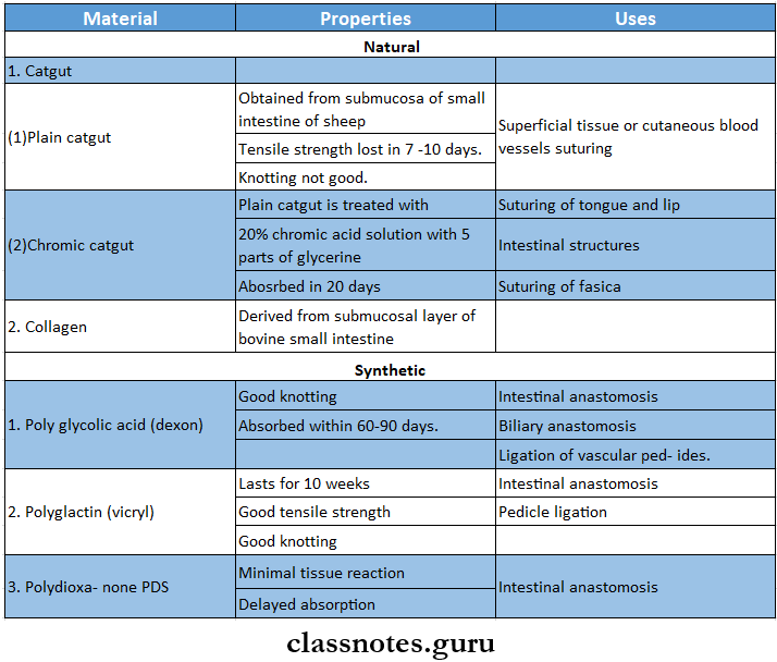
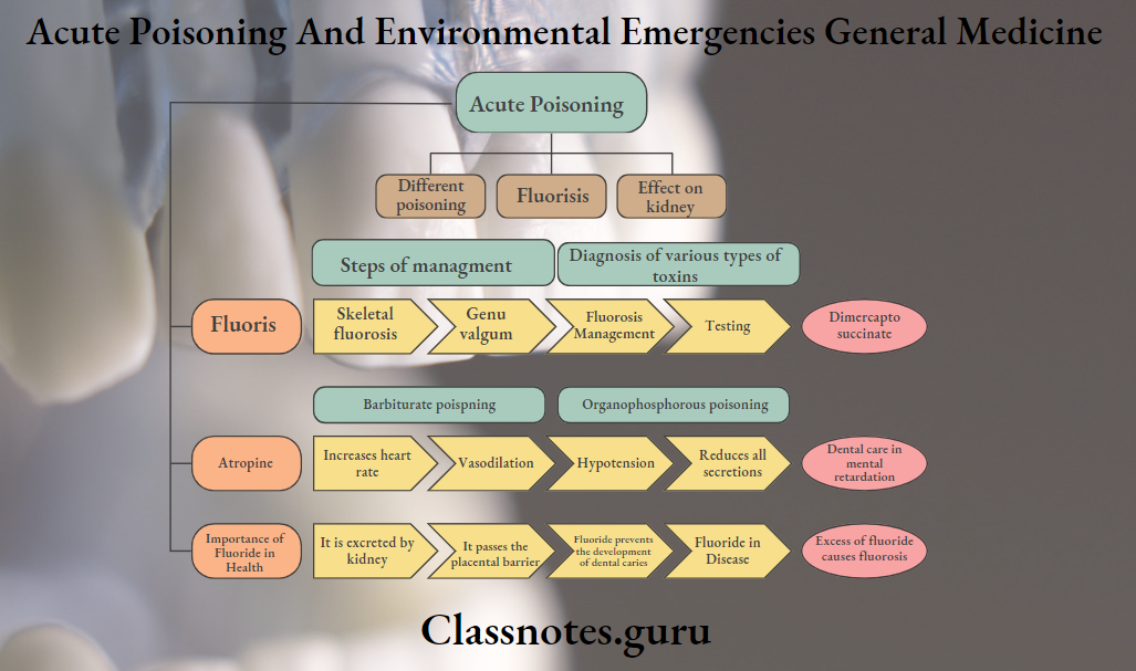
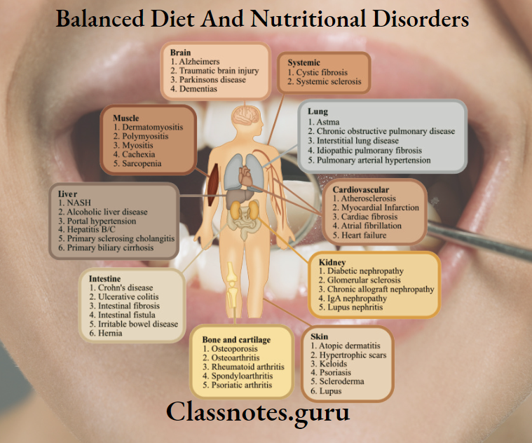
Immunological Factors In Disease Important Notes

  1. Hypersensitivity ReactionsImmunological Factors In Disease Hypersensitivity Reactions
  2. Anaphylactic Reaction
    • Anaphylactic Reaction is a state of rapidly developing immune response to an antigen mediated by IgE antibodies
    • Anaphylactic Reaction Clinical features:
      • Systemic anaphylaxis
      • Pruritus
      • Wheel and flare lesions
      • Bronchospasm
      • Tightness of chest
      • Wheezing
      • Laryngeal edema
      • Dyspnea
      • Cyanosis
      • Shock
      • Diarrhea
      • Pulmonary edema
      • If not treated death occurs Local anaphylaxis
      • Hay fever
      • Bronchial asthma
      • Food allergies
      • Contact dermatitis
      • Angioedema

Immunological factors in disease questions and answers

Read And Learn More: General Medicine Question and Answers

Immunological Factors In Disease Long Essays

Question 1. Classify allergic reactions. Describe clinical features and management of generalized anaphylaxis.

Answer:

Allergic Reactions

  • Hyperscusillvity/allergy In defined as an exaggerated/inappropriate state of normal Immune response with the onset of adverse effects on the body.
  • Lesions of hypersensitivity are a form of antigen-antibody reaction
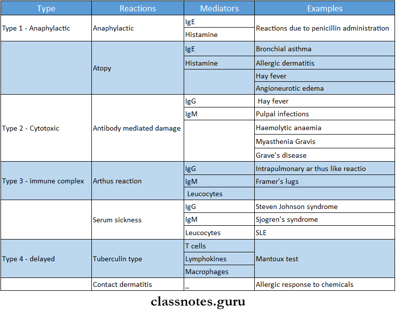
  • Hypersensllivlly reactions are classified into 4 types,
    • Type 1: Anaphylactic (atopic) reaction
    • Type 2: Cyloxlc (cytolytic) reaction
    • Type 3: Immune complex-mediated (arthus) reaction,
    • Type 4: Delayed hypersensitivity (cell-mediated) reaction.
  • Depending, upon the rapidity, duration, and type of immune response, they are classified into immediate type and delayed type.
    • Immediate type: On administration of antigen, reaction occurs immediately, This includes types 1, 2, and 3.
    • Delayed type: Reaction is slower in onset, develops within 24-48 hours and the effects is prolonged. Includes type 4 reaction.

Anaphylaxis:

  • Anaphylaxis or type 1 hypersensitivity is defined as a state of rapidly developing immune responses to an antigen (i.e., allergen) to which the individual is previously sensitized.
  • The reaction appears within 15 – 30 min of exposure to antigen.

Etiology:

  • Type 1 reaction is mediated by humeral antibodies of IgE type or regain antibodies in response to antigen.
  • The definite cause is not known, but the following may be responsible.
  • Environmental pollutants
  • Genetic basis
  • Concomitant factors Allergic response may be linked to the occurrence of certain viral infections of upper respiratory tract.

Anaphylaxis Effects:

  • Increased vascular permeability
  • Smooth muscle contraction
  • Vaaocontriction followed by vasodilation
  • Shock
  • Increased gastric secretion
  • Increased nasal and lacrimal secretions
  • Eosinophils and neutrophilia.

Anaphylaxis Examples:

  1. Reactions against mycobacterial infection.
    • Tuberculin reaction, the granulomatous reaction in tuberculosis, leprosy.
  2. Reaction against virally infected cells
  3. Reaction against malignant cells in the body.
  4. Reaction against organ transplantation, for example, transplant rejection, graft versus host reaction.

Short essay on immunological factors in disease

Immunological Factors In Disease Short Essays

Question 1. Anaphylaxis

Answer:

Anaphylaxis

  • Anaphylaxis or type 1 hypersensitivity is defined as a stale of rapidly developing immune response to an antigen (i.e., allergen) to which the individual is previously sensitised.
  • The reaction appears within 15 – 30 min of exposure to antigen.

Etiology:

  • Type 1 reaction is mediated by humoral antibodies of IgE type or regain antibodies in response to antigen.
  • The definite cause is not known, but the following may be responsible.
  • Environmental pollutants
  • Genetic basis
  • Concomitant factors – Allergic response may be linked to occurrence of certain viral infections of upper respiratory tract.

Anaphylaxis Effects:

  • Increased vascular permeability
  • Smooth muscle contraction
  • Vasoconstriction followed by vasodilation
  • Shock
  • Increased gastric secretion
  • Increased nasal and lacrimal secretions
  • Eosinophia and neutrophilia.

Anaphylaxis Examples:

  1. Reactions against mycobacterial infection.
    • Examples: Tuberculin reaction, granulomatous reaction in tuberculosis, leprosy.
  2. Reaction against virally infected cells
  3. Reaction against malignant cells in the body.
  4. Reaction against organ transplantation for example, transplant rejection, graft versus host reaction.

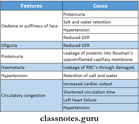
Question 2. Oedema- causes

Answer: Oedema

Oedema Causes:

  • Congestive heart failure
  • Nephrotic syndrome
  • Severe malnutrition
  • Hepatic cirrhosis
  • Acute or chronic renal failure
  • Hypothyroidism

Long essay on immunity and disease

Question 3. Diagnostic procedures for hypersensitive reactions

Answer:

Hypersensitive Reactions Diagnostic Tests:

  1. Precipitation test
    • When a soluble antigen reacts with its antibody in the presence of electrolytes at optimal temperature and pH, the antigen-antibody complex forms an insoluble precipitate called precipitation
    • Tests:
      • Ring test
        • Ring is formed at the junction of antigen and antibody
      • Flocculation test
        • When instead of sedimenting, the precipitate is suspended as floccules, the reaction is called flocculation
      • Immunodiffusion test
        • Gel is used in it
        • Types
          • Single diffusion in one dimension
          • Double diffusion in one dimension
          • Single diffusion in two dimension
          • Double diffusion in two dimension
          • Immunoelectrophoresis
          • Electroimmunodiffusion
    • Counter immune electrophoresis
    • Rocket electrophoresis
  2. Agglutination:
    • In it antigen combines with its antibody in presence of an electrolyte to form clumps of particles
    • Types:
      • Slide agglutination test
      • Tube agglutination test
      • Coombs test
      • Heterophile agglutination test
        • Weil Felix reaction
        • Paul-Bunnel test
        • Streptococcus MG agglutination test
      • Passive agglutination test
        • Latex agglutination test
        • Haemagglutination test
        • Coagglutination
  3. Complement fixation test:
    • Antigen-antibody complex formed fixes complement
  4. Neutralisation test
    • Bacterial exotoxins neutralises antibody
  5. Immunofluorescence:
    • Antigen-antibody complexes produce fluorescence

Immunology essay questions with answers

Immunological Factors In Disease Short Answers

Question 1. T cells

Answer:

T cells

  • T cells are lymphocytes
  • They are thymus-dependent

T cells Types:

  1. Regulatory T cells
    • T helper cells
      • Facilitates B cell response to produce immunoglobulin
      • The balance between T helper cells and T suppressor cells produces an optimum immune response
      • Overactivity of helper cells leads to autoimmunity
      • Decreased activity of helper cells causes an immunodeficiency state
    • T suppressor cells
      • They block the immune reaction
      • Overactivity of suppressor cells leads to an immunodeficiency state
      • Decreased activity of suppressor cells causes an autoimmunity state
    • Effector cells
      • Cytotoxic T cells
        • It can lyse specific target cells
      • Delayed-type hypersensitivity cells
        • Responsible for delayed hypersensitivity reaction

Question 2. Penicillin anaphylaxis

Answer:

Penicillin Anaphylaxis

  • Penicillin allergy develops within minutes and is called an immediate hypersensitivity reaction
  • Penicillin Anaphylaxis can induce both types of reactions
  1. Humoral-mediated immunity- causes
    • Type 1- Anaphylaxis
    • Type 2-Cytolytic reaction
    • Type 3- Arthus reaction
  2. Cell-mediated immunity- causes
    • Delayed hypersensitivity reaction
    • Induces synthesis of IgE antibodies
    • Formation of antigen-antibody complexes
    • Degranulation of mast cells
    • Release of inflammatory mediators
    • Bronchospasm
    • Laryngeal edema
    • Hypotension

Immunopathology essay type questions

Question 3. Serum sickness

Answer:

Serum Sickness Causes:

  • After the administration of foreign serum
  • Tetanus antitoxin
  • Rabies antiserum

Serum sickness Mechanism:

  • Antibodies form immune complexes in blood vessels with administered antigens
  • These complexes fix complement which attracts the leukocytes to the area causing direct tissue injury

Serum Sickness Features:

  • Fever
  • Swelling
  • Lymphadenopathy
  • Joint and muscle pain
  • Rash
  • Peripheral neuritis
  • Kidney disease
  • Myocardial ischemia

Question 4. Pemphigus Vulgaris

Answer:

Pemphigus Vulgaris

  • Pemphigus Vulgaris is the most common type of pemphigus

Pemphigus Vulgaris Features:

  • Age: 40-70 years
  • Sex: Common in females

Pemphigus Vulgaris Presentation:

  • Development of vesicles and bullae over skin and mucous membrane
  • Rupture of vesicles or bullae
  • Formation of painful ulcers that bleed profusely
  • Oblique pressure on the unaffected aeas around the lesion causes the stripping of the normal skin or mucous membrane called “Nikolsky’s sign”
  • Areas affected:
    • Skin lesions- over scalp, trunk, and umbilical areas
    • Orallesions- cheek, palate, and gingiva
      • Leads to excessive pain, excessive salivation, difficulty in intake of food, halitosis

Immunological basis of disease short answers

Question 5. Allergy

Answer:

Allergy

  • Allergy is a state of hypersensitivity induced by exposure to a particular antigen
  • This antigenic substance capable of inducing type 1 IgE-mediated immune response
  • The first dose of allergenic exposure sensitizes B lymphocytes
  • The subsequent exposure results in harmful immunologic activation resulting in expression of an allergic reaction

Question 6. Urticaria

Answer:

Urticaria Types:

  • IgE dependent urticaria
  • Complement mediated urticaria
  • Nonimmunologic urticaria
  • Idiopathic urticaria

Urticaria Features:

  • Formation of wheal and flare cutaneous lesions involving only superficial portions of the dermis
  • This results in circumscribed wheals with erythematous raised borders with blanched centers

Question 7. Angioedema

Answer:

Etiology:

  • Food or drug allergy
  • Biochemical abnormality
  • Absence of inhibitor of Cl esterase enzyme from serum
  • Formation of kinin-like substances

Angioedema Clinical Features:

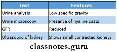
  • Exhibits as smooth, diffused, edematous swelling involving face, lips, chin, eyes, tongue, and extremities
  • Leads to edema of the glottis resulting in suffocation

Angioedema Treatment:

Immunological Factors In Disease Angioedema

Autoimmune diseases essay questions

Question 8. Steven Johnson syndrome

Answer:

Steven Johnson Syndrome

  • Steven Johnson is a severe form of erythema multiforme with widespread involvement, typically involving the skin, oral cavity, eyes, and genitalia

Steven Johnson syndrome Clinical Features:

  1. Symptoms:
    • Fever
    • Malaysia
    • Photophobia
    • Eruptions on oral mucosa, genital mucosa, and skin
  2. Skin Lesions:
    • Flemorrhagic
    • Vesicle and bullae are present
  3. Eye Lesions:
    • Photophobia
    • Conjunctivitis
    • Corneal ulceration
    • Keratoconjunctivitis
  4. Genital Lesions:
    • Non-specific urethritis
    • Balanitis
    • Vaginal ulcers
  5. Complications:
    • Trachea-bronchial ulceration
    • Pneumonia

Mechanism of immune system in diseases

Steven Johnson syndrome Treatment:

  • ACTH
  • Cortisone
  • Chlortetracycline

VIVA VOCE

  1. Adrenaline is the drug of choice in anaphylaxis

General Principles Of Operative Surgery Question And Answers

General Principles Of Operative Surgery Long Essays

Question 1. What is sterilisation? What are the methods of sterilizing surgical materials? Mention the merits and demerits.

Answer:

Sterilisation:

  • Sterilisation is defined as a process by which an article, object or surface is free of all micro-organisms in the vegetative form and in the spore state.

Sterilisation Physical Methods

General Principles Of Operative Surgery Sterilisation Physical Methods

Sterilisation Chemical Agents

General Principles Of Operative Surgery Sterilisation Chemical Methods

General principles of operative surgery Q&A

General Principles Of Operative Surgery Short Essays

Question 1. Autoclave.

Answer:

Autoclave

Autoclaving is the process of sterilization by saturated steam under high pressure above 100°C temperature.

Autoclave Principle:

  • Water boils when its vapour pressure equals that of the surrounding atmosphere.
  • When the atmospheric pressure is raised then the boiling temperature is also increased.
  • At normal pressure water boils at 100oC but when pressure inside a closed vessel increases, the temperature at which water boils also increases.
  • When steam comes into contact with a cooler surface, it condenses to water and gives up its Latent heat to the surface.

Read And Learn More: General Surgery Question and Answers

Sterilisation conditions:

General Principles Of Operative Surgery Sterilisation Conditions

Sterilisation Control:

  • Spores of bacillus stearothermophilus.
  • Brown’s tubes.
  • Thermocouples.
  • Autoclave tapes.

General Principles Of Operative Surgery A Simple Autoclave

Autoclave Uses:

  • Used for sterilisation of
    • Culture media.
    • Rubber articles.
    • Syringes and surgical instruments
    • OT gowns and dressing materials.
    • Endodontic instruments.

Question 2.Absorbable suture material.

Answer:

Absorbable Suture Material

  • Various processes like hydrolysis and proteolytic enzymatic degradation break down absorbable sutures.
  • They are made from either natural or synthetic polymers.

Absorbable Suture Material Classification:

General Principles Of Operative Surgery Absorbable Suture Material Classification

Operative surgery questions and answers

Question 3. Cryosurgery.

Answer:

Cryosurgery

Cryosurgery is the deliberate destruction of tissue by the application of extreme cold.

  • Barnard introduced it.
  • The temperature used for it is -20°C to -180°C.

Cryosurgery Methods Employed Are:

  • Spray freeze technique.
  • Applicator technique.
  • Cryoprobe method.
  • Thermocouple method.

Cryosurgery Technique:

  • Anaesthetize the area.
  • Freeze the exposed nerve for 2 min.
  • Wait for 5 min.
  • Next freeze it for another 3 min.

Cryosurgery Uses:

  • Keratotic lesions.
  • Granulomatous lesions.
  • Hyperplastic lesions.
  • Vascular and pigmented lesions.
  • Salivary gland lesions.
  • Gingival lesions.
  • Periodontal diseases.

Cryosurgery Effects:

  • Dehydration.
  • Denaturation of lipid molecules.
  • Necrosis of tissues, capillaries, small arterioles and venules.
  • Allows the destruction of the tumoral or any other pathological tissues.

Question 4. Antiseptics

Answer:

Antiseptics

Antiseptics is chemical that is applied to living tissues such as mucous membranes to reduce the number of micro-organisms present, through inhibition of their activity

Antiseptics Requirements:

  • A broad spectrum of activity
  • Fast acting
  • Not affected by physical factors
  • Non-toxic
  • Surface compactable
  • Easy to use
  • Odourless
  • Economical

Antiseptics Agents:

  1. Alcohols: ethyl alcohol
  2. Iodophors povidone iodine
    • Hepatitis virus not susceptible to it
    • Formulated as 1% iodine solution
    • Not stable at high temperature
  3. Hexachlorophene
    • Toxic
    • Used in patients who are sensitive to iodine
  4. Chlorhexidine 0.075%

Surgical principles MCQs with answers

Question 5. Sutures

Answer:

Sutures Ideal requirements

  • Should have
    • Adequate strength
    • Good knot-holding property
    • Least reactive
    • Easy to handle
    • Less memory
    • Easily available
    • Cost-effective

Sutures Classification

General Principles Of Operative Surgery Digested By Body Enzymes

Coated Or Non-Coated: Coated with biologically inert non-resorbable compound

General Principles Of Operative Surgery Short Answers

Question 1. Antisepsis.

Answer:

Antisepsis

Antisepsis is defined as the prevention of infection by inhibiting the growth and multiplication of bacteria in wounds or tissues.

  • Antiseptics are substances that kill or prevent the growth of microorganisms.
  • English physician Sir John Pringle first used the word antiseptic.
  • Commonly used antiseptics are:
    • Alcohol.
    • I-propanol.
    • Isopropanol.
    • Boric acid
    • Brilliant green.
    • Chlorhexidine gluconate.
    • Hydrogen peroxide.

Question 2. Asepsis.

Answer:

Asepsis

  • Asepsis means precautions taken before any surgical procedure, against the development of infection.
  • Elimination of infection is the goal of asepsis.

Asepsis Examples:

  • Washing the hands prior to delivery reduces puerperal fever.
  • Wearing gloves before any procedure.
  • Cleaning the operative site with iodine.
  • Sterilisation of instruments.

Question 3. Disinfection.

Answer:

Disinfection

Disinfection means the destruction of all pathogens or organisms capable of producing infection but not necessary spores.

  • Disinfection is less effective than sterilisation.
  • Disinfection only reduces the number of microorganisms.
  • Disinfection doesn’t need any strict protocol.
  • Disinfection is commonly used in daily life.
  • Thus, it prevents the spread of infection.

Short notes on operative surgery

Question 4. Sterilisation.

Answer:

Sterilisation Definition:

  • Sterilisation is defined as a process by which an article, object or surface is free of all micro-organisms in the vegetative and spore form.

Sterilisation Classification:

General Principles Of Operative Surgery Sterilisation

Sterilisation Uses:

  • Sterilisation of materials or instruments used in surgical and diagnostic procedures.
  • For media and reagents used in the microbiology laboratory.

Question 5. Sterilisation of hot air.

Answer:

Sterilisation Of Hot Air

  • Sterilisation Of Hot Air is a method of sterilization using dry heat.
  • Sterilisation Of Hot Air is the most widely used.

Temperature And Time:

  • 160°C for 2 hours.

Sterilisation Of Hot Air Advantages:

  • Safer to use – as it does not require water and high pressure.
  • Suitable to work in the laboratory.
  • Smaller in size.
  • Rapid method.

Sterilisation Of Hot Air Disadvantages:

  • Some organisms may not killed by it

Sterilisation Of Hot Air Uses:

  • Used for sterilisation of
    • Glasswares like glass syringes, flasks and test tubes.
    • Surgical instruments like scalpels, and scissors.
    • Chemicals such as liquid paraffin, and fats.

Question 6. Incineration.

Answer:

Incineration

Incineration is a waste treatment process that involves the combustion of organic substances contained in waste material.

  • It converts waste into ash, fuel gas and heat.
  • It is used to destroy soiled dressings, bedding, bandages, etc.
  • Pathogens and toxins can be destroyed by high temperatures by it.
  • An incinerator and a furnace is used for it.

Incineration Types:

  • Moving grate
  • Fixed grate
  • Rotary kiln.
  • Fluidized bed.

Question 7. Cidex,

Answer:

Cidex

  • Cidex is one of the brand names of glutaraldehyde.
  • It is effective against mycobacterium, fungi and viruses.

Cidex Advantages:

Doesn’t affect the lens of the instrument.

Cidex Disadvantages:

  • Toxic, oils liquid.
  • Harmful if inhaled or swallowed.
  • Irritating to eyes and respiratory tract.
  • Can cause severe damage to skin and eyes.

Cidex Uses:

  • Used to sterilise cystoscopes, bronchoscopes, endotracheal tubes and metallic instruments,

Question 8. Diathermy.

Answer:

Diathermy

Diathermy is a therapeutic treatment most commonly prescribed for joint conditions.

In it, a high-frequency electric current is delivered via shortwave, microwave or ultrasound to generate deep heat in body tissues.

Diathermy Effects:

  • Promotes blood flow.
  • Improves flexibility in stiff joints.
  • Decreases pain and inflammation.
  • Causes blood to coagulate and vessels to be sealed off or centralised.
  • Removes tumour cells.

Diathermy Types:

  • Shortwave diathermy.
  • Microwave diathermy.
  • Ultrasound diathermy.

Diathermy Side-effects:

  • Causes extreme heat in metal devices such as bone pins, dental fillings and metal sutures.
  • Causes burn in adjacent tissues.
  • Can cause shock or burn.

Diathermy Uses:

  • Arthritis.
  • Back pain.
  • Arthralgia.
  • Fibromyalgia.
  • Muscle spasms.
  • Neuralgia.
  • Bone injuries.
  • Bursitis.
  • In treatment of tumour.

Viva questions on principles of surgery

Question 9. Cryosurgery

Answer:

Cryosurgery

  • Cryosurgery is the deliberate destruction of tissue by the application of extreme cold
  • It was introduced by Barnard
  • Temperature used is -20 degree C to -180 degree C
  • Technique
    • Anaesthesize the area
    • Freeze the exposed nerve for 2 min
    • Wait for 5 min
    • Next freeze for other 3 min

Cryosurgery Uses:

  • Keratotic lesions
  • Granulomatous lesions
  • Hyperplastic lesions
  • Vascular and pigmented lesions
  • Salivary gland lesions
  • Gingival lesions
  • Periodontal lesions

Question 10. Types of biopsy

Answer:

Types Of Biopsy

Type of Biopsy is the removal of part of tissue for the purpose of histological examination and analysis

Biopsy Types:

1. Punch Biopsy: The sample is obtained with the help of a punch

Biopsy Indications:

  • Mucosal lesions
  • Inaccessible areas

2. Incisional Biopsy:

Indication: large lesions

Tumours Edge biopsy is taken where the tumour cells can be compared with the normal cells

3. Excisional Biopsy:

Indication: small lesions

The entire lesion is excised in a single sitting and sent for histological examination

4. Needle Biopsy: FNAC

Indication: cystic cavity: A 23-26 gauge needle is used to aspirate the contents of the lesion

Question 11. Drains

Answer:

Drains Need for Drainage:

  • Obliterate dead space
  • Removal of foreign bodies
  • Evacuate fluid/ blood/ urine from body

Drains Indications:

  • Abscess cavities
  • Surgical defects
  • Urinary retention
  • Gastric lavage
  • Osteomvelitic lesions

Drains Functions:

  • Escape of fluids from body
  • Introduce drugs into the body

Drains Types:

  1. Simple rubber catheter
  2. Corrugated rubber drain
  3. Infant feeding tube
  4. Foley’s catheter
  5. Nasogastric tube

Basic surgical techniques questions

Question 12. Name suture materials

Answer:

Suture Materials Classification:

Absorbable: Digested by body enzymes
/\
/\
/\
/\
Natural Synthetic

Non-absorbable: Cannot be digested
/\
/\
/\
Natural Synthetic

Coated Or Non-Coated: Coated with biologically inert non-resorbable compound


Acute Poisoning Question And Answers

Acute Poisoning And Environmental Emergencies Important Notes

  1. Different PoisoningAcute Poisoning And Environmental Emergencies Different Poisoning

  2. Fluorosis

    • Dental fluorosis is caused by excessive intake of fluoride during tooth development
    • Features:
    • Dental Fluorosis
      • Mottled enamel
      • Presence of hypoplastic areas
      • Mottled areas may stain yellow/ brown
      • Fluoride occurs symmetrically within dental areas, commonly affecting premolars
    • Skeletal Fluorosis
      • Severe pain in
        • Backbones
        • Joints
        • Hips
    • Stiffness in joints and spine
    • Knock-knee syndrome
      • Outward bending of legs and hands
      • Damage to fetus
      • Blocking and calcification of blood vessels
      • Cripping fluorosis
  3. Effect On Kidney
    • May aggravate renal disease

Acute poisoning viva questions

Management of Acute Poisoning

Acute Poisoning Steps Of Management:

  1. Resuscitation and initial stabilization
    • Maintain airway, breathing, and circulation
    • Blood sample collection for examination
    • Rectal temperature is obtained
    • Treatment of hypotension with crystalloids
    • Administration of a cocktail of 50% dextrose, naloxone, and thiamine
  2. Diagnosis of various types of toxins
    • History
      • Reveals type of poison and amount of overdose taken
    • Examination
      • Helps to detect a syndrome associated with certain poisons
    • Investigations
      • Colour of urine
      • Colour of blood
      • Crystals in urine
      • Ketonuria
      • Anion gap
  3. Nonspecific treatment
    • Reduces levels of toxin in the body
    • Gastric decontamination- includes
      • Removal of unabsorbed poison from the gut
      • Induction of emesis
      • Gastric lavage
      • Cathartics
      • Use of activated charcoal
      • Whole bowel irrigation
    • Enhancement of excretion of absorbed toxins from the body
      • Forced diuresis- alkaline diuresis
      • Use of multiple doses of activated charcoal
      • Peritoneal and hemodialysis
    • Dialysis
  4. Specific therapy
    • A specific antidote is administered

Important MCQs on acute poisoning

Read And Learn More: General Medicine Question and Answers

Signs, Symptoms, And Management Of Fluorosis

Fluorosis

  • Excessive intake of fluoride causes fluorosis

Fluorosis Types:

  1. Dental Fluorosis
    • Caused by fluoride intake above 2 ppm
    • Its symptoms are:
    • Mottling of enamel
    • Discoloration of teeth
    • Teeth become weak and rough
    • Brown or yellow patches appear on their surfaces
  2. Skeletal Fluorosis
    • Caused by fluoride intake above 20 ppm
    • It causes:
    • Pathological changes in the bone
    • Hypercalcification
    • Bone density of limbs, pelvis, and spine increases
    • Ligaments of the spine and collagen of bones get calcified
    • Neurological disturbances may also occur
  3. Genu valgum
    • It is an advanced stage of Fluorosis
    • In it, individuals are unable to perform their routine work
    • Joints become stiff
    • Individuals are crippled

Fluorosis Management:

  • Vomiting is induced wi a syrup of ipecac or digital or mechanical stimulation the of tongue or throat
  • Decrease the absorption of fluoride by administering fluoride-binding liquids like warm water, calcium hydroxide, antacids containing aluminum or magnesium hydroxide, or milk
  • The stomach should be thoroughly washed with additional lime water
  • Calcium gluconate should be administered intravenously along with lime to prevent shock

Short answer questions on acute poisoning

Atropine

  • Atropine is a natural anticholinergic drug

Atropine Mechanism Of Action:

  • Bind to muscarinic receptors
  • Blocks the effects of acetylcholine

Atropine Actions

  • Increases heart rate
  • Vasodilation
  • hypotension
  • Reduces all secretions
  • Muscle relaxation
  • Bronchodilation
  • Relaxes ureter
  • Produces mydriasis
  • CNS stimulant

Case-based questions on acute poisoning

Atropine Uses

  • Anti-spasmodic
  • Mydriatric and cycloplegic
  • Preanaesthetic medication
  • Organophosphorous poisoning
  • Bronchial asthma
  • Peptic ulcer
  • Parkinsonism
  • Motion sickness
  • During labor

Frequently asked questions in acute poisoning

Atropine Adverse Reactions

  • Blurring vision
  • Dry mouth
  • Dysphagia
  • Dry skin
  • Fever
  • Constipation
  • Urinary retention
  • Skin rashes
  • Palpitation
  • Flushing
  • Restlessness
  • Delirium
  • Hallucination
  • Psychosis
  • Convulsion
  • Coma

Acute Poisoning And Environmental Emergencies General Medicine Acute Poisoning

Arsenic Poisoning Features

  • Gingivitis
  • Stomatitis
  • Painful mucosal ulceration
  • Hyperpigmentation and hyperkeratosis
  • Excessive salivation
  • Vomiting
  • Diarrhea
  • Neurological disturbances

Oral exam questions on acute poisoning

Symptoms Of Lead Poisoning

Symptoms Of Lead Poisoning Features:

  • Excessive salivary secretions
  • Metallic taste in the oral cavity
  • Swelling of the salivary glands
  • Development of the dark lead line along the gingival margin
  • Convulsions
  • GI upset
  • Anaemia
  • Neuritis
  • Basophilic stippling of the RBC cells

Fluorides in Health and Disease

Importance Of Fluoride In Health:

  • The kidney excretes it
  • Fluoride passes the placental barrier
  • Fluoride prevents the development of dental caries
  • Fluoride converts hydroxyapatite to fluorapatite
  • Fluoride is mostly found in bones and teeth
  • Fluoride is deposited in other calcified tissues also
  • Fluoride is required for the proper development of bones
  • Fluoride inhibits the activities of certain enzymes
  • Sodium fluoride inhibits enolase in glycolysis
  • Fluoroacetate inhibits aconitase in TCA cycle

Fluoride In Disease:

  • Excess of fluoride causes fluorosis
  • Drinking water containing less than 0.5 ppm of fluoride causes development of caries in children

Previous year acute poisoning questions

 Dental care in mental retardation

Dental Care In Mental Retradation

  • Familiarise the patient with the office and dental personnel to reduce his/her fear of the unknown before undertaking any treatment
  • Speech must be slow and simple
  • Only one instruction at a time should be given
  • Tell, show, and do technique is used in mild cases and sedation in moderate cases
  • Carefully listen to the patient
  • Appointments should be short and scheduled during the early part of the day
  • Children should be managed with a blend of kindness and firmness
  • General anesthesia may be indicated in cases where adequate levels of cooperation cannot be achieved


Types of Fracture Short Question and Answers

Fractures General Principles Important Notes

  1. Fracture – It Is Loss Of Continuity Of Bone
  2. Types Of Fractures
    Fractures General Principles Types Of Fractures
  3. Stages Of Healing Of Fracture
    • Stage of hematoma formation
    • Stage of cellular proliferation
    • Stage of callus formation
    • Stage of new bone formation
    • Stage of remodeling
  4. Brain Injuries
    Fractures General Principles Brain Injuries
  5. Sequele Of Contusion And Laceration
    • Post traumatic amnesia
    • Cerebral irritation
    • Post contusional syndrome
    • Traumatic epilepsy of Jacksonian type
    • Cerebral compression
  6. Lucid Interval
    • In the case of extradural hemorrhage when the hematoma has reached a considerable size it causes a sufficient rise in intracranial pressure to cause cerebral compression
    • This causes unconsciousness due to pressure on the reticular system of the midbrain
    • The time taken to form such a big hematoma is known as a lucid interval
  7. Complications Of Head Injury
    • Early complication
      • Leakage of CSF
      • Aerocele
      • Meningitis
      • Fat embolism
      • Brain stem injury
      • Posterior fossa injury
      • Pituitary failure
    • Late complication
      • Chronic subdural hematoma
      • Post-traumatic epilepsy
      • Headache
      • Hydrocephalus

Read And Learn More: General Surgery Question and Answers

Fractures General Principles Short Answers

Question 1. Clicking jaw

Answer:

Clicking Jaw Causes:

  • Normal jaw mechanics
  • Temporomandibular joint disorders
  • Masticatory muscles disorders
  • Maxillo-mandibular alignment disorder
  • Occlusal discrepancies
  • Bruxism

Types of fractures questions and answers

Question 2. Black eye

Answer:

Black Eye

  • Feature of Lefort 2 fracture

Black Eye Appearance:

  • Presence of bilateral circumorbital edema
  • Presence of bilateral circumorbital ecchymosis

Black Eye Diagnosis:

  • Difficult due to rapid development of swelling of eyelids

Question 3. Lefort I fracture

Answer:

Lefort I Clinical Features:

  • Oedema of lower part of face
  • Ecchymosis in buccal vestibule
  • Bilateral epitaxis
  • Mobility of upper teeth
  • Disturbed occlusion
  • Pain
  • Upward displacement of fragment- telescopic fracture
  • ‘Cracked cup’ sound on percussion of upper teeth
  • ‘Guerin sign’- ecchymosis in the greater palatine region

Lefort I Management:

  1. Reduction
    • Reduction of the impacted fragment with the help of disimpaction forceps (Rowe’s and William’s forceps)
    • Placement of Rowe’s forceps:
    • The straight blade is placed into the nostrils
    • The curved blade is placed over the palate
    • Placement of William’s forceps:
    • Placed over the buccal aspect
    • Displaces maxilla in mesiodistal direction
  2. Fixation:
    • Zygomatic suspension fixation is done
    • Holes are drill over the zygomatic arch
    • Pass wire through it
    • Bring it up to the arches
    • Twisted over are arch bars
    • 3.Inter Maxillary Fixation
    • IMF done for 3-4 weeks

Question 4. Extradural hematoma

Answer:

Extradural Hematoma

  • Extradural Hematoma is the hemorrhage in the space outside the dura mater but inside the skull

Extradural Hematoma Causes:

  • Injury to the main trunk of the middle meningeal artery
  • Injury to middle meningeal vein
  • Bleeding in the posterior cranial fossa
  • Fractures of the anterior fossa
  • Bleeding from one of the venous sinuses

Extradural Hematoma Clinical Features:

  • Bleeding in the epidural spaces
  • They can quickly expand and compress the brain stem
  • Unconsciousness
  • Abnormal posture
  • Abnormal pupil responses to light

Extradural Hematoma Treatment:

  • Blood may be aspirated surgically to remove the mass and reduce the pressure on the brain
  • Hematoma is evacuated through a burr hole or craniotomy

Bone fracture types Q&A

Question 5. Subdural hematoma

Answer:

Subdural Hematoma

  • Subdural hematoma is a type of hematoma in which blood gathers within the outermost meningeal layer between the duranater which adheres to the skull and the arachnoid mater enveloping the brain

Subdural Hematoma Causes:

  • Laceration of the cortex
  • Rupture of superior cerebral veins

Subdural Hematoma Clinical Features:

  • Severe brain damage
  • No definite lucid interval
  • Early unconsciousness
  • Cerebral compression
  • When subdural hematoma is less dramatic and delayed by several days, it is called subacute subdural hematoma
  • When subdural hematoma appears further late, it is called chronic subdural hematoma

Subdural Hematoma Treatment:

  • Extensive craniotomy
  • Hemorrhagic vessels are secured and ligated
  • Hematomas are cleared off
  • Subdural Hematoma

Fractures General Principles Subdural Hematoma

Question 6. Temporomandibular Dislocation

Answer:

Temporomandibular Dislocation

  • Dislocation refers to the condition in which the condyle is placed anterior to the articular eminence with collapse of the articular space

Temporomandibular Dislocation Clinical Features:

  • Pain
  • Inability to close the mouth
  • Tense masticatory muscles
  • Difficulty in speech
  • Excessive salivation
  • Open bite
  • Protuding chin
  • Deviation of the lower jaw

Question 7. An immediate complication of fracture

Answer:

An Immediate Complication Of Fracture

  • Anesthesia
    • Anesthesia of lower lip occurs in case of neuropraxia, axonometric or, neurotmesis
  • Malunion
    • Improper alignment of the fracture ends leads to malunion
  • Infection
    • It may be initiated as a localized abscess but later progresses to osteomyelitis
  • Superior orbital fissure syndrome
    • Hematoma within the fissure causes damage to the 3rd, 4th and 6th cranial nerves
  • Nonunion
    • It is a lack of bony fusion of the fractured ends
  • Delayed union
    • If the fracture does not heal in 4-6 weeks, it is called delayed healing
    • It is a temporary condition and can be corrected
  • Derangement of occlusion
    • If there is traumatic occlusion it is corrected by selective grinding of teeth
  • Ankylosis of TMJ
    • Prolonged immobilization causes ankylosis
  • Other complications
    • Diplopia
    • Enophthalmos
    • Blockade of nares
    • Anosmia
    • epiphora

Short questions on fractures

Question 8. Head injury management

Answer:

Head Injury Management

  • Management of the head injury depends on the Glasgow Coma Scale
    • A less than 8 score- indicates severe injury
    • Score 9-12- moderate injury
    • Score 13-15- mild injury
  • Measures include
    • Examination of the wound
    • Continued ventilation
    • Intensive care unit management of intracranial pressure
    • Oxygenation
    • Frequent neurological examination
    • CT scan

Question 9. Artificial Respiration

Answer:

Artificial Respiration

  • Artificial respiration is required whenever there is arrest of breathing which occurs during
    • Accidents
    • Drowning
    • Asphyxia
    • Gas poisoning

Artificial Respiration Purpose:

  • Ventilation of alveoli
  • Stimulation of respiratory centers

Artificial Respiration Methods:

  • Manual method
    • Mouth to mouth method
    • Holger Neilson method
  • Mechanical methods
    • Drinker’s method
    • Ventilation method

Fracture classification Q&A

Question 10. Cardiopulmonary resuscitation

Answer:

Cardiopulmonary Resuscitation

  • Cardiopulmonary resuscitation is done by external cardiac compression which is a rhythmic application of pressure over the lower half of the sternum

Cardiopulmonary Resuscitation Steps:

  • Position yourself in kneeling position on the side of the patient
  • Place the heel of one hand on the position of pressure
  • Place the heel of other hand on top of first one and interlock the fingers
  • Apply pressure to depress the sternum at least 11/2 – 2 inches
  • The rate of compression should be 60 per min.
  • Apply 15 compressions on the chest followed by 2 full ventilation
  • Repeated to form 4 complete cycles in a minute

Cardiopulmonary Resuscitation Effects:

  • Pressure in the thorax increases
  • Thorax causes the blood from the periphery to flow back into the heart and refill the chambers
  • This increases cardiac output

Question 11. Malunion

Answer:

Malunion

  • Improper alignment of the fractured ends leads to malunion
  • Usually it does not require any treatment
  • But if it affects patient’s occlusion, function, and esthetics it should be treated

Malunion Treatment:

  • Osteotomy of fragment segments
  • Realignment
  • Fixation
  • Use of elastics to correct malocclusion

Question 12. Depressed fractured skull

Answer:

Depressed Fractured Skull

  • Depressed fractured skull can be of two types
    • Open
    • Closed
  • Depressed Fractured Skull may lead to
    • Dural tear
    • Pressure on the cerebral cortex
    • Underlying hemorrhage
    • Epilepsy
    • Pressure on dural venous sinuses

Depressed Fractured Skull Treatment:

  • Shaving of the head
  • Detect neurological deficit
  • Debridement of the scalp
  • A burr hole is made by the side of the fractured portion
  • Through it an elevator is introduced and the underlying dura is gently separated
  • The depressed fragments are lifted up and dura is inspected

Fracture types viva questions

Question 13. Cerebral concussion

Answer:

Cerebral Concussion

  • Cerebral Concussion is a type of brain injury

Cerebral Concussion Features

  • Temporary physiological paralysis of function without organic structural damage
  • Transient loss of consciousness, dizziness, or mild confusion followed by complete recovery
  • May last from 1 min to hours or even a day

Question 14. causes of nasal bleeding

Answer:

causes Of Nasal Bleeding

  • Trauma
  • Exposure to warm, dry air for long time
  • Nasal and sinus infection n Allergic rhinitis
  • Nasal foreign body
  • Vigorous nose blowing
  • Deviated nasal septum
  • Cocaine use
  • Use of anti-coagulant
  • Hypertension
  • Bleeding disorders

Important questions on bone fractures

Fractures General Principles Viva Voce

  1. The mass of new bone formation at the site of fracture is known as a callus
  2. Crepitus is a sensation of grating which may be felt or heard
  3. Perkin’s formula helps to estimate the time required for union of fracture and consolidation
  4. If time taken for union for fracture is unduly prolonged it is called a delayed union
  5. When bony union cannot takes place naturally without operation it is called non-union
  6. Meningitis is very common complication of skull fractures
  7. Post-traumatic amnesia is loss of memory for events after the occurrence of trauma
  8. Retrograde traumatic amnesia means loss of memory for events before the occurrence of the accidents


Disease Of Connective Tissues Bones And Joints

Disease Of Connective Tissues Bones And Joints Short Essays

Osteoporosis-Clinical Features And Treatment

Answer:

Osteoporosis

  • Osteoporosis is a disease characterized by increased porosity of the skeleton resulting from reduced bone mass

Osteoporosis Types:

  1. Localized
  2. Generalized
    • Primary
      • Postmenopausal
      • Senile
  3. Secondary
    • Endocrine disorders
    • Neoplasia
    • Gastrointestinal disorders

Diseases of connective tissue bones and joints

 Rheumatoid Arthritis

  • Rheumatoid arthritis is a systemic disease that usually affects many joints
  • Rheumatoid Arthritis is characterized by the progressive destruction of the joint structures

Etiology:

  • Increased serum IgG
  • Increased rheumatoid factor
  • Presence of antinuclear antibodies

Read And Learn More: General Medicine Question and Answers

Rheumatoid Arthritis Clinical Features

  • Age- third and fourth decade of life
  • Sex- females are more affected
  • fever
  • Malaise
  • Weight loss
  • Anaemia
  • Raised ESR
  • Joint becomes swollen and stiff
  • Presence of pain
  • Restricted jaw movements
  • Tenderness in joints
  • Malocclusion

Rheumatoid Arthritis Treatment:

  • Systemic steroid therapy
  • Antibiotics

Gout

  • Gout occurs due to increased production of uric acid or decreased renal excretion of uric acid

Gout Clinical Features:

  • Gout comprises of four stages
  • Onset may be insidious or sudden
  • Recurrent attacks
  • Aggregate deposits of monosodium urate monohydrate in and around the joints
  • Renal diseases
  • Uric acid nephrolithiasis

Gout Diagnosis:

  • Elevation of serum uric acid levels
  • Presence of urate crystals in synovial fluid

Gout Treatment:

  • NSAIDS are used

Connective tissue disorders list

Scleroderma

  • Scleroderma is characterized by progressive fibrosis and calcification of skin and mucosa

Scleroderma Etiology:

  • Endocrinal disturbances
  • Nervous disturbances
  • Vascular disturbances
  • Allergic reactions
  • Infections

Disease Of Connective Tissues Bones And Joints

Scleroderma Clinical Features

  • Cutaneous changes- indurated edema and erythema of the skin
  • Fixation of epidermis to underlying tissues
  • CVS disturbances
  • CNS disturbances
  • The tongue becomes stiff and broad
  • Difficulty in mouth opening
  • Xerostomia

Scleroderma Treatment:

  • Corticosteroid therapy

Connective tissue disease symptoms

COX-2 Inhibitors

  • COX-2 inhibitors are advantageous
  • Some of the COX-2 inhibitors are:
    • Meloxicam
    • Coxibs- celecoxib, rofecoxib, parecoxib, etori- coxib, valdecoxib
    • Nimesulide

COX-2 Inhibitors Properties

  • Analgesic
  • Anti-inflammatory
  • Anti-pyretic effect
  • Less gastric ulcerogenic effect
  • Do not inhibit platelet aggregation

Autoimmune connective tissue diseases

COX-2 Inhibitors Disadvantages

  • Increases risk of cardiovascular events
  • Increases cerebrovascular thrombotic events
  • Increases risk of myocardial infarction and stroke

Balanced Diet And Nutritional Disorders

Balanced Diet And Nutritional Disorders Important Notes

  1. Marasmus
    • Marasmus is a common form of protein energy malnutrition
    • Mostly occurs 6-12 months postnatally
    • Clinical features
      • Large head
      • Distended abdomen
      • Diarrhea
      • Stick like limbs
      • Reduced body weight
  2. Kwashiorkor
    • It is a disease of protein energy malnutrition occurring in the second year of life
    • Clinical features
      • Generalized edema
      • Change in the color of hair
      • Diarrhea
      • Palpable liver
      • Pigmentation of skin and ulceration
  3. Fat-Soluble Vitamins
    • Vitamin A
    • Vitamin D
    • Vitamin E
    • Vitamin K
  4. Water Soluble Vitamin
    • Vitamin B complex
    • Vitamin C

Balanced diet and nutritional disorders

Balanced Diet And Nutritional Disorders Long Essays

Question 1. Describe the various manifestations of avitaminosis with particular reference to the oral cavity. How will you treat them?

Answer: 

The Various Manifestations Of Avitaminosis With Particular Reference To The Oral Cavity

  • Vitamins are essential for growth and normal body functions
  • A deficiency of vitamins causes various clinical manifestations as follows

Balanced Diet And Nutritional Disorders Avitaminosis Treatment

Importance of balanced diet essay

Read And Learn More: General Medicine Question and Answers

Etiology, Clinical Features, Diagnosis, And Treatment Of Scurvy

Scurvy Etiology

  • Vitamin C deficiency results in scurvy.

Scurvy Clinical Features

  • Lesions and clinical manifestations of Vitamin C deficiency are seen more commonly in two extreme age groups i.e., early childhood and geriatric patients.

The following manifestations are seen in vitamin C deficiency.

  1. Hemorrhage diathesis – A marked tendency to bleed which is the characteristic of scurvy. There may be hemorrhages in the skin, mucous membranes, gums, muscles, joints, and underneath the periosteum.
  2. Skeletal lesions – There is a deranged formation of osteoid matrix but not deranged mineralization. Growing tubular bones as well as flat bones are affected.
  3. Delayed wound healing.
  4. Anaemia Normocytic normochromic type.
  5. Skin rash Hyperkeratotic and follicular rash may occur.
  6. Lesions in teeth and gums – Scurvy may interfere with the development of dentin.
    • The gums are soft and swollen, may bleed easily, and get infected commonly.

Scurvy Diagnosis

  • Through clinical features

Scurvy Treatment

  • 250 mg of vitamin C orally three times a day
  • Removal of the underlying cause

Nutritional deficiency diseases list

Etiology, Clinical Features, Diagnosis, And Treatment Of Thiamine Deficiency

Thiamine Deficiency

  • Occurs due to vitamin B1
  • It leads to beriberi

Thiamine Deficiency Types

  1. Wet beriberi
    • Characterized by edema of legs, face, trunk, and serous cavities
  2. Dry beriberi
    • Associated with neurological manifestations
  3. Infantile beriberi
    • Seen in infants

Thiamine Deficiency Clinical Features

  • Loss of appetite
  • Weakness
  • Constipation
  • Nausea
  • Mental depression
  • Peripheral neuropathy
  • Irritability
  • Numbness in tire legs

Thiamine Deficiency Diagnosis

  • Diagnosis is made from clinical manifestations

Malnutrition and undernutrition

Thiamine Deficiency Treatment

  • The initial dose of 50 mg intramuscularly is given for several days then 2.5-5 mg is given daily by mouth

Balanced Diet And Nutritional Disorders population Of Numbers

Malnutrition

Answer:

Malnutrition

  • Malnutrition occurs due to absolute or relative deficiency of energy and protein

Malnutrition Clinical Features

  • Mild growth retardation
  • Loss of weight
  • Thirst
  • Weakness
  • Feeling cold
  • Nocturia
  • Amenorrhoea
  • Pale and dry skin
  • Thinning of hair
  • Cold extremities
  • Muscle wasting
  • Oedema
  • Subnormal body temperature
  • Distended abdomen
  • Diminished tendon jerks
  • Apathy
  • Depression
  • Susceptibility to infections

Malnutrition Management

  • Extra feeding
  • Repletion of proteins, energy, vitamins, and micronutrients

Vitamins and minerals deficiency

Beriberi-Types And Clinical Features

Beriberi Types:

  1. Wet beriberi
    • Characterized by edema of legs, face, trunk, and serous cavities
  2. Dry beriberi
    • Associated with neurological manifestations
  3. Infantile beriberi
    • Seen in infants

Beriberi Clinical Features

  • Loss of appetite
  • Weakness
  • Constipation
  • Nausea
  • Mental depression
  • Peripheral neuropathy
  • Irritability
  • Numbness in the legs


Lesions In Vitamin A deficiency

  1. Ocular lesions:
    • Night blindness
    • Xerophthalmia, dry and scaly sclera, conjunctiva
    • Keratomalacia due to infections of corneal ulcers,
    • Pilots spots are focal triangular areas of opacities due to the accumulation of keratinized epithelium.
    • Blindness due to infection, scarring, and opacities.
  2. Cutaneous lesions: Xeroderma/toad-like appearance of skin because of papular lesions due to follicular hyperkeratosis and keratin plugging in the sebaceous glands.
  3. Other lesions:
    • Squamous metaplasia of
    • Respiratory epithelium,
    • Pancreatic ductal epithelium,
    • Urothelium
    • Long-standing metaplasia may progress to anaplasia.
    • Immune dysfunction,
    • Pregnant women may have an increased risk of maternal infection, mortality, and impaired embryonic development.

Night Blindness

  • Night blindness is a common symptom of vitamin A deficiency. It is a defective dark adaptation due to defective synthesis of rhodopsin in rods
  • Night blindness is supported by low plasma retinol concentration
  • If untreated the condition progresses with loss of the normal mucous cells from the cornea leading to xerophthalmia


Pellagra

  • Niacin deficiency causes Pellagra i.e., rough skin
  • The cardinal manifestations of pellagra are referred to as the three Ds ie, dermatitis, diarrhea, and dementia and if not treated may lead to 4th D i.e., death.
  • Dermatitis: Sun-exposed areas of skin developed erythemia resembling sunburn which may progress to chrome type with blister formation.
  • Diarrhoea This is seen along with stomatitis, glossitis, enteritis, nausea, and vomiting
  • Dementia: Degeneration of neurons of the brain of the spinal tract results in neurological symptoms such as dementia, peripheral neuritis, ataxia, and visual and auditory disturbances.
  • Oral findings include:
    • Bald tongue of sandwich,
    • Raw beefy tongue
    • Mucosa becomes fiery red and painful
    • Profuse salivation
  • Chronic alcoholics are at high risk of developing pellagra because, in addition to dietary deficiency, niacin absorption is impaired in them.


Vitamin B12

  • Vitamin B12 is cyanocobvilamine

Coenzyme Forms:

  • 5 Deoxyadenosyl cobalamin
  • Methylcobalamin

Coenzyme Functions

  1. Synthesis of methionine from homocysteine
    • Vitamin B12 is used as methylcobalamin in this reaction
  2. Isomerization of methyl malonyl CoA to succinyl CoA
    • It occurs m the presence of vitamin B12 coenzyme, deoxy adenosylcobalamin

Coenzyme Daily Requirements:

  • Adults- 3 micrograms/day
  • Children -0.5-1.5 micrograms/ day
  • During pregnancy and lactation- 4 micrograms/day

Coenzyme Deficiency Manifestation:

  • Cyanocobalamine or vitamin B12 deficiency leads to
  1. Pernicious anemia
    • Characterized by low hemoglobin levels, decreased number of erythrocytes, and neurological manifestations
  2. Neuronal degeneration
  3. Demyelination of the nervous system

Balanced diet chart for students

Riboflavin Deficiency

  • Riboflavin deficiency symptoms include
  1. Cheilosis
    • The presence of fissures at the corner of the mouth
  2. Glossitis
    • The tongue appears smooth and purplish
  3. Dermatitis

Balanced Diet

  • It is defined as a diet that contains different types of foods possessing the nutrients- carbohydrates, fats, proteins, vitamins, and minerals in a proportion to meet the requirements of the body
  • It supplies a little more of each nutrient than the minimum requirement to keep the body in a state of good health and protect against leanness
  • Its basic composition varies from country to country
  • Indian balanced diet is composed of
    • Cereals
    • Pulses
    • Roots and tubers
    • Fruits
    • Milk and milk products
    • Fats and oils
    • Sugar
    • Groundnuts
  • Meat, fish, and eggs in non-vegetarians
  • Additional amounts of milk and pulses in vegetarians

Scurvy

Scurvy Etiology:

  • Vitamin C deficiency results in scurvy.

Scurvy Clinical Features:

  • Lesions and clinical manifestations of Vitamin C deficiency are seen more commonly in two extreme age groups i.e., early childhood and geriatric patients.
  • The following manifestations are seen in vitamin C deficiency.
  1. Hemorrhage diathesis – A marked tendency to bleed which is characteristic of scurvy. There may be hemorrhages in the skin, mucous membranes, gums, muscles, joints, and underneath the periosteum.
  2. Skeletal lesions – There is a deranged formation of the osteoid matrix but not deranged mineralization. Growing tubular bones as well as flat bones are affected.
  3. Delayed wound healing.
  4. Anaemia Normocytic normochromic type.
  5. Skin rash Hyperkeratotic and follicular rash may occur.
  6. Lesions in teeth and gums Scurvy may interfere with the development of dentin.
    • The gums are soft and swollen, may bleed easily, and get infected commonly.

Rickets

  • Rickets is a clinical disorder seen in growing children from 6 months to 2 years of age due to a deficiency of Vitamin D.

Rickets Clinical Features:

  • The gross skeletal changes depend on the severity of the rachitic process, its duration, and in particular the stresses to which individual bones are subjected.
  • Craniotabes, are the earliest bony lesions due to small round unossified areas in the membranous bones of the skull.
  • Harrisons sulcus occurs due to in drawing of soft ribs on inspiration.
  • Pigeon chest deformity
  • Bow legs occur In ambulatory children due to weak bones of lower logs.
  • Knocked knees may occur due to enlarged ends of the femur, tibia, and fibula.
  • Lower epiphyses of the radius may be enlarged.
  • Lumbar lordosis due to involvement of the spine and pelvis.

Read And Learn More: General Medicine Question and Answers

Niacin

  • Niacin is vitamin M3
  • Niacin is a pyridine derivative

Coenzyme Forms:

  • Nicotinamide adenine dinucleotide, NAD+
  • Nicotinamide adenine dinucleotide phosphate, NADP+

Niacin Functions:

  • Involved in an oxidation-reduction reaction
  • Participates in almost all the metabolism

Niacin Deficiency Manifestation:

  • Pellagra

Nutritional disorders and prevention

Vitamin D

  • Vitamin D is a fat-soluble vitamin
  • Vitamin D resembles sterols in structure
  • Its best source is sunlight
  • Its active form is calcitriol
  • Vitamin D is involved in calcium metabolism
  • Required in bone formation
  • Its daily requirement is 400 IU
  • Its deficiency leads to rickets in children and osteomalacia in adults


Vitamin C

  • Vitamin C Is a water-soluble vitamin
  • Vitamin C plays an important role in human health and disease
  • Vitamin C is readily absorbed by the intestines and excreted in urine
  • About 60-70 mg of vitamin C is required per day Citrus fruits are the main sources of vitamin C
  • Vitamin C deficiency leads to scurvy


Vitamin E

  • Vitamin E is a fat-soluble vitamin

Vitamin E Sources:

  • Vegetable oils
  • Meal
  • Milk
  • Butler
  • Eggs

Vitamin E Requirements:

  • Males-10 mg
  • Females- 8

Vitamin E Deficiency Manifestations:

  • Neurological dysfunction


Bitot’s Spots

  • Bitot’s spot may appear as glistening white plaques of desquamated thickened conjunctival epithelium, usually triangular and firmly adherent to the underlying triangular and firmly adherent to the underlying conjunctival.
  • Bitot’s spots occur due to vitamin A deficiency.

Bitot’s Spots Treatment:

  • Oral administration of retinol 30 mg daily for 3 days
  • In advanced cases, vitamin A is given in the dose of 50,000 IU parenterally for three days

Bitot’s Spots Prevention:

  • Good nutrition
  • Intake of fresh leafy green vegetables
  • Intake of vitamin A


Vitamin B12

  • Vitamin B12 is cyancvohalamine

Vitamin B12 Coenzyme Forms:

  • 5-Deoxyadenosyl cobalamin
  • Methylcobalamin

Vitamin B12 Functions:

  1. Synthesis of methionine from homocysteine
    • Vitamin B12 is used as methylcobalamin in this reaction
  2. Isomerization of methyl malonyl CoA to succinyl CoA
    • It occurs in the presence of vitamin B12 coenzyme, deoxy adenosylcobalamin

Vitamin B12 Daily Requirements:

  • Adults- 3 micrograms/day
  • Children -0.5-1.5 micrograms/day
  • During pregnancy and lactation- 4 micrograms/day


Vitamin A deficiency


Vitamin A deficiency leads to

  1. Night blindness
  2. Xerophthalmia
  3. Keratomalacia
  4. Retarded growth

Vitamin K


Vitamin K is fat soluble vitamin

Vitamin K Sources:

  1. Animal sources
    • Egg yolk, meat, liver, cheese, and dairy products
  2. Plant sources
    • Cabbage, cauliflower, tomatoes, alfalfa, spinach

Vitamin K Functions:

  • Helps in coagulation
    • Daily Requirement- 70-140 micrograms/day

Vitamin K Deficiency Symptoms:

  • Diminished blood clotting
  • Increased prothrombin time

 Vitamin B6


Vitamin B6 Sources:

  • Milk
  • Liver
  • Meat
  • Legumes
  • Whole grains
  • Cereals
  • Nuts
  • Vegetables

Vitamin B6 Functions:

  • Acts as a cofactor for several enzymes involved in amino acid metabolism
  • Involved in the metabolism of fat, carbohydrates, and several vitamins
  • Involved in heme synthesis

Pyridoxine deficiency


Pyridoxine Deficiency Symptoms:

  • Glossitis
  • Cheilosis
  • Weakness
  • Peripheral neuropathy
  • Depression
  • Irritability
  • Microcytic, hypochromic anemia
  • Seizures

Pyridoxine Deficiency Treatment:

  • Oral administration of vitamin B6 in the dosage of 10-20 mg/day

Effects of unbalanced diet

Cyanocobalamine Deficiency

Cyanocobalamine or vitamin B12 deficiency leads to

  1. Pernicious anemia
    • Characterized by low hemoglobin levels, decreased number of erythrocytes, and neurological manifestations
  2. Neuronal degeneration
  3. Demyelination of the nervous system

Infectious Diseases Important Notes

Infectious Diseases Important Notes

  1. Gas Gangrene
    • Caused by clostridium perfrigens
    • Gas Gangrene is characterised by skin color change from pallor to bronze/ purple
    • Skin is tense and tender
    • Gas in the tissues is elaborated by crepitus or visible on radiograph
    • Treatment
    • Surgical debridement
    • Antibiotic therapy with high dose of 4 penicillin, clindamycin, and metronidazole
    • Hyperbaric oxygen therapy
  2. Hyperbaric Therapy Is Used For Treatment Of
    • Gas gangrene
    • Osteoradionecrosis
    • Chronic osteomyelitis
  3. Rubella ( German Measles)
    • Rubella is caused by a paramyxovirus
    • Rubella is spread by droplet infection
    • Rubella is characterised by Koplik’s spots on the buccal mucosa which is seen as small white spots surrounded by erythema
    • Rubella is followed by the appearance of rash first on the back of ears and at hairline and maximum on face
    • Rubella in early pregnancy causes
      • Congenital abnormalities like deafness
      • Spontaneous abortion
      • Congenital heart diseases like persistent patent ductus arteriosus, atrial septal defect, etc
    • Complications
      • Pneumonia
      • Encephalitis
      • Otitis media
  4. Hepatitis E
    • Hepatitis E is also called Non-A or Non-B hepatitis
    • Caused by HEV which is an RNA virus
    • Spreads by faeco oral route
    • Clinical illness resembles hepatitis A infection
    • Pregnant women are particularly liable to acute hepatic failure
    • Epidemics are almost exclusively caused by hepatitis E virus
  5. Viruses And Infections Caused By ThemInfectious Diseases Viruses And Infections Caused By Them
  6. Classification Of Virus

    • DNA Virus
      • Pox viridae
        • Small pox
        • Molluscus
        • Contagiosum
      • Herpesviridae
      • Adenoviridae
        • Major cause for nonbacterial pharyngitis and tonsillitis
      • Papovaviridae
      • Hepadnaviridae
    • RNA Virus

      • Picornaviridae
        • Polio
        • Coxsackie
        • Hepatitis A
        • Rhinovirus
      • Orthoviridae
        • influenza
    • Paramyxoviridae
    • Retrovirus
    • Togaviridae
    • Rhabdoviridae
    • Flaviviridae
    • Calciviridae
  7. Types Of Fever In Different Infections
    • Saddleback fever – dengue
    • Step ladder fever – typhoid
    • Pell Ebstein fever – brucellosis and Hodgkin’s disease
    • Double rise of temperature in a day – kalaazar
  8. Clinical Features Of Syphilis
    • Congenital syphilis
      • Hutchison’s triad
      • Saddle nose
      • Sabre tibia
      • Rhagades
      • Bossing of frontal and parietal bones
      • Hypoplastic maxilla
      • Salt and pepper scars on retina
    • Primary syphilis
      • Painless chancre
      • Painless palpable rubbery inguinal lymph nodes
    • Secondary syphilis
      • Fever, malaise
      • Maculopapular rash on trunks and limbs
      • Condylomalata
      • Mucous patches in the genitalia, mouth, and pharynx
      • Snail track ulcers in mouth
    • Tertiary syphilis
      • Gumma
      • Cardiovascular syphilis
      • Aortitis
      • Aortic aneurysm
      • Aortic incompetence
      • Neurosyphilis
      • Tabesdorsalis
      • Meningovascular disease
      • General paralysis
  9. Complications Of Different InfectionInfectious Diseases Complications Of Different Infection
  10. Rashes In Different DiseasesInfectious Diseases Rashes In Different Diseases
  11. Spots In Different Diseases

Read And Learn More: General Medicine Question and Answers

Infectious Diseases Important Notes

VIVA VOCE

  1. Clostridium perfrigens causes gas gangrene
  2. Clostridium tetani causes tetanus
  3. Clostridium difficile causes pseudomembranous colitis
  4. Koplik’s spots is a characteristic feature of measles
  5. Rubella is transmitted by aerosol infection
  6. Hepatitis A and E spread by fecal-oral route
  7. Hepatitis B, C, and D spread by parenteral route
  8. Hepatitis A affects children more than adults

Infectious Diseases Infaxt Population That access Are Vaccinated

important notes on infectious diseases for mbbs

Endocrine And Metabolic Diseases Short Question and Answers

Endocrine And Metabolic Diseases Important Notes

  1. Acromegaly:
    • GH excess after epiphyseal closure results in acromegaly
    • Clinical features:
      • Skin thickening
      • Enlarged nose and tongue
      • Macroglossia
      • Carpal tunnel syndrome
      • Large hands and feet
      • Prognathic lower jaw
      • Diabetes mellitus
      • Hypertension
  2. Thyroid Storm/ Thyrotoxic Crisis, Thyrotoxicosis
    • Thyroid Storm is an acute life-threatening hypermetabolic state induced by the excessive release of thyroid hormones
    • Clinical features
      • Fever
      • Heat intolerance
      • Exophthalmos
      • Tachycardia
      • Increased appetite
      • Excessive Sweating
      • Weight loss
      • Systolic hypertension
      • Cardiac arrhythmias
  3. Diabetes Mellitus
    • Features
      • Polyuria, polyphagia, polydipsia
      • Glycosuria
      • Ketoacidosis
      • Kussmaul breathing
      • Circulatory shock, coma
      • Bone resorption, loosening of teeth
      • Acetone breath
    • Types
      • Type 1 (Insulin-Dependent Diabetes Mellitus) 
        • Occurs in young age groups
        • Occurs due to deficiency of insulin
          • Destruction of beta cells during autoimmune diseases
          • Destruction of beta cells by viral infec­tion
          • Congenital disorder
        • Associated with acidosis ketosis or coma
      • Type 2 (Non-Insulin-Dependent Diabetes Mellitus)
        • Occurs after the age of 40 years
        • Also called maturity-onset diabetes
        • The structure and functions of beta cells are normal
        • Occurs due to a reduced number of insulin receptors
        • Associated with obesity and hereditary
        • Rarely, associated with ketosis
    • Diagnosis
      • Fasting blood sugar > 126 mg/ dl or random blood sugar > 200 mg/dl is suggestive of diabetes
  4. Blood Glucose Test
    Endocrine And Metabolic Diseases Blood Glucose Test
  5. Cardinal Features Of Diabetic Ketoacidosis
    • Hyperglycaemia
    • Hyperketonaemia
    • Metabolic acidosis
  6. Hyperthyroidism And Hypothyroidism
    Endocrine And Metabolic Diseases Hyperthyroidism And Hypothyroidism
  7. Hypoglycemia-Features
    • Sweating
    • Palpitation
    • Hunger
    • Confusion
    • Drowsiness
    • Incoordination
  8. Tetany
    • Occurs when plasma calcium level falls below 6 mg%
    • Signs of TetanyEndocrine And Metabolic Diseases Tetany
  9. Risk Factors Of Osteoporosis
    • Diet or calcium intake
    • Immobility
    • Thyrotoxicosis, hyperparathyroidism
    • Rheumatoid arthritis
    • Corticosteroids
    • Smoking and alcoholism
  10. Cretinism And DwarfismEndocrine And Metabolic Diseases Cretinism And Dwarfism

endocrine system short questions and answers

Endocrine And Metabolic Diseases Short Answers

Question 1. Tetany

Answer:

Tetany

  • Tetany is a clinical condition characterized by low levels of ionized calcium leading to increased neuromuscular excitability

Tetany Clinical Features:

  1. In children
    • Characteristic triad- carpopedal spasm, stridor and convulsion
    • Carpopedal spasm- flexion at metacarpophalangeal joints and extension at interphalangeal joints with the opposition of the thumb
    • Stridor- closure of glottis
  2. In adults
    1. Tingling sensation in peripheral parts of limbs or around the mouth
    2. Painful carpopedal spasm
    3. Rarely stridor and convulsions
  3. Signs
    • Trousseau’s sign
      • Raising the blood pressure above systolic level by inflation of the sphygmomanometer cuff produces carpal spasm within 3-5 minutes
    • Chvostek’s sign
      • A tap at the facial nerve at an angle of the jaw produces twitching of facial muscles

Read And Learn More: General Medicine Question and Answers

Question 2. Causes Of Tetany

Answer:

Causes Of Tetany

  1. Hypocalcaemia
    • Malabsorption
    • Osteomalacia
    • Hypoparathyroidism
    • Chronic renal failure
    • Acute pancreatitis
  2. Alkalosis andhypokalaemia
    • Repeated vomiting
    • Excessive intake of alkalies
    • Primary hyperaldosteronism
    • Hypomagnesaemia

Question 3. Treatment Of Tetany

Answer:

Treatment Of Tetany

  1. Treatment of hypocalcemia
    • Injection of 20 ml of 10% calcium gluconate
  2. Treatment of alkalosis
    • Intravenous administration of isotonic saline
    • Withdrawal of alkalies
    • Inhalation of 5% C02 in oxygen- to treat hyperventilation
    • Psychotherapy

Endocrine Disorders Short Answer Questions

Question 4. Diabetes mellitus-complications

Answer:

Acute Metabolic Complications:

  1. Diabetic ketoacidosis
    • Develop in patients with severe insulin deficiency
    • Clinical Features:
      • Nausea, vomiting, anorexia
      • Deep and fast breathing
      • Mental confusion
      • Coma
  2. Hyperosmolar hyperglycemia non-ketotic coma
    • It is a complication of type 2 diabetes mellitus
    • Caused by severe dehydration which leads to sustained hyperglycemia diuresis
  3. Hypoglycaemia
    • Develop in type 1 diabetes mellitus

Late Systemic Complications:

  1. Atherosclerosis
    • Common in both type 1 and type 2 diabetes mellitus
    • Atherosclerosis may lead to Myocardial in fraction cerebral stroke
    • Gangrene of toes and feet
  2. Diabetic microangiopathy
    • It is the basement membrane thickening of small blood vessels and capillaries of different organs and tissues
    • Occurs due to increased glycosylation of hemoglobin and other proteins
  3. Diabetic nephropathy
    • It is a severe complication of diabetes mellitus
    • Occurs in both types
  4. Diabetic neuropathy
    • Effects all parts of the nervous system
  5. Diabetic retinopathy
    • It is the cause of blindness
  6. Infections
    • Diabetic patients are more susceptible to infections like tuberculosis, pyelonephritis, otitis, carbuncles, and diabetic ulcers

Question 5. Oral complications of diabetes mellitus

Answer:

Oral Complications Of Diabetes Mellitus

  1. Periodontium
    • Alter response of the periodontal lesion to local irritants
    • Retards healing of tissues
    • GCF contains more glucose
    • Periodontal abscess formation
    • Tooth mobility
    • Severe and rapid bone resorption
  2. Tongue
    • Altered taste sensation
    • Median rhomboid glossitis
    • Impaired local immune response
    • Decreased Langerhans cell
  3. Oral candidiasis
    • Alveolar bone
    • Localized osteitis
  4. Mouth
    • Burning mouth syndrome
    • Dvsgeusia
    • Dysesthesia
    • Xerostomia
    • Increased caries activity
  5. Diabetic siaiadenosis- involving trigeminal nerve
  6. Angular cheilosis
  7. Oral lichen planus

Question 6. Diagnosis of diabetes mellitus

Answer:

Diagnosis Of Diabetes Mellitus

  1. Detection of glycosuria
    • It is detected by a dipstick test
    • The green color indicates urinary glucose concentration between 10-20 mg% or more
  2. Urine for ketone bodies
    • Ketonuria indicates diabetes
  3. Oral glucose tolerance test
    • Advise tire patient to take an unrestricted carbohydrate diet for 3 days before the test
    • Overnight fast
    • Collect a fasting sample of blood
    • Administer 75 g of glucose dissolved in 300 ml of water
    • Collect blood and urine samples at half-hour intervals for 2 hours

Endocrine And Metabolic Diseases Diagnosis Of Diabetes Mellitus

Short questions on metabolic disorders

Question 7. Glucosuria

Answer:

Glucosuria

  • Glucosuria is the condition of glucose excretion in urine
  • Glucose appears in urine when the plasma glucose concentration exceeds the renal threshold for glucose

Glucosuria Types:

  1. Renal glycosuria
    • It is a benign condition
    • Occurs due to a reduced renal threshold for glucose
    • It is unrelated to diabetes
  2. Alimentary glllucosuria
    • In certain individuals, blood glucose rapidly increases after meals which gets excreted in urine
    • This is known as alimentary glucosuria
    • It is observed in
      • Normal individuals
      • Individuals with
        • Hepatic diseases
        • Hyperthyroidism
        • Peptic ulcer

Question 8. Insulin

Answer:

Insulin

  • Insulin is a hormone required for the regulation of blood glucose level

Insulin Secreted By:

  • Beta cells of the islets of Langerhans of the pancreas
  • Functions:
  • Lowers blood glucose level
  • Promotes glucose utilization and storage
  • Inhibits glucose production
  • Required for glucose uptake
  • Increases glycolysis
  • Decreases gluconeogenesis
  • Promotes lipogenesis from glucose
  • Reduces lipolysis and ketogenesis
  • Enhances protein synthesis

Question 9. Oral hypoglycaemic drugs

Answer:

Oral Hypoglycemic Drugs Classification:

  1. Sulphonylureas
    • First generation
      • Tolbutamide
      • Chlorpropamide acetohexamide
      • Tolazamide
    • Second generation
      • Clibendamide
      • Glipizide
      • Gliclazide
  2. Biguanides- Metformin
  3. Meglilinides- repaglinide, nateglinide
  4. 4.Thiazolidinediones- Troglitazone, rosiglitazon
  5. Alpha-glucosidase inhibitors- Acarbose, miglitol
  6. Newer drugs
    • Amylin analog- pramlintide
    • GLP-1 analog- exenatide
    • DPP-4 inhibitor- Sitagliptin

Oral Hypoglycemic Drugs Features:

  1. They are used in mild and early non-insulin-dependent diabetes mellitus
  2. Oral Hypoglycemic Drugs lowers blood glucose levels
  3. They are noninvasive drugs

Question 10. Sulphonylurea

Answer:

Sulphonylurea

  • Sulphonylurea were the first oral hypoglycaemic drugs

Sulphonylurea Classification:

  1. First generation
    • Tolbutamide
    • Chlorpropamide acetohexamide
    • Tolazamide
  2. Second generation
    • Glibenclamide
    • Glipizide
    • Gliclazide

Sulphonylurea Mechanism of Action:

  • Sulphonylurea reduces the blood glucose levels by:
    • Stimulating the release of insulin from the pancreatic beta cells
      • Increasing the sensitivity of peripheral tissues to insulin
      • Increases the number of insulin receptors
      • Suppresses hepatic gluconeogenesis

endocrine system viva questions with answers

Question 11. Metformin

Answer:

Metformin

  • Metformin is biguanide

Metformin Mechanism of Action:

  • Suppresses hepatic gluconeogenesis
  • Inhibits glucose absorption from the intestines

Metformin Use:

  • In obese patients with type-2 diabetes mellitus either alone or in combination with sulphonylureas

Metformin Adverse Effects:

  • Nausea
  • Diarrhea
  • Metallic taste
  • Mild lactic acidosis
  • Anorexia
  • Loss of appetite

Question 12. Glibenclamide

Answer:

Glibenclamide

  • Glibenclamide is second generation sulphonylurea

Glibenclamide Mechanism of Action:

  • Glibenclamide reduces the blood glucose levels by:
  1. Stimulating the release of insulin from the pancreatic beta cells
  2. Increasing the sensitivity of peripheral tissues to insulin
  3. Increases the number of insulin receptors
  4. Suppresses hepatic gluconeogenesis
    • DOSE- 5-15 mg
    • HALF-LIFE- 4-6 hours
    • DURATION OF ACTION-18-24 hours

Question 13. Treatment of diabetic ketoacidosis

Answer:

Treatment Of Diabetic Ketoacidosis

  1. Correction of hyperglycemia
    • Administration of regular insulin 0.1 U/kg bolus followed by 0.1 U/kg/hour by continuous 4 infusion till the patient recovers
  2. Correction of dehydration
    • Normal salinelitersion of 1 litre m the first hour
    • Then 1 litre over the next 4 hours
    • Then quantity is titrated
  3. Correction of acidosis
    • Use of sodium bicarbonate
  4. Potassium
    • 10-20 mEq/ hour potassium chloride is added to the drip for rapid correction of hyperglycemia

Question 14. Prevention of tetanus

Answer:

Prevention Of Tetanus

  1. Surgical
    • Removal of foreign bodies, blood clots
    • Cleansing
    • Radical excision
  2. Antibiotics
    • Long-acting penicillin injection or erythromycin may be given
  3. Immunization
    • Active immunization
      • DPT vaccine
      • It is combination of Diphtheria toxoid, pertusis vaccine and tetanus toxoid
      • Route Of Adminis Tration:
        • Intramuscular
      • Dose:
        • Initial dose- 6 weeks
        • Three doses are completed at intervals of 46 weeks
        • Booster doses-18 months and 5 years
    • Passive immunization
      • Antitetanus serum is used in a dose of 1500 IU by intramuscular route
    • Combined prophylaxis

Question 15. Cretinism

Answer:

Cretinism

  • Cretinism is a characteristic feature of infantile hypothyroidism

Cretinism Clinical Features:

  • Mental retardation
  • Delayed milestones of development
  • Protruding tongue
  • Flat nose
  • Dry skin
  • Sparse hairs
  • Enlarged skull
  • Generalised edema
  • Hypotension
  • Atrophy of sweat glands
  • Protruded abdomen

Cretinism Oral Manifestations:

  • Delayed eruption and exfoliation of deciduous teeth
  • Macroglossia
  • Thick lips
  • Constant drooling of saliva
  • Malocclusion
  • Underdevelopment of mandible
  • Wide face

important short questions in endocrinology

Question 16. Albuminuria

Answer:

Albuminuria

  • The presence of albumin in the urine is known as albuminuria
  • The Dipstick test is a standard test for it
  • It identifies the presence of renal disease or urinary infection in diabetic individuals
  • It detects urine albumin greater than 300 mg/1 and even smaller amounts of urinary albumin

Question 16. Phenylketonuria

Answer:

Phenylketonuria

  • Phenylketonuria is a common metabolic disorder

Phenylketonuria Causes:

  • Deficiency of the hepatic enzyme phenylalanine hydroxylase

Phenylketonuria Mechanism:

Endocrine And Metabolic Diseases Diabetic Ketoacidosis Pathogenesis

2. Phenylalanine is diverted to alternate pathways

Phenylketonuria Clinical Features:

  1. Effects on CNS
    • Mental retardation
    • Failure to walk or talk
    • Retarded growth
    • Seizures and tremors
    • Low IQ
  2. Effect on pigmentation
    • Hypopigmentation
  3. Urine
    • Contains phenylalanine and its metabolic products
    • Mousthe ey odor due to press ence of phenylacetate

Phenylketonuria Treatment:

  • Intake of diet with low phenylalanine content
  • Use of synthetic amino acid preparation
  • Provide tyrosine in the diet
  • Administration of 5-hydroxytryptophan and dopa in serious conditions

Question 17. Hyperpituitarism

Answer:

Hyperpituitarism

  • Hyperpituitarism in infancy leads to gigantism and among adults it produces acromegaly

Hyperpituitarism  Causes:

  • Hypersecretion of growth hormone
  • Increased function of the anterior pituitary

Hyperpituitarism  Gigantism:

  • Generalized symmetric overgrowth of the body
  • Gentital underdevelopment
  • Excessive sweating
  • Headache
  • Lassitude
  • Joint and muscle pain
  • Defective vision

Question 18. Goitre

Answer:

Goitre

  • Goitre refers to enlargement of the thyroid gland irrespective of its cause

Goitre Classification:

  1. Simple goitre
    • Diffuse hyperplastic goitre
    • Nodular goitre
    • Colloid goitre
  2. Toxic goitre
    • Diffuse toxic goitre
    • Toxic nodular goitre
    • Toxic nodule
  3. Neoplastic goitre
    • Benign tumours
    • Malignant tumours
  4. Thyroiditis
  5. Other rare conditions

Question 19. Exophthalmos

Answer:

Exophthalmos

  • Exophthalmos is defined as abnormal protrusion of the eyeball anteriorly out of the orbit

Exophthalmos Causes:

  1. Inflammatory/Infection:
    • Graves’ disease
    • Orbital cellulitis
    • Mucormycosis
    • Orbital pseudotumor
    • High-altitude cerebral edema
    • Wegener’s granulomatosis
  2. Neoplastic:
    • Leukemias
    • Meningioma, (of the sphenoid wing)
    • Nasopharyngeal angiofibroma
    • Hemangioma, cavernous
  3. Cystic:
    • Dermoid cyst
  4. Vascular:
    • Carotid-cavernous fistula
    • Aortic insufficiency
  5. Others:
    • Orbital fracture: apex, floor, medial wall, zygomatic
    • Retrobulbar hemorrhage
    • Cushing’s syndrome

Exophthalmos Complications:

  • Corneal dryness and damage
  • Keratoconjunctivitis
  • Blindness due to compression of optic nerve and vessels

short answer type questions endocrine system

Question 20. Hyperparathyroidism

Answer:

Hyperparathyroidism

  • Hyperparathyroidism is an endocrine disorder occurring due to an excess of circulating parathyroid hormone

Hyperparathyroidism Types:

  1. Primary hyperparathyroidism
    • Occurs due to tumor of glands
  2. Secondary hyperparathyroidism
    • Occurs in response to hypocalcemia
  3. Tertiary hyperparathyroidism
    • Occurs after long-standing secondary hyperparathyroidism

Hyperparathyroidism Clinical Features:

Age and sex- common in middle-aged women

  1. Classic triad
    • Kidney stones
    • Bone resorption
    • Duodenal ulcers
  2. Renal symptoms
    • Renal calculi
    • Hematuria
    • Back pain
  3. Psychological symptoms
    • Emotionally unstable
  4. GIT symptoms
    • Anorexia
    • Nausea, vomiting
  5. Skeleta
    • Bone pain
    • Pathologic fractures
    • Bone deformities
    • Hypercalcemia
  6. Generalised symptoms
    • Muscle weakness
    • Fatigue
    • Weight loss
    • Insomnia
    • Headache
    • Olydipsiaand polyuria
  7. Oral manifestations
    • Intraoral and extraoral swelling
    • Gradual loosening of teeth
    • Drifting and loss of teeth
    • Malocclusion

Question 21. Risus sardonicus

Answer:

Risus sardonicus

  • Risussardonicus or rictus grin is a highly characteristic, abnormal, sustained spasm of the facial muscles that appear to produce grinning.

Risus sardonicus Causes:

  • Tetanus
  • Poisoning with strychnine

Question 22. Gynaecomastia

Answer:

  • Gynaecomastia is the presence of glandular breast tissues in males

Risus sardonicus Causes:

  • Idiopathic
  • Physiological
  • Drug-induced
  • Hypogonadism
  • Androgen resistance syndromes
  • Oestrogen excess

Risus sardonicus Investigations:

  • Ultrasonography
  • Mammography
  • Random blood sample

Risus sardonicus Treatment:

  • Self regressing
  • Surgical excision for cosmetics reasons
  • Androgen replacement

Question 23. Thyroxin

Answer:

Thyroxin

  • Thyroxin is a hormone secreted by the thyroid gland
  • The thyroid gland secretes mainly thyroxin, T4, and small amount of triiodothyronine, T3
  • T3 is the active form of the hormone, Most of the T4 is converted into T3 in peripheral tissues
  • Thyroid hormones are carried in plasma in the bound form with a plasma protein while a small amount circulates unbound
  • Free form enters cells and exerts its metabolic effects
  • its level is measured by thyroid function tests

endocrinology short questions mbbs

Question 24. Anti-thyroid drugs

Answer:

Anti-Thyroid Drugs

  • These are used to restore the patient to a euthyroid state and maintain it
  • They are:
  1. Propylthiouracil
    • Dose: 100-300 mg every 6-8 hours
  2. Carbimazole
    • Dose: 10-20 mg every 6-8 hours

Anti-Thyroid Drugs Side Effects:

  • Skin rashes
  • Fever
  • Peripheral neuritis
  • Polyarteritis
  • Agranulocytosis
  • Aplastic anemia
  • Prothrombin deficiency

VIVA VOCE

  1. Acetone breath is seen in diabetes mellitus
  2.  Congestive heart failure occurs in hypothyroidism
  3. Carpal tunnel syndrome occurs in hypothyroidism
  4. Metformin is a biguanide hypoglycaemic drug
  5. Severe neurological and cardiac changes occur at calcium levels> 16 mg/ dl

Haemorrhage Shock And Blood Transfusion Notes

Haemorrhage Shock And Blood Transfusion Important Notes

  1. Classification Of Haemorrhage
    • Based on the nature of the vessel involved
      • Arterial
      • Venous
      • Capillary
    • Based on the timing of the haemorrhage
      • Primary
      • Reactionary
      • Secondary
    • Based on the duration of the haemorrhage
      • Acute
      • Chronic
    • Based on the source of the haemorrhage
      • External
      • Internal
  2. Methods Of Measuring Blood Loss
    • Weighing swab
    • Measurement of swelling in a closed fracture
    • Measuring a blood clot
  3. Methods To Stop Bleeding
    • Rest
    • Pressure and packing
    • Ligation of vessels
  4. HemophiliaHaemorrhage Shock And Blood Transfusion Hemophilia
  5. Shock
    • Definition
      • Shock is a condition in which circulation fails to meet the nutritional needs of the cells and fails to remove the metabolic waste products
    • Classification
      • Haematogenic or hypovoJaernic shock
      • Traumatic shock
      • Neurogenic shock
      • Cardiogenic shock
      • Septic shock
      • Miscellaneous
        • Anaphylactic shock
  6. Blood Transfusion
    • Indications
      • Acute haemorrhage
      • Major surgery
      • Deep burns
      • Pre-operative and post-operative in anaemia
      • In malnutrition
      • In coagulation disorders
      • In erythroblastosis details
      • During chemotherapy in malignant diseases
    • Complications
      • Transfusion reactions
        • Incompatibility
        • Pyrexia reactions
        • Allergic reactions
      • Transmission of diseases
      • Reactions caused by massive transfusion
        • Acid-base imbalance
        • Hyperkalaemia
        • Citrate toxicity
        • Hypothermia
        • Failure of coagulation
      • Complications of over-transfusion
        • Congestive cardiac failure
      • Other complications
        • Thrombophlebitis
        • Air embolism
  7. Blood Products
    • Plasma and its derivatives
      • Fresh frozen plasma
      • Platelet-rich plasma
      • Fibrinogen
      • Albumin
      • Cryoprecipitate
    • Synthetic solution
      • Dextran
      • Gelatin
      • Hydroxyethyl starch
      • Fluorocarbons

Haemorrhage shock and blood transfusion notes

Haemorrhage Shock And Blood Transfusion Short Essays

Question 1. Vasovagal shock.

Answer:

Vasovagal Shock

  • Vasovagal shock is a type of neurogenic shock.

Read And Learn More: General Surgery Question and Answers

Pathophysiology:

Haemorrhage Shock And Blood Transfusion Haemorrhage shock And Blood Transfusion

Vasovagal Shock Clinical features:

  • Reduced blood flow to the brain.
  • Cerebral hypoxia.
  • Bradycardia.
  • Hypotension
  • Unconsciousness
  • Pallor

Vasovagal Shock  Treatment:

  • Place the patient flat or in head low position.
  • Maintain airway
  • Use of 4 atropine.

Notes on haemorrhage and its classification

Question 2. Reactionary bleeding.

Answer:

Reactionary Bleeding

Reactionary bleeding is defined as bleeding that occurs within 24 hours of injury or operation.

Reactionary Bleeding Causes:

  • Dislodgement of a blood clot.
  • Slipping of ligatures.

Reactionary Bleeding  Precipitating Factors:

  • Rise in blood pressure
  • Restlessness.
  • Coughing
  • Vomiting
  • A rise in venous pressure.

Question 3. Haemophilia.

Answer:

Haemophilia

  • Haemophilia is a sex-linked inherited disorder.
  • Haemophilia is carried by a recessive gene.
  • Transmitted through female carriers.

Haemophilia Cause:

  • Lack of factor 8, a coagulation factor.

Haemophilia Clinical features:

  • Bleeding after mild trauma.
  • Repeated haemorrhages into joints.
  • Spontaneous retroperitoneal bleeding.
  • Severe abdominal pain and tenderness.
  • Haematuria.
  • Permanent damage to the articular cartilages and articular surfaces.
  • Disorganisation of the joints.

Haemophilia Treatment:

  • Periodic infusion of cryoprecipitate.
  • Transfusion of fresh blood or fresh frozen plasma.

Shock types and management notes

Haemorrhage Shock And Blood Transfusion Viva Voce

  1. Albumin is not used in chronic renal disease, chronic liver disease and to treat malnutrition
  2. Cryoprecipitate is a rich source of factor 8
  3. Fluorocarbon is considered to be a red cell substitute
  4. Blood is stored in blood banks at 4°C
  5. Cold citrate-containing blood changes storage
  6. Blood is separated into individual components to optimize therapeutic potency
  7. Styptics are local hemostatic agents used to stop bleeding from the local approaching site
  8. Hypotension manifests when blood loss exceeds 30%

Diseases Of The Kidneys And Genitourinary System Long Essays

Diseases Of The Kidneys And Genitourinary System Important Notes

  1. Rennin
    • Rennin is an enzyme released from the chief cells of fundic glands of the stomach
    • Rennin is milk milk-curdling enzyme
    • Rennin is absent in man
  2. Polyuria is seen in
    • Hypercalcemia
    • Glycosuria
    • Hypokalemia
  3. Acute nephritic syndrome
    1. Acute nephritic is characterized by sudden onset of renal failure and oliguria
    2. Renal blood flow and glomerular filtration rate falls
  4. Nephritic syndrome
    • Nephritic is characterized by protein loss in the urine of more than 3.5 g proteinuria/day, fluid retention or edema, hypercoagulability, hypercholesterolemia, and infections
    • Clinical features
      • Splenomegaly
      • Ascites
      • Varices and collateral vessel formation
  5. Urine examination
    • Discoloration of urine

Diseases Of The Kidneys And Genitourinary System Urine Examination Discoloration Of Urine

Kidney diseases long answer questions

Diseases Of The Kidneys And Genitourinary System Long Essays

Question 1. Write about pathogenesis and treatment of acute glomerulonephritis.
(or)
Mention etiology, clinical features, complications, diagnosis, and management of acute glomerulonephritis.
(or)
A young person develops sudden facial puffiness. How do you investigate a case of acute glomerulonephritis, outline complications and treatment?
(or)
What are the causes and management of glomerulonephritis?
(or)
Describe the etiology, clinical features, complications, investigations, and management of acute nephritis.
(or)
Define acute nephritic syndrome. Discuss the clinical features and treatment of post-streptococcal acute glomerulonephritis.

Answer:

Glomerulonephritis/Acute Nephritis:

  • It is an inflammation of the glomeruli and to a lesser extent the tubules of the kidney

Acute Nephritis Causes:

  1. Infectious diseases
    • Post-streptococcal glomerulonephritis
    • Non-streptococcal glomerulonephritis
      • Bacterial
        • Infective endocarditis
        • Staphylococcal and pneumococcal infections
      • Viral
        • Hepatitis B
        • Infectious mononucleosis
      • Parasitic
        • Malaria
  2. Systemic disorders
    • Systemic lupus erythematosus
    • Vasculitis
  3. Primary glomerular diseases
    • Mesangiocapillary glomerulonephritis
  4. Miscellaneous
    • Serum sickness
    • IgA nephropathy

Pathogenesis:

Diseases Of The Kidneys And Genitourinary System Pathogenesis

Acute Nephritis Clinical Features:

Diseases Of The Kidneys And Genitourinary System Glomerulonephritis Or Acute Nephritis Clinical features

Acute Nephritis Complications:

  • Hypertensive encephalopathy
  • Pulmonary edema
  • Uraemiaete

Long essay on renal and urinary tract diseases

Read And Learn More: General Medicine Question and Answers

Acute Nephritis Diagnosis:

Diseases Of The Kidneys And Genitourinary System Glomerulonephritis Diagnosis

Acute Nephritis Management:

  1. Bed rest
    • To improve acute symptoms
  2. Diet
    • Restriction on dietary protein, sodium, and potassium intake
    • Restriction on fluid intake
  3. Treatment of infection
    • Procaine penicillin IM for 6 days is used to treat streptococcal infections
  4. Treatment of hypertension
    • Diuretics are used
    • Enalapril 2.5-10 mg daily is used
  5. Dialysis
    • Required if there is presence of fluid overload

Acute Nephritic Syndrome or Acute Glomerulonephritis:

  • It is an inflammation of the lomeruli and to la esser extent the tubules of the kidney

Post Streptococcal Acute Glomerulonephritis:

  • It follows an acute streptococcal infection of the throat or skin

Question 2. Describe clinical features, diagnosis, and management of nephrotic syndrome.
(or)
Definenephroticc syndrome. Describe clinical features, investigations, and treatment of nephrotic syndrome.
(or)
Describe ethe tiology, clinical features, investigations, and management of the nephrotic syndrome.
(or)
Mention the causes of nephrotic syndrome. Describe clinical features, diagnosis, and treatment.

Answer:

Nephrotic Syndrome:

Nephrotic Syndrome Definition:

  • It refers to massive proteinuria of ore than 3.5 g/day mainly of albumin, reduced albumin concentration, edema, hhyperlipidemia lipiduria, and hypercoagulability

Nephrotic Syndrome Causes:

  1. Primary renal disorders
    • Minimal lesion
    • Membranous glomerulonephritis
    • Mesangioproliferative glomerulonephritis
    • Focal glomerulosclerosis
  2. Secondary nephrotic syndrome
    • Infections- malaria
    • Following hepatitis B infection
    • Complication of infective endocarditis
    • Syphilis
    • Following collagen disease
    • Metabolic diseases- diabetes mellitus
    • Hereditary
    • Drugs- Gold, mercury, penicillamine, captopril, antitoxins

Nephrotic Syndrome Clinical Features:

  • Edema-puffiness of eyelids or periorbital edema
  • Generalised anasarca
  • Ascites
  • Bilateral pleural effusion
  • Pulmonary edema
  • Fever due to infection
  • Arterial and venous thrombosis
  • Pulmonary embolism
  • Renal vein thrombosis
  • Hypertension
  • Hematuria

Nephrotic Syndrome Diagnosis:

Diseases Of The Kidneys And Genitourinary System Nephrotic Syndrome Diagnosis

Nephrotic Syndrome Management:

  1. Relief of edema
    • Restriction of salt
    • Use of diuretics
    • Frusemide-80-120 mg/ daily
    • In severe cases ,salt-free albumin- 20 g in 100 ml in 1 hour or plasma albumin intravenous infusion is used
  2. Control of proteinuria
    • ACE inhibitors, angiotensin receptor blockers, and NSAIDs are used
  3. Treatment of hyperlipidemia
    • Lipid-lowering agent, atorvastatin 20 mg is used
  4. Immunosuppressive therapy
    • Corticosteroids are given for 6-8 weeks at the dose of 1 mg/ kg daily
  5. Control of infection
    • Antibiotics like cephalexin iareused
  6. Anti-coagulants

Genitourinary system disorders long essays

Diseases Of The Kidneys And Genitourinary System Short Essays

Question 1. Uraemia

Answer:

Uraemia

  • Uremia is a clinical state in which the blood urea nitrogen level, an indicator of nitrogen waste products, is elevated.

Uraemia Clinical Features:

  • Confusion,
  • Loss of consciousness,
  • Low urine production,
  • Dry mouth,
  • Fatigue,
  • Weakness,
  • Pale skin or pallor,
  • Bleeding problems,
  • Rapid heart rate (tachycardia),
  • Edema (swelling), and
  • Excessive thirst.
  • Uremia may also be painful.

Uraemia Treatment:

  • Immediate treatment for uremia (within the first 24 hours after diagnosis) is needed to stabilize the patient and address the cause of uremia. Immediate treatment for uremia may include:
    • Blood products
    • Blood transfusions
    • Fluid therapy
    • Hemodialysis (Filtering blood outside the body)
    • Hospitalization
    • Intravenous fluid
    • Medication to increase blood pressure and cardiac output such as dopamine
  • Ongoinguremia treatment
  • Long-term treatment for uremia may include:
    • Dialysis
    • Dietary modification
    • Medication

Uraemia Complications:

  • Complications of uremia include:
    • Anemia
    • Bleeding disorders (delayed blood clotting, platelet dysfunction)
    • Cardiac arrest
    • Fragile bones
    • Kidney failure
    • Malnutrition
    • Respiratory failure
    • Sexual dysfunction


Question 2. Proteinuria

Answer:

Proteinuria

  • Urine containing more than 150 mg of proteins is called proteinuria

Proteinuria Tests:

  • Heat coagulation method
  • Electrophoresis of proteins
  • Dipstick test
  • Immunoelectrophoresis
  • Radioimmunoassay

Etiopathogenesis:

  1. Tubular proteinuria
    • Produces damage more to tubules than to glomeruli
    • Urine contains more than 1-3 g/day of proteins
  2. Glomerular proteinuria
    • Glomerular injury occurs
    • Albuminuria occurs
  3. Asymptomatic proteinuria
    • Common in younger individuals
    • It is postural or exercise-induced
    • There is presence of hypertension, haematuria, and impaired renal function
  4. Orthostatic proteinuria
    • Persons passa an amount of proteins in the urine during the day assuming an  upright posture
  5. Microalbuminuria
    • Contains 30-300 mg/ day albumin in urine
  6. Bence-Jones proteinuria
    • Contains Bences Jonces proteins in urine
    • Identified by immunoelectrophoresis of urine

Question 3. Diuretics

Answer:

Diuretics

  • Drugs that increase urine and solute excretion causing loss of sodium and water from the body are called diuretics.

Diuretics Classification:

  1. High efficacy or loop diuretics Furosemide, Bumetanide, Torasemide
  2. Medium efficacy diuretics
    • Benzothiadiazine or thiazides Hydrochlorothiazide, Benzthiazide
    • Thiazides like drugs Chlorthalidone, Metolazone
  3. Low efficacy or weak diuretics
    • Carbonic anhydrase inhibitors Acetazolamide
    • Potassium-sparing diuretics Aldosterone antagonist- spironolactone Inhibitors of renal epithelial sodium channel- Triamterene, amiloride
    • Osmotic diuretics Mannitol, isosorbide, Glycerol

Chronic kidney disease (CKD) long essay

Question 4. Management of renal colic

Answer:

Management Of Renal Colic

  • Treatment for renal colic and kidney stones involves reducing pain and breaking up the stones.
  • In some cases, kidney stones may pass on their own, allowing symptoms of renal colic to resolve.
  • However, it is common for kidney stones to recur.
  • Drinking plenty of fluids may help the stone to pass and may lessen the pain of renal colic.

Renal Colic Medications Used Are:

  • Allopurinol (for uric acid kidney stones)
  • Alpha-blocker medications to help stones pass
  • Antibiotics to clear infections
  • Cystine control medications to reduce cystine levels in urine
  • Diuretics
  • Pain medications
  • Potassium citrate or sodium bicarbonate regulates urine pH and ppreventsstone formation
  • Sodium cellulose phosphate to bind calcium in the intestine
  • Other treatments for renal colic
  • Heat therapy (For pain)
  • Lithotripsy (Use of ultrasonic vibration to break down kidney stones)
  • Surgery to remove large stones
  • Ureteral stent placement, to keep the urine tubes from the kidneys to the bladder (Ureters) open
  • Ureteroscopy (Minimally invasive surgery)

Diseases Of The Kidneys And Genitourinary System Short Answers

Question 1. Frusemlde

Answer:

Frusemlde

  • It is high efficacy’- diuretics

Frusemlde Uses:

  • Edema
  • Hypertension
  • Forced diuresis
  • Hypercalcaemia
  • Renal stones
  • During blood transfusions

Frusemlde Adverse Effects:

  • Acute salt depletion
  • Hepatic coma
  • Photosensitivity
  • Headache
  • Giddiness
  • Nausea, vomiting
  • Paresthesia, impotence

Urinary tract infection (UTI) long essay

Question 2. Renal colic

Answer:

Renal Colic Clinical Features

  • Pain, especially in the back, side, or groin;
  • Blood in the urine; abnormally colored urine;
  • Fever;
  • chills; and
  • Nausea with or without vomiting.

Renal Colic Causes:

  • Bowel disease
  • Surgery
  • Genetics
  • Certain dietary’ factors, or
  • Diseases, such as ccystinuria

Question 3. Haematuria

Answer:

Haematuria Causes:

  1. Renal disorders
    • Trauma
    • Glomerular disease
    • Carcinoma
  2. Extrarenal disorders
    • Trauma
    • Injections
    • Urethritis
    • Prostatitis
  3. Systemic disorders
    • Vasculitis
    • Bleeding diseases

Question 4. Trace elements

Answer:

Trace elements

Diseases Of The Kidneys And Genitourinary System Trace Elements

Glomerulonephritis long answer question

Question 5. Nephrotic syndrome

Answer:

Nephrotic Syndrome Definition:

  • Nephrotic Syndrome refers to massive proteinuria mof ore than 3.5 g/ day mainly of albumin, reduced albumin concentration, oedema hyperlipidemia, lipiduria, and hypercoagulability

Nephrotic Syndrome Clinical Features:

  • Edema-puffiness of eyelids or periorbital edema
  • Generalised anasarca
  • Ascites
  • Bilateral pleural effusion n Pulmonary edema
  • Fever due to infection
  • Arterial and venous thrombosis
  • Pulmonary embolism
  • Renal vein thrombosis
  • Hypertension
  • Hematuria

Question 6. Uraemia

Answer:

Uraemia

  • Uremia is a clinical state in which the blood urea nitrogen level, an indicator of nitrogen waste products, is elevated.

Uraemia Clinical Features:

  • Confusion
  • Loss of consciousness
  • Low urine production
  • Dry mouth
  • Fatigue
  • Weakness
  • Pale skin or pallor
  • Bleeding problems
  • Rapid heart rate (tachycardia)
  • Edema (swelling), and
  • Excessive thirst
  • Uremia may also be painful.

Question 7. Complications of acute nephritis

Answer:

Complications Of Acute Nephritis

  • Hypertensive encephalopathy
  • Pulmonary edema
  • Uraemiaete

Question 8. Acute renal failure

Answer:

Acute Renal Failure Definition

  • Acute Renal Failure is defined as sudden and usually reversible deterioration of renal function developing ooverdays or weeks with rapid rise in blood urea

Acute Renal Failure Causes:

  1. Pre renal causes
    • Hemorrhage
    • Severe burns
    • Crushing injuries
    • Shock
    • Hypovolaemia
    • Septicaemia
    • Cardiac failure
  2. Intra renal causes
    • Vasculitis
    • Renovascular obstruction
    • Acute tubular necrosis
  3. Urinary tract obstruction

Acute Renal Failure Clinical Features:

  • Oliguria
  • Anuria
  • Reduced GFR
  • Septicaemia
  • Acute ischemia

Question 9. Chronic renal failure- three laboratory abnormalities

Answer:

Diseases Of The Kidneys And Genitourinary System Chronic Renal Failure Three Laboratory Abnormalities

Nephrotic syndrome long essay for exams

Question 10. Treatment of chronic renal failure

Answer:

Treatment Of Chronic Renal Failure

  1. Treatment of hypertension
    • Salt restriction
    • Use of diuretics and ACE inhibitors or angiotensin receptor blockers
  2. Diet
    • Protein restriction
    • Salt restriction
    • Fluid restriction
  3. Treatment of anemia
    • Blood transfusion when hemoglobin level falls below 5 g/dl
  4. Treatment of metabolic acidosis
    • Use of intravenous sodium bicarbonate 1 g/8 hourly
  5. Treatment of infections
    • Use of antibiotics
  6. Treatment of renal osteodystrophy
    • Administration of 1 alpha-hydroxycholecalciferol or 1,25 di- hydroxycholecalciferol- 0.25-1 microgram/day
  7. Dialysis and renal transplantation
    • In severe cases

Question 11. Diabetic nephropathy

Answer:

Diabetic Nephropathy

  • It is a evere complication of diabetes mellitus
  • Occurs in both types

Diabetic Nephropathy Features:

  • Asymptomatic proteinuria
  • Nephrotic syndrome
  • Progressive Renal failure
  • Hypertension

Question 12. Acute tubular necrosis

Answer:

Acute Tubular Necrosis

  • There are clinically three distinct stages of acute tubular necrosis
  • They are:
  1. Oliguric phase
    • Patient passes less than 500 ml of urine per day
    • Urine contains proteins, casts, and cells
    • SThe specificgravity of urine is about 1.010 or more
    • Phase persists for 10-14 days
  2. Diuretic phase
    • Patient passes large amount of solute-free urine 3-5 litres/day for 3-5days
  3. Recovery phase
    • It occurs after 7-20 days
    • Urine volume becomes normal

Question 13. Define- nephrotic syndrome

Answer:

Nephrotic syndrome

  • It refers to massive proteinuria mof ore than 3.5 g/day mainly of albumin, reduced albumin concentration, oedema hyperlipidemia, lipiduria, and hypercoagulability

Question 14. Importance of urine analysis

Answer:

Importance Of Urine Analysis

  • Urine analysis is saimple procedure used for diagnosing, screening, and treating infections
  • For it, urine is collected in

Urine Analysis Uses:

  • For urinary tract infections
  • For kidney disorders
  • in chrome disorders like diabetes mellitus
  • In infections like polyuria
  • In combination owithother tests

Urine Analysis Importance:

  • Gross examination of urine- color ami consistency
  • Microscopic examination- casts, cells present in urine
  • prof urine
  • The specific gravity of urine
  • Presence of proteins
  • Presence of glucose

Renal failure pathophysiology long essay

Question 15. Drugs causing kidney damage

Answer:

Drugs Causing Kidney Damage

  • Drugs causing damage to kidneys are:
  • Cyclosporine
  • Aminoglycosides antibiotics
  • Cisplatin
  • Amphotericin B
  • Beta lactam antibiotics
  • Indomethacin

VIVA VOCE

  1. The presence of albumin m urine is a sign of glomerular abnormally
  2. Glomerular filtration «eases when systolic blood pressure falls below 70 mm of Hg