
Testis
The External Features Of The Testis And Explain In Detail The Coverings Of The Testis

- Male gonad
- Homologous with ovary in female
- It is suspended in the scrotal sac by spermatic cord
- Lies obliquely in both half of the scrotum, such that the upper pole is tilted forwards and medially
- Function: Secretion of testosterone, production of spermatozoa
- Oval in shape, weight: 10–15 g
- Measurements
- Length: 4–5 cm
- Breadth: 2.5 cm
- Anteroposterior Diameter: 3 cm
Read And Learn More: Abdomen And Pelvis
Testis External Features: Testis has
- Two Poles: Upper and lower
- Two Borders: Anterior and posterior
- Two Surfaces: Medial and lateral
- Two poles are convex and smooth
- The upper pole provides attachment to the spermatic cord
Testis Borders
- Anterior Border
- Convex and smooth
- Completely covered by tunica vaginalis
- Posterior Border
- Straight
- Partially covered by tunica vaginalis
Testis Relations:
- Epididymis lies on its lateral aspect
- Both are separated by an extension of the cavity of tunica vaginalis known as the sinus of the epididymis
- Two Surfaces (Medial And Lateral): Convex and smooth.
Anatomy Of Testes
Appendix Of The Testis:
- Small oval body attached to the upper pole of the testis
- It is the remnant of the paramesonephric duct.
Coverings of Testis

1. Tunica Vaginalis
- Tunica Vaginalis is a serous sac
- Represents the lower persistent portion of processus vaginalis
- Tunica Vaginalis is invaginated by the testis from behind
- As a result, it has two layers (parietal and visceral) with a cavity between them
- Tunica vaginalis completely covers the testis, except for its posterior border.
2. Tunica Albuginea
- The thick, dense, white fibrous layer
- Tunica Albuginea completely covers the testis
- Tunica Albuginea is enclosed by the visceral layer of tunica vaginalis except posteriorly where testicular nerves and vessels enter into testis
- Mediastinum Testis: Vertical septum formed by the thickened posterior border of the tunica albuginea
- Numerous incomplete fibrous septa extend from the mediastinum into the inner aspect of Tunica albuginea
- These septa divide testis in to 200–300 lobules.
3. Tunica Vasculosa
- Innermost vascular layer
- It lines the lobules.
Testis Blood Supply
- Arterial Supply
- Testicular Artery
- Branch of abdominal aorta given of at level of L2 vertebrae
- Descends through posterior abdominal wall
- Reach deep inguinal ring
- Enters spermatic cord
- Reaches posterior border of testis
- Divides Into:
- Two large branches—medial and lateral
- Small branches
- Medial and lateral branches Pierces the tunica albuginea
- Ramify in tunica vasculosa.
- Artery To Vas (Sometimes)
- Testicular Artery
- Venous Drainage
- By pampiniform plexus of veins
- Thy condenses into two veins at the deep inguinal ring and accompanies testicular artery
- Finally, two veins fuse together forming one vein which drains into the inferior vena cava.
Testis Lymphatic Drainage: Preaortic and para-aortic lymph nodes.
Testis Nerve Supply: By sympathetic fibers from the T10 segment.
Function Of Testes
Development of Testis
The Development Of Testis
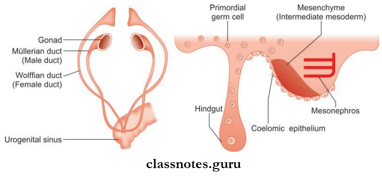
The development of the testis and ovaries begins in a similar manner but parts way at a particular point.
Development Of Testis
- Sex cords increase in length and extend into the medulla of the developing gonad. Sex cords are now called as medullary cords
- The sex cords anastomose with each other and canalize resulting in the formation of seminiferous tubules
- The ends of seminiferous tubules anastomoses with one another giving rise to rete testis
- Two Types Of Cells Line The Seminiferous Tubules:
- Spermatogenic cells: Formed from primordial germ cells
- Sertoli cells: Formed from coelomic epithelium
- A dense fibrous layer is formed by mesoderm which separates the sex cords from coelomic epithelium, known as the tunica albuginea.
- Mesoderm Also Gives Rise To:
- Leydig cells
- The connective tissue around seminiferous tubules
- Mediastinum testis
- The canal of the epididymis and vas deferens develop from the mesonephric duct. The development of the testis and ovaries begins in a similar manner but parts way at a particular point
- Gonads develop from three sources:
- Intermediate mesoderm—which is present medial to the middle part of the mesonephros
- Coelomic epithelium—which covers the intermediate mesoderm
- Primordial germ cells from the wall of the yolk sac near the allantois
- Coelomic epithelium begins to proliferate and it gets thickened
- Mesoderm below the coelomic epithelium condenses due to the thickening of coelomic epithelium
- Both these processes lead to the formation of the genital ridge
- Coelomic epithelial cells continue to proliferate and they invade the condensed mesoderm in the form of solid cords, known as the ‘sex cords’
- Primordial germ cells from the wall of the yolk sac migrate along the dorsal mesentery of the hindgut toward the developing gonad
- Sex cords and primordial germ cells get intermixed
- Till this point, the development of the testis and ovaries are the same.
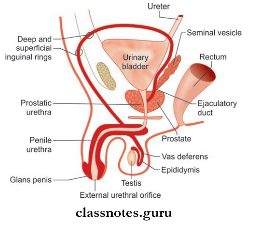
Male Reproductive System Anatomy
Descent of Testis
Descent Of Testis
- Testis which develops in relation to the lumbar region of the posterior abdominal wall starts to descend
- It gradually descends to the scrotum through the iliac fossa (3rd month) and the inguinal canal (7th month), finally reaching the scrotum by the end of 8th month.
- It is a mandatory developmental process to ensure that the mature testis promotes normal spermatogenesis
- Some Factors Responsible For The Descent Of The Testis Are:
- Increased intra-abdominal pressure
- Gubernaculum: A guiding force for the descent
- Differential growth of body wall.
Vas Deferens
The Features And Course Of Vas Deferens
- Also known as ductus deferens
- Thick-walled muscular tubes
- Two in number
- Length: 45 cm
- Lumen: Narrow, but the terminal part (ampulla) is sacculated.
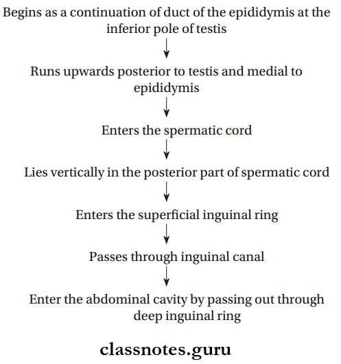
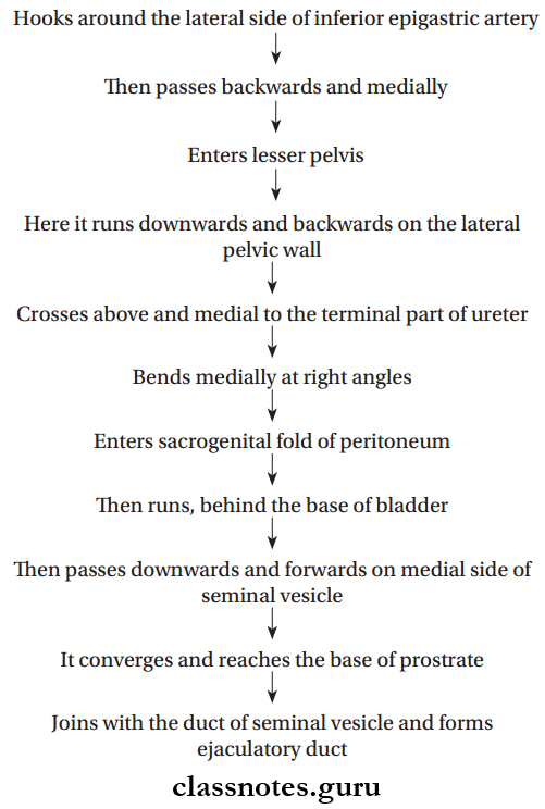
Vas Deferens Course: It has
- External course
- Internal course.
Vas Deferens External Course
Vas Deferens Internal Course
Testes Structure And Function
Vas Deferens Blood Supply
1. Arterial Supply
- From artery to vas deferens
- This artery can arise from either
- Superior vesical artery (common)
- Inferior vesical artery or
- Middle vesical artery.
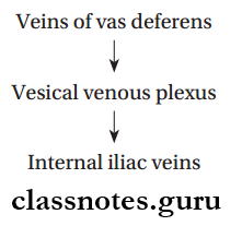
2. Vas Deferens Venous Drainage
3. Vas Deferens Nerve Supply
Pelvic splanchnic nerves—parasympathetic.
Male Internal Genital Organs
- Penis
- Scrotum
Male External Genital Organs
- Testis
- Epididymis
- Vas deferens
- Prostate
- Seminal vesicles
- Bulbourethral glands
Male Reproductive System Anatomy
Layers Of The Scrotum From Outside To Inside
The Layers Of The Scrotum From Outside To Inside
- Skin
- Dartos muscle (which replaces the superficial fascia)
- External spermatic fascia
- Cremasteric muscle and fascia
- Internal spermatic fascia.
Mnemonics: ‘Some Damn Englishman Called It scrotum’
Male Genital Organs Multiple Choice Questions
Question 1. The coverings of the testis are:
- Tunica vasculosa
- Tunica albuginea
- Tunica vaginalis
- All of the above
Answer: 4. All of the above
Question 2. Which of the following arteries gives blood supply to vas deferens?
- Middle rectal artery
- Inferior epigastric artery
- Cremasteric artery
- Superior vesical artery
Answer: 4. Superior vesical artery
Question 3. Which of the following statements are true about testis?
- It has no parasympathetic supply T cannot find it in books but Blitz reckons it has a vagal supply
- Appendix is inferior
- Vas deferens in somewhere
- Epididymis is somewhere else
- Drains to para-aortic and inguinal nodes
Answer: 1. It has no parasympathetic supply T cannot find it in books but Blitz reckons it has a vagal supply
Question 4. Lymph from the vas deferens drains into nodes:
- Superficial inguinal
- External iliac
- Internal iliac
- Lumbar
Answer: 2. External iliac
Testes Location In Body
Question 5. All of the following statements regarding ductus deferens are true, except:
- It is separated from the base of the bladder by the peritoneum
- It passes lateral to inferior epigastric artery at deep inguinal ring
- It crosses the ureter in the region of the ischial spine
- The terminal part is dilated to form an ampulla
Answer: 1. It is separated from the base of the bladder by the peritoneum