
Nerves Of Upper Limb Question And Answers
Question 1. Outline the nerve supply of upper limb.
Answer:
- The brachial plexus (C5, C6, C7, C8, T1) provides nerve supply to the upper limb.
- The major nerves originating from the brachial plexus are:
- Axillary nerve
- Musculocutaneous nerve
- Radial nerve
- Median nerve
- Ulnar nerve
Question 2. Write in detail about the brachial plexus and make notes on Erb’s and Klumpke’s paralysis.
Answer:
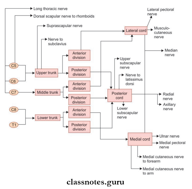
- Brachial plexus is the plexus of nerves formed by the anterior (ventral) rami of the last four cervical and first thoracic spinal nerves. (C5, C6, C7, C8, T1)
- It is divided into five subunits:
- Roots
- Trunks
- Division
- Cords
- Branches.
Mnemonic: Brachial Plexus Subunits
- ‘Randy Travis Drinks Cold Beer’:
- Roots
- Trunks
- Divisions
- Cords
- Branches
- Roots
- They constitute anterior primary rami of C5 to T1 spinal nerves.
- They are located in the neck.
- Trunks
- Upper trunk is formed by the union of C5 and C6 roots
- Middle trunk is formed by C7 alone
- Lower trunk is formed by union of C8 and T1 roots
- They are also located in the neck.
- Divisions
- Each trunk is divided to form anterior and posterior divisions.
- They are located behind the clavicle
- Cords
- Lateral cord is formed by union of the anterior division of upper and middle trunk.
- Medial cord is a continuation of anterior division of lower trunk.
- The posterior cord is formed by the union of the posterior division of all trunks.
- Cords are located in the axilla.
- Branches of Brachial Plexus
- From roots
- Long thoracic nerve/nerve to serratus anterior
- Dorsal scapular nerve/nerve to rhomboids
- From trunks
- Suprascapular nerve
- Nerve to subclavius
- From cords
- From lateral cord
- Lateral pectoral nerve
- Lateral root of median nerve
- Musculocutaneous nerve
- From medial cord
- Medial pectoral nerve
- Medial cutaneous nerve of arm
- Medial cutaneous nerve of forearm
- Medial root of median nerve
- Ulnar nerve
- From lateral cord
- From roots
Read And Learn More: Anatomy Question And Answers
-
-
- From posterior cord
- Radial nerve
- Axillary nerve
- Thracodorsal nerve/nerve to latissimus dorsi
- Upper subscapular nerve
- Lower subscapular nerve
- From posterior cord
-
Mnemonic: Brachial plexus: Branches of the posterior cord
STAR
- Subscapular [upper and lower]
- Thoracodorsal
- Axillary
- Radial
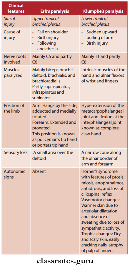
Clinical Anatomy: Two types of lesions occurring in brachial plexus are important.
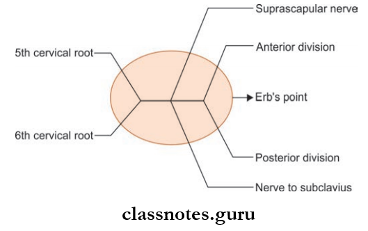
- Erb’s paralysis:
- There is a point in the brachial plexus where six nerves meet called Erb’s point. These meeting nerves are:
- 5th and 6th cervical roots
- Upper trunk formed by the union of these nerve roots
- Suprascapular nerve and nerve to subclavius branching from the upper trunk
- Any accident which causes an increase in angle between the head and shoulder can cause injury to the upper brachial plexus most commonly at Erb’s point.
- It results in a specific type of paralysis of the upper limb known as Erb’s paralysis
- For example A fall on the shoulder, birth injury, following anesthesia, etc.
- The clinical features are given in the table.
- There is a point in the brachial plexus where six nerves meet called Erb’s point. These meeting nerves are:
- Klumpke’s paralysis.
- It is another type of paralysis of the upper limb caused by accidents that increases angle between the trunk and shoulder making injury to lower brachial plexus.
- For example Sudden upward pulling of the arm, birth injury, etc.
Question 3. Explain in detail about axillary nerve under headings—origin, root value, course, branches, and innervation. Make a note on the injury to the nerve.
Answer:
- It is called axillary because it runs through the upper part of the axilla.
- It is called circumflex because it courses around the surgical neck of humerus.
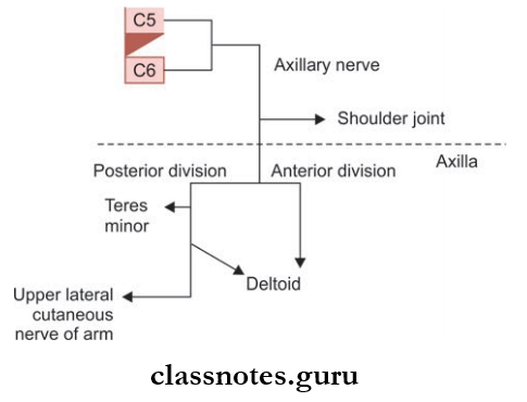
Axillary Nerve Origin
- It is smaller terminal branch of posterior cord of brachial plexus.
Axillary Root Value
- Ventral rami of C5, C6 segments.
Axillary Nerve Course
- From the posterior cord, it passes backward through the quadrangular intermuscular space.
- After reaching back, it divides into anterior and posterior divisions below the capsule of the shoulder joint.
- The posterior division again divides and one part continues as upper lateral cutaneous nerve and the other part goes to supply deltoid and teres minor with a pseudoganglion in it.
- The anterior division supplies deltoid muscle and skin over its anteroinferior part (regiment badge).
Axillary Nerve Branches and Innervation
- Trunk of axillary nerve
- Articular branch to shoulder joint
- Anterior division
- Muscular branch to deltoid-cutaneous branch to the skin over deltoid’s anteroinferior part (regimen badge)
- Posterior division
- The cutaneous branch continues as upper cutaneous nerve of arm.
- Muscular branch to teres minor and posterior part of the deltoid.
Axillary Nerve Clinical Anatomy
- The axillary nerve can easily get injured in inferior dislocation of the humerus or in injury to the surgical neck of the humerus.
- The presentation will be:
- Impaired abduction.
- Loss of contour of shoulder due to deltoid muscle wasting.
- Loss of sensation over the lower half of the deltoid (regimen badge).
Question 4. Explain in detail about musculocutaneous nerve under headings—origin, root value, course, branches, and innervation. Make a note on the injury to the nerve.
Answer:
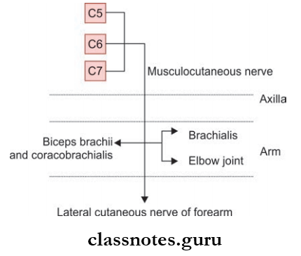
It is the nerve of front of arm.
Musculocutaneous Nerve Origin
- Lateral cord of brachial plexus in the axilla.
Musculocutaneous Nerve Root Value
- C5, C6, and C7 spinal segments.
Musculocutaneous Nerve Course
- From lateral cord of the brachial plexus, it runs downwards and laterally piercing coracobrachialis by supplying it.
- From there, it further descends downwards in between biceps brachii and brachialis muscles by innervating them.
- It reaches the lateral aspect of biceps tendon to pierce the deep fascia just above the elbow joint.
- From there, it continues down as lateral cutaneous nerve of forearm.
Musculocutaneous Nerve Branches and Innervation
- Muscular branches supplying
- Biceps brachii
- Coracobrachialis
- Brachialis
- Cutaneous branch (lateral cutaneous nerve of forearm)
- Supplies skin over the front and lateral aspect of forearm.
- Articular branch
- To the elbow joint
Musculocutaneous Nerve Clinical Anatomy
- Injury to the musculocutaneous nerve though rare can cause loss of
- Flexion of elbow
- Biceps tendon reflux
- Loss of sensation over the lateral aspect of the forearm.
Question 5. Explain in detail about radial nerve under headings—origin, root value, course, branches, and innervation. Write a note on injury to the nerve.
Answer:
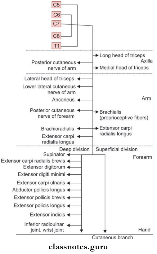
- It is the largest nerve of the brachial plexus.
- In general, it is the nerve of the dorsum of the arm, forearm, and hand.
Radial Nerve Root Value
- C5–C8, T1 spinal segments.
Radial Nerve Origin
- It is the continuation of the posterior cord of the brachial plexus.
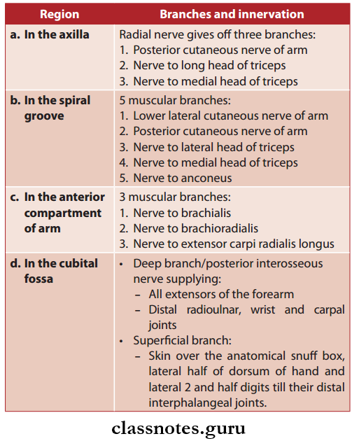
Radial Nerve Course
- From the posterior cord, the radial nerve descends downwards to enter the axilla.
- In the axilla radial nerve gives of three branches:
- The posterior cutaneous nerve of arm
- Nerve to the long head of triceps
- Nerve to medial head of triceps
- From the axilla, the radial nerve enters the posterior compartment of arm through the intermuscular space between the medial and long head of triceps.
- Radial nerve reaches the spiral groove of humerus and gives of 5 branches:
- Lower lateral cutaneous nerve of arm
- Posterior cutaneous nerve of arm
- Nerve to lateral head of triceps
- Nerve to medial head of triceps
- Nerve to anconeus.
- At the lower end of the spiral groove, the radial nerve pierces the lateral intermuscular septum to reach in between the brachialis and brachioradialis.
- It descends further down to run between the brachialis and extensor carpi radialis longus and enters the cubital fossa.
- In the anterior compartment of the arm, above the level of the lateral epicondyle, it gives of 3 branches:
- Brachialis
- Nerve to brachioradialis
- Nerve to extensor carpi radialis longus
- It then divides into superficial and deep branches at the level of the lateral condyle of humerus in the cubital fossa.
- The deep branch or posterior interosseous nerve supplies 2 muscles:
- Extensor carpi radialis brevis
- Supinator
- The deep branch further descends down to enter the posterior compartment and supplies:
- All extensors of the forearm
- Distal radioulnar, wrist, and carpal joints.
- The superficial branch passes down to the hand over the tendons of the anatomical snuff box to terminate as a cutaneous branch.
Radial Nerve Branches and Innervation
Mnemonic: Radial Nerve: Muscles Innervated
- ‘Try A Big Chocolate Chip Sundae, Double Dip Cherries And Peanuts Preferably Included’:
- In order of their innervation, proximal to distal:
- Triceps
- Anconeus
- Brachioradialis
- Extenstion Carpi radialis longus
- Extension Carpi radialis brevis
- Supinator
- Extension Digitorum
- Extension Digiti minimi
- Extension Carpi ulnaris
- Abductor poll. longus
- Extension Poll. brevis
- Extension Poll. longus
- Extension Indicis
- For the neighboring words that start with the same letter (for example, chocolate and chip), notice that the longer word in the mnemonic.
- Corresponds to the longer of the two muscle names (example: extensor carpi radialis longus and ext. carpi radialis brevis).
Radial Nerve Clinical Anatomy
- Radial nerves can get injured at various sites.
- Injury to the radial nerve in axilla can occur due to pressure from the upper end of crutch called as crutch palsy.
- Injury to radial nerve in radial or spiral groove can occur due to:
- Midshaft fracture of humerus
- Pressure was applied on radial nerve against the humerus by a drunker who is asleep with the medial aspect of his arm resting over the chair (Saturday night palsy).
- In such situations, the presentation will be:
- Loss of extension of wrist and fingers (wrist drop).
- Loss of supination when arm is extended.
- Sensory loss is restricted only to a small area over the dorsum of the hand in between first and second metacarpals.
- In such situations, the presentation will be:
- Injury to radial nerve at the elbow
- It is called radial tunnel syndrome.
Question 6. Explain in detail about ulnar nerve under headings— origin, root value, course, branches, and innervation. Write a note on injury to the nerve.
Answer:
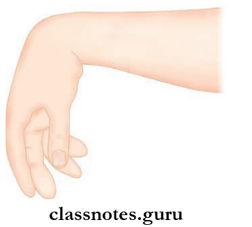
- It runs along the ulnar side of the upper limb hence the name.
- It is the nerve of fie movement and is called the musicians nerve.
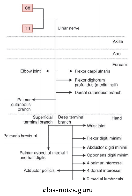
Ulnar Nerve Origin
- Medial cord of brachial plexus.
Ulnar Nerve Root Value
- C8 and T1 spinal segments mainly. C7 also contributes.
Ulnar Nerve Course
- In the axilla: Ulnar nerve lies medial to the third part of the brachial artery.
- In the arm:
- It enters the arm along the medial side of the brachial artery and runs up to the level of the mid-arm.
- The ulnar nerve pierces the medial intermuscular septum to reach the back of the arm.
- From there, the ulnar nerve descends down to pass through the cubital tunnel formed by the medial epicondyle and the fibrous band extending from the medial epicondyle to the olecranon process.
- The ulnar nerve has no branches in the axilla as well as in the arm.
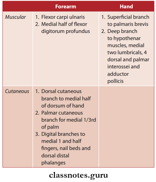
- In the forearm:
- In the upper 1/3rd, ulnar nerve passes between the two heads of flexor carpi ulnaris to reach underneath the muscle and runs vertically down.
- In the lower 2/3rd, it becomes superficial and runs downwards along with the ulnar artery being on the lateral side.
- Branches of the ulnar artery in the forearm are:
- Proximal forearm Muscular branch:
- Flexor carpi ulnaris
- Medial half of flxor digitorum profundus
- Mid forearm
- Cutaneous branch: Palmar cutaneous branch
- Distal forearm
- Cutaneous branch: Dorsal cutaneous branch
Ulnar Nerve In the Hand
- The ulnar nerve enters the hand superficial to the carpal tunnel through the ulnar tunnel and divides into superficial and deep terminal branches.
- The superficial branch gives sensory supply to the palmar aspect of the medial ½ digits and motor supply to palmaris brevis muscle.
- Deep branches give off
- Articular branches to the wrist
- Muscular branches to:
- 2 medial lumbricals
- 4 palmar interossei
- 4 dorsal interossei
- Flexor digiti minimi (hypothenar muscle)
- Abductor digiti minimi (hypothenar muscle)
- Opponents digiti minimi (hypothenar muscle)
- Adductor pollicis (thenar muscle)
Ulnar Nerve Branches and Supply
Ulnar Nerve Clinical Anatomy: Injury to the ulnar nerve can occur in the elbow or wrist.
- Injury to the ulnar nerve at the elbow
- Common causes are:
- Compression of the nerve in between the two heads of flexor carpi ulnaris
- Fracture and dislocation of medial epicondyle
- Valgus deformity of the elbow
- Thickening of the fibrous roof of the cubital tunnel (cubital tunnel syndrome).
- Common causes are:
The clinical presentation will be:
-
-
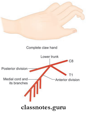
- Claw hand deformity affecting ring and little fingures where the metacarpophalangeal joints are extended while the interphalangeal joints are fixed (its actually not a complete claw hand, complete claw hand is seen when both ulnar nerve and medial nerve are injured simultaneously resulting in the hyperextended wrist and metacarpophalangeal joints and fixed interphalangeal joints).
- Flattening of the hypothenar eminence due to atrophy.
- Adduction and abduction of figers are affcted.
- Thmp can not be adducted.
- Loss of sensation over the palmar and dorsal surface of the medial 1/3rd of hand and medial 1-and-a-half
- fingers
- Foments sign will be positive when the integrity of the palmar interossei is tested.
- The ulnar nerve can also get injured in the wrist. Here, the clawing is more pronounced known as the ulnar paradox.
-
Question 7. Explain in detail about median nerve under headings—origin, root value, course, branches, and innervation. Write a note on injury to the nerve.
Answer:
- Median nerve is called median because it runs in the median plane of forearm.
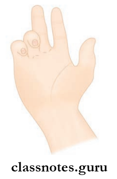
- It is called the eye of the hand because it gives sensory supply to the pulp of thumb and index fingers.
- It is also known as laborers nerve.
Median Nerve Origin
- By the union of medial and lateral cord of the brachial plexus.
Median Nerve Root Value
- C5, C6, C7, C8, T1.
Median Nerve Course
- In the axilla:
- Median nerve goes down to enter in the arm from the lateral side of 3rd part of the axillary artery.
- In the arm:
- The median nerve lies lateral to the brachial artery.
- But at the level of the middle of the arm, it crosses the brachial artery and runs medial to enter the cubital fossa.
- In the cubital fossa:
- It gives of muscular branches to flexor carpi radialis, flxor digitorum superfiialis and palmaris longus.
- The median nerve leaves the cubital fossa between two heads of pronator teres and then deep to the fibrous arch of the flexor digitorum superficial.
- In the forearm:
- In the forearm, it is adhered to the deep surface of the flexor digitorum superficial and leaves the muscle along its lateral border.
- It runs deep to the palmaris longus and gives of the palmar cutaneous branch before getting under the carpal tunnel.
- In the palm:
- After passing through the carpal tunnel, median nerve divides into lateral and medial divisions.
- Lateral division supplies 3 out of 4 thenar muscles, 1st and second lumbricals.
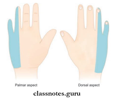
- Medial division along with the remaining fingers of lateral division provides cutaneous supply to the lateral 3 and half digits and their nail beds including the skin of distal phalanges on their dorsal aspect.
Median Nerve Branches and Innervations
Mnemonic: Median nerve: Hand muscles innervated
‘The LOAF muscles’:
- lumbrical 1 and 2
- Opponents pollicis
- Abductor pollicis brevis
- Flexor pollicis brevis
- Alternatively: LLOAF, with 2 L’s, to recall there is 2 lumbricals.
- To remember that these are the Median nerve muscles, think ‘Meat LOAF’.
Median Nerve Clinical Anatomy
- Injury to the median nerve can occur at various levels and the clinical features vary accordingly.
- Injury at the level of the elbow
- It can be due to:
- Supracondylar fracture of humerus.
- Entrapment of nerve between the two heads of pronator teres during its course.
- Tight tourniquet usage during venipunctures.
- The clinical features are:
- Loss of pronation as pronator teres is paralyzed.
- Weak wrist flexion due to paralysis of flexors supplied by the median nerve.
- The wrist will be adducted due to weakening of the flexor carpi radialis and unopposed action of the flexor carpi ulnaris and medial half of flexor digitorum profundus.
- It can be due to:
- Benedicts deformity:
- Median nerve injury leads to no flexion at interphalangeal joints of the index and middle fingers due to paralysis of the flexor digit running the superficial and lateral half of the flexor digitorum profundus. It results in placing of hand in a position comparable to Benedict’s hand.
- Ape thumb deformity:
- It is characterized by flttening of thenar eminence with lateral rotation and adduction of thumb due to paralysis of thenar muscles supplied by the median nerve
1. Injury at the level of the forearm
2. Injury at the carpal tunnel:
- Median nerve can get compressed in the tightly packed carpal tunnel due to:
- Myxedema
- Tenosynovitis of flxor tendons
- Dislocation of lunate bone following fracture
- Retention of fluid in pregnancy
- Osteoarthritis in the wrist joint
- It is presented with:
- Burning sensation over the area of sensory supply of median nerve in the hand (lateral 3½ digits) more during night
- Weakening of thenar muscles
- Ape thumb deformity, if left untreated
- Reduced conduction velocity in nerve conduction studies
- Phalen’s test and Tinel’s test positive.
Mnemonic: Carpal tunnel syndrome causes
MEDIAN TRAP
- Myxoedema
- Edema premenstrually
- Diabetes
- Idiopathic
- Acromegaly
- Neoplasm
- Trauma
- Rheumatoid arthritis
- Amyloidosis
- Pregnancy
Mnemonic fits nicely since the median nerve is trapped.
Question 8. Write a note on cutaneous supply of upper limb.
Answer:
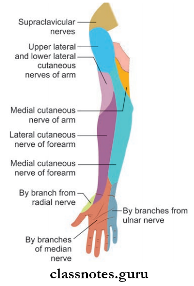
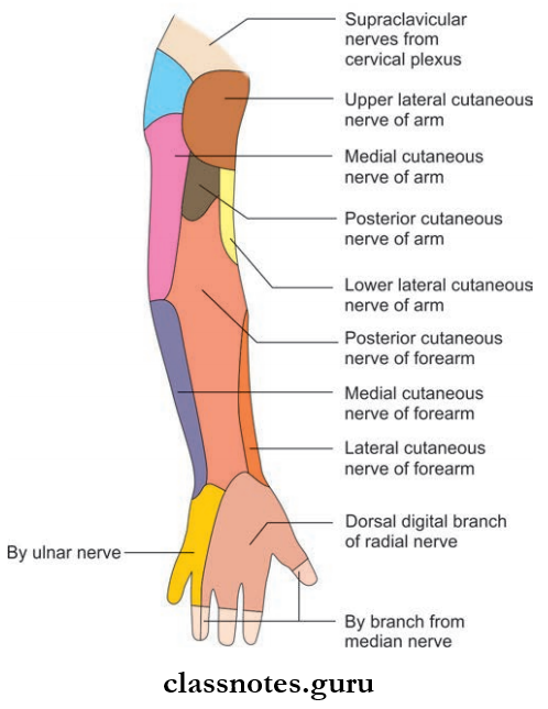
- Upper limb is supplied by C3–T2 spinal segments
- This is via:
- Supraclavicular nerves (C3 and C4)
- Nerves from brachial plexus (C5–T1)
- Intercostobrachial nerve (T2)
- Pectoral Region
- Above the 2nd rib by supraclavicular nerves (C3, C4)
- Below the 2nd rib by intercostal nerves (T2–T6).
- Axilla
- Intercostobrachial nerve (T2)
- Small branches from (T3).
- Shoulder
- Upper half of deltoid by supraclavicular nerves (C3, C4)
- Lower half of the deltoid by upper lateral cutaneous nerve of the arm.
- Arm
- Upper medial part by the intercostobrachial nerve (T2)
- Lower medial part by the medial cutaneous nerve of arm (T1, T2)
- Upper lateral half by upper lateral cutaneous nerve of arm
- Lower lateral part by lower lateral cutaneous nerve of arm (C5, C6)
- Posterior aspect of arm by the posterior cutaneous nerve of arm (C5).
- Forearm
- Medial side of the forearm by the medial cutaneous nerve of forearm (C8, T1)
- The lateral side of the forearm by lateral cutaneous nerve of the forearm (C5, C6)
- Posterior aspect of forearm by the posterior cutaneous nerve of forearm (C6, C7, C8).
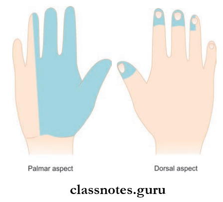
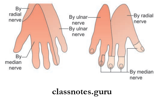
- Hand
- Palmar surface
- Lateral 2/3 rd of the palm by a palmar cutaneous branch of median nerve
- Medial 1/3rd of the palm is supplied by palmar cutaneous branch of the ulnar nerve.
- Dorsal surface
- Lateral 2/3 rd of the dorsum by superfiial terminal branch of radial nerve
- Medial 1/3 rd by dorsal branch/posterior cutaneous branch of ulnar nerve.
- Palmar surface
- Digits
- Palmar surface
- Lateral 3½ digits upto distal half of the middle phalanges by digital branches of median nerve
- Medial 1½ digits upto distal half of the middle phalanges by palmar digital branch of ulnar nerve
- Dorsal surface
- Lateral 3½ digits up to the proximal half of their middle phalanges by digital branches of radial nerve
- Lateral 3½ digits upto distal half of the middle phalanges by digital branches of median nerve
- Medial 1½ digits upto their middle phalanges by digital branches of ulnar nerve
- Medial 1½ digits up to the distal half of the middle phalanges by a palmar digital branch of the ulnar nerve.
- Palmar surface
Question 9. Draw the dermatomes of upper limb.
Answer:
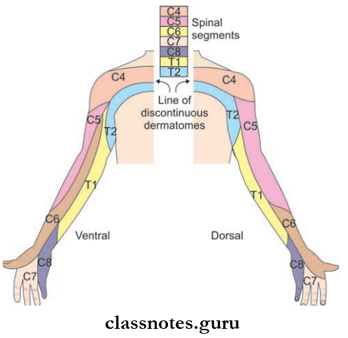
- The area of the skin supplied by one spinal segment is called as a dermatome.
- Dermatomes of the upper limb are given in the picture.
Nerves Of Upper Limb Multiple Choice Questions
Question 1. What is the continuation of ventral rami of 7th spinal cord called?
- Medial cord
- Upper trunk
- Middle trunk
- Lateral cord
Answer: 3. Middle trunk
Question 2. A patient presents with loss of abduction and weakness of lateral rotation of the arm. This is due to injury to a nerve caused by a fracture of the humerus at:
- Anatomical neck
- Midshaft
- Surgical neck
- Medial epicondyle
Answer: 3. Surgical neck
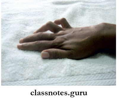
Question 3. Which nerve is injured if on trying to make a circle by touching the tip of index finger and thumb, the approximation of palmar spaces of distal phalanx occurs (as in pinching)?
- Median nerve at wrist
- Anterior interosseous nerve
- Recurrent branch of median nerve
- Deep branch of ulnar nerve
Answer: 2. Anterior interosseous nerve
Question 4. A sportsman with a severe injury to their right leg had to use crutches for several months. Subsequently, his doctor found that he had restricted abduction of shoulder and extension of the elbow. What is the site of injury to the brachial plexus?
- Middle trunk
- Posterior cord
- Lateral cord
- Medial cord
Answer: 2. Lateral cord
Question 5. Which dermatome overlies the thumb?
- T1
- C8
- C7
- C6
Answer: 4. C6
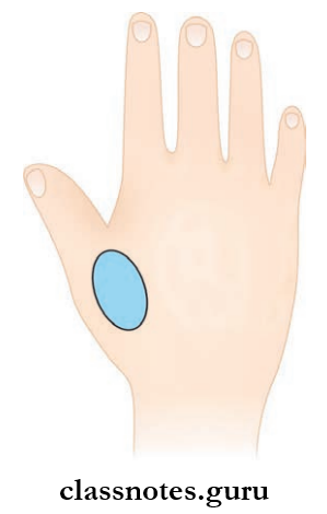
Question 6. The skin overlying the thenar eminence is supplied by:
- Recurrent branch of the median nerve
- Palmar cutaneous branch of ulnar nerve
- Palmar cutaneous branch of median nerve
- Lateral proper digital branch of median nerve
Answer: 3. Palmar cutaneous branch of the median nerve