
Sole Of Foot Question And Answers
Question 1. Give a description of the sole of foot with the peculiarities of skin, superficial fascia, and deep fascia.
Answer:
The Sole Of Foot
The region in the upper limb corresponding to the sole of the foot is hand. However, due to functional difference, sole and hand differ in their anatomical arrangements.
Sole Of Foot Skin of Sole
- It is specialized because:
- It is thick and hairless
- It is creased for grip
- Firmly adherent to the underlying deep fascia
- It has numerous sweat glands.
Sole Of Foot Superficial Fascia
- It is thick and dense over weight-bearing points. It contains:
- Subcutaneous fat
- Cutaneous nerves.
Read And Learn More: Anatomy Question And Answers
Sole Of Foot Deep Fascia
- It is modifid to form:
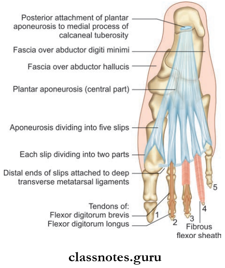
- Plantar Aponeurosis
- It is the thickened part of the fascia covering the sole
- It represents the distal part of plantaris muscle
- It is triangular in shape
- The apex is proximal and the base is distal
- At the base, it is divided into 5 processes
- Each process splits into the superficial and deep slip
- Superficial slip is attached to the skin
- Deep slip again splits and embraces the flexor retinaculum.
- Functions:
- It fies the skin to the sole
- It protects deeper structure in the sole
- Helps to maintain the longitudinal arch of the foot
- It gives origin to the first layer of the sole.
- Deep transverse metatarsal ligaments.
- Fibrous flexor sheaths.
- Plantar Aponeurosis
Sole Of Foot Clinical Anatomy
Plantar fasciitis can occur due to stretching of palmar aponeurosis in policemen.
Question 2. List the muscles of the sole of the foot. Give their nerve supply and actions.
Answer:
The Muscles Of Sole Of Foot
- There are 18 intrinsic muscles and 4 extrinsic tendons in the sole.
- Intrinsic muscles are arranged in 4 layers.
- They are chiefly concerned about maintaining the arches of the foot.
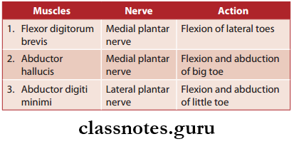
Muscles of First Layer of Sole of Foot
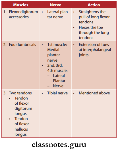
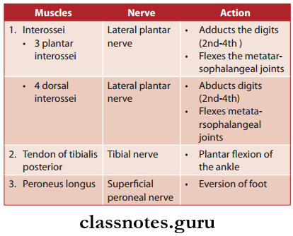
Muscles Of The Second Layer Of The Sole Of The Foot
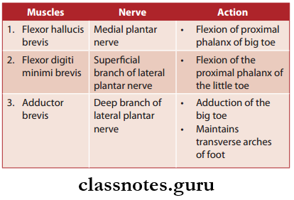
Muscles of Third Layer of Sole of Foot
Muscles of Fourth Layer of Sole of Foot
Question 3. Explain in detail about the arches of the foot by classifying and comparing them.
Answer:
Arches Of Foot
- The human foot is uniquely architectured to perform complex functions.
- Arches of the foot help in weight bearing, fast walking, running, and jumping.
- These arches are maintained by intrinsic and extrinsic muscles of the sole in addition to ligaments, aponeurosis, and the shape of bones.
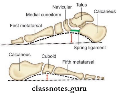
- Arches of the foot are classified into:
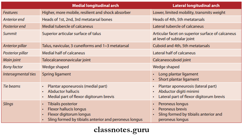
- Longitudinal:
- Medial
- Lateral
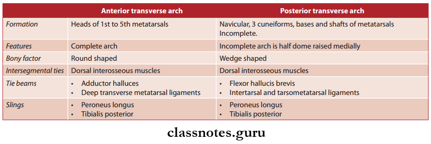
- Transverse:
- Anterior
- Posterior
- Longitudinal:
Arches Of Foot Functions
- It distributes body weight to the weight-bearing parts of the sole.
- They act as springs and help in walking and running.
- They also act as shock absorbers while stepping and jumping.
- The soft tissue of the sole is protected due to the concavity created by the arches.
Arches Of Foot Clinical Anatomy
- Flat Foot (Pes Planus): Occurs due to medial longitudinal arch deformity.
- High-Arched Foot (Pes Cavus): Occurs due to exaggeration of the longitudinal arch of the foot.
- Club Foot/Talipes: Combined defect of ankle and foot resulting in an inability to walk normally. For example, talipes equinus, talipes varus, talipes valgus, etc.
Mnemonics
- Tarsal bones of ankle ‘Tiger Cubs Need MILC’:
- Superior, then clockwise on right foot:
- Talus
- Calcaneus
- Navicular
- Medial cuneiform
- Intermediate cuneiform
- Lateral cuneiform
- Cuboid
Sole Of Foot Multiple Choice Question
Question 1. Which is the main joint of the medial longitudinal arch?
- Calcaneocuboid
- Subtalar
- Talocalcaneonavicular
- Ankle
Answer: 3. Talocalcaneonavicular