
Developmental Disturbances Of Oral And Paraoral Structures Short Question And Answers
Question 1. Gardener’s syndrome
Answer:
Gardener’s syndrome
It is a hereditary disorder characterized by colorectal polyps in association with various other lesions involving skin, eyes, teeth, and skeletal system
Gardener’s Syndrome Clinical Features:
- Multiple intestinal polyps
- Multiple osteomas of the skin, paranasal sinuses, and jaw
- Facial deformity
- Difficulty in mouth opening
- Multiple supernumerary teeth, impacted teeth, and odontomas
- Desmoid tumors of soft tissue and dermoid cysts of skin are present
- Pigmented lesion in ocular fundus
Gardener’s Syndrome Treatment:
- Prophylactic colectomy
- Surgical removal of osteomas and dermoid cyst
Question 2. Ramsay Hunt syndrome
Answer:
Ramsay Hunt syndrome
It is a zoster infection of geniculate ganglion with involvement of external ear and oral mucosa
Read And Learn More: Oral Pathology Questions and Answers
Ramsay Hunt syndrome Clinical Features:
- Facial paralysis
- The pain of external auditory meatus
- Pinna of the ear
- Vesicular eruption in the oral cavity and oropharynx
- Hoarseness of voice
- Tinnitus
- vertigo
Question 3. Melkersson-Rosenthal syndrome
Answer:
Melkersson-Rosenthal syndrome
- The melkersson-Rosenthal syndrome consists of
- Recurrent attacks of facial paralysis identical to Bell’s palsy
- Nonpitting, non-inflammatory painless edema of the face
- Chelitis granulomatosa
- Scrotal tongue
- Persistent unilateral edema of orbit and eyelid
Question 4. Hairy tongue
(or)
Black hairy tongue
Answer:
Black hairy tongue Etiology:
- Formation of excess keratin
- Infections- like candidiasis
Black hairy tongue Clinical Features:
- Elongation of filiform papillae
- Color- white to yellow
- Located on the posterior dorsal surface of the tongue
- Poor oral hygiene
- Bad taste in the mouth
Black hairy tongue Treatment:
- Elimination of predisposing factors
- Cleaning of the dorsal surface of the tongue with a soft toothbrush
- Treat candidiasis
Question 5. PeutJeghers syndrome
Answer:
PeutzJeghers syndrome Features:
- Recurrent abdominal pain due to familial intestinal polyps
- Cutaneous pigmentation in the perioral region
- Precocious puberty
- Gastrointestinal bleeding
- Pigmentation of buccal mucosa
Question 6. Xerostomia
Answer:
Xerostomia
It refers to the subjective sensation of dry mouth associated with salivary hypofunction
Xerostomia Etiology:
- Developmental- salivary aplasia
- Water or metabolic imbalance
- Iatrogenic causes
- Medications- antihistamines, decongestants, antidepressants, antihypertensives
- Radiation
Xerostomia Clinical Features:
- Reduction in salivary secretion
- Residual saliva is foamy or thick
- Fissured dorsum of the tongue
- Atrophy of filiform papilla
- Difficulty in mastication and swallowing
- Susceptibility to infection
- Dry mouth
- More prone to dental caries
Question 7. Turner’s hypoplasia
Answer:
Turner’s hypoplasia
- Turner’s hypoplasia is enamel hypoplasia occurring due to trauma or infection to the deciduous dentition
- Commonly affects incisors or premolars
- Periapical infection of deciduous teeth affects the ameloblastic layer of underlying permanent teeth
- As a result, permanent teeth get discolored or pitted
Question 8. Dilaceration
Answer:
Dilaceration
It refers to an angulation or sharp bend or curve anywhere along the root portion of the tooth
Dilaceration Clinical Features:
- Involves both dentition
- Seen at the coronal portion of the teeth
- The tooth looks like hook-shaped due to bending in the root
Treatment: - Extraction of involved teeth
Question 9. Talons cusp
Answer:
Talons cusp
It is an anomalous projection from the lingual aspect of the maxillary and mandibular permanent incisors
Talons cusp Clinical Features:
- It arises from the cingulum area of the tooth which extends to the incisal edge as a prominent T-shaped projection
- Asymptomatic
- Cosmetic problems
- Susceptible to caries
- Consist of normal-appearing enamel, dentin, and vital pulp tissue
Associated Syndrome:
Rubinstein Taybi syndrome
Talons cusp Treatment:
Restorative measures- to prevent caries
Question 10. Taurodontism
Answer:
Taurodontism
Taurodontism is a peculiar developmental condition in which the crown of the tooth is enlarged at the expense of its roots
Taurodontism Pathgenesis:
- It occurs due to failure of the Hertwig’s epithelial root sheath to invaginate at the proper horizontal level
Taurodontism Clinical Features:
- It involves both the sex
- It commonly affects multi-rooted permanent molar teeth and sometimes premolar
- It rarely occurs in primary dentition
- Common in Neanderthal men
- The affected tooth exhibits an elongated pulp chamber with rudimentary roots
- Teeth are usually rectangular with minimum constriction at the cervical area
- The furcation area of the teeth is more apically placed
- Teeth often have a greater apical-occlusal height
Question 11. Unerupted teeth
Answer:
Unerupted teeth
- It is an uncommon condition
- Causes delayed eruption of permanent teeth
Unerupted teeth Causes:
- Retained deciduous teeth
- Failure of eruption of permanent teeth
- Lack of eruptive force
- Cleidocranial dysplasia
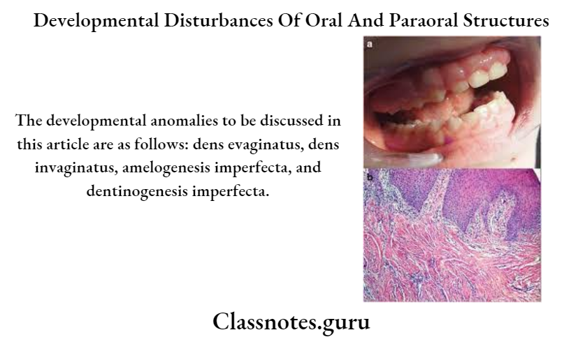
Question 12. Amelogenesis imperfecta
Answer:
Amelogenesis imperfecta
It is a developmental anomaly characterized by defective enamel formation
Amelogenesis imperfecta Clinical Features:
- Affects both dentition
- Color- chalky white to yellow
- Prone to disintegration
- Open contact points due to loss of enamel
- Abraded occlusal surfaces and incisal edges
- Abrasion of dentin
- Cheesy consistency of enamel
- Alteration in the eruption process
- Anterior open bite
- Presence of grooves and wrinkles on enamel surfaces
- The presence of some white opaque flecks at incisal margins gives Snow-capped teeth appearance
Question 13. Shell teeth
Answer:
Shell teeth
- In type 3 dentinogenesis imperfect the dentin appears very thin and pulp chambers and root canals are extremely large
- Because of this, the teeth appear thin shells of enamel and dentin
- Thus it is described as shell teeth
- Seen in association with enamel aplasia
Question 14. Ghost Teeth
Answer:
Ghost Teeth
- It is a radiographic feature of regional odontodysplasia
- It involves both dentition
- Permanent teeth show delayed eruption and defective mineralization
- There is a marked decreased radiodensity
- Enamel and dentin are very thin
- Pulp chambers are extremely large and open
- This results in the ghostly appearance of the involved teeth
Question 15. Tooth ankylosis
Answer:
Tooth ankylosis
Fusion between the tooth and bone is called ankylosis
Tooth ankylosis Clinical Features:
- Asymptomatic
- Produces dull, muffled sound on percussion
- Loss of periodontal ligament
- Mild sclerosis of the bone
- Blending of bone with tooth root
- This leads to difficulty in the extraction
Question 16. Submerged teeth
Answer:
Submerged teeth
Submerged teeth are ankylosed deciduous teeth
Submerged teeth Causes:
- Trauma
- Infection
- Disturbed local metabolism
- Genetic factor
Submerged teeth Clinical Features:
- Commonly affects mandibular second molars
- It prevents the exfoliation of deciduous teeth and the eruption of their successor
- It is located below the occlusal level of other teeth
- There is a lack of physiological mobility in the teeth
- It imparts solid sound on percussion
Radiographic Features:
- Absence of periodontal ligament
- Blending of tooth root and bone
Question 17. Enameloma
Answer:
Enameloma
- The ectopic formation of enamel in the form of globule on the root surface is referred to as Enameloma
- It is usually located in the cementoenamel junction or the cervical third of the root surface
Enameloma Clinical features
- It appears as yellowish white, spherical, or globular structure adherent to the furcation areas of the root surface
- Diameter ranges from 1-3 mm
- More common in the roots of maxillary molars
- May produce localized periodontal destruction in molars
- Radiological features
- Appears as hemispherical dense radio parities projecting from the root surface.