Syndromes Short Question And Answers

Oral Medicine Syndromes Important Notes

1. Features of various syndromes

Oral Medicine Syndromes Features of various syndromes
Oral Medicine Syndromes Features of various syndromes.

Oral Medicine Syndromes Short Essays

Question 1. Steven Johnson Syndrome.

Answer:

Steven Johnson Syndrome

It is the severe form of erythema multiform with wide-spread involvement, typically involving the skin, oral cavity, eyes, and genitalia

Steven Johnson Syndrome Clinical Features:

  • Symptoms:
    • Fever, malaise
    • Photophobia
    • Eruptions on oral mucosa, genital mucosa and skin
  • Skin lesions:
    • Hemorrhagic
    • Vesicle and bullae are present
  • Eye lesions:
    • Photophobia
    • Conjunctivitis
    • Corneal ulcerations
    • Keratoconjunctivitis
  • Genital lesions:
    • Non-specific urethritis
    • Balanitis
    • Vaginal ulcers
  • Complications:
    • Trachea-bronchial ulceration
    • Pneumonia
  • Oral manifestations:
    • Oral mucosa may be extremely painful
    • Mastication becomes impossible
    • Mucosal vesicles or bullae occur
    • They rupture and leave a surface covered with thick, white or yellow exudates
    • Erosions of the pharynx are also common
    • Lip may exhibit ulcerations with bloody crusting.

Steven Johnson Syndrome Management:

  • ACTH, cortisone, and chlortetracycline are advised

Question 2. Behcet’s syndrome.

Answer:

Behcet’s syndrome

  • It is a triad of recurring oral ulcers, recurring genital ulcer,s and eye lesions

Etiology:

  • Immunological
    • It is caused by immune complexes
  • Environmental antigen
    • It may be associated with streptococci, viruses, pesticides, and heavy metal

Behcet’s Syndrome Classification:

  • Mucocutaneous – oral, genital, and skin lesions
  • Arthritic
  • Neuro-ocular

Behcet’s Syndrome Clinical Features:

  • Age and Sex – Common in males with a mean age of 30 years
  • Recurring oral lesions
    • It may be mild/deep large scarring
    • It may appear anywhere on the oral and pharyngeal mucosa
    • They are painful lesions

Read And Learn More: Oral Medicine Question and Answers

    • Size – varies from several mm to cm.
    • They have erythematous borders and are covered by exudates
  • Recurrent genital lesions:
  • Site: Scrotum and penis in males
    • Labia in females
  • They are small and painful
  • Eye lesions:
    • Uvetitis
    • Retinal internal edema
    • Vascular occultation
    • Optic atrophy
    • Conjunctivitis
    • Keratitis
  • Skin lesions: Small pustules/papules occur
  • Arthritis:
  • Neurologic degeneration:
  • Others:
    • Thrombophlebitis
    • Intestinal ulceration
    • Venous thrombosis
    • Renal and pulmonary disease

Behcet’s Syndrome Management:

  • Topical and intralesional steroids – Useful for oral mucosal lesion
  • In severe cases – a combination of immune-suppressive drugs and systemic corticosteroids are used
  • Cyclosporine and colchicines combination – useful in mucocutaneous and GIT manifestations
  • Plasmapheresis – This is used in emergencies

Question 3. Gardener’s syndrome.

Answer:

Gardener’s syndrome

  • Gardener’s syndrome is inherited as an autosomal dominant trait

Gardener’s Syndrome Clinical Features:

  • Intestinal polyposis
  • Multiple osteomas
  • Multiple impacted supernumerary teeth
  • Connective tissue tumors
  • Thyroid carcinomas
  • Hypertrophy of pigmented epithelium of retina
  • Dental hypercementosis
  • Congenital missing teeth
  • Fused molar roots
  • Long and tapered molar roots
  • Hypodontia
  • Compound odontomas
  • Multiple carious lesions
  • Difficulties in extraction due to ankylosis

Oral Medicine Syndromes Short Answers

Question 1. Trotter’s Syndrome.

Answer:

Trotter’s Syndrome

  • The patient complains of neuralgic pain in the mandible, side of the head, tongue, and ear.
  • It is associated with middle ear deafness and defective mobility of the soft palate
  • Trismus

Trotter’s Syndrome Cause:

  • Tumor of the nasopharynx, starting in the pharyngeal wall involving the Eustachian tube.

Question 2. Grinspan syndrome.

Answer:

Grinspan syndrome

  • It is triad of oral lichen planus, diabetes mellitus and hypertension

Question 3. Ramsay Hunt syndrome.

Answer:

Ramsay Hunt syndrome

  • It is a zoster infection of the geniculate ganglion with involvement of external ear and oral mucosa
  • Facial paralysis, pain of external auditory meatus and pinna of the ear.
  • Vesicular eruptions in the oral cavity and oropharynx
  • Hoarseness of voice
  • Tinnitus
  • Vertigo

Question 4. Albright’s syndrome.

Answer:

Albright’s syndrome

  • Sex: It is found exclusively in females
  • It is a severe form of fibrous dysplasia involving nearly all the bones in the body
  • It is accompanied by pigmentations of skin along with endocrine disorders
  • Endocrine disorders:
    • Precocious puberty
      • Pubic hair, axillary hair, and breast development occur at the age of 5 years
  • Goiter
  • Hyperthyroidism
  • Hyperparathyroidism
  • Cushing’s syndrome
  • Acromegaly
  • Skin pigmentations:
    • These are coffee with milk color spot
    • There is an irregular flat area of increased skin pigmentation
  • Vaginal bleeding occurs
  • Long bones are frequently affected
  • Prognosis
    • The active period ceases in adult life
    • If growth continues the rate of activity is reduced
    • Calcification of fibrous tissues continues

Question 5. Behcet’s Syndrome

Answer:

Etiology:

  • Immunologicalcaused by immune complexes formed by immunocompetent T lymphocytes and plasma cells
  • Environmental: associated with bacteria such as streptococci, viruses, pesticides, and heavy metals

Behcet’s Syndrome Clinical Features:

  • Age: 10-45 years
  • Sex: Common in males

Behcet’s Syndrome Presentation:

  • Genital lesions:
    • Consist of recurrent aphthae
    • Larger than mucosal lesions
  • Site: in malespenis, inner thigh, scrotum
  • In females: vulva, painful in females
    • Healing of these lesions may lead to severe scarring
  • Skin lesions:
    • Small pustules or papules appear on the trunk or limbs
  • Eye lesions:
    • Initially, ocular lesions develop
    • Consist of conjunctivitis, keratitis, uveitis, optic atrophy, retinal edema
    • This may lead to visual damage and eventually blindness
    • Photophobia may also occur
  • Oral manifestation:
    • Recurrent aphthae ulcers develop
    • They vary in size
    • They are deep and painful
    • They are well-demarcated
    • Have an erythematous border
    • Covered by gray or yellow exudates
    • They can occur anywhere in the oral mucosa
    • Other features:
    • Neurological degeneration
    • Intestinal ulceration
    • Arthralgia
    • Visceral involvement

Behcet’s Syndrome Diagnosis:

  • Presence of eye lesions
  • Recurrent genital lesions
  • Skin lesions
  • Positive pathergy test

Behcet’s Syndrome Management:

  • Administration of topical and intralesional steroids
  • Use of immunosuppressor drugs like cyclosporine, azathioprine
  • interferon alpha
  • Plasmapheresis is used in emergency

Question 6. Eagle’s Syndrome.

Answer:

  • Synonym: DISH Syndrome

Eagle’s Syndrome Types:

  • Classic type: Occurs after tonsillectomy
  • Carotid artery syndrome: Results from calcification of stylohyoid ligament
  • Traumatic Eagle’s syndrome: Develops after fracturing of stylohyoid ligament

 Eagle’s SyndromeClinical Features:

  • Age: seen commonly in adults
  • Presentation:
    • The elongated styloid process can be visualized
    • Pain in the lateral pharyngeal area and side of the lower face and neck
    • Difficulty in swallowing
    • Sore throat
    • Glossodynia
    • Headache
    • Dull to severe hemifacial pain
    • Blurred vision
    • Vertigo
    • Pan while opening of mouth wide or while swallowing

Eagle’s Syndrome Radiographic Features:

  • Elongation of the styloid process is seen

Eagle’s Syndrome Management:

  • Topical anesthesia
    • Infiltration of anesthetic agent will relieve pain
    • Surgical treatment
    • Surgical resection or segmentation of elongated styloid process
  • Corticosteroid injection
    •  It provides relief

Question 7. Melkerson Rosenthel Syndrome.

Answer:

Melkerson Rosenthel Syndrome

  • It is a triad of
  • Chelitis granulomatosa
  • Facial paralysis
  • Scrotal tongue

Question 8. Heerfordt’s syndrome.

Answer:

Heerfordt’s syndrome

  • It is characterized by
    • Firm, painless, bilateral enlargement of the parotid gland
    • Inflammation of the uveal tract of the eye
    • Facial palsy

Question 9. Ascher’s syndrome.

Answer:

Ascher’s syndrome

  • It consists of
    • Double lip
    • Blepharochalasis – It is the drooping of the tissue between the eyebrow and the edge of the upper eyelid
    • Non-toxic thyroid enlargement

Question 10. Sturge Weber syndrome.

Answer:

Sturge Weber syndrome

  • It is a congenital disorder

Sturge Weber syndrome Classification:

  • Complete presymptomatic
  • Incomplete symptomatic
  • Incomplete monosymptomatic

Sturge Weber syndrome Etiology:

  • Dysplasia of the embryonal vascular system
  • Development insult
  • Aberrant migration

Sturge Weber syndrome Clinical Features:

  • Port wine stain present at the distribution of trigeminal nerve
  • There is angioma
  • The patient is present with seizures
  • The patient may feel weakness
  • Headaches can also occur
  • Glaucoma may be present
  • There is the presence of vascular hyperplasia of a purple color
  • Gingival enlargement can also occur

Sturge Weber syndrome Management:

  • Anticonvulsant – To control seizures
  • Anti – glaucoma
  • Dentally managed

Question 11.Papillion Lefevre Syndrome.

Answer:

Papillion Lefevre Syndrome

  • It is an autosomal recessive disorder
  • It is a triad of
    • Hyperkeratosis of palms of the hand and soles of feet
    • Extensive prepubertal destruction of the periodontal bone supporting the dentition, usually extensive generalized horizontal bone loss
    • Calcification of dura

Oral Medicine Syndromes Viva Voce

  1. Sturge-Weber syndrome is associated with vascular lesions
  2. Hypopigmentation of skin and hair occurs in Che- diak- Higashi syndrome

Endocrine Disorders Short Essays

Endocrine Disorders Important Notes

1. Addison’s disease

  • Occurs due to insufficiency of adrenal cortex hormone
  • Manifested as pale brown to deep chocolate pigmentation of oral mucosa spreading to the buccal mucosa, gingiva, tongue, and lips
  • Bronzing of skin
  • Hypotension
  • GIT disturbances occur
  • Other features include lethargy, fatigue, muscular weakness, severe anemia
  • Biopsy shows acanthosis with silver-positive granules in the cells of stratum germinativum

2. Gigantism and acromegaly

  • Occurs due to excessive secretion of growth hormone
  • There is an increase in the number of granules in the acidophilic cells
  • If the increase in granules occurs before the closure of the epiphysis of long bones, gigantism results
  • If the increase occurs after epiphyseal closure acromegaly develops
  • Gigantism occurs in children and acromegaly occurs in adults
  • Acromegaly
    • Lips become thick
    • The tongue becomes enlarged and shows indentations on the side
    • The mandible becomes large and macrognathic
    • Mandibular teeth are buccally tipped

3. Hypophosphatemia

  • Occurs due to deficiency of alkaline phosphatase enzyme in serum
  • Oral manifestations are
    • Formation of globular, hypocalcified dentin in region of pulp horns
    • Pulp horns are elongated
    • Abnormal cementum
    • Lamina dura is absent

Endocrine Disorders Short Essays

Question 1. Precautions to be taken during dental treatment of cardiac patients
(or)
Dental Considerations for Ischaemic Patients.

Answer:

Dental Considerations for Ischaemic Patients

  • Ischaemic heart disease patients should be taken up for early appointments in the morning and not made to wait
  • Patient with known diseases should be asked to carry their rescue medication
  • In patients with moderate to severe angina, prophylactic nitroglycerine may be used 5 minutes before starting the dental procedure
  • If a patient gets an acute episode of angina during a dental procedure
    • Stop the dental procedure for a few minutes
    • Administer tab nitroglycerine 0.5 mg sublingually
    • Monitor pulse and BP
    • If pain is not relieved in 8-10 minutes, the nitroglycerine dose may be repeated

Question 2. Hyperparathyroidism/Brown’s tumor.

Answer:

Hyperparathyroidism

  • It is an endocrine disorder occurring due to an excess of circulating parathyroid hormone

Brown’s Tumor Types:

  1. Primary Hyperparathyroidism: Occurring due to tumor of glands
  2. Secondary Hyperparathyroidism: Occurs to response to hypocalcemia
  3. Tertiary Hyperparathyroidism: Occurs after long-standing secondary hyperparathyroidism

Brown’s Tumor  Clinical Features:

  • Age and Sex – Common in middle-aged women

Brown’s Tumor  Classic Triad:

  • Kidney stones
  • Bone resorption
  • Duodenal ulcers

Brown’s Tumor  Renal Symptoms:

  • Renal calculi
  • Hematuria
  • Back pain

Read And Learn More: Oral Medicine Question and Answers

  • Psychological symptoms – Emotionally unstable
  • GIT symptoms – Anorexia, nausea, vomiting

Skeletal:

  • Bone pain
  • Pathologic fractures
  • Bone deformities
  • Hypercalcemia

Generalized Symptoms:

  • Muscle weakness
  • Fatigue
  • Weight loss
  • Insomnia
  • Headache
  • Polydipsia and polyuria

Oral Manifestations:

  • Brown tumor – Intraoral/Extraoral swelling appears

Brown’s Tumor  Teeth:

  • Gradual loosening
  • Drifting and loss of teeth
  • Malocclusion

Brown’s Tumor  Radiographic Features:

  • Radiodensity-radiolucentlesion
  • There may be a normal, granular, or ground-glass appearance
  • Moth-eaten appearance with varying density
  • Osteitis fibrosa generalisation – Localised bone destruction
  • Brown tumor – represents ill-defined radiolucency
  • Thinning of cortical tables
  • Pepper pot skull
  • Demineralization of the inferior border of the mandibular canal
  • Thinning of outlines of the maxillary sinus
  • Loss of lamina dura

 Brown’s Tumor Management:

  • Surgical – Hyperplastic tissue is removed
  • Vitamin D – Oral administration of vitamin D
  • Parathyroidectomy
  • Restriction of dietary phosphate, phosphate binding agent, and aluminum salts

Question 3. Oral manifestations of diabetes mellitus.
(Or)
Diabetes Oral manifestations and dental management.

Answer:

Periodontium:

  • Alter response of the periodontal lesion to local irritants
  • Retards healing of tissues
  • GCF contains more glucose
  • Periodontal abscess formation
  • Tooth mobility
  • Severe and rapid bone resorption

Diabetes Oral manifestations Tongue:

  • Altered taste sensation
  • Median rhomboid glossitis
  • Impaired local immune response
  • Decreased Langerhans cell

Oral Candidiasis:

  • Alveolar bone
  • Localized osteitis

Diabetes Oral manifestations Mouth:

  • Burning mouth sensation
  • Dysgeusia
  • Dysesthesia
  • Xerostomia
  • Increased caries activity

  Diabetic Sialadenosis:

  • Nerve: Diabetes neuropathy involving the trigeminal nerve
  • Angular cheilosis
  • Oral lichen planus

Dental Management:

  • Acute musculoskeletal pain, painful dental lesions
    • Patients with diabetes need to maintain very good oral hygiene as they are prone to odontogenic, periodontal, and other infections
    • In well-controlled diabetics, dental procedures generally do not require any special precautions
    • Antibiotic coverage may be required
    • In uncontrolled diabetics, infections and wound healing may be a problem even after minor procedures
    • If major dental procedures or surgery is to be undertaken, prior control of diabetes by the physician is needed
    • If the patient is on only oral antihyperglycemic drugs, he should start insulin
    • Dental procedures may often reduce food intake and diabetics may go to hypoglycemia
    • Dose adjustments may be required

Question 4. Serum Sickness.

Answer:

Serum Sickness Causes:

  • After the administration of foreign serum
  • Tetanus antitoxin
  • Rabies antiserum

Serum Sickness Mechanism:

  • Antibodies form immune complexes in blood vessels with administered antigens
  • These complexes fix complement which attracts the leukocytes to the area causing direct tissue injury

Serum Sickness Features:

  • Fever
  • Swelling
  • Lymphadenopathy
  • Joint and muscle pain
  • Rash
  • Peripheral neuritis,
  • Kidney disease
  • Myocardial ischemia

Question 5. Name 2 conditions with increased serum alkaline phosphate.

Answer:

2 conditions with increased serum alkaline phosphate.

  • Malignancy
  • Abscess of liver
  • Amyloidosis
  • Leukemia
  • Sarcoidosis

Endocrine Disorders

Endocrine Disorders Viva Voce

  1. Rapid weight loss with salt and water depletion is found in type 1 diabetes
  2. Chronic fatigue and malaise are found in type 2 diabetes
  3. Median rhomboid glossitis is a pathognomonic oral manifestation

Red And White Lesions Short Question And Answers

Oral Medicine Red And White Lesions Short Answers

Question 1. Etiology and risk factors for oral cancer

Answer:

Etiology and risk factors for oral cancer

Oral medicine Red And White lLsions Etiology and risk factors for oral cancer

Question 2. Local and general predisposing factors for oral candidiasis

Answer:

  • Local predisposing factors
    • Changes in oral microbial flora
      • Occurs owing to administration of antibiotics, excessive use of antibacterial mouth rinses, xerostomia, secondary to anticholinergic agents
    • Local irritant
      • Chronic irritants due to dentures, orthodontic appliances, and heavy smoke
  • Systemic factors
    • Drug therapy
      • Administration of corticosteroids, cytotoxic drugs, immunosuppressive agents
    • Acute and chronic diseases
      • Leukemia, lymphoma, diabetes, and tuberculosis
    • Malnutrition
    • Age- Infancy, pregnancy, and old age
    • Endocrinopathy- Hypoparathyroidism, hypotony- iodism, Addison’s disease
    • Immunodeficiency states

Question 3. Traumatic keratosis

Answer:

Traumatic keratosis

It refers to an isolated area of thickened whitish oral mucosa

Etiology:

  • Local irritants- 111 fitting dentures, sharp clasp, and rough edges of restoration
  • Cigarette smoking

Clinical Features:

  • Commonly involves are lip and buccal mucosa
  • Appears as an isolated thickened whitish area
  • Glassblower’s white patch- it is a variant of traumatic keratosis affecting the cheek and lips that occurs in a glass factory

Management:

  • Removal of local Irritants

Question 4. Carcinoma in situ.

Answer:

Carcinoma in situ

It is the most severe stage of epithelial dysplasia, which involves the entire thickness of the epithelium with the basement membrane intact

Carcinoma in situ Clinical Features:

  • Age: Elderly patients
  • Sex: common in males

Carcinoma in situ Presentation:

  • Appears as white plaques or ulcerated areas
  • Site: Floor of the mouth, tongue, lip, etc
  • Appears as leukoplakia or erythroplakia

Carcinoma in situ Treatment:

  • Surgery
  • Radiotherapy
  • Electrocautery

Question 5. Id reaction.

Answer:

Id reaction

  • A person with chronic Candida infection may develop a secondary response characterized by localized or generalized sterile vesicopapular rash
  • It may be due to an allergic response to Candida antigen.

Question 6. White sponge nevus.

Answer:

Etiology:

  • An autosomal dominant genetic defect

White sponge nevus Features:

  • Diffuse, thickened white lesion
  • Has a corrugated appearance
  • Occurs bilaterally over Buccal mucosa
  • Occasionally involves the tongue, the floor of the mouth, and labial mucosa

Oral Medicine Red And White Lesions

Read And Learn More: Oral Medicine Question and Answers

  • It is a painless lesion
  • Persistent in nature
  • Usually present during early childhood
  • May also involve mucosa of the larynx, esophagus, and vagina

White sponge nevus Differential Diagnosis:

  • Leukoedema
  • Frictional keratosis
  • Plaque type lichen planus
  • Chronic hyperplastic candidiasis

White sponge nevus Treatment:

  • No treatment necessary

Question 7. Types of oral candidiasis.
(or)
 Classification of oral candidiasis.

Answer:

Types of oral candidiasis

  • Oral Candidiasis
  • Acute
    • Acute pseudomembranous candidiasis
    • Acute atrophic candidiasis
  • Chronic
    • Chronic atrophic candidiasis
      • Denture stomatitis
      • Median rhomboid glossitis
      • Angular cheilitis
  • ID reaction
  • Chronic hyperplastic candidiasis
  • Chronic mucocutaneous candidiasis
    • Familial CMC
    • Localized CMC
    • Diffused CMC
    • Candidiasis endocrinopathy syndrome

Question 8. Oral hairy leukoplakia.

Answer:

Etiology:

  • Hyperplasia of the oral epithelium with a production of excess keratin caused by Epstein- Burr virus infection
  • The surface of the lesion is frequently infected with Candida albicans
  • Occurs most commonly in individuals with compromised immunity secondary to HIV

Oral Hairy Leukoplakia Features:

  • Adherent white plaque is located most often on the lateral borders of the tongue
  • The surface of each lesion is characteristically corrugated or shaggy in appearance
  • Typically bilateral
  • Usually painless
  • Persistent lesion
  • More common in young adult males

Oral Hairy Leukoplakia  Differential Diagnosis:

  • Frictional keratosis
  • Plaque type lichen planus
  • Chronic hyperplastic candidiasis
  • Smoking related leukoplakia

Oral Hairy Leukoplakia  Treatment:

  • Some patients respond to high doses of Acyclovir
  • Lesions often recur when treatment is stopped

Question 9. Linea alba.

Answer:

Linea alba

It refers to the line of keratinization, found on the buccal mucosa parallel to the line of occlusion.

Etiology:

  • Diet: Hard Food
  • Frictional irritation- Due to contact of teeth with Mucosa.
  • Increased overjet
  • Other: Smoking and environmental irritants.

Linea alba Clinical Features:

  • Site: Buccal mucosa at the line of occlusion.
  • Presentation:
    • Palate and gingiva appear whiter.
    • The line extends from the commissure to most posterior teeth.

Linea alba Management:

  • Spontaneous treatment.

Oral Medicine Red And White Lesions Viva Voce

  1. Tobacco is the major causative factor of leukoplakia
  2. Nonsmokers have a high percentage of leukoplakia at the border of the tongue
  3. The floor of the mouth has the highest rate of malignant transformation of leukoplakia
  4. Wickham’s striae are found in the reticular type of oral lichen planus
  5. Leukoplakia is the most common precancerous lesion
  6. Krythroplakia is the most malignant precancerous lesion
  7. Civatte bodies is seen in lichen planus
  8. Civatte bodies are often present in spinous and basal layers
  9. Saw tooth retepegs are seen in lichen planus
  10. Wickham’s striae is a feature of lichen planus Candida that occurs in three forms: pseudohyphae, yeast, and chlamydospores
  11. Thrush is prone to occur in debilitated or chronically ill patients
  12. Angular cheilitis is due to chronic atrophic candidiasis

Red And White Lesions Short Essays

Red And White Lesions

Question 1. TNM staging
Answer: TNM staging

It is the staging of malignancy which measures 3 major parameters of cancer

  • T- the size of the tumor
    • N- lymph node involvement
    • M-distant metastasis
  • T- Primary tumor
    • The tx-primary tumor cannot be assessed
    • To-No evidence of primary tumor
    • This- carcinoma in situ o Ti- Tumour size- 2 cm or less in diameter
    • T2– Tumour size- 2-4 cm in diameter
    • T3– Tumour size- more than 4 cm in diameter
    • T4– Tumour invades adjacent structures
  • N- Regional lymph node
    • Nx – Regional lymph node cannot be assessed
    • N0-No regional lymph node metastasis
    • N1– Metastasis in a single ipsilateral lymph node, 3 cm or less in dimension
    • N2– Metastasis in the single ipsilateral lymph node, more than 3 cm but less than 6 cm
    • N2a– Metastasis in the single ipsilateral lymph node, 3-6 cm in dimension
    • N2b – Metastasis in multiple ipsilateral lymph nodes, not more than 6 cm
    • N2c– Metastasis in bilateral or contralateral lymph nodes, not more than 6 cm
    • N3 – Metastasis in the lymph node, more than 6 cm in dimension
  • M- Distant metastasis
    • Mx – The presence of distant metastasis cannot be assessed
    • M0 – No distant metastasis Mi – Presence of metastasis
    • M1– Presence of metastasis

Read And Learn More: Oral Medicine Question and Answers

Question 2. Treatment of cancer
Answer: Aims Of Cancer Treatment:

  • Cure of the patient
  • Palliation
  • Preservation of function
  • Cosmetic function
  • Treatment of lymph node
  • Treatment of advanced tumors

Role Of Chemotherapy:

    • Cisplatin is the most effective drug
    • In advanced cases, chemotherapy is given before surgery or radiotherapy
    • This is called induction chemotherapy

Radiotherapy:

    • Radiotherapy preserves anatomical parts and functions
    • 6500-7500 cGy units are required to eradicate cancer

Role Of Surgery:

    • It may be in the form of wide excision or wide excision with removal of the bone
    • Radical neck dissection is done in case of lymph node involvement

Question 3. Lichenoid reactions
Answer: Lichenoid reactions

It has a clinical picture similar to lichen planus

Etiology:

  • Disorders: lichen planus
  • Drugs:
    • Antimicrobial: tetracycline
    • Anti par asltit thlornquine
    • Antihypertensive methyl dopa
    • Anti ills gold

Lichenoid Reactions Clinical Features:

    • Lichenoid mm positive over oral mucosa
    • Lichenoid dermatitis: over the skin
    • Lichenoid gingivitis.: over gingiva

Lichenoid reactions Management: discontinuation of the drug

Question 4. Erythroplakia.
Answer:

Erythroplakia

  • It is a red patch or plaque in the oral mucosa which cannot be characterized clinically or pathologically as any other condition and which has no apparent cause

Etiology:

  • Use of tobacco
  • Alcohol
  • Candida infection
  • Idiopathic

Erythroplakia Clinical Features:

  • Age: a fifth-seventh decade of life
  • Sex: both sexes are equally affected
  • Site
    • The floor of the mouth
    • Retromolar area
    • Buccal mucosa
    • Gingiva
    • Tongue
    • Soft palate
  • Presentation
  • It appears as a small or extensive reel lesion
  • It has well-defined borders

Erythroplakia Types:

  • Homogeneous
    • Has uniform red patches all over
  • Erythroplakia with interspersed patches of leukoplakia
    • Has a few white leukoplakic patches along with a red patch
  • Speckled leukoplakia
    • It is characterized by the presence of soft irregular, raised, erythematous areas with a granular surface

Erythroplakia Differential Diagnosis:

  • Candidiasis: Lesson can be rubbed off
  • Denture stomatitis: The commonly involved site is the palate
  • Tuberculosis: Present of tubercular ulcers
  • Histoplasmosis:  It is common in farmers

Erythroplakia Management

  • Elimination of the causative agent
  • Mucosal stripping of the lesion
  • Laser ablation
  • Electrocoagulation
  • Cryotherocoagulation
  • Maintenance by periodic recall visits every 3 months

Red And White Lesions.

Question 5. Diagnosis of oral lichen planus.
Answer:

Diagnosis of oral lichen planus

  • Clinical
    • The presence of bilateral interlacing white striae
    • Presence of Wickham striae and Koebner phenomenon
  • Laboratory diagnosis
    • Hyperorthokeratosis
    • Hyperparakeratosis
    • Acanthosis with intercellular edema
    • Civatte bodies
    • The sawtooth appearance of the rete pegs
  • Immunofluorescence
    • Positive reactions with IgA, IgM, and IgG antisera
    • Presence of subepithelial deposits of fibrinogen and antigenically related substances

Question 6. Atrophic candidiasis.
Answer:

Atrophic candidiasis

Synonym: Antibiotic sore mouth

Atrophic Candidiasis Clinical Features:

  • Site: tongue, tissue underlying the prosthesis
  • Presentation
    • The lesion appears red or erythematous
    • Patients usually have vague pain or a burning sensation
    • Lesion reveals a lew white thickened foci, that are rubbed off leaving a painful surface
    • It closely resembles erosive lichen planus and erythroplakia

Atrophic Candidiasis  Differential Diagnosis:

  • Chemical burn: History of chemicals
  • Drug reaction: Diminished host response
  • Syphilitic mucous patch– Skin lesion is also present
  • Necrotic ulcer and gangrenous stomatitis: Ulcer is deeper
  • Traumatic ulcer: History of trauma present

Atrophic Candidiasis  Management:

  • Elimination of causative agent:
    • Replacement of denture
    • Relining of denture
    • The denture must be cleaned thoroughly and regularly
    • It should be left out of out of the mouth at night in a hypochlorite solution
      • Topical application
  • Clotrimazole:
    • It is an effective topical treatment when dissolved in the mouth for five minutes daily
    • Nystatin Preparation:
      • Dissolves only in the mouth for 5 minutes a day.
  •  Amphotericin B:
      • 5-10 ml of oral solution was used as a rinse.

Question 7. Erosive lichen planus.
Answer:

Erosive lichen planus

  • It clinically exhibits a mixture of erythematous, ulcerated, and white pseudomembranous areas
  • A faint white zone resembling radiating striae is frequently seen at the junction where the erosive area meets the normal epithelium
  • Most of the lesions develop on the buccal mucosa and the vestibule
  • Patients often complain of severe pain and burning sensation at the time of taking hot and spicy food
  • Patients may restrict themselves to only the bland liquid diet
  • Palpation of the affected mucosa often elicits pain and bleeding
  • The areas of mucosa where the lesion has already healed up exhibit melanotic hyperpigmentation.


Red And White Lesions Long Essays

Oral Medicine Red And White Lesions Long Essays

Question 1. Classify red and white lesions of the oral cavity. Write etiology, clinical features, and management of oral submucous fibrosis and candidiasis.

Answer.

Classification:

  1. Red lesions:
  • Inflammatory Conditions:
    • Inflammation associated with traumatic injury
    • Mechanical- cheek biting, ill-fitted denture
    • Chemical- aspirin, formoterol
    • Thermal- hot food, hot beverages
    • Radiation- mucositis
    • Infection
      • Bacterial
      • Scarlet fever
      • Gonococcal stomatitis
      • Vincent infection
      • Fungal
      • Atrophic candidiasis
      • Angular cheilitis
      • Viral
      • Measles
      • Herpes simplex infection
      • Herpes zoster
      • Herpangina
      • Chickenpox
      • Allergic
      • Pyogenic granuloma
      • Giant cell epulis
      • Pregnancy tumor
  • Congenital:
    • Hemangioma
    • Sturge-Weber syndrome
    • Median rhomboid glossitis
    • Geographic tongue
  • Vascular diseases:
    • Purpura
    • Polycythemia
    • Agranulocytosis
    • Leukemia

Read And Learn More: Oral Medicine Question and Answers

  • Dermatological:
    • Pemphigus
    • Erythema multiforme
    • Steven Johnson’s syndrome
    • Lichen planus
    • Psoriasis
  • Other diseases:
    • Uremic stomatitis
    • Diabetes stomatitis
    • Scurvy
    • Pernicious anemia
  • Premalignant and malignant lesions:
    • Atrophic leukoplakia
    • Erythroplakia
    • Carcinoma in situ
    • Kaposi’s sarcoma

2. White lesions:

  • Variation in structure and appearance of normal mucosa
    • Leukoedema
    • Fordyce’s granules
    • Linea alba
  • A white lesion with precancerous potential
    • Leukoplakia
    • Erythroplakia
    • Lupus erythematous
    • Carcinoma in situ
    • Lichen planus
  • White lesion without precancerous potential
    • Traumatic lesions
    • Focal epithelial dysplasia
    • White sponge nevus
    • Stomatitis nicotine
    • Hairy leukoplakia
  • Nonkeratotic lesion
    • White hairy tongue
    • Burns
    • Pemphigus
    • Desquamative gingivitis
    • Candidiasis
    • Koplik’s spots

Oral Submucous Fibrosis:

  • An insidious chronic disease affecting any part of the oral cavity and sometimes the pharynx.
  • Although occasionally preceded by and /or associated with vesicle formation, it is always associated with juxtaepithelial inflammation reaction followed by fibroelastic changes of lamina pro¬pria with epithelial atrophy leading to stiffness of oral mucosa and causing trismus and inability to eat

Etiology:

  • Use of chilies due to the presence of capsaicin in it
  • Tobacco- acts as a local irritant
  • Lime used along with betel nut
  • Nutritional deficiency- Vitamin B complex deficiency
  • Defective iron metabolism
  • Bacterial infections
  • Collagen disorders like scleroderma, rheumatoid arthritis
  • Immunological disorders like raised ESR and globulin levels
  • Altered salivary composition- Increase salivary pH, Increase salivary amylase, low levels of calcium, increase in alkaline phosphatase
  • Genetic susceptibility

Oral Submucous Fibrosis Clinical Features:

  • Age and sex: third fourth-decade males are more effected
  • Initially patient complaints of a burning sensation on having spicy and hot food
  • It is followed by vesicles formation
  • There may be excessive or reduced salivation
  • Gradual stiffness of the mucosa occurs
  • Oral mucosa becomes blanched
  • Palpation of the mucosa reveals many vertical fibrous bands
  • There may be difficulty in deglutition
  • Referred pain in the ear
  • Depapillation of tongue
  • Restricted movement of floor of the mouth
  • Shrunken uvula
  • Restricted mouth opening

Oral Submucous Fibrosis Management:

  • Quit the habit
  • Antioxidant- Oxyace-1 capsule/ day
  • Multivitamin therapy
  • Steroid- Betnovate 0.12%
  • Tumeric application
  • Intralesional injection of Hyaluronidase- 1500 U
  • Physiotherapy
  • Splitting of fibrous bands
  • Laser

Oral Submucous Fibrosis Candidiasis:

  • Candidiasis is the most common fungal infection of the oral cavity

Etiology/Causative Organisms:

  • Candidiasis is caused by
  1. Mainly Candida albicans
  2. Other species- C.tropicalis, C.glabrata, C. Krusie, C.pscudotropicalia

Causative Organisms Clinical Features:

Candidiasis may range from mild superficial mucosal involvement to severe, fatal, disseminated form

Oral medicine Red And White lLsions oral cavity clinial types and features

Causative Organisms Management:

1. Removal of causative agents

  • Replacement or relining of denture

2. Topical applications

  • Clotrimazole- oral troche- 10 mg tablet dissolved in mouth five times daily
  • 1% gentian violet
  • Nystatin preparations
  • Vaginal tablets- one tablet, 1,00,000 units dissolved in mouth 3 times a day
  • Oral pastilles 2,00,000 units dissolved slowly in mouth 5 times a day
  • Oral suspension- 1,00,000 units/cc, 1 teaspoon is mixed with 1/4 cup of water, used as an oral rinse
  • Amphotericin B- 0.1 mg/ml used as rinse
  • Mycostatin cream- 1 lac unit or lactose-containing vaginal tablet

3. Systemic treatment

  • Nystatin- 250 mg TDS for 2 weeks followed by 1 troche per day for the third week
  • Ketoconazole- 200 mg tablet once daily
  • Fluconazole-100 mg tablet once daily for 2 weeks
  • Itraconazole-100 mg capsules for 14 days.

Red And White Lesions

Question 2. Enumerate oral precancerous lesions and discuss differential diagnosis of oral leu¬koplakia

Answer:

Precancerous Lesions:

  • It is defined as morphologically altered tissue in which cancer is more likely to occur than its normal counterparts
  • For example,
  • Leukoplakia
  • Erythroplakia
  • Mucosal changes associated with smoking habits
  • Carcinoma in situ
  • Bowens disease
  • Actinic keratosis

Leukoplakia:

Differential Diagnosis:

1. Lichen planus

  • There is the presence of Wickham’s striae

2. Chemical burn

  • Gives a history of burn
  • Painful lesion
  • Appears as an irregularly shaped, white pseudonym-brane-covered lesion
  • Gentle lateral pressure causes the white material to slide away exposing painful central red ulceration

3. Syphilitic mucous patches

  • Presence of split papule or condyloma latum

4. White sponge nevus

  • It occurs soon after birth or at least by puberty
  • It is widely distributed over the oral mucous membrane
  • The familial pattern is seen

5. Discoid lupus erythematosus

  • Central atrophic area with a small white dot and slightly elevated border zone or radiating white striae

6. Psoriasis

  • Positive Au spitz’s sign
  • Skin lesions are present

7. Leukoedema

  • Occurs on buccal mucosa covering most of the oral surface of the cheek and extending onto labial mucosa
  • The presence of a faint milky appearance with a folded and wrinkled pattern

8. Hairy leukoplakia

  • Associated with AIDS patients
  • Corrugated leukoplakic lesions occurring on the lateral and ventral surface of the tongue

9. Verruca Vulgaris

  • Commonly occurs in the oral cavity as a small,, raised white lesion, more than 0.5 cm in diameter

10. Verrucous carcinoma

  • Lesions are elevated

11. Cheek biting lesion

  • The presence of local irritants as a causative factor
  • Removal of cause relieves the condition

12. Electrogalvanic white lesion

  • Disappears when different metal restorations are replaced with composite restorations

Question 3. Define premalignant lesion conditions. Describe oral lichen planus in detail.

Answer:

Premalignant Lesions Definition:

Premalignant Lesion:

  • It is defined as morphologically altered tissue in which cancer is more likely to occur than its nor¬mal counterparts
  • For example,
  • Leukoplakia
  • Erythroplakia
  • Mucosal changes associated with smoking habits
  • Carcinoma in situ
  • Bowen’s disease
  • Actinic keratosis

Preremalignant Condition:

  • It is defined as a generalized state or condition associated with a significantly increased risk for cancer development
  • For example:
    • Oral submucous fibrosis
    • Syphilis
    • Sideropenic dysplasia
    • Dyskeratosis congenital
    • Lupus erthymetosis

Oral Lichen Planus:

  • It is a relatively common dermatological disorder occurring on skin and oral mucous membranes and refers to lace-like patterns produced by symbolic algae and fun-gal colonies on the surface of rocks in nature

Etiology:

  • Cell-mediated immune response:
  • It is associated with wadi lymphocyte epidermal interactions
  • Autoimmunity:
    • The activated T lymphocytes also secrete gamma interferon which induces keratinocytes to increase differentiation

Immune deficiency:

  • There is decreased serum levels of IgG, IgA, or IgM in lichen planus

Genetic factor:

  • It has a familial trait

Infection:

  • It may be caused due to Spirochetes and rod-like bodies

Psychogenic factor

  • Stress may be related to the occurrence of lichen planus

Habits:

  • It Is associated with tobacco chewing and betel nut chewing habits

Miscellaneous:

  • It Is associated with deficiency of Vitamin HI, HO, and C and also associated with electric potential difference, anemia, and patient with secondary syphilis

Types:

  • Reticular
  • Papular
  • Plaque
  • Atrophic
  • Classical
  • Erythematous
  • Ulcerative
  • Hypertrophic
  • Erosive
  • Bullous
  • Annular
  • Actinic
  • follicular
  • Linear

Clinical Features:

  • Site:
    • Buccal mucosa
    • Tongue
    • Lips
    • Gingiva
    • The floor of the mouth
    • Palate
    • Presentation
  • Initially, there is a burning sensation of the oral mucosa
  • It appears as radiating white and grey velvety thread-like papules in a linear, angular or retiform arrange¬ment forming typical lacy, reticular patterns, rings, and streaks
  • Wickham’s striae- tiny white elevated dots are present at the intersection of white lines
  • It may be superimposed to candidal infections

Types:

  • Reticular type:
    • It is bilateral
    • It consists of slightly elevated fine whitish lines that produces lace-like patterns
  • Papular type:
    • Whitish elevated lesions of 0.5-1 mm in size
    • It is well seen in keratinized areas
    • They are seen at the periphery of the reticular pattern
  • Plaque type:
    • It is soon on the dorsum of the tongue and buccal mucosa
    • It loads lo disappearance of papillae
    • It spreads In concentric peripheral growth
  • Atrophic type:
    • It appears as smooth, red, poorly defined area
    • The attached gingival is frequently involved
    • Patient may complain of pain and burning In the areas of Involvement
  • Bullo us type:
    • It consists of vesicles and bullae which are short-lived
    • They rupture leaving an ulcerated surface
  • Hypertrophic form:
    • It appears as well-circumscribed, elevated white le¬sion resembling leukoplakia
  • Annular form:
    • It appears as a round or ovoid, white outline with either pink or reddish-pink center

Differential Diagnosis:

  • Leukoplakia:
    • Absence of Wickham’s striae
  • Candidiasis:
    • Pseudomembrane can be rubbed off
  • Lupus erythematous:
  • It has the feathery appearance of the lesion

Management:

  • Removal of the causative agent
  • Steroid therapy.
  • Steroid spray.
    • Use of beclomethasone dipropionate spray, triamcinolone acetonide in gel or cream form over small and moderately sized painful lesions
  • Steroid coating:
    • Some painful lesions are treated with topical application of topical steroids
  • Topical delivery
  • Prednisolone:
    • 30 mg/day for the first week
    • 15 mg/day for the second week
    • 5 mg/day for the third and final week
  • Intralesional injection of methylprednisolone 40 mg/ml
  • Topical application of fluocinolone acetonide for 4 weeks
    • Nystatin and ketoconazole for treating fungal infections
    • Vitamin supplements
  • Surgical therapy:
    • Cryosurgery and cauterization
  • Psychotherapy:
    • Tranquilizers are tried to reduce the anxiety
  • Dapsone therapy:
    • To control the lymphocytes-mediated progress of li-chen planus
  • PUVA therapy
    • High-intensity long wave ultraviolet (PUVA) light is used as a therapeutic agent
  • Fluocinonide
    • It is used as an adhesive base
  • Symptomatic therapy
    • As topical analgesic, topical anesthetic and antihistaminic

Question 4. Classify white lesion. Describe in detail the etiology, classification, clinical features, and management of leukoplakia.

Answer:

Classification:

1. Variation in structure and appearance of normal mucosa

  • Leukoedema
  • Fordyce’s granules
  • Linea alba

2. White lesion with precancerous potential:

  • Leukoplakia
  • Erythroplakia
  • Lupus erythematosus
  • Carcinoma in situ
  • Lichen planus

3. White lesion without precancerous potential

  • Traumatic lesions
  • Focal epithelial dysplasia
  • White sponge nevus
  • Stomatitis nicotina
  • Hairy leukoplakia

4. Nonkeratotic lesion

  • White hairy tongue o Burns
  • Pemphigus
  • Desquamative gingivitis
  • Candidiasis
  • Koplik’s spots

Definition:

  • It is a whitish patch or plaque that cannot be characterized, clinically or pathologically, as any other disease and which is not associated with any other physical or chemi¬cal causative agent except the use of tobacco.

Etiology:

  • Tobacco
    • Smokeless tobacco
    • Smoking tobacco
  • Alcohol
  • Chronic irritation
  • Candidiasis
  • Galvanism
  • Vitamin deficiency
  • Xerostomia
  • Nutritional deficiency
  • Hormones: sex hormones
  • Drugs: Anticholinergic, antimetabolic
  • Virus: herpes simplex and HIV
  • Idiopathic

Clinical Features:

  • Age and sex: in older age males it occurs commonly i.e in the age of 35-45 years
  • Sites
    • Buccal mucosa
    • Commissures
    • Lip
    • Tongue
  • Presentation
    • Oral leukoplakia often represents solitary or multiple white patches
    • They can be nonpalpable, faintly translucent, white areas over the mucosa
    • Many lesions can be thick, fissured, indurated or papillomatous in nature
    • The size of the lesion varies from a small well localized patch measuring about a few mm in diameter to a dif¬fuse large lesion, covering a wide mucosal surface
    • The surface of the lesion may be smooth or finely wrinkled or even rough on palpation, and the lesion cannot be removed by scraping
    • The lesions are usually white or grayish or yellowish-white in color and in some cases, due to the heavy use of tobacco, they may take brownish-yellow color
    • Some lesions may exhibit a pumice-like surface, which occurs due to the presence of multiple discrete keratotic striae, on the surface of these lesions
    • Leukoplakia of the floor of the mouth sometimes has an ebbing tide pattern of appearance
    • The thickness of the patch may vary from only faint to considerably thick
    • In most of the cases, these lesions are asymptomatic, however in some cases, they may cause pain, a feeling of thickness and a burning sensation, etc
  • Types:
    • Homogenous leukoplakia
    • Ulcerative leukoplakia
    • Nodular or speckled leukoplakia

Staging Of Leukoplakia:

According to size, clinical aspect, and pathological features

Size: It is denoted by L

  • L1: size is less than 2 cm
  • L2: size is in the range of 2-4 cm
  • L3: size is more than 4 cm
  • Lx: size is not specified

Clinical Aspect: it is denoted by C

  • C1– homogenous
  • C2– nonhomogenous
  • Cx-not specified

Pathological Features: It is denoted by P

  • P1– no dysplasia
  • P2– mild dysplasia
  • P3– moderate dysplasia
  • P4– severe dysplasia
  • Px– not specified

Site: it is denoted by S

  • S1– all sites excluding floor of the mouth, tongue
  • S2– floor of the mouth and tongue
  • S3-not specified

Management:

  • Elimination of etiological factor
  • Prohibition of smoking
  • Removal of chronic irritants
  • Elimination of other etiologic factors like syphilis, alcohol dissimilar metal restoration, etc
  • Conservative treatment
  • Vitamin therapy:
    • Vitamin A: ‘3000 to 300000 H1 tor 3 months
    • Vitamin A + Vitamin K therapy to inhibit metabolic degradation
  • Nystatin therapy
    • it is given o00000 ID twin’ daily pins .’0’V borax glycerol or t ‘V gentian violet or month rinses with chlorogenic solution
  • Panthenol
    • Lingual tablets and oral sprays may be used against glossitis and glossodynia
  • Ksttvgen:
  • it can be helpful

Surgical Management:

  • Conventional surgery
    • Affectxsi area is undermined and disserted from the underlying tissue
    • Sliding unusual dap is prepared for covering the wound
    • After proper mobilization of the unusual dap, it is advanced, and multiple interrupted Mark silk sutures are used
    • Post-operative application of ioehags at the sife is ad’ vised to minimize bleeding and swelling:
  • Cryosurgery
    • Tissue is exposed to extreme cold to produce irreversible cell damage
    • Liquid nitrogen or pressurized nitrogen oxide is used at -d0” Celsius
  • Fulguration
    • It’s a technique in which there is the destruction of tissues by high voltage current and action is controlled by movable electrodes

Laser:

  • CO2 lasers are most commonly used in oral lesions due to their great affinity for any tissue with high water content and their minimum penetration depth. U\ tVd- 0. 3 mm in oral tissue
  • It is used in the form of
    • Biopsy
    • Laser peel
    • Ablation

Miscellaneous:

  • Radiation therapy is only used in neoplastic tissues
  • Chemotherapeutic agents-
    • Bleomycin and human fibroblast interferon are used

Question 5. Classify white lesion. Describe in detail the clinical features, differential diagnosis & management of oral submucous fibrosis.

Answer:

Classification Of White Lesions:

  • Variation in structure and appearance of normal mucosa
    • Leukoedema
    • Fordyce’s granules
    • Linea alba
  • A white lesion with precancerous potential
    • Leukoplakia
    • Erythroplakia
    • Lupus erythematosus
    • Carcinoma in situ
    • Lichen planus
  • White lesion without precancerous potential
    • Traumatic lesions
    • Focal epithelial dysplasia
    • White sponge nevus
    • Stomatitis nicotina
    • Hairy leukoplakia
  • Nonkeratotic lesion
    • White hairy tongue
    • Burns
    • Pemphigus
    • Desquamative gingivitis
    • Candidiasis
    • Koplik’s spots

Oral Submucous Fibrosis Clinical Features:

  • Age and sex: Third – fourth-decade males are more affected
  • Gradual stiffness of the mucosa occurs
  • Oral mucosa becomes blanched
  • Palpation of the mucosa reveals many vertical fibrous bands
  • There may be difficulty in deglutition
  • Referred pain in the ear
  • Depapillation of tongue
  • Restricted movement of floor of the mouth
  • Shrunken uvula
  • Restricted mouth opening

Differential Diagnosis:

  • Scleroderma
  • Chemical burns

Management:

  • Quit the habit
  • Antioxidant- Oxyace- 1 capsule/ day
  • Multivitamin therapy
  • Steroid-Betnovate 0.12%
  • Tumeric application
  • Intralesional injection of Hyaluronidase- 1500 U
  • Physiotherapy
  • Splitting of fibrous bands
  • Laser

Blood Disorders Question and Answers

Oral Medicine Blood Disorders Important Notes

1. Plummer Vinson syndrome 

  • Iron deficiency anemia
  • Carcinoma of hypopharynx
  • Koilonychias

2. Types of anemia

Oral Medicine Blood Disorders Types of anaemia

3. Hair on-end appearance is seen in

  • Thalassemia
  • Sickle cell anemia

4. Anitschow cells

  • They are modified epithelial cells with
    • Elongated nuclei
    • Linear bar of chromatin
  • Seen in
    • Sickle cell anemia
    • Iron deficiency anemia
    • Aphthous ulcer

Oral Medicine Blood Disorders Short Essays

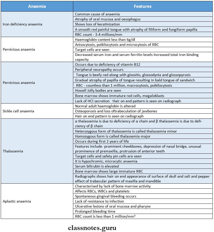
Question 1. Define anemia. Write oral manifestations of iron deficiency anemia
(or)
Iron Deficiency Anaemia.

Answer:

  • Anaemia:
    • Anemia is defined as hemoglobin concentration in blood below the lower limit of the normal range for the age and sex of the individual.
  • Iron Deficiency Anaemia
    • It is a chronic, microcytic, hypochromic type of anemia, which occurs either due to inadequate absorption or excessive loss of iron from the body.

Iron Deficiency Anaemia Causes:

  • Inadequate intake of iron in the diet
  • Malabsorption of iron due to diarrhea
  • The increased requirement for growing children and pregnancy
  • Increased loss of iron due to injury, epistaxis, and peptic ulcer
  • Gastrotomy

Iron Deficiency Anaemia Clinical Manifestations:

  • Age and Sex – Women in the 4th and 5th decade
  • Tiredness, headache, paresthesia, and lack of concentration
  • Koilonychia – nails become brittle, flattened and spoon-shaped
  • Lemon-tinted paler skin
  • Dizziness and sensitivity to cold

Read And Learn More: Oral Medicine Question and Answers

  • The liver and spleen may be palpable
  • Tingling sensation in extremities
  • Knuckle pigmentation is seen on the dorsum of the hand at the metacarpophalangeal joint

Iron Deficiency Anaemia Oral Manifestations:

  • The pallor of the oral mucosa and gingival with atrophy and loss of keratinization
  • Tongue – Glossitis, loss of papillae, and glossodynia
  • The tongue appears bald and red with a glazed appearance
  • Mucosa – Recurrent aphthous ulcer
  • Candidiasis
  • Abnormal bleeding
  • Faulty wound healing
  • Angular cheilitis
  • Associated syndrome – Plummer-Vinson syndrome

Iron Deficiency Anaemia Laboratory Diagnosis:

  • Hemoglobin level – decreased
  • Total RBC count – < 3 million per mm3
  • MCV, MCH, MCHC – reduced
  • Serum iron level – decreased to 10 mg
  • Hemosiderin – absent

Iron Deficiency Anaemia Management:

  • Iron supplement
  • Ferrous sulfate 300 mg 3-4 times/day for 6 months orally
  • Iron sorbitol 1.5 mg/kg body weight given parenterally.

Oral Medicine Blood Disorders

Question 2. Thalassemia Major.

Answer:

Thalassemia Major

  • It occurs when the patient is homozygous
  • It is also called Cooley’s anemia
  • It is an autosomal recessive disease

Thalassemia Major Clinical Manifestations:

  • Age – occurs in the first two years of life
  • The patient often suffers from jaundice
  • Fever, chills, marked anemia, malaise, generalized weakness, and lethargy.
  • Splenomegaly, leg ulcers
  • Most of the patients have a mongoloid facies with a prominent forehead, depressed nasal bridge, and slant-ing eyes
  • It leads to organ failure

Thalassemia Major Oral Manifestations:

  • Jaw bone – Excessive growth of the maxilla
  • Mucosa-Yellow tinted palor mucosa
  • Alveolar process – Hyperplastic
  • Teeth – Bimaxillary protrusion
  • Spacing and flaring of maxillary anterior
  • Open bite
  • Discoloration of teeth
  • Chipmunk facies – Characterized by saddle nose, prominent malar bone, and pneumatization of maxil¬lary sinus
  • Poor healing after dental treatment

Thalassemia Major Radiographic Appearance:

  • The skull bones exhibit a typical “hair-on-end” appearance on the radiograph
  • Generalized rarefaction of the alveolar bone, thinning of the cortex, and enlarged marrow spaces are found
  • It shows the “salt-and-pepper effect” by coarsening of some bony trabeculae and blurring of others
  • Spike-shaped and short roots of teeth
  • Antrum is reduced in size

Thalassemia Major Diagnosis:

  • RBC – microcytic, hypochromic
  • Bone marrow – shows increased erythropoietic activity
  • Reticulocyte count-increased
  • Serum bilirubin and urobilinogen – elevated
  • Fetal hemoglobin – elevated

Thalassemia Major Management:

  • Repeated blood transfusions are necessary
  • Allogenic bone marrow transplantation can be done
  • Splenectomy is indicated
  • Supplementary folic acid is required
  • Surgical correction in patients with abnormal facial appearance

Question 3. Agranulocytosis.

Answer:

Agranulocytosis

  • It is a serious acute disease characterized by a significant decrease in the number of granular leukocytes.

Etiology:

  • Toxic effects of some drugs like sulfonamides
  • Due to the hypersensitivity to drugs
  • Long-term administration of analgesics
  • Ionizing radiation, tuberculosis, typhoid, malaria

Agranulocytosis Clinical Features:

  • Initially high fever, chills, and sore throat
  • Skin appears pale and anemic
  • Persistent infections with regional lymphadenitis
  • Severe dysphagia
  • Weak and rapid pulse
  • Urinary tract infections
  • Vaginal and rectal ulceration
  • Patient dies within 3-5 days due to toxemia and septicemia

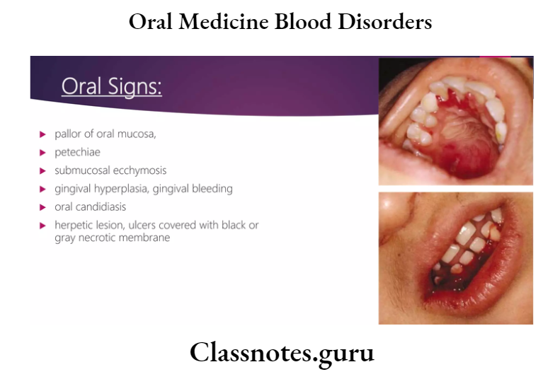
Agranulocytosis Oral Manifestations:

  • Mucosa-necrotizingulceration
  • They are covered by a yellow/grey membrane
  • Gingival bleeding spontaneously
  • Excessive salivation
  • Halitosis
  • Fungal infections
  • The lesion appears as ragged and necrotic
  • It is covered with grey black membrane
  • Destroys supporting structures along with teeth loss.

Agranulocytosis Diagnosis:

  • WBC count – reduced – 500 to 3000/mm³
  • Bone marrow – the absence of granulocytes
  • Granulocyte count < 100 cells/mm³

Agranulocytosis Treatment:

  • Removal of the offending agent
  • Transfusion of red cell constituent when hemoglobin is < 10 gm/dl.
  • Antibiotics – To control septicemia
  • A combination of drugs is given
  • Granulocyte-macrophage colony-stimulating factors
  • Dental management – for ulcers, 5% dyclonine and 5% Benadryl mixed with magnesium hydroxide or Kaolin with pectin is useful.

Question 4. Cyclic Neutropenia.

Answer:

Cyclic Neutropenia

It is an idiopathic disease characterized by episodic defects in neutrophil maturation in the bone marrow, resulting in periodic falls in circulating neutrophils

Cyclic Neutropenia Clinical Features:

  • Recurrent upper respiratory tract infections
  • Short-term fevers, sore throat
  • Headache, arthritis, conjunctivitis
  • Amyloidosis
  • Regional lymphadenopathy, Gingivitis, ragged painful ulcers

Cyclic Neutropenia Diagnosis:

  • WBC count – decreases to 3000 cells/cu mm
  • Absence of infiltrating neutrophils

Cyclic Neutropenia Treatment:

  • Antibiotics to control infection
  • Oral hygiene maintenance
  • Cytokine granulocyte colony-stimulating factor
  • Splenectomy and nutritional supplement

Question 5. Hemophilia.

Answer:

Hemophilia

It is a potentially fatal inherited bleeding disorder characterized by profound hemorrhage due to the deficiency of clotting factors

Etiology:

  • Hereditary
  • Sex-linked recessive trait
  • Spontaneous mutations

Hemophilia Types:

  1. Hemophilia A – Due to factor 8 deficiency
  2. Hemophilia B – Due to factor 9 deficiency
  3. Hemophilia C – Due to factor 11 deficiency
  4. Von Willebrand’s disease

Hemophilia Clinical Features:

  • Initially diagnosed in infancy
  • Characterized by
  • Easy bruising
  • Prolonged bleeding after trauma
  • Bleeding into the subcutaneous tissue
  • Hematoma formation
  • Nose-fatal epistaxis
  • GIT – Gastric hemorrhage
  • Joints – Recurrent hemarthrosis
  • Osteoporosis
  • Brain – Intracranial hemorrhage
  • Urinary tract – hematuria

Hemophilia Oral Manifestations:

  • Gingiva-Severe hemorrhage
  • Soft tissue – Hematoma formation
  • Jaw – Recurrent subperiosteal hematoma
  • Tumor-like malformations
  • Teeth – High caries index
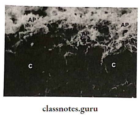
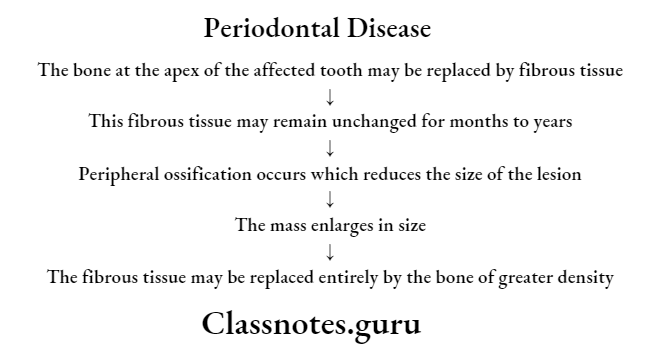
  • Severe periodontal disease
  • Oropharyngeal bleeding
  • Severe bleeding at the injection site

Hemophilia Treatment:

  • Replacement therapy by the use of fresh frozen plasma
  • Administration of Factor 8
  • Prevention of complications by the use of a synthetic analog of anti-diuretic l-diamino-8-d-arginine vaso¬pressin
  • Genetic counseling – To prevent inheritance of the condition

Question 6. Lab Investigations for anemia.

Answer:

1. Haemoglobin Estimation:

  • If the hemoglobin value is below the lower limit of the normal range for a particular age and sex, the patient is said to be anemic
  • Several methods are available but the most reliable and accurate is the cyanmethaemoglobin method utilizing Drab Kin’s solution and spectrophotometer
  • Normal values
    • Males – 13-18 g/dl
    • Females – 11.5 – 16.5 g/dl

2. Peripheral blood film examination:

The blood smear is evaluated in an area where there is neither rouleaux formation nor so thin as to cause red cell distortion

  •  Variation in size:
    • Normally, red cell diameter varies from 6.7 to 7.7 pm
    • Macrocytes (larger than normal) are found in megaloblastic anemia, aplastic anemia
    • Microcytes (smaller than normal) are found in iron deficiency anemia, thalassemia
  • Variation in shape (Poikilocytosis):
    • It is produced in megaloblastic anemia, iron deficiency anemia, hemolytic anemia
  •  Inadequate hemoglobin formation:
    • Lower hemoglobin content develops in iron deficiency anemia
    • Increased hemoglobin content develops in megaloblastic anemia

3. Red cell indices:

  • In iron deficiency anemia, MCV, MCH, and MCHC are reduced
  • In megaloblastic anemia, MCV is raised

4. Erythrocyte Sedimentation rate:

  • Anaemia causes a rise in the ESR

5. Reticulocyte count:

  • To assess the marrow erythropoietic activity

Question 7. Diagnostic Test of Pernicious Anaemia.

Answer:

Diagnostic Test of Pernicious Anaemia

  • Hypergastrinaemia:
  •  Pentagastrinaemia:
  •  Haematologic findings:
    • Haemoglobin – below the normal range
    • Red cells – Macrocytes
    • Reticulocyte count – lower than normal
    • Red cell indices – MCV – elevated
      • MCH – elevated
      • MCHC – Normal or reduced
    • Leukocytes – Reduced
    • Platelets – moderately reduced
  • Biochemical alterations:
    • Serum bilirubin, LDH, haptoglobin, ferritin, and iron – increased
    • Red cell folate – reduced
  • Schilling test: Abnormal due to intrinsic factor deficiency

Question 8. Diagnostic Test for Bleeding Disorders.

Answer:

1. Investigations of disordered vascular hemostasis

  • Bleeding Time:
    • It is based on the principle of formation of a hemostatic plug following a standard incision on the volar surface of the forearm and the time the incision takes to stop bleeding is measured
    • Normal – 3-8 minutes
  •  Hess Capillary Resistance test:
    • This test is done by typing the sphygmo-manometer cuff to the upper arm and raising the pressure in it between diastolic and systolic for 5 minutes.
    • After deflation, the number of petechiae appearing in the next 5 minutes in a 3cm² area over the cubital fossa is counted
    • The presence of more than 20 petechiae is considered a positive test

2. Investigation of blood coagulation:

  • Screening Tests
    • Whole blood coagulation time:
      • The estimation of whole blood coagulation time done by various capillary and tube methods
      • Normal value – 4-9 minutes
    • Activated partial thromboplastin time (PTTK):
      • This test is used to measure the intrinsic system factors as well as factors common to both intrinsic and extrinsic systems
      • One-stage prothrombin time (PT) – It measures the extrinsic system factor VII as well as factors in the common pathway.
  • Special Tests:
    • Coagulation factor assays: These are based on the results of PTTK or PT tests.
    • Quantitative assays: The coagulation factors can be quantitatively assayed by immunological and other chemical methods

Question 9. Oral manifestations of Anaemia.

Answer:

Oral manifestations of Anaemia

  • Mucosa – There is pallor of oral mucosa
  • Generalized atrophy of mucosa occurs
  • Tongue – Redness, soreness, or burning of tongue, Assuring of tongue
    • Atrophy of papillae of the tongue
  • Gingiva – Gingival enlargement
  • Lip – Vermilion borders of the lip are very thin
    • There is cracking and Assuring at the corner of the mouth
  • Ulcers – Recurrent aphthous ulceration and candidiasis occur
  • The width of the mouth is narrowed
  • Throat – spasm in the throat
    • Food sticking in the throat occurs
  • Teeth – There may be delayed eruption
    • Deposition of blood pigment in enamel and dentin
    • Enamel hypoplasia
  • Jaws – The patient is more prone to develop osteomyelitis due to hypervascularity of the bone marrow
  • Nerves – Patient may present with paresthesia of men¬tal nerve
    • Hypersensitivity and paresthesia
  • Dysphagia
  • Xerostomia – dryness of mouth

Question 10. Oral Manifestation of Leukemia.

Answer:

Acute Leukemia:

  • Lymph nodes – submental, cervical, and pre and post-auricular lymph nodes may be enlarged and tender
  • Lower lip – Paresthesia and crusting of the lower lip
  • Chin – Paresthesia of chin
  • Mucosa – The oral mucosa shows pallor, ulceration with necrosis, petechiae, ecchymosis, and bleeding ten¬dency
    • There may be massive necrosis of lingual mucosa with sloughing
  • Gingiva – It shows hypertrophy and cyanotic discolora-tion
    • The gingiva appears boggy, edematous, and deep red bleeds easily due to ulceration of the sulcus epithelium and necrosis of underlying tissue
  • Teeth – mobility of permanent teeth
  • Infection – Candidial, viral, and bacterial infections occur frequently

Chronic Leukemia:

  • Gingiva-Hypertrophy of gingiva
    • There may be ulceration with necrosis and gangre¬nous degeneration
    • Dark brown exudates and foul fetor oris are pre¬sent
  • Tongue – It is frequently swollen and dark
  • Lymph nodes – Regional lymphadenopathy
  • Teeth – Rapid loosening of teeth
    • Necrosis of periodontal ligament
    • Destruction of alveolar bone

Question 11. ESR (Erythrocyte Sedimentation Rate).

Answer:

ESR (Erythrocyte Sedimentation Rate)

  • It is a measure of the rate at which RBCs sediment in a tube of plasma
  • It helps follow the progress of some chronic infections as well as diseases characterized by altered globulins such as collagen diseases, nephritis, rheumatic fever, and dysproteinemias

Erythrocyte Sedimentation Rate Technique:

  • Mechanism
    • The rate is accelerated when there are changes in the physicochemical properties of plasma or the red cell surface or when the changes in plasma protein cause the RBCs to aggregate
  • Westergren Method:
    • A graduated sedimentation tube is filled with oxalate blood and placed in a vertical position
  • Results:
    • The erythrocyte level is read at 10-minute intervals and the end of an hour.

Question 12. Lazy-leucocyte syndrome.

Answer:

Lazy-leucocyte syndrome

  • It is a result of the loss of chemotactic function of the neutrophils

Lazy-leucocyte syndrome Clinical Features:

  • Age – Apparent at the age of 1-2 years

Lazy-leucocyte Syndrome Symptoms:

  • Stomatitis
  • Otitis media
  • Bronchitis
  • Recurrent infection
  • Periodontitis

Lazy-leucocyte syndrome Diagnosis:

  • Total WBC count – low
  • Neutrophil count – 100-200 cells/mm3
  • Bone marrow – Normal number of mature neutrophils

Lazy-leucocyte syndrome Management:

  • Antibiotics – to control infections

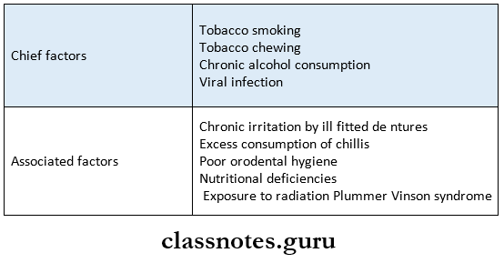
Question 13. Plummer-Vinson Syndrome/Paterson-Brown- Kelly syndrome.

Answer:

Plummer-Vinson Syndrome

  • It is characterized by dysphagia, iron deficiency anemia, dystrophy of nails, and glossitis

Plummer-Vinson Syndrome Clinical Features:

  • Age – It is found in middle age women

Plummer-Vinson Syndrome Symptoms:

  • Dysphagia
  • Angular cheilitis
  • The patient may complain of spasms in the throat
  • Sore mouth
  • A smooth, red, and enlarged tongue
  • Oral mucosa is pale and painful
  • Dry mouth
  • Spoon shaped nails

Plummer-Vinson Syndrome Diagnosis:

  • Biopsy – It shows
  • Atrophic epithelium
  • Atrophy of lamina propria and muscles

Plummer-Vinson Syndrome Management:

  • Correction of anemia
  • Esophageal dilation

Question 14. Control of Bleeding.

Answer:

Control of Bleeding

  • Bleeding in the surgical site around 48 hours after the surgical procedure is known as reactionary hemorrhage
  • This is usually due to a local rise in blood pressure
  • Bleeding seen after a week of the procedure is known as secondary hemorrhage

Bleeding Management:

  • Pressure pack:
    • A moist gauze with saline may be used to apply firm finger pressure on the area
  • Local anesthetic pack:
    • Local anesthesia with adrenaline can be used to temporarily control bleeding
    • A gauze piece soaked in epinephrine may be placed
  • Sutures:
    • Bleeding from soft tissues may be controlled by placing sutures
    • This helps to compress the mucosa against the bone and reduce bleeding
  • Cautery:
    • Bleeding from bone may be controlled by cauterization
    • The exact bleeding spot is located first
    • The area is dried as much as possible and a hot ball burnisher may be used to cauterize
    • Electrocautery may also be used
  • Ligation:
    • If a major artery has been severed, it may need to be ligated.
  • Gelfoam:
    • This is a gelatin-based sponge that acts by disrupting platelets and establishing a framework with fibrin strands to create a clot
    • It gets absorbed within 4-6 weeks
  • Oxidized cellulose:
    • It releases cellulosic acid
    • This has a marked affinity for hemoglobin leading to the formation of an artificial clot
    • These substances may be placed in the socket to enhance clot formation and control bleeding
  • Bone wax:
  • This is a mechanical agent to block the bleeding vessel
  • Bleeding from bone may be occluded by placing a small piece of bone wax firmly on the spot of the bleeding
  • The patient should be made to wait for half an hour in the dental clinic to confirm the absence of bleeding.

Question 15. Dental management of Haemophilia B.

Answer:

Dental management of Haemophilia B

  • Anesthesia
    • Local anesthesia is contraindicated in severe hae- -mesophilic patients
    • If required, it should be given intramurally, in-parliamentary, and papillary
    • Sedation with diazepam of nitrous oxide can be given
  • Endodontic treatment
    • Common endodontic procedures can be provided
    • Hemorrhage into the canal can be controlled with 1:1000 aqueous epinephrines
  • Restorative
    • Restorative procedures can be carried out
    • Avoid the use of rubber dams to prevent trauma to the gingiva and other soft tissue
    • The epinephrine-impregnated hemostatic cord can be used for gingival retraction
  • Prosthodontics treatment
    • Complete dentures and partial dentures are well tolerated in hemophilic patients
  • Periodontal therapy
    • Conservative therapy is acceptable
    • Surgery requires replacement therapy
  • Oral surgical procedure
    • Local hemostatic agents and techniques are used
    • After packing, the tooth socket must be protected by a mechanical splint to prevent disturbances to the clot
    • The anti-fibrinolytic agent is used postoperatively
  • Drugs precaution
    • Aspirin and other NSAIDs are avoided due to their antiplatelet function
    • Intramuscular injection should be avoided owing to the risk of hematoma formation
  • Electrosurgery
    • Avoided as it causes tissue necrosis and haemor¬rhage

Oral Medicine Blood Disorders Short Answers

Question 1. Cyclic neutropenia

Answer:

Cyclic neutropenia

It is an idiopathic disease characterized by episodic defects in neutrophil maturation in the bone marrow, resulting in periodic falls in circulating neutrophils

Cyclic neutropenia Clinical Features:

  • Recurrent upper respiratory tract infections
  • Short-term fevers, sore throat
  • Headache, arthritis, conjunctivitis
  • Amyloidosis
  • Regional lymphadenopathy, Gingivitis, ragged painful ulcers

Question 2. Four oral manifestations of aplastic anemia.

Answer:

Four oral manifestations of aplastic anemia

  • Petechiae and frank hematoma formation in the oral cavity
  • Gingiva – Spontaneous bleeding
  • Mucosa – Multiple ulcerations
  • Persistent oral infections
  • May lead to bacteremia and septicemia

Question 3. Oral manifestation of oral leukemia.

Answer:

Oral manifestation of oral leukemia

  • Gingiva:
    • Hypertrophy of gingiva
    • Ulceration with necrosis and gangrenous degenera¬tion
    • Dark brown exudates
    • Foul fetor oris
  • Tongue – Swollen and dark
  • Lymph nodes – Regional lymphadenopathy
  • Teeth-Loosening of teeth
    • Necrosis of periodontal ligament
    • Destruction of alveolar bone

Oral Medicine Blood Disorders Viva Voce

  1. Purplish discoloration of oral mucosa is found in primary polycythemia
  2. Delayed eruption and hypoplasia of dentition are found in sickle cell anemia
  3. The color of skin in thalassemia patients is ashen gray
  4. The bimaxillary protrusion is seen in Cooley’s anemia
  5. The beefy red tongue is found in pernicious anemia
  6. Spontaneous hemorrhage from the gingiva is a feature of aplastic anemia
  7. Spasm in the throat occurs in Plummer Vinson syn-drome
  8. Platelet count is used to evaluate primary hemostasis
  9. Haemophilia A is caused by a deficiency of Factor 8
  10. Microvascular infarcts are found in thrombotic thrombocytopenic purpura
  11. The labial frenum exhibits the most frequent bleeding in hemophilia
  12. DIC is treated by intravenous heparin
  13. The bald tongue of the sandwich is a feature of pernicious anemia
  14. Howell Jolly bodies are seen in pernicious anemia
  15. Safety pin cells are seen in thalassemia

Bacterial Infections Notes And Short Essays

Oral Medicine Bacterial Infections Important Notes

1. Important features of different stages of syphilis

Oral Medicine Bacterial Infections Importances features of different stages of Syphilis

2. Hutchison’s triad

  • Mulberry molars and notched incisors
  • Interstitial keratitis
  • 8th nerve deafness

3. Actinomycosis

  • It commonly Involves the cervicofacial region
  • There Is the development of multiple abscesses which drain through the skin
  • Pus contains sulfur granules

4. Scarlet fever

  • Caused by a hemolytic type of streptococci
  • Appears as a bright skin rash
  • Small papule erupts through these rashes
  • The tongue exhibits a white coating at the fungiform papilla that projects above the surface
  • Known as strawberry tongue
  • Later the coating is lost and the tongue becomes red and smooth called raspberry tongue

Read And Learn More: Oral Medicine Question and Answers

5. Tuberculosis

  • Primary tuberculosis of the skin is called lupus vulgaris
  • Primary oral tuberculosis is associated with regional lymphadenopathy mostly involving submandibular and cervical implodes
  • It involves gingiva
  • Present as diffuse, nodular, or papillary proliferation of gingival tissues
  • Secondary oral tuberculosis can affect any site but the most common tongue and palate
  • Present as superficial or deep, irregular painful ulcer

6. Psoriasis with Monro abscess is seen in

  • Benign migratory glossitis
  • Psoriasis
  • Reiter’s syndrome

7. Snail track ulcers are seen in

  • Secondary syphilis
  • Pyostomatitis vegetans

Oral Medicine Bacterial Infections

Oral Medicine Bacterial Infections Short Essays

Question 1. Congenital syphilis.

Answer:

Congenital syphilis

It is an infection of the fetus established by the passage of spirochaetes from the mother, through the placenta

Clinical Features:

1. Within the first 2 weeks of life

Features: Rhinitis

  • Chronic nasal discharge
  • Loss of weight
  • Bullae, vesicle formation
  • Superficial desquamation with cracking and scaling of reddened soles and palms
  • Petechiae, mucous patches

2. Age – After 2 years

Features:

  • Interstitial keratitis
  • Vascularization of cornea
  • 8th nerve deafness
  • Arthropathy
  • Neurosyphilis
  • Anterior tibial bowing
  • Higoumenakis sign – Irregular thickening of a sternoclavicular portion of the cheek

Oral Manifestation:

  • Postrhagadic Scarring:
    • These are linear lesions found around oral and anal orifices.
    • These are seen from 3rd- 7th week after birth
    • They appear as red or copper-colored linear areas covered with a soft crush
  • Syphilitic Rhagades:
    • They are radially arranged and perpendicular to the mucocutaneous junction
  • Teeth:
    • Retarded root resorption of deciduous
    • Marring of permanent incisors
    • Spacing between cuspid and incisors o Malocclusion and the open bite is present
    • The Crown of the molar is irregular
    • The occlusal surface appears to be arranged in an agglomerate mass of globules
    • Incisor-screw driver shaped
    • Constriction of crown towards the incisal edge
    • The incisal edge is usually a notch
    • Rounding of mesial and distal incisal line angles
    • Jaws – Hypoplastic maxilla
    • Frontal bossing
    • Saddle nose deformity

Management:

  • Antibiotics:
    • Benzathine penicillin – 2.4 million units/IM
    • Aqueous crystalline penicillin
    • Tetracycline hydrochloride – 500 mg orally 4 times a day
    • Patient allergic to penicillin – Erythromycin 500 mg orally Q1D for 15 days
    • Follow up – Repeated examination is to be done at 1, 3, 6, 9,12,18, and 24 months
    • Prevention – It can be achieved by subjecting pregnant women to antenatal and post-natal checkups.

Oral Medicine Bacterial Infections Short Answers

Question 1. Chancre.

Answer:

Chancre

  • It is one of the clinical findings of primary syphilis
  • Incubation period – 3 – 90 days
  • Site – Lesion develops at the site of inoculation
    • It most frequently occurs on the penis in males and the vulva or cervix in females

Features:

  • It is slightly raised over the surface
  • It becomes ulcerated
  • It is a non-tender, non-bleeding firm plaque
  • Shape – Round and indurated
  • Size – Varies from 5 mm to several centimeters
  • Edges – Rolled raised edges are seen
  • Prognosis – It disappears without treatment after 10 days

Question 2. Hutchison’s Triad.

Answer:

Hutchison’s Triad

  • It is a feature of congenital syphilis
  • It includes
    • Interstitial keratitis – It results from opacification of the corneal surface with resultant loss of vision
    • Enamel hypoplasia of permanent incisors and 1st permanent molars
    • Eight nerve deafness

Question 3. Split Papule.

Answer:

Split Papule

  • It is a feature of secondary syphilis
  • It is a double papule that occurs at skin folds and the angle of the mouth

Question 4. Koplik’s spots.

Answer:

Koplik’s spots

  • It is one of the important clinical features of measles
  • Site: buccal mucosa
  • Presentation
    • The mucosa becomes inflamed
    • Over it, there is the presence of white or white-yellow pinpoint papules

Oral Medicine Bacterial Infections Viva Voce

  1. Purulent discharge with sulfur granules is characteristic of actinomycosis
  2. Tuberculosis is also referred as an acid-fast infection
  3. Scrofula refers to tuberculous involvement of cervical lymph nodes
  4. Paraesthesia in lips and tongue is found in tertiary syphilis
  5. Syphilitic rhagades are found in congenital syphilis
  6. Peg-shaped laterals and mulberry molars are found in congenital syphilis
  7. Chancre lesions are a characteristic feature of primary syphilis
  8. Strawberry tongue is a feature of scarlet fever
  9. A lumpy jaw is seen in actinomycosis
  10. Sulfur granules are diagnostic of actinomycosis
  11. Actinomycosis is also called ray fungus

Bone Disorders Notes And Short Essays

Oral Medicine Bone Disorders Important Notes

1. Fibrous dysplasia:

  • Classification:
    • Monostotic – Only one bone is involved
    • Polyostotic – More than one bone is involved
      • Jaffe’s type – Polyostotic along with cafe-au-lait-skin pigmentation
      • Albright syndrome – is characterized by polyostotic fibrous dysplasia, cafe-au-lait skin pigmentation, and endocrine disturbances

2. Cleidocranial dysplasia:

  • It is characterized by abnormalities of the skull, shoulder girdle, jaws, and teeth
  • Skull – delayed closure of sutures and wormian bones
  • Shoulder – the partial or complete absence of clavicles
  • Teeth – prolonged retention of deciduous and delayed eruption of permanent
  • Numerous supernumerary teeth are found in the mandibular premolar and incisor areas.

3. Paget’s disease:

  • It is characterized by excessive and abnormal remodeling of bone
  • Affects the adult skeleton
  • Patients suffer from deafness, blindness, and facial paralysis
  • There is a progressive enlargement of the skull and maxilla because of which the patient has to change the hats and dentures frequently

4. Cherubism:

  • Manifests by the age of 3-4 years
  • Painless symmetric swelling of the mandible or maxilla occurs
  • Results in chubby face appearance
  • The deciduous teeth shed prematurely and numerous teeth are absent
  • X-ray shows numerous unerupted teeth floating in cyst-like spaces

Read And Learn More: Oral Medicine Question and Answers

5. Eagle’s syndrome: elongation of styloid process or ossification of stylohyoid ligament leading to

  • Dysphagia
  • Sore throat
  • Otalgia
  • Glossodynia

6. Albright syndrome:

  • Precocious puberty
  • Polyostotic fibrous dysplasia
  • Cafe-au- lait pigmentation

7. Marfan’s syndrome:

  • Long thin extremities
  • Hyperextensibility of joints
  • Spidery fingers
  • Arachnodactyly
  • Bifid uvula
  • CVS complications

8. Cafe-au-lait pigmentation:

  • It has brownish pigmentation
  • Seen in
    • Neurofibromatosis
    • Fibrous dysplasia
    • Peutzjeghers syndrome
    • Hypothyroidism

9. Blue sclera is seen in:

  • Osteogenesis imperfect
  • Marfan’s syndrome
  • Cherubism
  • Ehlers Danlos syndrome
  • Osteopetrosis
  • Fetal rickets

10. Ground glass appearance is seen in:

  • Primary hyperparathyroidism
  • Monostotic fibrous dysplasia
  • Cherubism

11. Cotton wool appearance is seen in:

  • Paget’s disease
  • Chronic sclerosing diffuse osteomyelitis
  • Fibrous dysplasia
  • Cemento-osseous dysplasia

12. Hypermobility of joints is seen in:

  • Ehler Danlos syndrome
  • Marfan’s syndrome
  • Osteogenesis imperfecta
  • Down’s syndrome

13. Radiographic features in different diseases:

Oral Medicine Bone Disorders Radiographic feature in different disease

14. Laboratory investigations

Oral Medicine Bone Disorders Laboratory investigations

Oral Medicine Bone Disorders Short Essays

Question 1. Fibrous Dysplasia.

Answer:

Fibrous Dysplasia Definition:

It is an idiopathic condition, in which an area of normal bone is gradually replaced by abnormal fibrous connec¬tive tissue, which then again undergoes osseous metaplasia, and eventually, the bone is transformed into a dense lamellar bone.

Fibrous Dysplasia Classification:

  • Monostotic – Only one bone is involved
  • Polyostotic – More than one bone is involved
    • Jaffe’s type: Polyostotic along with cafe-au-lait-skin pigmentation
    • Albright syndrome: Characterized by polyostotic fibrous dysplasia, cafe-au-lait skin pigmentation, and endocrine disturbances

Etiology:

  • Developmental – Developmental anomaly occurring during the growth period of life
  • Endocrine disturbances – Complex endocrine disturbances
  • Liver damage
  • Infections
  • Trauma
  • Abnormal osteoclastic maturation of bone-forming – mesenchyme

Fibrous Dysplasia Clinical Features:

  • Age – It commonly occurs in the first and second decade of life
  • Sex – It commonly occurs in females

Fibrous Dysplasia Sites:

  • Commonly affected sites are
  • Skull
  • Facial bones: Maxilla more affected than the mandible
  • Clavicles
  • Pelvic bones
  • Long bones – femur, tibia, humerus

Fibrous Dysplasia Presentations:

  • Skeletal lesions
    • Unilateral distribution of the lesion
    • Swelling is seen on the affected side
    • Recurrent bone pain is common
    • There may be a cessation of growth but proliferation may occur
    • Spontaneous fracture is a common complication
  • Skin lesion
    • Cafe-au-lait pigmentation is seen
    • It consists of irregularly, pigmented, light brown, flat, melanotic spots
  • Oral symptoms
    • Slow enlarging, painless, unilateral swelling of the jaw
    • This leads to facial deformity
    • Expansion and distortion of the cortical plate
    • Displacement of teeth
    • Disturbances in tooth eruption
    • Severe malocclusion
      • Maxillary lesions
      • They extend into the maxillary sinus and upto orbital floor leading to exophthalmos proptosis and nasal obstructions
    • Mandibular lesions
    • Protuberances in the premolar-molar area result in increased depth of the jaw
    • Other – Precocious puberty leading to premature vaginal bleeding, breast development, and axillary and pubic hair at the age of 2-3 years.

Oral Medicine Bone Disorders

Fibrous Dysplasia Radiographic Appearance:

  • Radio density – Unilocular or multilocular radiolucent lesion
  • Borders – Ill-defined
  • Lamina dura – Loss of lamina dura
  • Teeth – Resorption of roots occurs
  • Jaw – Expansion and distortion of cortical plates
  • Granular appearance – Surrounding the margins of the radiolucent area, there may be a wider band of increased density, but granular in appearance
  • Ground glass appearance – It may demonstrate areas of whorled amorphous partially calcified materials that are well-circumscribed

Fibrous Dysplasia Differential Diagnosis:

  • Lesion and their Distinguishing Feature:
    • Traumatic bone cyst – Absence of cortical expansion
    • Aneurysmal bone cyst – Hemorrhagic aspirate
    • Chronic osteomyelitis – Occurs in older age

Fibrous Dysplasia Management:

  • Surgical – Removal of lesion surgically
  • Osseous contouring – for esthetic purpose

Question 2. Pathogenesis and management of Osteoradionecrosis.

Answer:

Pathophysiology:

  • When small particles of high-energy radiation strike tumor cells or normal tissue cells, the cellular water undergoes radiolysis and is converted into free radicals
  • These free radicals interact with the DNA of these cells and disrupt them
  • This resultant DNA destruction is manifested differently at various levels
  • On a cellular level, this is seen as a chromosomal break-age or disintegration
  • The cell may then change like
    • The cell may die
    • It may repair its DNA to survive with impaired function
    • It may repair DNA damage and function normally
  • The tissues responsible for developing osteoradionecrosis are endothelium, bone, periosteum, and fibrous connective tissue of mucosa, and skin.
  • At the tissue level, these radiation effects are seen as endothelial death, hyalinization, and thrombosis of the vessel
  • Fibrosis of marrow spaces takes place
  • At the organ level, it is seen as a composite tissue that is hypocellular, hypovascular, and hypoxic.
  • This irradiated bone is thus unable to replace the nor¬mal cell loss with new cells and wound healing is thus severely compromised
  • This creates a wound in which the demand for oxygen is much more than what can be provided
  • Therefore there is tissue breakdown and it manifests as a chronic non-healing wound.

Management Of Osteoradionecrosis:

  • The patient is made to breathe 100% oxygen via a pilot’s face mask
  • The patient is exposed to 24 atmospheres of absolute pressure of chamber compression
  • The oxygen exposure is for 90 minutes, once a day for 5 days a week
  • Each exposure to hyperbaric oxygen (HBO) is called a dive
  • Protocol

Stage 1 – 30 dives

Condition improved

The next 30 dives are given

No improvement

Stage 2 – Sequestrictomy

Mucosal closure

60 dives are given

If the condition doesn’t improve

Stage 3 – 30 dives

Resection

30 dives

After 10 weeks
Additional 60 dives
are given

Oral Medicine Bone Disorders Short Answers

Question 1. Cherubism.

Answer:

Cherubism

It was first described by Jones in 1933

Cherubism Classification:

  • Based on the severity and location of the lesion
    • Grade 1 – Affects ramus of mandible
      • This leads to bilateral and symmetrical expansion of bone
    • Grade 2 – Affects ramus and body of mandible maxillary tuberosity
    • Grade 3 – Affects maxilla and mandible entirely

Etiology:

  • Autosomal dominant trait
  • Hormonal – latent hyperparathyroidism
  • Trauma
  • Disturbance in bone-forming mesenchyme

Cherubism Clinical Features:

  • Age and sex – 2-3 years males are affected
  • Site – Angle of mandible bilaterally
  • Appearance
    • Bilateral, painless, symmetrical swelling giving a chubby appearance
    • Swelling is firm to hard on palpation
  • Associated symptoms
    • Maxillary swelling pressurizes the floor of the orbit
    • This leads to an upward turn of the pupils of the eyes referred to “heavenward look”
    • Difficulty in speech, deglutition, mastication, and respiration
    • Limited jaw movement
    • Expansion and widening of the alveolar ridge
    • Flattening of palatal vault
    • Chronic lymphadenopathy
    • Malocclusion
  • Syndrome Associated: Noonan syndrome

Cherubism Radiographic Features:

  • Well-defined, well-corticated cyst-like radiolucency of the mandible
  • Teeth appear as hanging in the air
  • Displacement of the inferior alveolar canal
  • Destruction of maxillary autrium
  • Expansion of cortical plates
    • Displacement of teeth

Question 2. Cleidocranial Dysplasia.

Answer:

Cleidocranial Dysplasia

It is a hereditary disorder characterized by abnormal growth of the bones in the face, skull, and clavicles with a tendency for the failure of tooth eruption

Cleidocranial Dysplasia Clinical Features:

  • Absence or hypoplasia of one/both clavicles
  • Hypermobility of shoulder joints
  • Elongated frontal and occipital skull plates
  • Underdeveloped entire midface
  • Delayed closure of fontanelles
  • High and narrow arched palate
  • Underdeveloped paranasal sinuses
  • Photophobia
  • Multiple unerupted and impacted teeth

Radiographic Features – Radiograph Reveals:

  • Open sutures
  • Open fontanelles
  • Partial/complete loss of clavicles
  • Multiple impacted teeth
  • Thin roots of teeth

Cleidocranial Dysplasia Treatment

  • No treatment is possible

Question 3. Enumerate diseases with cafe-au-lait.

Answer:

Pigmentation:

  • Albright syndrome
  • Von Recklinghausen’s Neurofibromatosis
  • Bloome’s syndrome
  • Fanconi anemia
  • Cowden’s syndrome
  • Tuberculosis sclerosis
  • Watson’s syndrome
  • Ataxia telangiectasia

Oral Medicine Bone Disorders Viva Voce

  1. Paget’s disease shows the cotton wool appearance
  2. The development of osteosarcoma is the most serious complication of Paget’s disease
  3. Central giant cell granuloma commonly involves mandible
  4. Cherubism gives a floating tooth appearance
  5. Cherubism shows eye to heaven look appearance
  6. In osteopetrosis, there is endosteal production of bone
  7. Mosaic bone is due to partially resorbed and repaired bone
  8. Osteogenesis imperfecta is characterized by extreme fragility and porosity of bones

Periodontal Disease Notes And Short Essays

Oral Medicine Periodontal Disease Important Notes

1. Widening of PDL space is seen in

  • Osteosarcoma
  • Chondrosarcoma
  • Scleroderma
  • Trauma from occlusion

2. Obliteration of PDL space is seen in

  • Hypercementosis
  • Ankylosis
  • Hypofunction of teeth
  • Paget’s disease

3. Papillon Lefevre syndrome

  • Juvenile periodontitis
  • Palmar plantar keratosis
  • Calcification of falx cerebri

4. Desquamative gingivitis represents oral manifestations of

  • Lichen planus
  • Cicatricial pemphigoid
  • Pemphigus

Oral Medicine Periodontal Disease Long Essays

Question 1. Describe clinical features, differential diagnosis, and treatment of acute necrotizing ulcerative gingivitis.

Answer:

Acute Necrotising Ulcerative Gingivitis

  • It is a relatively rare condition and is characterized clinically by necrosis of the free gingival margin the crest of the gingiva and the interdental papillae

Acute Necrotising Ulcerative Gingivitis Clinical Features:

  • Age – 15-35 years
  • Sex – Common in males
  • Initially, the gingiva becomes red and painful
  • Further, punched-out erosion of the interdental papillae occurs
  • Gingiva is covered by a “pseudo-membrane”
  • Bleeding occurs spontaneously or on slight provocation
  • It produces an extremely unpleasant odor
  • The gingival lesion may extend to the mucosal surfaces of the soft palate and tonsils
  • It leads to difficulty in deglutition, mastication
  • Increased salivation and a metallic taste in the mouth

Read And Learn More: Oral Medicine Question and Answers

Acute Necrotising Ulcerative Gingivitis Generalized Symptoms:

  • Headache
  • Fever, malaise
  • Lymphadenopathy
  • Leukocytosis
  • Tachycardia
  • Gastrointestinal disturbances

Acute Necrotising Ulcerative Gingivitis Differential Diagnosis:

  • Primary acute herpetic gingivostomatitis
    • Occurs more frequently in children
    • Characterized by vesicular eruption
      • Erosive lichen planus
      • Drug allergy

Acute Necrotising Ulcerative Gingivitis Treatments:

  1. Removal of pseudomembrane by gently swabbing the area with a cotton pellet
  2. Rinsing the mouth with a glassful of an equal mixture of warm water and 3% hydrogen peroxide
  • Twice daily rinse with 0.12% chlorhexidine
    • Antibiotics:
      • Penicillin V – 250-500 mg or erythromycin 250-500 mg 6 hourly for 7 days
      • Metronidazole 400 mg 8 hourly for 7 days
    • Gingival curettage: After the active phase of the disease
    • Supportive treatment:
      • Fluid consumption
      • Administration of nutritional supplements

Oral Medicine Periodontal Disease Short Essays

Question 1. Pericoronitis.

Answer:

Pericoronitis

  • It refers to inflammation of the gingiva about the crown of an incompletely erupted tooth

  Pericoronitis Types:

  • Acute
  • Sub-acute
  • Chronic

Pericoronitis Clinical Features:

  • It occurs most often in the mandibular third molar area
  • The space between the crown of the tooth and the overlying gingival flap leads to the accumulation of food debris and bacteria
  • Thus it is chronically inflamed and infected
  • Various degrees of ulceration occurs
  • The lesion is red, swollen, and suppurating
  • It is tender on palpation
  • The pain radiates to the ear, throat, and floor of the mouth
  • Presence of foul taste
  • Inability to close the jaws
  • Swelling of the cheek in the region of the angle of the jaw
  • Lymphadenitis
  • Fever, leukocytosis, and malaise occur

Pericoronitis Treatment:

  • Antibiotics
  • Phenoxymethyl penicillin 250 mg 4 times daily
  • Metronidazole 200 mg 3 times daily for 7 days
  • Drainage
  • To allow evacuation of pus
  • Extraction – during the sub-acute phase
  • Operculectomy – If retention of 3rd molar is preferred
  • Extraction of maxillary 3rd molar – when mandibular 3rd molar fully erupts

Question 2. Gingival Enlargement.

Answer:

Gingival Enlargement

  • An increase in the size of the gingiva is termed gingival enlargement

Gingival Enlargement Types:

1. Inflammatory enlargement:

  •  Chronic:
    • It originates as a slight ballooning of the interdental papilla and marginal gingiva
    • In early stages, it produces a life-preserver-shaped bulge around the involved teeth
    • This bulge can increase in size until it covers part of the crown
    • It can be localized or generalized
  • Acute Inflammatory enlargement:
    • Gingival abscess:
      • It is a localized, painful, rapidly expanding lesion that is usually of sudden onset
      • It is generally limited to the marginal gingiva or interdental papilla
      • Initially, it appears as a red swelling with a smooth, shiny surface
      • Within 24-48 hours the lesion usually becomes fluctuant and pointed with a surface orifice from which purulent exudates may be expressed
    • Periodontal abscess: It produces enlargement of the gingiva along with the supporting periodontal tissues

Oral Medicine Periodontal Disease Vertical section through a 4 day human plaque sample.

Oral Medicine Periodontal Disease Vertical section through a 4 day human plaque sample.

Oral Medicine Periodontal Disease Diagram depicting the plaque bacteria association with tooth surface and periodontal tissues.

2. Drug-induced enlargement: Characterized by bead-like enlargement of the interdental papilla and extends to the facial and lingual gingival margins
Oral Medicine Periodontal Disease Scanning electron photomicrograph of cross section of cementum

3. Enlargements associated with systemic diseases:

  • Conditioned enlargement:
    • Types:
      • Hormonal (Pregnancy, Puberty)
      • Nutritional (Vit. C deficiency)
      • Allergy
    • Features:
      • Gingival edema
      • Increased inflammatory response to dental plaque
      • Gingiva is bright red/magenta in color
      • It is soft and friable
      • It has a smooth, shiny surface
      • Bleeding occurs spontaneously or on slight provocation
      • The lesion appears as a discrete, mushroom-like, flattened spherical mass that protrudes from the gingival margin
  •  Enlargement associated with the systemic disease: An existing inflammation initiated by dental plaque

Oral Medicine Periodontal Disease Scsnning electron micrograph of cocci and filaments associated with surface of pocket epithelium in a case of marginal gingivitis

4. Neoplastic enlargement:

  • Benign Tumor of Gingiva:
    • Fibroma
    • Papilloma
    • Leukoplakia
  • Malignant tumor of Gingiva:
    • Carcinoma
    • Malignant melanoma

Oral Medicine Periodontal Disease Histologic section of sub gingival plaque

5. False enlargement: They may appear as a result of an increase in the size of the underlying osseous or dental tissues

Question 3. Halitosis.

Answer:

Halitosis

  • It is an unpleasant odor exhaled in breathing

Halitosis Classification:

  • Physiologic halitosis
  • Pathologic halitosis
  • Pseudo halitosis

Halitosis Causes:

  • Physiologic
    • Mouth breathing
    • Medications
    • Aging
    • Fasting, starvation
    • Tobacco
    • Food
  • Pathology:
  • Periodontal infection
  • Tongue coating
  • Stomatitis
  • Faulty restoration
  • Unclean dentures
  • Pathologic lesions
  • Abscess

Halitosis Diagnosis:

  • Subjective organoleptic method: It is used as a benchmark
  • Gas chromatography: Measure molecular levels of volatile sulfur compounds
  • Halimeters: Measure the level of sulfide gas
  • BANAtest:

Halitosis Treatment:

  • Improvement of oral hygiene and periodontal health
  • Tongue brushing should be advised
  • Rinsing or gargling with an effective mouthwash
  • Halita – It reduces volatile sulfur compound levels

Question 4. Investigation and management of Primary Herpetic Gingivostomatitis.

Answer:

Primary Herpetic Gingivostomatitis Investigations:

  • Patient history
  • Clinical findings
  • Direct smear
    • The material is obtained from the base of the lesion and smeared and stained
    • The finding of multinucleated cells with swelling, ballooning, and degeneration is adequate for the diagnosis
    •  Inoculation of the virus from a suspected site to tissue culture
  • Serological studies

Primary Herpetic Gingivostomatitis Management:

  • Local applications:
    • Using 8% zinc chloride, Talbot’s iodine, phenol, riboflavin, thiamine, etc.
    • Chlortetracycline is used as a mouthwash.
  • Palliative treatment:
    • Plaque, food debris, and superficial calculus are removed
    • Relief in pain is obtained with diclonine hydrochloride
  • Supportive treatment: Copious fluid intake and systemic antibiotic therapy i.e. aspirin is administered

Oral Medicine Periodontal Disease Short Answers

Question 1. Name drugs causing gingival enlargement.

Answer:

Drugs causing gingival enlargement

  • Nifedipine
  • Cyclosporine
  • Dilantin sodium

Question 2. Epulis.

Answer:

Epulis

  • It refers to solid swelling situated on the gum

Epulis Types:

1. Granulomatous Epulis:

  • Precipitating factors
    • Caries tooth
    • Denture
  • Poor oral hygiene
  • It manifests as a mass of granulation tissue around the teeth on the gums
  • It is soft to firm fleshy mass and bleeds on touch

2. Fibrous Epulis:

  • It is the commonest form
  • It arises from the periodontal membrane
  • It is present on the gum
  • It may undergo a sarcomatous change
  • It is a firm polypoidal mass, slow-growing, and non-tender

3. Giant cell Epulis:

  • It is an osteoclastoma arising in the jaw
  • It presents as hyperemic vascular oedematous, soft to firm gums with indurated underlying mass due to expansion of the bone
  • It may ulcerate and result in hemorrhage
  • X-ray shows bone destruction with ridging of walls

4. Carcinomatous Epulis:

  • This is an epithelioma arising from the mucous membrane of the alveolar margin
  • Typically, it presents as a non-healing, painful ulcer
  • It slowly infiltrates the bone

Question 4. Drug-induced gingival enlargement

Answer:

drugs causing gingival enlargement:

Gingival Enlargement Clinical Features

  • It occurs 3 months after initiation of phenytoin therapy
  • Common in younger individuals
  • Generalized distribution but severe in the maxilla
  • The site involved- marginal gingiva and interdental papilla
  • Appears as a painless, bead-like enlargement
  • Interferes with occlusion
  • Has lobulated surface
  • Firm to resilient in consistency
  • No tendency to bleed

Oral Medicine Periodontal Disease Viva Voce

  1. Drug-inducing gingivitis – phenytoin, cyclosporine, and nifedipine
  2. Phenytoin-induced gingivitis starts in interdental papillae
  3. Herpetic gingivostomatitis usually occurs in children and young adults
  4. Pseudomembrane formation occurs in ANUG

Pulpal Diseases Question And Answers

Oral Medicine Pulpal Diseases Important Notes

1. Periapical granuloma

  • Many cases are usually asymptomatic
  • Some may complain of mild pain on biting/ chewing solid food
  • The tooth may be slightly elongated in the socket
  • Histologically it mainly composed of macrophages, lymphocytes, and plasma cells
  • Less frequently contains mast cells and eosinophils

2. Condensing ostetitis

  • It shows radiopacity which is not attached to the tooth
  • Lamina dura is intact
  • Periodontal space is widened
  • The tooth may be vital or nonvital

3. Chronic hyperplastic pulpitis

  • It is the excessive, exuberant proliferation of chronically inflamed pulp tissue
  • Occurs in children and young adults
  • Commonly involves large, open carious lesions
  • Teeth commonly involved are deciduous molars and first permanent molars

4. Osteomyelitis

  • Acute osteomyelitis do not show any radiographic changes in the early stages
  • Later diffuse lytic changes appear in the bone
  • Individual trabeculae become fuzzy
  • Indistinct and radiolucent areas begin to appear
  • Chronic osteomyelitis gives a mottled appearance in the early stages
  • Established cases give a moth-eaten appearance due to the enlargement of medullary spaces and widening of Volk- Mann’s canal

Read And Learn More: Oral Medicine Question and Answers

5. Streptococcus

  • It is a potent producer of hyaluronidase
  • This dissolves hyaluronic acid and helps in the spread of infection
  • Staphylococcus are not potent producers of hyaluronidase, thus they remain localized and do not spread infection

Oral Medicine Pulpal Diseases Short Essays

Question 1. Periapical abscess.

Answer:

Periapical abscess

It can be defined as a localized acute or chronic suppurative infection in the periapical region of a tooth.

Periapical abscess Etiology:

  • Extension of pulpal infection into periapical tissue
  • Fracture of the tooth with pulp exposure
  • Accidental perforation of the apical foramen during root canal treatment
  • Extension of periodontal infection into the periapical tissues
  • Anachoretic infection of the periapical tissues

Periapical abscess Clinical Features:

  • Acute abscess produces severe pain in the affected tooth
  • There will be localized swelling and an erythematous change in the overlying mucosa
  • The pain aggravates during percussion and when pressure is applied
  • It causes extrusion of the tooth from its socket
  • The associated tooth is non-vital and sometimes it can be mobile also
  • The affected area of the jaw may be tendered on palpation
  • The application of heat intensifies the pain
  • Pus discharging sinus often develops
  • A chronic periapical abscess often produces dull pain

Periapical abscess Complications:

  • Space infections
  • Septicaemia
  • Ludwig’s angina
  • Cavernous sinus thrombosis
  • Osteomyelitis

Periapical abscess Radiographic Features:

  • Widening of periodontal ligament space
  • There is a loss of lamina dura
  • An area of diffuse periapical rarefaction is seen
  • Margins vary from well-defined to poor defined
  • In advanced cases, the trabeculae are destroyed
  • Radiolucency may involve adjacent tooth
  • Osteitis can occur at the side of the root
  • Maxillary posterior teeth may lead to the destruction of a portion of the antral floor
  • Roots of the affected teeth may show resorption

Periapical abscess Management:

  • Emergency opening of the pulp chamber through passing file into the periapical region
  • Through and through the drain is placed in the abscess and irrigated with a 1:1 mix of 3% H2O2 and normal saline solution
  • Antibiotics
    • Penicillin 500 mg QID for 5 days
  • Endodontic treatment
    • Root canal treatment or extraction of the offending tooth as required, is carried out in 24-48 hours.
  • Warm saline mouth rinse

Question 2. Clinical features and management of acute suppurative osteomyelitis.

Answer:

  • Acute suppurative osteomyelitis is a serious type of diffusely spreading acute inflammation of the bone characterized by extensive tissue necrosis.

Acute Suppurative Osteomyelitis Clinical Features:

  • Age – It occurs after 30 years of age
  • Sex – It is common among males
  • Site – Mandible is commonly affected
  • Presentation
    • It often causes severe pain
    • There is the presence of diffuse, large swelling of the jaw
    • Often there is loosening and soreness of the regional teeth with difficulty in food intake
    • Patients often complain of excessive salivation, difficulty in mouth opening, and bad breath
    • Multiple intraoral or extraoral pus discharging sinuses often develop
    • Regional lymph nodes are enlarged and tendered o Paresthesia of the lip
    • Reddening of the overlying skin or mucosa

Acute Suppurative Osteomyelitis Generalized Features:

  • Fever, malaise
  • Anorexia,, vomiting
  • Metastatic spread of infection
  • Pathologic fracture

Acute Suppurative Osteomyelitis Management:

  • Incision and drainage
  • It is done over the fluctuant areas under antibiotic cover
  • Irrigation and debridement of the area
    • Debride any foreign bodies, necrotic tissue or sequestra
    • Irrigate the area with hydrogen peroxide and saline
  • Antibiotic therapy
    • Regimen 1- Aqueous penicillin 2 million units 4 4 hourly + oxacillin 1 gm IV 4 hourly
    • Regimen 2 – Penicillin V 500 mg, 6 hourly, Dicloxacil- lin 250 mg, 4 hourly for 2 – 4 weeks
  • Extraction – Extraction of the offending tooth
  • Sequestrectomy – It is the removal of sequestra which are small pieces of necrotic bone that are avascular and harbor micro-organisms.

Question 3. Periapical Cemental Dysplasia.

Answer:

Periapical Cemental Dysplasia

  • It is a reactive fibro-osseous lesion derived from the odontogenic cells in the periodontal ligament

Periapical Cemental Dysplasia Etiology:

  • Local factors – trauma, chronic irritation
  • Nutritional deficiency
  • Metabolic disturbances

Periapical Cemental Dysplasia Pathogenesis:

Periapical Cemental Dysplasia Pathogenesis

Periapical Cemental Dysplasia Clinical Features:

  • Age and Sex – It is common in women of middle age group
  • Site – The mandibular anterior region is commonly affected
  • Features
    • The involved tooth is vital
    • It produces pain and paresthesia in the area
    • Hypercementosis is usually associated with it

Periapical Cemental Dysplasia Radiographic Features:

  • Site – It usually lies at the apex of the tooth
  • Margins – Margins are well defined

Oral Medicine Pulpal Diseases Acute suppurative osteomyelitis of radiographic features

Periapical Cemental Dysplasia Management:

  • Maintenance phase – The patient is observed periodically
  • Enucleation – It is carried out in case of larger lesions

Question 4. Internal Resorption.

Answer:

Internal Resorption

  • It is a condition starting in the pulp, in which the pulp chamber or the root canals or both, of the tooth expand by resorption of the surrounding dentin

Internal Resorption Etiology:

  • Inflammatory – Pulpal inflammatory hyperplasia
  • Iatrogenic-pulpal treatment may stimulate odontoblast formation
  • Idiopathic
  • Trauma to teeth

Internal Resorption Types:

  1. Internal inflammatory resorption – It occurs due to intense inflammatory reaction within the pulp tissue
  2. Internal replacement resorption – In this pulpal and dentinal walls are resorbed and replaced by bone or cementum-like bone.

Internal Resorption Clinical Features:

  • Age and Sex – Common in the 4th and 5th decade of life in males
  • Sites – It may affect any tooth of primary as well as permanent dentition
  • Symptoms – It is asymptomatic
    • The affected tooth appears pink called the “pink tooth of mummery”
    • It is due to the filling of the resorbed area by the hyperplastic pulp tissue

Internal Resorption Radiographic Features:

  • Site – It is situated entirely on one side of the root
  • Radiodensity – Homogenous radiolucency
  • Pulp canal – There is an enlargement of the pulpal canal
  • Root shape – Round, oval, inverted pear shape or irregular shape
  • Margins – well defined

Internal Resorption Management:

  • Root canal treatment of offending tooth
  • Extraction, if perforation occurs

Oral Medicine Pulpal Diseases Short Answers

Question 1. Condensing Osteitis.

Answer:

Condensing Osteitis Clinical Features:

  • Age – It occurs in young persons
  • Site – The commonly affected tooth is the mandibular first molar
  • Symptoms – Tooth is usually asymptomatic
    • The patient may experience pain or tenderness on percussion in rare cases

Condensing Osteitis Radiographic Features:

  • Site – Periapical area of the tooth
  • Margins – vary from well-defined to diffuse
  • Alveolar bone – The alveolar bone may be sclerosed between two adjacent teeth
  • Surrounding structures – Narrowing of the inferior dental canal

Condensing Osteitis Management:

  • Root canal treatment is carried out of the affected tooth
  • Extraction of the hopeless tooth can be done

Oral Medicine Pulpal Diseases Viva Voce

  1. Condensing osteitis causes widening of PDL space
  2. Streptococcus is a potent producer of hyaluronidase
  3. Foam cells are present in periapical granuloma