Haemorrhage Shock And Blood Transfusion Short Question and Answer

Question 1. Types of shock.

Answer:

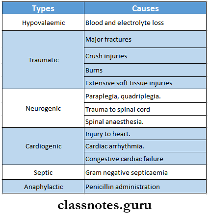
Various types of shock are as follows:

Haemorrhage Shock And Blood Transfusion Types Of Shock

Question 2. Septic shock.

Answer:

Aetiology:

  • Release of endotoxin by gram-negative organisms.
  • Severe septicaemia.
  • Peritonitis.
  • Meningitis

Question 3. Neurogenic shock.

Answer:

Neurogenic Shock Causes:

  • Paraplegia, quadriplegia.
  • Trauma to the spinal cord.
  • Spinal anaesthesia.

Neurogenic Shock Clinical Features:

  • Skin remains warm, pink and well-perfused.
  • Urinary output – normal.
  • Heart rate-rapid.
  • Blood pressure is decreased.

Question 4. Cardiogenic shock.

Answer:

Cardiogenic Shock Causes:

  • Injury to heart
  • Myocardial infarction.
  • Cardiac arrhythmia.
  • Congestive cardiac failure.

Cardiogenic Shock Clinical Features:

  • Initially
    • Skin is pale and cool.
    • Urine output reduced.
  • Later
    • Rapid pulse.
    • Hypotension
    • Distended neck veins.
    • The liver is enlarged.
    • The heart becomes enlarged.

Question 5. Hypovolaemic shock.

Answer:

Hypovolaemic Shock Causes:

  • Blood and plasma loss
  • Electrolyte loss.

Question 6. Crush Syndrome.

Answer:

Crush Syndrome

It is a symptom complex in which a portion of the body becomes crushed due to a heavy weight falling on that portion and is kept there for some time to crush all the tissues in that portion.

Crush Syndrome Causes:

  • Earthquakes
  • Mine injuries
  • Air raids
  • Collapse of building.
  • Use of tourniquet for a longer period.

Crush Syndrome Clinical Features:

  • Extravasaion of blood into muscles.
  • Muscles are crushed and swollen.
  • Acute renal tubular necrosis.
  • Reduced urinary output.
  • Tense and painful extremities.

Crush Syndrome Treatment:

  • Application of tourniquet.
  • Parallel incisions are made to relieve tension.
  • Administration of intravenous fluid.
  • Catheterization of bladder.
  • Hemodialysis is a severe condition.

Read And Learn More: General Surgery Question and Answers

Question 7. Hypokalaemia.

Answer:

Hypokalaemia

Potassium deficiency in diet is called hypokalemia.

Hypokalaemia Causes:

  • Following trauma.
    • Starvation
    • Loss of gastrointestinal secretion.

Hypokalaemia Clinical features:

  • Gradual onset of drowsiness.
  • Slow and slurred speech.
  • Irritability.
  • Muscular hypotonia and weakness.
  • Absence of deep reflexes.
  • Slow pulse rate.
  • Diminished intestinal motility.
  • Low BP.
  • Skin remains warm and dry.

Hypokalaemia Treatment:

  • Replacement of potassium deficit.

Question 8. Acidosis.

Answer:

Acidosis

An increase in pH leads to acidosis.

Acidosis Types:

  1. Metabolic acidosis.
    • In it, there is a gain or retention of fixed acids or loss of base.
    • Cause:
      • Diabetic acidosis.
      • Lactic acidosis.
      • Renal insufficiency.
      • Rapid transfusion of bank blood.
      • Diarrhoea.
    • Clinical Features:
      • Increase in rate and depth of breathing.
      • Rapid and noisy respiration.
      • Raised pulse rate and blood pressure.
      • Urine becomes strongly acidic.
    • Treatment:
      • Administration of ringer’s lactate solution.
  2. Respiratory acidosis.
    • Causes:
      • Lung disorders.
      • Peritonitis
      • Crush injury.
      • Depressed respiratory centre.
      • Airway obstruction.

Acidosis Clinical Features:

  • Slow rise in BP.
  • Abnormal respiration.
  • Restlessness.
  • Hypertension
  • Tachycardia.

Acidosis Treatment:

  • Mechanical ventilation
  • Endotracheal intubation.
  • Avoid over-sedation and over-use of muscle relaxants.

Question 9. Haemorrhage.

Answer:

Haemorrhage

Haemorrhage is defined as the escape of blood from blood vessels.

Haemorrhage Classification:

  1. Based on the bleeding haemorrhage.
    • External and internal.
    • Arterial, venous or capillary.
  2. Based on the time of appearance.
    • Primary – Secondary – Reactionary.

Question 10. Secondary Haemorrhage.

Answer:

Secondary Haemorrhage

It is a bleeding that occurs usually after 7-14 days of injury.

Secondary Haemorrhage Causes:

  • Infection.
  • Sloughing part of the arterial wall.

Secondary Haemorrhage Clinical features:

  • Preceded by warning haemorrhage staining the dressings.
  • Followed by moderate to severe haemorrhages.

Secondary Haemorrhage Types:

  • Externally – example: at operation site of haemorrhoids- tomy.
  • Internally Example: haematemesis following peptic ulcer operation.

Question 11. Hemophilia.

Answer:

Hemophilia

Hemophilia is an X-linked recessive disorder of coagulation factors.

Hemophilia Types:

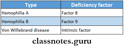
  1. Hemophilia A-classic haemophilia – due to factor 8 deficiency.
  2. Hemophilia B-Christmas disease -due to factor 9 deficiency.
  3. Von Willebrand’s disease due to deficiency of von Willebrand factor.

Hemophilia Clinical features:

  • Easy bruising
  • Prolonged bleeding.
  • Spontaneous bleeding into subcutaneous tissue.
  • GIT bleeding.
  • Hpitaxis.
  • Recurrent haemarthrosis.
  • Hematuria.
  • Intracranial haemorrhage.

Question 12. Disseminated intravascular coagulation (DTC)

Answer:

Disseminated intravascular coagulation (DTC)

Disseminated intravascular coagulation is a complex thrombo-hemorrhagic disorder occurring as a secondary complication in some systemic diseases.

Pathogenesis:

  • Includes
  1. Activation of coagulation
  2. Thrombotic phase
  3. Consumption phase.
  4. Secondary fibrinolysis.

Question 13. Blood groups.

Answer:

Blood groups

Blood groups are classified based on the presence or absence of specific agglutinogen or antigen on the surface of RBC.

Major blood groups are:

1. ABO system
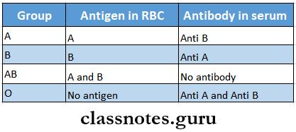
Haemorrhage Shock And Blood Transfusion Blood Groups

2. Rh blood group.

    • Rh factor is an antigen present in RBC.
    • Persons having D antigen are called Rh-positive.

Question 14. Rh factor.

Answer:

Rh factor

  • Rh factor is an antigen present in RBC.
  • It was first discovered by Landsteiner and Weiner in rhesus monkey.
  • It is detected only in RBCs
  • It is inherited from both parents as homozygous positive DD, heterozygous negative Dd or homozygous negative dd.
  • Rh Antibody is absent in plasma.
  • But its production can be evoked by.
    • Transfusion with Rh-positive blood.
    • Entrance of Rh-positive blood from foetus into circulation of Rh-negative mother.

Question 15. Blood Transfusion.

Answer:

Blood Transfusion

It is a process of transferring blood or blood-based products from one person into the circulatory system of another.

Blood Transfusion Types:

  1. Typical stored CPD blood from the blood bank.
  2. Filtered blood-filtered through a membrane with 40 |im pores.
  3. Warm blood is used in cardiopulmonary operations.
  4. Autotransfusion – preserving and then transfusing one’s blood.
  5. Replacement transfusion – used in newborns.
Haemorrhage Shock And Blood Transfusion

Question 16. Indications of blood transfusion.

Answer:

Acute haemorrhage.

  • During operations, preoperatively and postoperatively.
  • Anaemia.
  • Malnutrition.
  • In severe burns.
  • In coagulation disorders.
  • In erythroblastosis fetalis.
  • During chemotherapy.

Question 17. Complications of blood transfusion.

Answer:

Complications of blood transfusion

  1. Transfusion reactions
    • Incompatibility
    • Pyrexia reactions
    • Allergic reactions
    • Sensitisation to leucocytes and platelets
  2. Transmission of diseases
    • Serum hepatitis
    • AIDS
  3. Reactions caused by massive transfusion.
    • Acid-base imbalance.
    • Hyperkalaemia.
    • Citrate toxicity.
    • Hypothermia.
    • Failure of coagulation.
  4. Complication of over-transfusion.
    • Congestive cardiac failure.
  5. Other complications.
    • Thrombophlebitis
    • Air embolism.

Question 18. Types of haemorrhage

Answer:

Types of haemorrhage

  1. According to the source
    • External haemorrhage – seen externally
    • Internal haemorrhage – not seen externally
  2. According to the vessels involved
    • Arterial haemorrhage – haemorrhage coming out of the artery
    • Venous haemorrhage – haemorrhage coming out of vein
    • Capillary haemorrhage – haemorrhage coming out of the capillary
  3. According to the time of appearance
    • Primary haemorrhage – at the time of injury
    • Reactionary haemorrhage – within 24 hours of injury
    • Secondary haemorrhage – after 7-14 days of injury

Question 19. Reactions to blood transfusion

Answer:

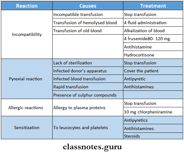
Reactions to blood transfusion

Haemorrhage Shock And Blood Transfusion Reactions To Bllod Transfusion

Haemorrhage Shock And Blood Transfusion Notes

Haemorrhage Shock And Blood Transfusion Important Notes

  1. Classification of haemorrhage
    • Based on the nature of the vessel involved
      • Arterial
      • Venous
      • Capillary
    • Based on the timing of the haemorrhage
      • Primary
      • Reactionary
      • Secondary
    • Based on the duration of the haemorrhage
      • Acute
      • Chronic
    • Based on the source of the haemorrhage
      • External
      • Internal
  2. Methods of measuring blood loss
    • Weighing swab
    • Measurement of swelling in a closed fracture
    • Measuring a blood clot
  3. Methods to stop bleeding
    • Rest
    • Pressure and packing
    • Ligation of vessels
  4. HemophiliaHaemorrhage Shock And Blood Transfusion Hemophilia
  5. Shock
    • Definition
      • Shock is a condition in which circulation fails to meet the nutritional needs of the cells and fails to remove the metabolic waste products
    • Classification
      • Haematogenic or hypovoJaernic shock
      • Traumatic shock
      • Neurogenic shock
      • Cardiogenic shock
      • Septic shock
      • Miscellaneous
        • Anaphylactic shock
  6. Blood transfusion
    • Indications
      • Acute haemorrhage
      • Major surgery
      • Deep burns
      • Pre-operative and post-operative in anaemia
      • In malnutrition
      • In coagulation disorders
      • In erythroblastosis details
      • During chemotherapy in malignant diseases
    • Complications
      • Transfusion reactions
        • Incompatibility
        • Pyrexia reactions
        • Allergic reactions
      • Transmission of diseases
      • Reactions caused by massive transfusion
        • Acid-base imbalance
        • Hyperkalaemia
        • Citrate toxicity
        • Hypothermia
        • Failure of coagulation
      • Complications of over-transfusion
        • Congestive cardiac failure
      • Other complications
        • Thrombophlebitis
        • Air embolism
  7. Blood products
    • Plasma and its derivatives
      • Fresh frozen plasma
      • Platelet-rich plasma
      • Fibrinogen
      • Albumin
      • Cryoprecipitate
    • Synthetic solution
      • Dextran
      • Gelatin
      • Hydroxyethyl starch
      • Fluorocarbons

Haemorrhage Shock And Blood Transfusion Short Essays

Question 1. Vasovagal shock.

Answer:

Vasovagal shock

  • Vasovagal shock is a type of neurogenic shock.

Pathophysiology:

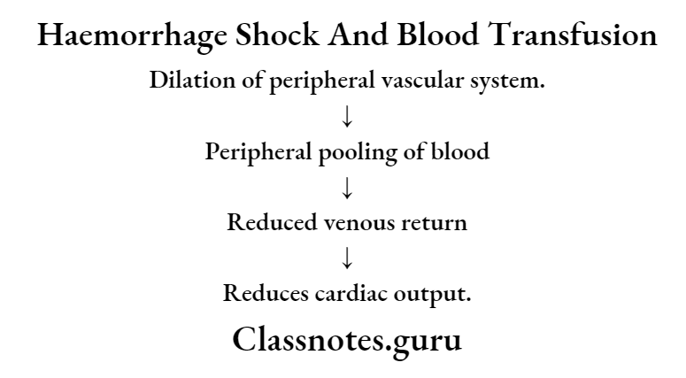
Haemorrhage Shock And Blood Transfusion Haemorrhage shock And Blood Transfusion

Vasovagal Shock Clinical features:

  • Reduced blood flow to the brain.
  • Cerebral hypoxia.
  • Bradycardia.
  • Hypotension
  • Unconsciousness
  • Pallor

Vasovagal Shock  Treatment:

  • Place the patient flat or in head low position.
  • Maintain airway
  • Use of 4 atropine.

Question 2. Reactionary bleeding.

Answer:

Reactionary bleeding

Reactionary bleeding is defined as bleeding that occurs within 24 hours of injury or operation.

Reactionary Bleeding Causes:

  • Dislodgement of a blood clot.
  • Slipping of ligatures.

Reactionary Bleeding  Precipitating Factors:

  • Rise in blood pressure
  • Restlessness.
  • Coughing
  • Vomiting
  • Raise in venous pressure.

Read And Learn More: General Surgery Question and Answers

Question 3. Haemophilia.

Answer:

Haemophilia

  • Hemophilia is a sex-linked inherited disorder.
  • It is carried by a recessive gene.
  • Transmitted through female carriers.

Haemophilia Cause:

  • Lack of factor 8, coagulation factor.

Haemophilia Clinical features:

  • Bleeding after mild trauma.
  • Repeated haemorrhages into joints.
  • Spontaneous retroperitoneal bleeding.
  • Severe abdominal pain, and tenderness.
  • Haematuria.
  • Permanent damages to the articular cartilages and articular surfaces.
  • Disorganization of the joints.

Haemophilia Treatment:

  • Periodic infusion of cryoprecipitate.
  • Transfusion of fresh blood or fresh frozen plasma.

Haemorrhage Shock And Blood Transfusion Viva Voce

  1. Albumin is not used in chronic renal disease, chronic liver disease and to treat malnutrition
  2. Cryoprecipitate is a rich source of factor 8
  3. Fluorocarbon is considered to be a red cell substitute
  4. Blood is stored in blood banks at 4°C
  5. Cold citrate-containing blood changes storage
  6. Blood is separated into individual components to optimize therapeutic potency
  7. Styptics are local hemostatic agents used to stop bleeding from the local approaching site
  8. Hypotension manifests when blood loss exceeds 30%

Haemorrhage Shock And Blood Transfusion Long Essays

Haemorrhage Shock And Blood Transfusion Long Essays

Question 1. Classify shock. Describe the pathophysiology, clinical features and management of shocks.

Answer:

Shock:

  • Shock is a condition in which circulation fails to meet the nutritional needs of the cells and fails to remove the metabolic waste products.
  • It is characterized by hypoperfusion and severe dysfunction of vital organs.

Shock Classification:

  1. Haematogenic or hypovolaemic shock.
    • Occurs due to loss of blood, plasma or body water and electrolytes.
    • Caused by haemorrhage, vomiting, diarrhoea, dehydration, etc.
  2. Traumatic shock.
    • Caused by major fractures, crush injuries, bums, extensive soft tissue injuries and intraabdominal injuries.
  3. Neurogenic shock.
    • Caused by paraplegia, quadriplegia, trauma to the spinal cord and spinal anaesthesia.
  4. Cardiogenic shock.
    • Caused by injury to heart, myocardial infarction or congestive cardiac failure.
  5. Septic shock.
    • Occurs due to gram-negative septicaemia.
  6. Miscellaneous types – includes:
    • Anaphylactic shock.

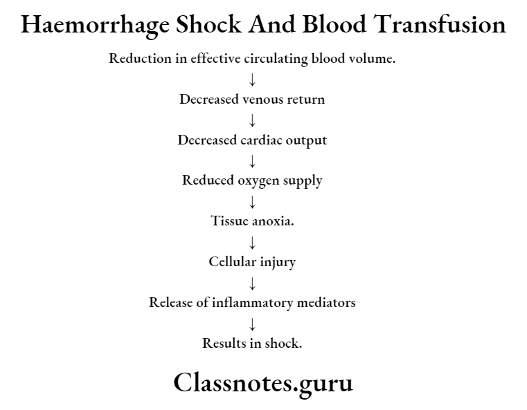
Physiology: It can be described as 2 processes.

  1. Reduced effective circulating volume.
    • May result either.
      • By actual loss of blood volume or
      • By decreased cardiac output.
  2. Tissue anoxia.

Haemorrhage Shock And Blood Transfusion Reduction In effective Circulating Blood Volume

Shock Clinical features:

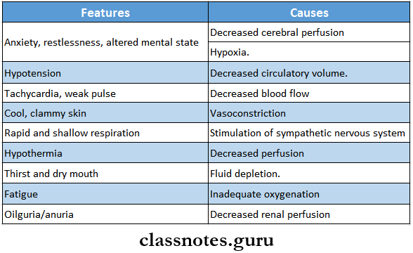
Haemorrhage Shock And Blood Transfusion Shock Clinical Features

Management: AIMS:

  • To increase cardiac output.
  • To improve tissue perfusion to vital organs.

Shock Treatment:

  1. Maintenance.
    • Maintain patent airway and oxygen.
    • Head position – At low position with face turned to one side
  2. Control of haemorrhage.
    • Done by elevation, compression bandages or by ligation of blood vessels.
  3. Extracellular fluid replacement.
    • Nonsugar, nonprotein crystalloid is preferred.
    • Normal saline or ringer’s lactate should be started first.
  4. Correct acid-base disturbance.
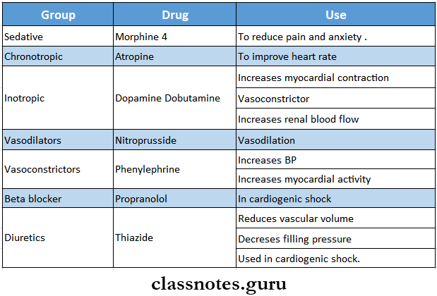
  5. Drugs.

Haemorrhage Shock And Blood Transfusion Shock Treatment

Question 2. Describe the pathophysiology, clinical features and treatment of septic shock.

Answer:

Septic Shock:

  • Septic shock is caused due to release of endotoxin in blood mostly by Gram-negative organisms.
  • Occurs in cases of severe septicaemia, peritonitis or meningitis.
  • Pathophysiology.

Presence of gram-positive and gram-negative organism

Local inflammation occurs

Release of endotoxins from organism

Activation of neutrophils, monocytes & macrophages.

Release of inflammatory mediators.

Cellular chemotaxis.

Endothelial injury

Activation of the coagulation cascade

Massive fluid loss

Septic shock

Septic Shock Clinical Features:

  • Initially, chills and fever above 100oC occurs.

Septic Shock Types:

  1. Early warm shock.
    • There is cutaneous vasodilation.
    • Body temperature increases
    • Cutaneous vasodilatation occurs.
    • Arterial blood pressure falls.
    • Cardiac output increases.
    • Skin remains warm, pink and well-perfused.
    • Pulse rate increases
  2. Late cold shock.
    • There is increased vascular permeability
    • Cardiac output decreasing.
    • Hypovolemia occurs.

Read And Learn More: General Surgery Question and Answers

Septic Shock Treatment:

  • Removal of septic focus.
    • Drainage of pus under anaesthesia.
    • Closure of perforation.
    • Resection of gangrene.
  • Antibiotis.
    • Administered after antibiotic sensitivity tests.
    • Initial antibiotics are
      • Cephalothin – 6 – 8 gm/day IV in 4 – 6 divided doses.
      • Gentamicin – 5 mg/kg/day.
      • Clindamycin
  • Fluid replacement.
    • Crystalloids such as isotonic saline as Ringer’s lactate may be used.
    • Blood transfusion – to maintain haemoglobin level to 10 mg%.
  • Supportive care.
    • Oxygenation.
    • Mechanical ventilation.
    • Endotracheal intubation.
  • Steroids.
    • Short-term, high-dose steroid therapy is used.
    • Initial dose of 15 – 30 mg/ kg body weight of methylprednisolone is given.
    • Same dose repeated within 4 hours.
    • Vasoactive drugs.
  • Vasodilators such as phenoxybenzamine is used along with fluid replacement.
    • Inotropic agents such as isoproterenol is used to restore adequate circulation.
    • It produces mild peripheral vasodilation.
    • There is slight fall in BP.

Question 3. Describe the pathophysiology, clinical features and management of haemorrhage or hypovolaemic shock.

Answer:

Haemorrhage shock/Hypovolaemic shock: Such shock occurs due to sudden loss of blood volume or loss of fluid from the vascular space.

Pathophysiology:

Haemorrhage

Loss of blood

Decreased filling of right heart.

Decreased filling of the pulmonary vasculature

Decreased filling of left atrium and ventricle

Decrease in stroke volume.

Drop in arterial blood pressure

Hypovolaemic Shock Clinical Features:

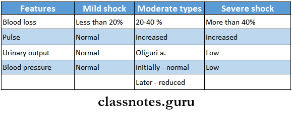
  • Depending on the degree of blood loss it can be described into three types.

Haemorrhage Shock And Blood Transfusion Haemorrhage Or Hypovolaemic Shock

Hypovolaemic Shock Management:

  1. Resuscitation.
    • Maintain airway with adequate ventilation and oxygenation.
    • Lower the head with jaw support.
  2. Immediate control of bleeding.
    • Raise the footend of the bed.
    • Use of compression bandages.
  3. Extracellular fluid replacement.
    • Ringer’s lactate, Ringer’s acetate or normal saline supplemented with 1-2 ampules of sodium bicarbonate is used.
    • 1000 – 2000 ml solution is given within 45 min intravenously.
    • Blood transfusion done if required.

Question 4. Describe neurogenic shock and its management.

Answer:

Neurogenic Shock Causes:

  • Paraplegia.
  • Quadriplegia.
  • Trauma to spinal cord
  • Spinal anaesthesia.

Pathophysiology:

Blockade of sympathetic nervous system

Loss of arterial and venous tone

Peripheral pooling of blood.

Decrease in cardiac filling
i
Decrease in stroke volume.

Decrease in pulmonary blood volume.

Decrease in cardiac output.

Shock

Neurogenic Shock Clinical Features:

  • Skin remains warm, pink and well-perfused.
  • Urinary output – normal.
  • Heart rate-rapid.
  • Blood pressure – decreased

Neurogenic Shock Management:

  1. Elevation of the legs to correct peripheral pooling of blood.
  2. Fluid administration to increase cardiac output.
  3. Use of vasoconstrictor drug.
    • It increases BP and myocardial activity.

Question 5. Classify haemorrhage and its management and Describe the causes, clinical features, How will you manage a case of primary haemorrhage after a dental extraction?.

Answer:

Haemorrhage: Haemorrhage is defined as escape of blood from blood vessels.

Haemorrhage Classification:

  1. According to the source:
    • External haemorrhage.
      • Seen externally.
    • Internal haemorrhage.
      • Not seen externally, it is hidden,
      • Example: GIT bleeding.
    • Arterial haemorrhage.
      • It is haemorrhage coming out of artery.
      • It is bright red in colour.
    • Venous haemorrhage.
      • It is haemorrhage coming out of vein.
      • It is dark red in colour.
    • Capillary haemorrhage.
      • It is haemorrhage coming out of capillary
      • It is bright red in color and it oozes out
  2. According to the time of appearance:
    • Primary haemorrhage.
      • Occurs at the time of injury.
    • Reactionary haemorrhage.
      • Occurs within 24 hours of injury.
      • Secondary haemorrhage.
    • Occurs after 7-14 days of injury.

Haemorrhage  Management:

  • To stop blood loss.
  1. Rest.
    • Use of sedative and analgesics.
    • Morphine is administered IM/IV.
    • Inj. Pethidine is better than morphine.
  2. Position of patient.
    • Head end of bed is raised in haemorrhage oc- curing after thyroidectomy.
    • Foot end of bed raised in case of haemorrhage from varicose veins.
  3. Pressure and packing.
    • Use of sterile gauze pieces and pressure bondage.
    • At home, it can be done by clean linen cloth.
  4. Operative methods.
    • Haemorrhage can be controlled by.
      • Use of artery forceps.
      • Ligation of blood vessels.
      • Smaller vessels are coagulated with diathermy.
      • Bigger vessels are sutured
      • In case of oozing blood-following is used
        • Oxycel or gelatine sponge.
        • Gauze soaked in adrenaline (1:1000)
        • Bone wax for bleeding occurring from the bone.

Haemorrhage  Causes:

  • Bleeding disorders.
  • Low platelet count
  • Anticoagulant medication.
  • Broken or ruptured blood vessels.
  • Severe trauma
  • After surgery.
  • After childbirth.

Haemorrhage  Clinical Features:

  • Blood loss
  • Increased pulse rate
  • Thready pulse
  • Low blood pressure
  • Pallor Restlessness
  • Deep respiration
  • Cold and calmmy extremities
  • Empty veins
  • Low urinary output.

Management of Primary Haemorrhage:

1. Post-extraction bleeding.

Removal of clots with gauze

Placement of gauze pad or tea bag over socket.

Patient is instructed to bite over it for 1 hour

Repeated 2-3 times.

Prevent disruption of clot

2. If bleeding continues.

Anaesthesize the area

Curette the socket

Remove existing clot and freshen the bone

Irrigate with normal saline
i
Place local haemostatic agent into socket

Suture under gentle tension

Question 6. Describe indications and complications of blood transfusion.

Answer:

Blood Transfusion: It is the process of transferring blood or blood-based products from one person into circulatory system of another.

Blood Transfusion Indications:

  • Acute haemorrhage – external or internal.
  • Certain major operations – like radical mastectomy.
  • In deep burns.
  • Preopera lively in anaemic patients.
  • Postoperatively in septicaemia.
  • In anaemia.
  • In severe malnutrition.
  • In coagulation disorders like hemophilia.
  • In treatment of erythroblastosis foetalis.
  • During chemotherapy of malignant diseases.

Blood Transfusion Complications:

  1. Transfusion reactions:
    • Incompatibility
      • Causes:
        • Incompatible transfusion.
        • Transfusion of hemcolyzed blood.
        • Transfusion of old blood.
      • Clinical features:
        • Fever, rigor.
        • Headache.
        • Nausea, vomiting.
        • Pain in the loins.
        • Tingling sensation in the extremities.
        • Feeling of tightness of chest
        • Dysponea.
        • Diminished urinary output.
        • Haemoglobunuria.
        • Jaundice
      • Treatment:
        • Stop the transfusion immediately.
        • Administration of 4 fluids.
        • Alkalization of blood with 10 ml of isotonic solution of sodium lactate and simultaneously 10 ml of saturated solution of sodium bicarbonate 4.
        • Use of 80 -120 mg frusemide IV to provoke diuresis.
        • Antihistamine and hydrocortisone may be prescribed.
    • Pyrexial reactions.
      • Causes:
        • Lack of sterilization
        • Infected donor’s apparatus
        • Iniected blood transfusion,
        • Rapid transfusion,
        • Presence of sulphur compounds in rubber tubing.
      • Clinical Features:
        • Pyrexia.
        • Rigor, chills.
        • Restlessness.
        • Headache.
        • Increased pulse rate.
        • Nausea and vomiting.
      • Treatment:
        • Stop transfusion immediately.
        • Cover the patient with blanket.
        • Antipyretic and antihistaminic drugs are injected.
    • Allergic reaction:
      • Cause:
        • Allergic reaction to plasma product
      • Features:
        • Mild tachycardia.
        • Urticarial rash.
        • Fever
        • Dysponea
        • Circulatory collapse.
      • Treatment:
        • Stop transfusion
        • Administer 10 mg chlorpheniramine.
    • Sensitization to leucocytes and platelets:
      • Use of antipyretics, antihistamines and steriods.
  2. Transmission of diseases:
    • Diseases transmitted ae.
      • Serum hepatitis
      • AIDS
      • Bacterial infections.
  3. Reactions caused by massive transfusion:
    • Acid base imabalance – alkalosis.
    • Hyperkalaemia.
    • Citrate toxicity.
    • Hypothermia.
    • Failure of cogulation.
  4. Complications of over-transfusion:
    • Congestive cardiac failure occurs.
  5. Other complications:
    • Thrombophlebitis
    • Air embolism.

Question 7. Define shock. Describe the pathophysiology and classification of shock. Discuss management of hypovolaemic shock

Answer:

Shock Definition: Shock is a condition in which circulation fails to meet the nutritional needs of the cells and fails to remove the metabolic waste products

Pathophysiology

  • Reduced effective volume
    • It may result either
      • By actual loss of blood volume or
      • By decreased cardiac output
  • Tissue anoxia
    • Reduction in effective circulating blood volume
    • Reduced venous return
    • Decreased cardiac output
    • Decreased oxygen supply
    • Tissue anoxia
    • Cellular injury
    • Release of inflammatory mediators
    • Results in shock

Shock Classification

  • Haematogenic or hypovolaemic shock
  • Traumatic shock
  • Neurogenic shock
  • Cardiogenic shock
  • Septic shock
  • Miscellaneous
    • Anaphylactic shock

Management of hypovolemic shock

  • Resuscitation
    • Maintain airway with adequate ventilation and oxygenation
    • Lower the head with jaw support
  • Immediate control of bleeding
    • Raise the foot end of the bed
    • Use of compression bandages
  • Extracellular fluid replacement
    • Ringer’s lactate, Ringer’s acetate or normal saline supplemented with 1-2 ampules of sodium bicarbonate is used
    • 1000-2000 ml solution is given within 45 min intravenously
    • Blood transfusion done if required

Question 8. What are blood components? Write in detail about the indications, contraindications and complications of blood transfusion.

Answer:

Blood components

  • There are four main components of blood
    • Plasma
    • Red blood cells or erythrocytes
    • White blood cells or leukocytes
    • Platelets

Blood transfusion

  • Indications
    • Acute haemorrhage
    • Major surgery
    • Deep burns
    • Pre-operative and post-operative in anaemia
    • In malnutrition
    • In coagulation disorders
    • In erythroblastosis fetalis
    • During chemotherapy in malignant diseases
  • Contraindications
    • Infections
    • Aortic stenosis
    • Angina
    • Significant cardiac or pulmonary disease
    • Coronary heart disease
    • Cyanotic heart disease
    • Uncontrolled hypertension
  • Complications
    • Transfusion reactions
      • Incompatibility
      • Pyrexial reactions
      • Allergic reactions.
    • Transmission of diseases
    • Reactions caused by massive transfusion
      • Acid base imbalance
      • Hyperkalaemia
      • Citrate toxicity
      • Hypothermia
      • Failure of coagulation
  • Complications of over transfusion
    • Congestive cardiac failure
  • Other complications
    • Thrombophlebitis
    • Air embolism