Asepsis In Endodontics Question And Answers

Asepsis In Endodontics Definitions

  • Sterilization
    • It is the complete destruction of agents that are capable of causing infections, including spores.

Asepsis In Endodontics Important Notes

  • Methods of sterilization
    • Physical agents
      • Sunlight
      • Drying
      • Dry heat
      • Moist heat
      • Filtration
      • Radiation
    • Chemical agents
      • Alcohol
      • Aldehyde
      • Chlorines
      • quaternary ammonium compound
      • Phenolic compounds
      • Antiseptics

Asepsis In Endodontics

Asepsis In Endodontics Long Essays

Question 1. Discuss sterilization of endodontic instruments.
Answer.

Sterilization: It is the complete destruction of agents that are capable of causing infections, including spores.

Read And Learn More: Endodontics Question and Answers

Sterilization Of Endodontic Instruments Methods:

Physical Agents:

  • Sunlight:
    • Natural means
    • Has bactericidal activity
  • Drying:
    • Unreliable
    • Spores are unaffected
  • Dry Heat:
    • Consists of
      • Flaming – for culture tubes, glass
      • Red heat – needles
      • Incineration – Dressings
      • Hot air oven – At 160°C for 2 hours
    • Metallic, glass particles, powder
  • Moist Heat:
    • Causes denaturation of proteins
  • Filtration:
  • Radiation:
    • Causes:
      • Inhibition of DNA replication
      • Damaging structure of DNA
      • Denaturation of protein
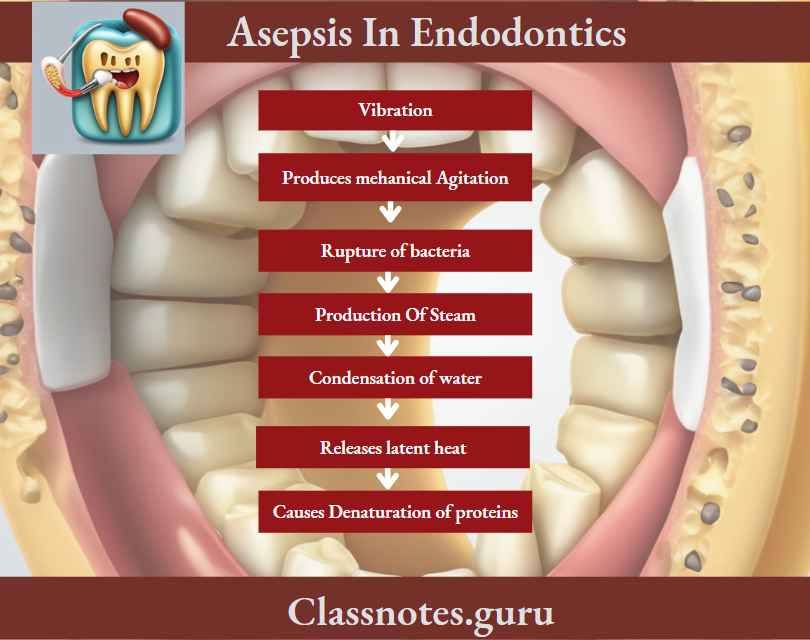
  • Ultrasonic Vibration:

Sterilization Of Endodontic Instruments Chemical agents:

  • Alcohol Ex. Ethyl alcohol
    • Bactericidal
    • Corrosive
    • Fast acting
  • Aldehyde – Ex.Glutaraldehyde
    • High-level disinfectant
    • Non-corrosive
    • Long activated life
  • Chlorines
  • Quaternary Ammonium Compound
    Ex:

    • Benzalkonium chloride
    • Low-level disinfectant
  • Phenolic compounds:
    • Low-level disinfectant
    • Used for floors, walls, and furniture
  • Antiseptics
    • Iodophor Ex. Povidoneiodone
      • Broad spectrum
      • Formulated as 1% I2 solution
    • Chlorhexidine

Sterilization Of Endodontic Instruments:

Endodontics Of Asepsis In Endodontics Chemical Agents Instruments

Sterilization In Endodontics

Sterilization Of Endodontic Instruments Steps:

  • Pre-soaking in water to soften organic debris
  • Cleaning – Hand washing or ultrasonic cleaning is done
  • Drying – To prevent corrosion
  • Packaging – Wrapping of instruments in clothes
  • Sterilization
  • Drying/cooling
  • Storage
  • Distribution
  • Sharpening of instruments

Asepsis In Operative Dentistry

Asepsis In Endodontics Short Questions And Answers

Question 1. Disinfection.
Answer.

Disinfection

  • It is the destruction of pathogenic micro-organisms
  • It permits adequate removal of pulp tissue and debris
  • This leads to the enlarging of the canal by biomechanical means and the clearing of its contents by irrigation

Question 2. Glass Bead Sterilizer/Salt Bead Stabilizer.
Answer.

Glass Bead Sterilizer

Rapid method of sterilization

Glass Bead Sterilizer Materials Used:

  • Usually salt
  • Salt is replaced by glass beads smaller than 1 mm in diameter

Glass Bead Sterilizer Reason For Smaller Beads:

  • Efficient in transferring heat to instruments
  • Time required – 5-15 seconds
  • Temperature – 437 – 465°F

Glass Bead Sterilizer Advantages:

  • Easily available
  • Salt is replaced by glass beads smaller than 1 mm in diameter

Glass Bead Sterilizer Disadvantage:

  • Handle portion is not sterilized

Endodontic Disinfection Methods

Question 3. Autoclave.
Answer.

Autoclave

Moist heat sterilization method

Autoclave Principle:

Asepsis In Endodontics

Endodontics Of Asepsis in Endodontics Autoclave Principles

Autoclave Advantages:

  • Effective
  • Accurate
  • Rapid

Autoclave Disadvantages:

  • Causes corrosion
  • Melts rubber
  • Unsuitable for oils

Autoclave Factors Effecting It:

  • Cleaning of instruments
  • Direct flow of steam
  • Periodic monitoring of sterilizer

Asepsis In Operative Dentistry

Asepsis In Endodontics Viva Voce

  • The hottest part of the glass bead sterilizer is along its outer rim and the temperature is lowest in the center of the surface layer of salt

Endodontics Treatment Of Traumatized Teeth Question And Answers

Treatment Of Traumatized Teeth Long Essays

Question 1. An 18-year-old patient reports to the clinic with a fracture of maxillary central incisor involving dentin. The trauma happened one month back, discuss your treatment options.
Answer.

Enamel-Dentin Fracture

Enamel-Dentin Fracture Definition:

  • A fracture confined to enamel and dentin with loss of tooth structure, but not exposing the pulp

Enamel-Dentin Fracture Clinical Features:

  • Not tender to percussion
  • Normal mobility
  • Positive pulp sensitivity

Enamel-Dentin Fracture Radiographic Appearance:

  • Visible enamel dentin loss

Enamel-Dentin Fracture Aims Of Treatment:

  • Elimination of discomfort
  • Preservation of vital pulp
  • Restoration of fractured crown

Read And Learn More: Endodontics Question and Answers

Enamel-Dentin Fracture Treatment Options:

  • Restorative procedure composite resin restoration is preferred
  • Fractured segments can be approximated and bonded back by dentin bonding agents
  • The use of indirect veneer is done to achieve esthetics

Endodontic Treatment Of Traumatized Teeth

Enamel-Dentin Fracture Follow-Up:

  • Clinical and radiographic control at 6-8 weeks and 1 year
  • The tooth should be periodically tested with an electric pulp tester

Treatment Of Traumatized Teeth Short Questions And Answers

Question 1. Hank’s balanced salt solution (HBSS).
Answer.

Hank’s Balanced Salt Solution (HBSS)

  • John H.Hank formulated it
  • It is a solution made to a physiological pH and salt concentration
  • It is a collective group of salts rich in bicarbonate ions
  • Used for washing tissues and cells
  • It provides the cells with water and inorganic ions while maintaining physiological pH and osmotic pressure

Management Of Dental Trauma

Question 2. Management of Avulsed Teeth.
Answer.

Avulsed Teeth Storage Media:

  • Saliva For 2 hours:
    • Milk for upto 6 hours
    • Water/saline – ineffective
    • Hanks balanced salt solution upto 72 hours

Roof Surface:

  • If Clean-Replant It:
    • If dirty-clean with tap water/wet sponge
      Periodontal Ligament:
  • If Vital – Replant It:

Endodontics Treatment Of Traumatized Teeth If vital Replant it

Socket:

  • Irrigate it
  • Do not curette the socket

Treatment Of Traumatized Teeth Viva Voce

  • Pulpal death is the most common cause of discoloration
  • Hydrogen sulfide produced by bacteria combines with pigments and darkens the tooth
  • Microabrasion is a method used to remove surface stains or defects
  • Mc Inn’s solution is used for the removal of endemic fluorosis stains
  • Composites should be delayed for 2-3 weeks after bleaching to allow for dissipation of residual peroxides

Pulp Space Anatomy And Access Cavities Question And Answers

Endodontics Of Anatomy Of Pulp Cavity And Its Access Opening Definitions

  • Anatomic Apex:
    • The Anatomic Apex is the tip or end of the root determined morphologically
  • Radiographic Apex:
    • Radiographic Apex is the tip or end of the root determined radiographically
  • Apical Foramen:
    • It is the main apical opening of the root canal
    • Frequently located away from the anatomic or radiographic apex
  • Apical Constriction:
    • Apical Constriction is the apical portion of the root canal having the narrowest diameter

Endodontics Of Anatomy Of Pulp Cavity And Its Access Opening Important Notes

  • Endodontic Anatomy

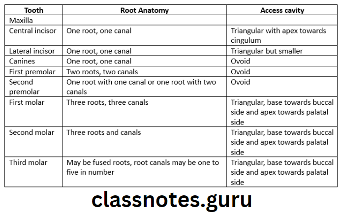
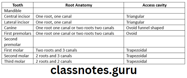
Endodontics Anatomy Of Pulp Cavity And Its Access Opening Endodontic Anatomy 1

Read And Learn More: Endodontics Question and Answers

Endodontics Anatomy Of Pulp Cavity And Its Access Opening Endodontic Anatomy 2

  • Access cavity of mandibular first molar
    • Mesiobuccal orifice is under mesiobuccal cusp
    • mandibular first molar is gained access from mesiobucco apical direction
    • Mesiolingual orifice is below the central groove
    • mandibular first molar is explained from distobuccal direction
    • The distal orifice is present in the center of tooth buccolingually
    • mandibular first molar is explained from mesial direction
  • Access cavity of maxillary first molar
    • The orifice of mesiobuccal canal is gained access from distopalatal direction
    • The distobuccal canal is gained access from mesiolingual direction
    • The palatal root is gained access from buccal direction

Pulp Space Anatomy

Anatomy Of Pulp Cavity And Its Access Opening Short Essays

Question 1. Access cavity preparation in mandibular permanent first molar.
Answer.

Mandibular Permanent First Molar Anatomy:

  • Average tooth length – 21.9mm
  • Pulp chamber
    • Roof is often rectangular in shape
    • Mesial wall is straight
    • Distal wall is round
    • Buccal and lingual walls converge to meet mesial and distal walls
    • Roof has four pulp horns – mesiobuccal, mesiolingual, distobuccal and distolingual
    • Three distinct orifices are present in the pulpal floor – mesiobuccal, mesiolingual and distal
  • Root and root canals
    • Two well-determined roots are present – Mesial and distal
    • Mesial root curves distally and distal root is straight
    • Mesial root has two canals
    • Distal root has one canal

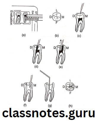
Mandibular Permanent First Molar Access Opening:

  • Enamel and dentin are pentrated in the central fossa with the bur angled towards the distal root
  • Bur is penetrated until pulp chamber is reached
  • A drop of the bur into the pulp chamber is left
  • Remove the bulk of the roof of the pulp chamber is felt
  • A trapered-cylinder bur is used to remove it
  • Walls of access cavity are refined with diamond bur
  • Access opening is usually trapezoidal with round corners or rectangular
  • The access opening extends towards the mesiobuccal cusp to uncover the mesiobuccal canal, lingually slightly beyond the central groove and distally slightly beyond the buccal groove

Endodontics Anatomy Of Pulp Cavity And Its Access Opening Steps in the access

Anatomy Of Pulp Cavity And Its Access Opening Short Answers

Question 1. Apical foramen.
Answer.

Apical Foramen

  • Apical foramen is an aperture at or near the apex of a root through which th eblood vessels and nerves of the pulp enter or leave the pulp cavity
  • In young, incompletely developed teeth, the apical foramen is funnel shaped with wider portion extending outwards
  • Mouth of the funnel is filled with periodontal tissue
  • As root developes apical foramen becomes narrower

Apical Foramen Variations:

  • Apical foramen is not always most constricted portion of the root canal
  • Apical constrictions are found 0.5-1 mm away from the root apex
  • Apical foramen is not always located in the center of the root apex

Access Cavity Preparation In Endodontics

Question 2. Nerve fibres of pulp.
Answer.

Nerve Fibres Of Pulp

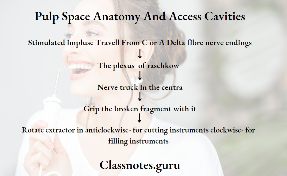
80% of the nerves of the pulp are C fibres and rest are A delta fibres

  • C Fibres:
    • They are unmyelinated and fine sensory afferent fibres
    • Diameter 0.3-1.2 micrometer
    • Conduction is slow – 0.4 – 2m/s
    • Distributed through out the pulp tissues
    • Experienced as a dull, poorly localized and lingering pain
    • Conduct throbbing and aching pain associated with pulp tissue damage
  • A Delta Fibres:
    • They are myelinated axons
    • Conduction is fast 6-30m/s
    • Diameter 2-5 micrometer
    • Present at the pulpal periphery and inner dentin
    • Interpreted as short, well-localized, sharp and pricking pain
    • Associated with dental pain

Question 3. Pain pathway.
Answer.

Pain Pathway

Endodontics Anatomy Of Pulp Cavity And Its Access Opening Pain Pathway

Root Canal Access Opening Techniques

Anatomy Of Pulp Cavity And Its Access Opening Viva Voce

  • The mesiobuccal canal of the maxillary first molar is the most difficult to prepare
  • The access cavity of a mandibular first molar is usually triangular
  • Bifurcations and trifurcations are most common in mandibular 1st premolar
  • The cervical cross-section of the maxillary 1st premolar is elliptical or kideny-shaped
  • Accessory canals are common in the apical third of the root
  • Among anterior teeth accessory canals are common in the mandibular central incisor
  • Among posteriors accessory canals are common in the mandibular first molar
  • Mandibular 1st premolar contains a prominent buccal cusp and a smaller lingual cusp that gives the crown a lingual tilt of 30°
  • Among single-rooted teeth bifurcated roots are commonly seen in mandibular 1st premolars followed by incisors and canines
  • In the maxillary molar, the mesiobuccal root has the greatest distal curvature and is the narrowest of all three canals
  • The pulp chamber of the maxillary 1st molar is the largest in the dental arch

Endodontic Emergencies Question And Answers

Endodontic Emergencies Short Essays

Question 1. Endodontic Emergency.
Answer.

Endodontic Emergency Definition:

  • Endodontic Emergency is defined as pain and/or swelling caused by inflammation or infection of the pulp and/or periradicular tissues necessitating an emergency visit to the dentist for immediate treatment.

Endodontic Emergencies

Endodontic Emergency Classification:

Endodontic Emergency Before Treatment:

  • Endodontic emergencies presenting with pain and/or swelling
    • Cracked tooth syndrome
    • Symptomatic reversible pulpitis
    • Symptomatic irreversible pulpitis
    • Phoenix abscess
    • Acute alveolar abscess
    • Celluitis
  • Traumatic injuries
    • Crown/root fracture
    • Luxation injuries
    • Tooth avulsion

Endodontic Emergency Treatment

Read And Learn More: Endodontics Question and Answers

Endodontic Emergency During Treatment:

  • Hot tooth
  • Endodontic flare-ups

Endodontic Emergency After Treatment:

  • Postobturation pain
  • Vertical root fracture

Question 2. Vertical Root Fracture.
Answer.

Vertical Root Fracture

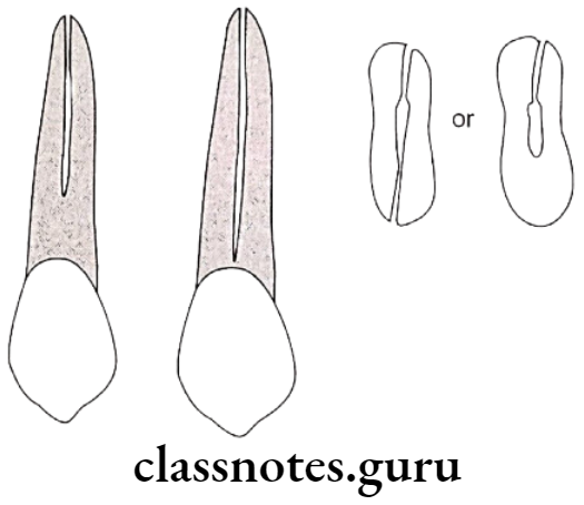
  • These are longitudinal fractures that originate in the roots of teeth

Acute Pulpitis Treatment

Vertical Root Fracture Etiology:

  • Over instrumentation
  • Over filling
  • Persistent pain
  • Hyper occlusion
  • Poor coronal seal

Vertical Root Fracture Clinical Features:

  • Site – Faciolingual plane
  • Sign – Crunching sound
  • Symptom – Pain at the site

Endodontic Emergencies Vertical root fracture

Dental Abscess Emergency Care

Vertical Root Fracture Diagnosis:

  • Radiograph shows a widening of PDL

Vertical Root Fracture Treatment:

  • Retreatment done
    • Repair
    • Root resection
    • Extraction

Cleaning And Shaping Of Root Canal Question And Answers

Cleaning And Shaping Of Root Canal Important Notes

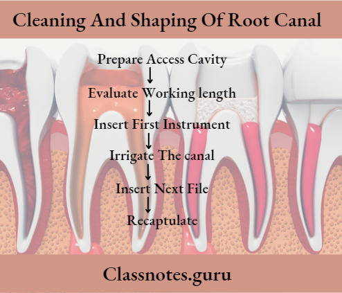
  • Cleaning And Shaping Of Root Canal Step Back Method
    • It is also called flare telescopic or serial root canal preparation
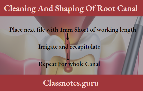
    • Once the canal has been enlarged in the apical third to at least non-25, each consecutive larger root canal instrument used for shaping the canal is placed short of the apex
    • This results in apical enlargement and marked taper from apical to corona;
      Advantages:
    • Less chances of periapical trauma
    • Facilitates removal of more debris
    • The development of an apical matrix or apical stop prevents overfilling of the root canal
    • Greater condensation pressure can be exerted which often fills lateral canals with the sealer
      Disadvantages:
    • Apical extrusion of the debris through the apex
  • Cleaning And Shaping Of Root Canal Modified Stepback
    • The preparation is completed in the apical area and then the step back begins 2-3mm up the canal
    • This gives a short almost parallel retention form to receive primary gutta-percha cone
  • Cleaning And Shaping Of Root Canal Step-Down Technique
    • It is called the crown down pressure less technique
    • Gates Glidden drills or large-sized files are used in the control 2/3rd of the canal and progressively smaller files are used from the coronal preparation until the desired length is obtained
      Advantages:
    • Eliminates the extrusion of the debris through the apex during instrumentation
    • Achieves complete cleansing of the canal
    • It helps in achieving a biocompatible seal at the apex
    • Prevents post treatment discomfort
    • Provides a coronal escape way that reduces the piston in-cylinder effect responsible for debris extrusion from the apex

Read And Learn More: Endodontics Question and Answers

Obturation Of Root Canal

  • Cleaning And Shaping Of Root Canal Hybrid Technique
    • Proposed by Goeing and Buchanan
    • Uses both step down and step back technique
    • The coronal portion is enlarged by step down technique
    • The apical portion is enlarged by the back technique
  • Cleaning And Shaping Of Root Canal Balanced force concept
    • Uses flex-R-file with the non-cutting tip
    • Reaming action using clockwise insertion and counter clockwise cutting and removal with apical force
    • The entire preparation steps down beginning with flaring of coronal and mid thirds of the canal with Gates Glidden drills 1-6
    • It involves placement, cutting, and removal using only rotary motion
    • Apical pressure application is adjusted to match the file strength
    • Clockwise rotation which sets the instrument should never exceed 180°
    • Counterclockwise rotation with apical pressure is 120° or greater
    • By these actions, the instrument advances toward the apex
    • With this technique problems of instrument breakage and root perforations are encountered
  • Cleaning And Shaping Of Root Canal Cleaning And Shaping Of Root Canal Principles Of Root Canal Preparation
    • Outline form
    • Convenience form
    • Toilet of cavity
    • Retention form
    • Resistance form
    • Extension for prevention
  • Cleaning And Shaping Of Root Canal Schilder Objectives
    • Cleaning And Shaping Of Root Canal Mechanical Objectives
      • Continuous tapering canal shape with the narrowest cross-sectional diameter apically and widest diameter coronally
      • Walls should taper evenly towards the apex
      • To give the prepared root canal the quality of flow
      • Should keep the apical foramen as small as practical
    • Cleaning And Shaping Of Root Canal Biologic Objectives
      • To debride and disinfect the root canal system
      • Necrotic debris should not be forced periodically
      • Sufficient space for intracanal medicaments and irrigants should be created

Cleaning And Shaping Of Root Canal

Cleaning And Shaping Of Root Canal Long Essays

Question 1. Classify techniques for root canal preparation. Discuss the crown-down technique.
Answer.

Techniques For Root Canal Preparation:

  • Step-back technique:
    • Conventional step-back
    • Passive step-back
  • Crown down or step down technique and its modifications
    • Crown down pressure less
    • Double flare
    • Balanced force
  • Hybrid technique

Cleaning And Shaping Of Root Canal Crown Down Technique:

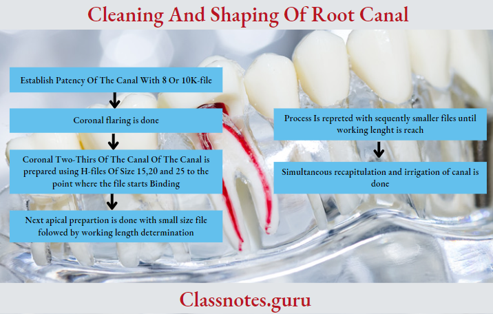
  • It involves the preparation of the coronal two-thirds of the canal first followed by a middle and apical third of the canal

Techniques For Root Canal Preparation Procedure:

Cleaning And Shaping Of Root Canal

Techniques For Root Canal Preparation Advantages

  • Shaping is easier
  • Elimination of the bulk of the tissue, debris, and micro-organisms from the the coronal and middle third before apical shaping.
  • Minimizes debris extrusion
  • Better access and control over apical enlarging instruments
  • Better penetration of irrigants

Question 2. Enumerate various techniques of canal preparation. Write in detail about the step-back technique.
Answer.

Obturation Of Root Canal

Techniques For Root Canal Preparation:

  • Step-back technique:
    • Conventional step-back
    • Passive step-back
  • Crown down or step down technique and its modifications
    • Crown down pressure less
    • Double flare
    • Balanced force
  • Hybrid technique

Techniques For Root Canal Preparation Step Back Technique:

  • It involves the preparation of the coronal two-thirds of the canal first followed by a middle and apical third of the canal

Techniques For Root Canal Preparation Step Back Technique Stage 1:

Endodontics Cleaning And Shaping Of Root Canal Step Back Technique Stage

Techniques For Root Canal Preparation Step Back Technique Stage 2:

Endodontics Cleaning And Shaping Of Root Canal Step Back Technique Stage 2

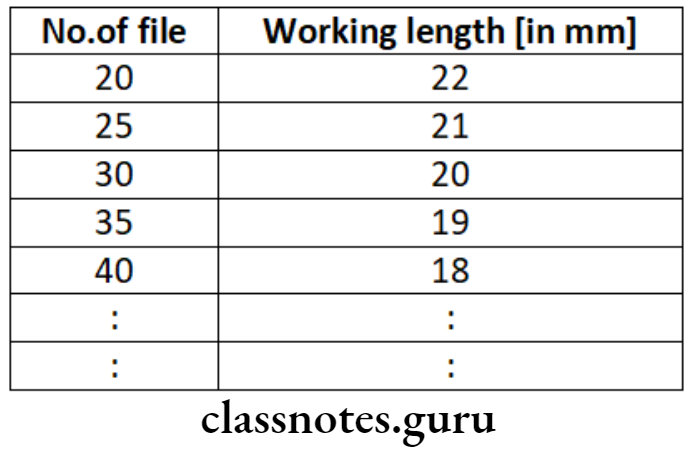
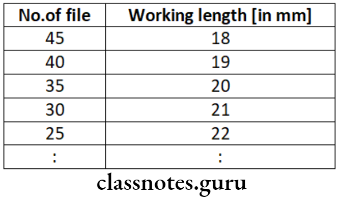
Example: For working length 22mm and the first file used is No-20

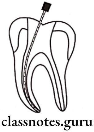
Endodontics Cleaning And Shaping Of Root Canal Length 22 mm and first file

Root Canal Preparation Techniques

Techniques For Root Canal Preparation Step Back Technique Advantage:

  • More flare at the coronal part
  • Popular technique
  • Ability to prepare a proper apical stop before preparation of the middle third and coronal third of the root canal

Techniques For Root Canal Preparation Step Back Technique Disadvantages:

  • Difficult to irrigate
  • Chances of pushing debris periodically
  • Time-consuming
  • Iatrogenic errors

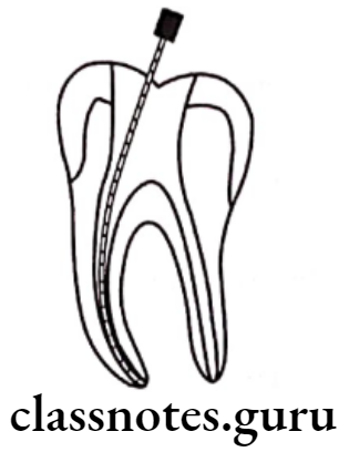
Endodontics Cleaning And Shaping Of Root Canal File at working length

Endodontics Cleaning And Shaping Of Root Canal File 2 mm short of working length

Cleaning And Shaping Of Root Canal Short Essays

Question 1. Crown Down Technique.
Answer.

Crown Down Technique

It involves the preparation of the coronal two-thirds of the canal first followed by a middle and apical third of the canal.

Crown Down Technique Advantages

  • Shaping is easier
  • Elimination of the bulk of the tissue, debris, and micro-organisms from the coronal and middle third before apical shaping
  • Minimizes debris extrusion
  • Better access and control over apical enlarging instruments
  • Better penetration of irrigants
    • Enhances tactile sensation
    • Coronal flaring
    • Removes debris coronally
    • Straight line access
    • Decreased frequency of blockage

Obturation Of Root Canal

Endodontics Cleaning And Shaping Of Root Canal Length 45 mm and first file

Root Canal Instrumentation Methods

Question 2. Balanced force technique.
Answer.

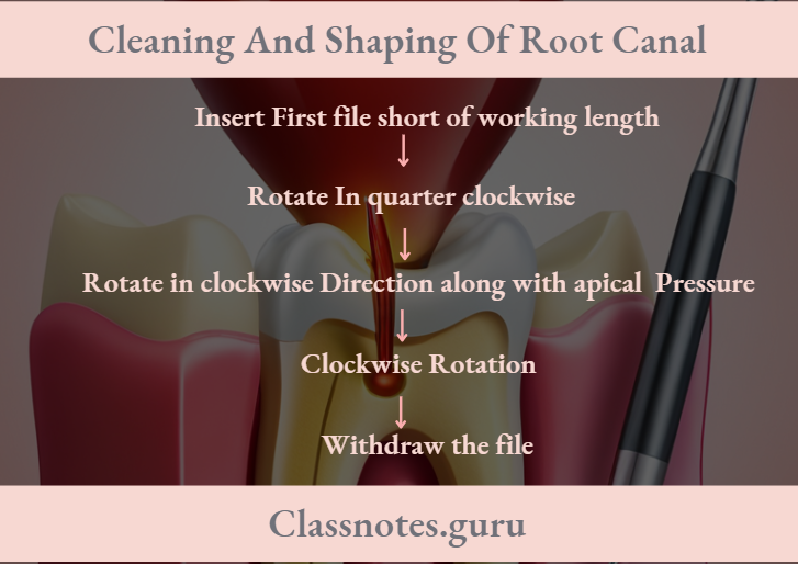
Balanced Force Technique Steps:

Endodontics Cleaning And Shaping Of Root Canal Balanced force technique

Balanced Force Technique  Uses:

  • Engage a small amount of dentin
  • Shear off dentin
  • Loosen the debris

Balanced Force Technique  Advantages:

  • Lesser canal transportation
  • Can manipulate files at any point
  • File cutting at the apical end

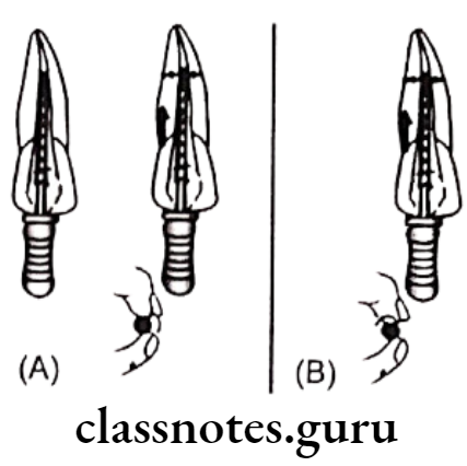
Endodontics Cleaning And Shaping Of Root Canal Engaging dentin with quater clockwise turn cutting action

Endodontics Cleaning And Shaping Of Root Canal Now file is turned quarter clockwise

Endodontic Shaping And Cleaning

Cleaning And Shaping Of Root Canal Viva Voce

  • Recapitulation is returning to a small instrument from time to time before advancing to a larger size
  • Recapitulation prevents the packing of dentin filling and ensures patency of the root canal through the apical foramen
  • Biomechanical preparation should provide smooth, funnel-shaped tapered walls for obturation
  • Step back technique is beginning the preparation at the apex and working back up to the canal coronally with larger and larger instruments
  • Step down technique begins coronally and the preparation is advanced apically using smaller and smaller instruments

Cephalometrics Question And Answers

Cephalometrics Important Notes

  • Landmarks

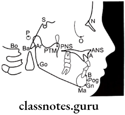
Orthodontics Cephalometrics Land Marks 1

Orthodontics Cephalometrics Land Marks 2

  • Planes

Orthodontics Cephalometrics Planes

  • Angle of convexity

Orthodontics Cephalometrics Angle of convexity 1

  • Angles

Orthodontics Cephalometrics Angles

  • Wit’s analysis
    • It is a measure of the extent to which the maxilla and mandible are related to each other in antero-posterior direction
    • It is used when the ANB angle is not reliable
  • Different analysis

Orthodontics Cephalometrics Different Analysis

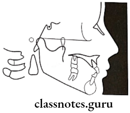
  • Tweed’s analysis
    • Forms a triangle by use of three planes

Orthodontics Cephalometrics Tweed's analysis

Read And Learn More: Orthodontics Short And Long Essay Question And Answers

Cephalometrics Long Essays

Question 1. What are diagnostic aids? Describe cephalometrics and its uses.
Answer.

Diagnostic Aids

Comprehensive orthodontic diagnosis is established by the use of clinical implements called diagnostic aids

Cephalometrics:

  • Introduced by Broadbent in USA and Hofrath in Germany in 1931
  • It describes the analysis and measurements made on the cephalometric radiographs

Types Of Cephalometrics:

  • Lateral cephalogram: Provides a lateral view of the skull
  • Frontal cephalogram: Provides an antero-posterior view of the skull

Technical Aspects:

  • Apparatus – X-ray source
    • Cephalostat – two ear rods
    • Orbital pointer
    • Forehead clamp

Landmarks:

  • Used for quantitative analysis and measurements

Types Of Landmarks:

  • Anatomic landmarks: Represent actual anatomic structures of the skull
  • Derived landmarks: Obtained from anatomic structures

Requirements Of Land Marks:

  • Easy to view
  • Reproducible
  • Provide valid measurements

Examples Of Landmarks:

  • Nasion
  • Orbitale
  • Porion
  • Sella

Lines And Planes:

  • Obtained by connecting two landmarks

Types Of Lines And Planes:

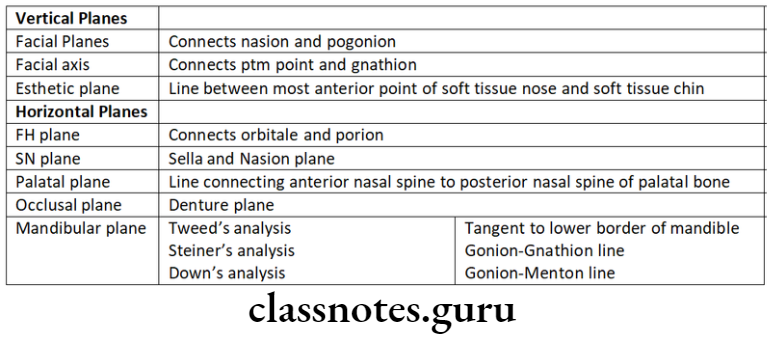
  • Horizontal Planes
    • Sella-Nasion Plane: Connecting sella and nasion
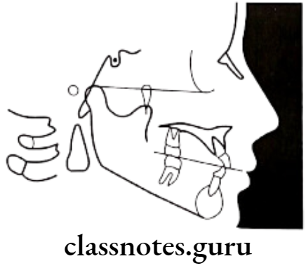
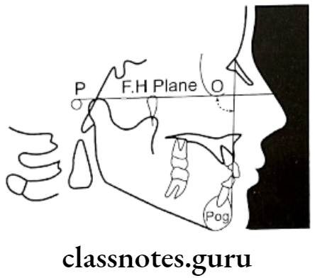
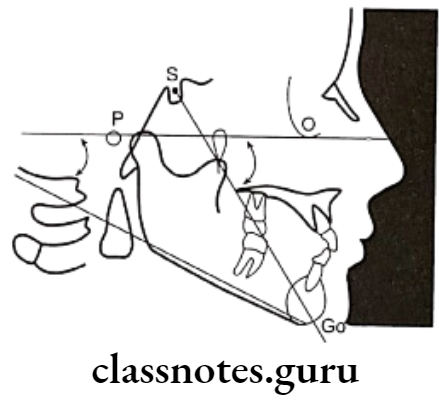
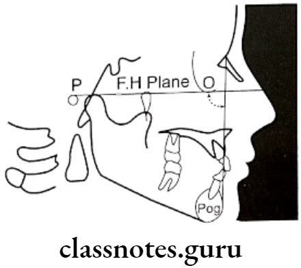
    • Frankfort Horizontal Plane: Connecting orbitale and porion
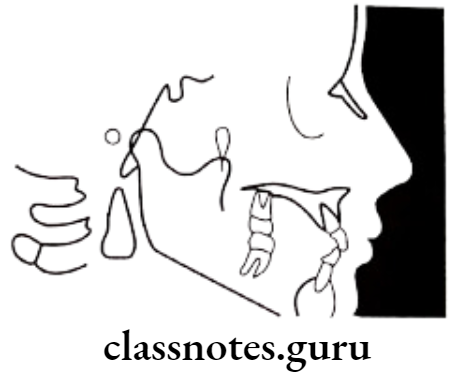
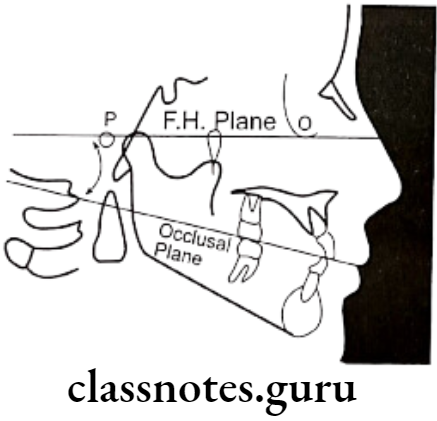
    • Occlusal Plane: Connecting occlusal surfaces of posterior to anteriors
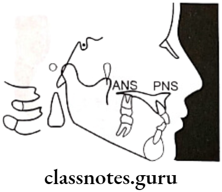
    • Palatal Plane: Connecting ANS and PNS
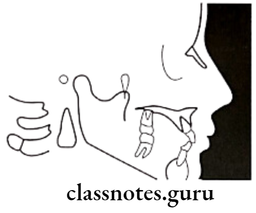
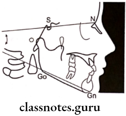
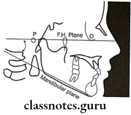
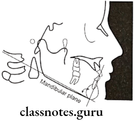
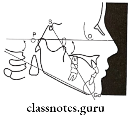
    • Mandibular Plane:
      • Tangent to lower border of mandible – Tweed
      • The line connecting gonion & gnathion – Steiner
      • The line connecting gonion & mention – Down’s
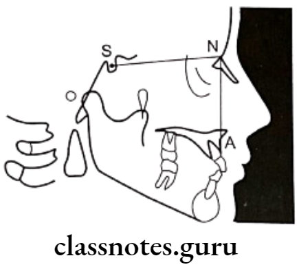
Orthodontics Cephalometrics Sella-Nasion plane

Orthodontics Cephalometrics Frankfort Horizontal plane

Orthodontics Cephalometrics Palatal plane

Orthodontics Cephalometrics Occlusal plane

Orthodontics Cephalometrics Mandibular plane

Orthodontics Cephalometrics Basion-Nasion plane

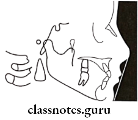
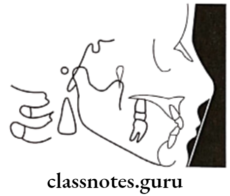
Orthodontics Cephalometrics Esthetic plane

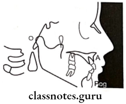
Orthodontics Cephalometrics A Pogonion plane

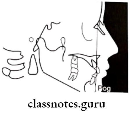
Orthodontics Cephalometrics Facial plane

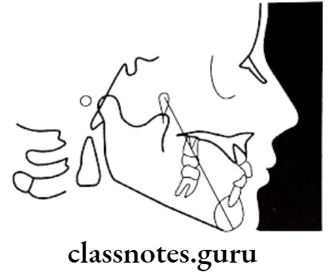
Orthodontics Cephalometrics Facial axis

  • Basion – Nasion Plane: Connecting Basion and Nasion
  • Bolton’s Plane: Connecting Bolton’s Point and Nation

Vertical Plane

  • A.Pog line: Point A to Pogonion
  • Facial Plane: Nasion to pogonion
  • Facial axis: Ptm point to gnathion
  • E.Plane: Soft tissue nose to the soft tissue of the chin

Cephalometrics Uses:

  • For diagnosis
  • Study of dental and soft tissue structures
  • Classify skeletal and dental abnormalities
  • Assess facial type
  • For treatment planning
  • For presuming the changes
  • For predicting growth-related changes
  • For research work

Orthodontics Cephalometrics Source mid sagittal plane distance of 5 feet

Orthodontics Cephalometrics Important lateral cephalometric landmarks

Orthodontics Cephalometrics Sella-Nasion plane

Orthodontics Cephalometrics Frankfort Horizontal plane

Orthodontics Cephalometrics Palatal plane

Orthodontics Cephalometrics Occlusal plane

Orthodontics Cephalometrics Mandibular plane

Orthodontics Cephalometrics Basion-Nasion plane

Orthodontics Cephalometrics Esthetic plane

Orthodontics Cephalometrics A Pogonion plane

Orthodontics Cephalometrics Facial plane

Orthodontics Cephalometrics Facial axis

Cephalometrics Short Essays

Question 1. Cephalogram.
Answer.

Cephalogram

  • Cephalogram is a cephalometric radiography introduced by Holly Broadbent and Herbert Hofrath in 1931

Types Of Cephalogram:

  • Lateral cephalogram
    • This provides a lateral view of the skull
  • Frontal cephalogram
    • Provides an antero-posterior view of skull

Cephalogram Uses:

  • For diagnosis
  • Study of dental and soft tissue structures
  • Classify skeletal and dental abnormalities
  • Assess facial type
  • For treatment planning
  • For presuming the changes
  • For predicting growth-related changes
  • For research work

Cephalogram Limitations:

  • Exposure to a patient is harmful
  • Provides two three-dimensional views of three-dimensional structures
  • Not well standardized
  • Absence of anatomic references
  • Has limited relevance

Question 2. Uses of Cephalometrics
Answer.

Uses of Cephalometrics

  • For diagnosis
  • Study of dental and soft tissue structures
  • Classify skeletal and dental abnormalities
  • Assess facial type
  • For treatment planning
  • For presuming the changes
  • For predicting growth-related changes
  • For research work

Question 3. Cephalostat.
Answer.

Cephalostat

  • It is head holding device

Orthodontics Cephalometrics Cephalostat

  • Position: The distance between the X-ray source and the mid-sagittal plane of the patient is 5 feet
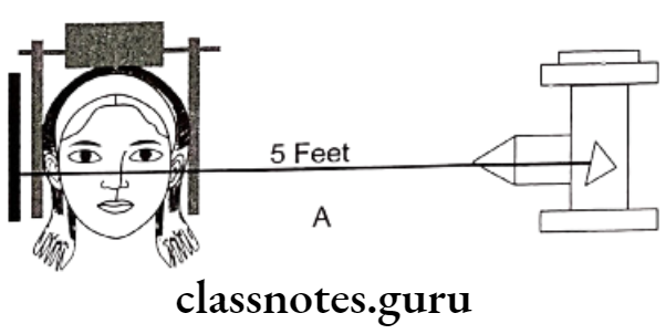
Orthodontics Cephalometrics Source mid sagittal plane distance of 5 feet

Uses Of Cephalostat:

  • For standardizing radiography
  • Comparing serial radiographs

Question 4. Steiner Analysis.
Answer.

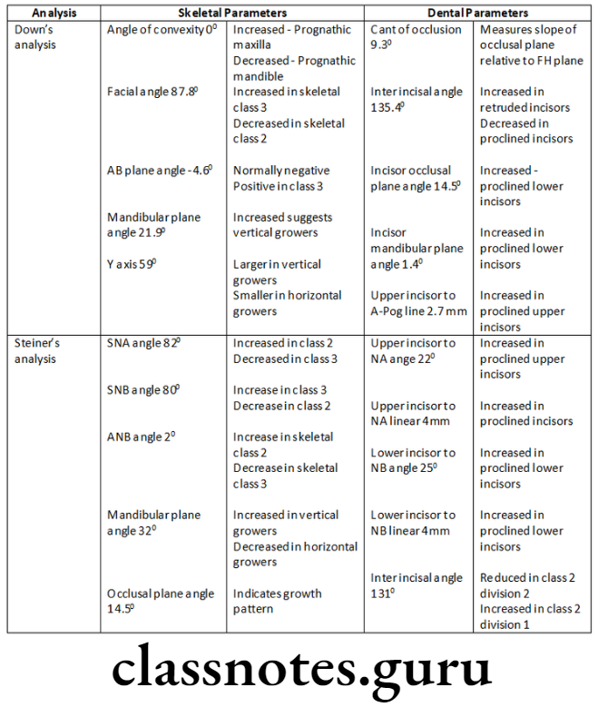
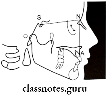
Parameter:

Skeletal

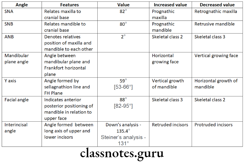
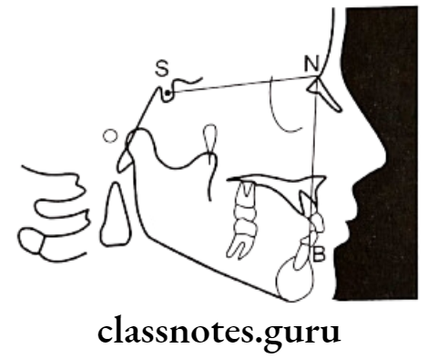
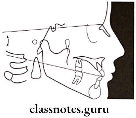
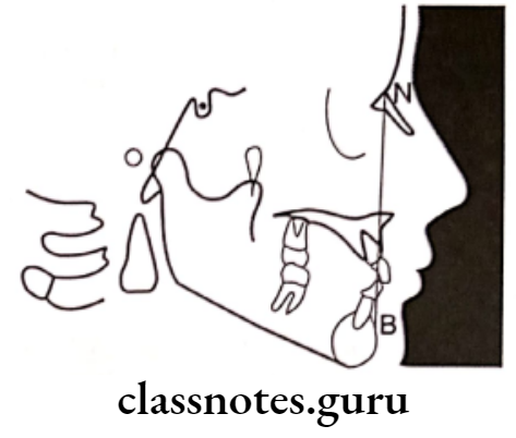
  • SNA angle:
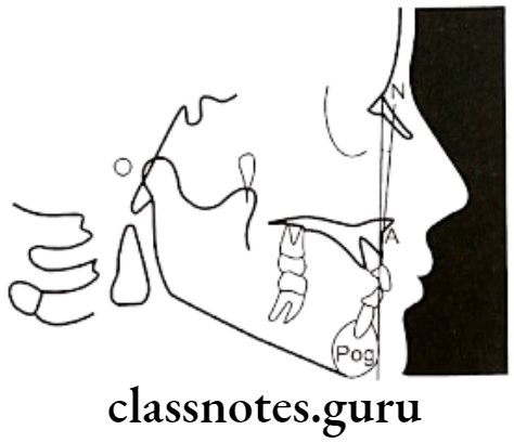
    • SN plane and line joining nasion and point A – 82°
    • Increased – Class 2; Decreased – Class 3
  • SNB angle:
    • Between SN plane and the line joining nasion and point B – 80°
    • Increased – Class 3; Decreased – Class 2
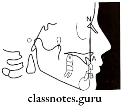
  • ANB angle:
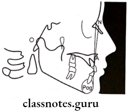
    • Between the line joining Maison and point A and Nation and point B – 2°
    • Increased – Skeletal Class 2; Decreased – Skeletal Class 3
  • Mandibular Plane angle:
    • Between SN plane and mandibular plane – 32°
    • Low angle – Horizontal grower; High angle – vertical grower
  • Occlusal plane angle:
    • Between occlusal plane and SN plane – 14.5°
    • Indicates growth pattern of individual

Dental:

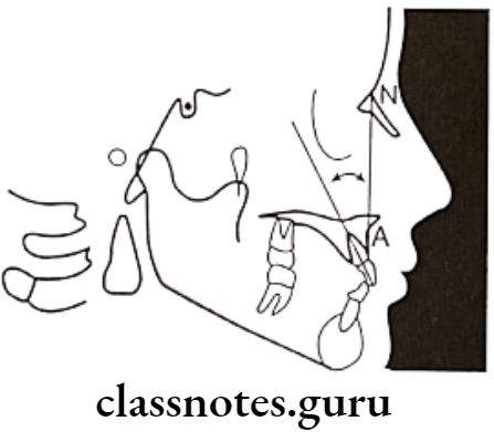
  • Upper Incisor to N-A:
    • Between the long axis of the upper incisor and nasion to point A – 22°
    • Increased – proclination of upper incisors
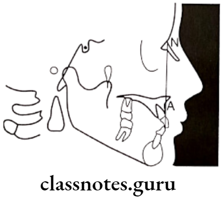
  • Upper Incisor to N-A:
    • Linear measurement between labial surface of upper incisor and line joining nasion to point A – 4mm
  • Lower Incisor to N-B:
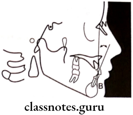
    • Between long axis of lower incisor and nasion to point B – 25°
    • Increased – Proclined lower incisor
  • Lower Incisor to N-B:
    • The linear distance between the labial surface of the lower central incisor and the line joining nasion to point B – 4mm
    • 131°
  • Inter-incisor angle:
    • Between the long axis of the upper and lower central incisor
    • Increased – Class 2 div 2, Decreased – Class 2 div 1, Class 1

Soft Tissue analysis

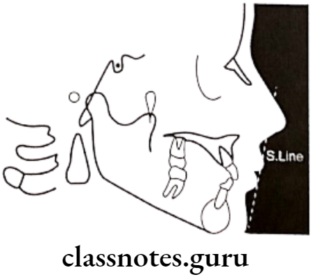
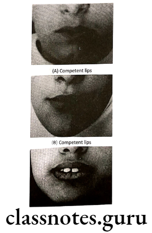
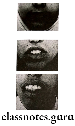
According to Steiner the lips in a well-balanced face should touch a line extending from the soft tissue of the chin to the middle of the ‘S’ formed by the lower border of the nose

  • Protrusive – Lips beyond this line – Convex profile
  • Retrusive – Lips behind this line – Concave profile
Orthodontics Cephalometrics SNA angle

Orthodontics Cephalometrics SNB angle

Orthodontics Cephalometrics ANB angle

Orthodontics Cephalometrics Mandibular plane angle

Orthodontics Cephalometrics Occlusal plane angle

Orthodontics Cephalometrics Upper incisor to NA angle

Orthodontics Cephalometrics Upper incisor to NA linear

Orthodontics Cephalometrics Lower incisor to NB

Orthodontics Cephalometrics Lower incisor to NB linear

Orthodontics Cephalometrics Inter-incisal angle

Orthodontics Cephalometrics S line

Question 5. Down’s Analysis.
Answer.

Down’s Analysis
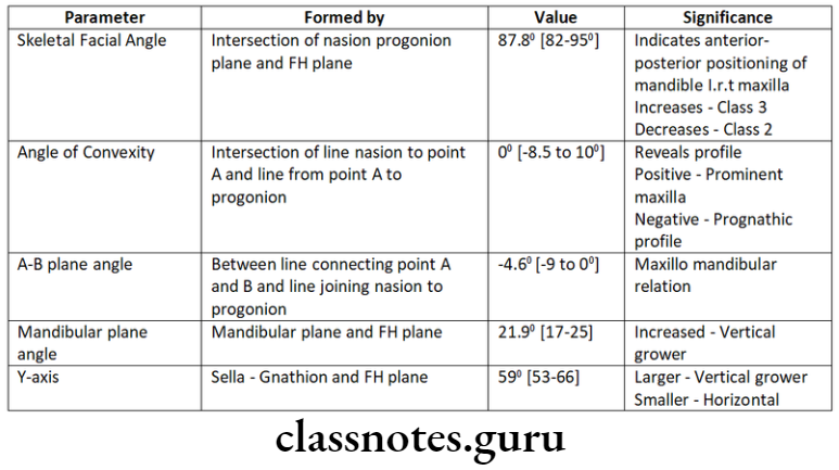
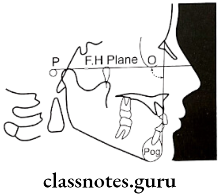
Orthodontics Cephalometrics Down's analysis

Dental

Orthodontics Cephalometrics Dental

Orthodontics Cephalometrics Facial angle

Orthodontics Cephalometrics Angle of convexity

Orthodontics Cephalometrics A-B plane angle

Orthodontics Cephalometrics Mandibular plane angle 1

Orthodontics Cephalometrics Y-axis

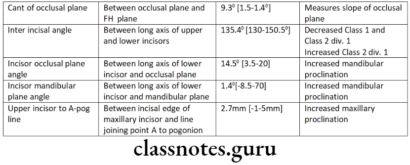
Orthodontics Cephalometrics Cant of occlusal plane

Orthodontics Cephalometrics Inter-incisal angle

Orthodontics Cephalometrics Incisor occlusal plane angle

Orthodontics Cephalometrics Incisor mandibular plane angle

Orthodontics Cephalometrics Upper incisor to A - Pog line

Question 6. Visual treatment objective (VTO).
Answer.

Types Of Visual Treatment Objectives

Clinical VTO:

  • It is an aid to decide the type of appliance in skeletal class 2 malocclusion
  • The procedure consists of asking the patient to bring the mandible to an edge-to-edge bite relation and note the changes in appearance of the patient at two levels
    • Edge-to-edge relation
    • Midway between existing occlusion and edge-to-edge position
  • Interpretation:
  • If the profile worsens at the edge-to-edge position, the fault lies in the maxilla
  • If the profile improves at the edge-to-edge position, the fault lies in the mandible
  • If the profile improves at the midway position, there is a fault in both the maxilla and mandible

Cephalometric VTO:

  • It is like a visual plan to predict the normal growth of a patient and anticipate the effects of the treatment for an individual patient
  • It permits the development of alternative treatment plans and to set goals in advance for the treatment

Question 7. Tweed’s triangle.
Answer.

Tweed’s triangle

Orthodontics Cephalometrics Tweed's analysis

Question 8. Cephalometrics uses, advantages, and disadvantages.
Answer.

Uses Of Cephalometrics:

  • For diagnosis
  • Study of dental and soft tissue structures
  • Classify skeletal and dental abnormalities
  • Assess facial type
  • For treatment planning
  • For presuming the changes
  • For predicting growth-related changes
  • For research work

Advantages Of Cephalometrics:

  • Tangible records that are relatively permanent
  • Nondestructive and noninvasive
  • Easy to store, transport, and reproduce
  • Yield accurate information on the spatial relationship between surface and deep structures
  • Low physiological cost

Disadvantages Of Cephalometrics:

  • It gives a two-dimensional view of a three-dimensional object
  • There can be errors while developing which can limit measuring accuracy to 0.5mm
  • Needs a special X-ray apparatus with precise calibration
  • Needs complex patient positioning arrays to avoid errors

Cephalometrics Short Questions And Answers

Question 1. Frankfort plane.
Answer.

Frankfort plane

Connects lowest point of orbit and superior border of external auditory meatus i.e. orbitale to portion

Significance Of Frankfort plane: Important in Down’s analysis

  • Used in both dental and skeletal parameters

Question 2. Cephalostat.
Answer.

Cephalostat

It is head holding device

Orthodontics Cephalometrics Cephalostat

Position: The distance between the X-ray source the and mid-sagittal plane of the patient is 5 feet

Question 3. SNB angle.
Answer.

SNB angle

Used in Steiner’s Analysis

Angle: Between SN plane and the line joining nasion and point B

Importance: Indicates relative antero-posterior positioning of the mandible about the cranial base

Value: 80°

Changes:

  • Increased – Prognathic mandible [Class 3]
  • Decreased – Retrognathic mandible [Class 2]

Question 4. Registration Point.
Answer.

Registration Point

  • It is derived from landmarks obtained on cephalogram
  • It is the midpoint of the perpendicular drawn from the center of Sella Tursica to the Bolton plane

Question 5. Mandibular Plane.
Answer.

Mandibular Plane

Based on different analysis. There are several mandibular planes

Common are:

  • Tangent to lower border of mandible – Tweed
  • Line connecting gonion and gnathion – Steiner
  • The line connecting Gonion and mention – Down’s

Question 6. SNA angle.
Answer.

SNA angle

Used in Steiner’s Analysis

Angle: Between SN plane and the line joining nasion and point A

Importance: Indicates relative antero-posterior positioning of the maxilla about the cranial base

Value: 82°

Changes:

  • Increased – Prognathic maxilla [Class 2]
  • Decreased – Retrognathic maxilla [Class 3]

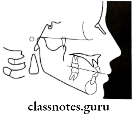
Question 7. Occlusal Plane.
Answer.

Occlusal Plane

It is a denture plane

Extend: Occlusion of posterior to interiors

Significance Of Occlusal Plane:

  • The angle formed between the occlusal plane and the SN plane is known as the occlusal plane angle
  • It indicates the relation of the occlusal plane to the cranium and face
  • It has a mean value of 14.5 degrees

Question 8. Cant of Occlusion.
Answer.

Cant of Occlusion

  • Used in Down’s analysis
  • The angle between the occlusal plane and the FH plane
  • Value: 9.3°
  • Range: 1.5 – 14°
  • It measures the slope of the occlusal plane relative to the FH plane
Orthodontics Cephalometrics Occlusal plane

Orthodontics Cephalometrics Basion-Nasion plane

Question 9. ANB angle.
Answer.

ANB angle

Used in Steiner’s analysis

Angle: Between line joining nasion to point A and nasion to point B

Significance Of ANB angle: Relative position of maxilla to the mandible

Value:

Changes:

  • Increase – Skeletal Class 2
  • Decrease – Skeletal Class 3

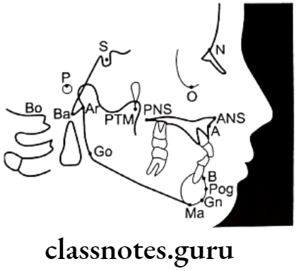
Question 10. Point A.
Answer.

Point A

It is an anatomic landmark of cephalometric

  • The deepest point in the midline between the anterior nasal spine and alveolar crest between two central incisors
  • Synonym: Subspinale
  • Line/Plane derived from it:
    • A.Pog line
    • A.B plane angle – Used in Down’s Analysis
    • The angle of convexity – Used in Down’s Analysis
    • SNA angle – Used in Steiner’s Analysis
    • ANB angle – Used in Steiner’s Analysis
Orthodontics Cephalometrics Important lateral cephalometric landmarks

Question 11. Key Ridge.
Answer.

Key Ridge

  • It is an anatomic landmark of cephalometric
  • It is the lowermost important point on the contour of the anterior wall of the infratemporal fossa
  • In edentulous cases, it is useful in determining occlusion

Question 12. Y axis.
Answer.

Y-axis

Used in Down’s analysis

Synonym: Growth axis

Angle – Between sella – gnathion line and FH plane

Value: 59° [53-66°]

Variations Of the Y axis:

  • Increased – Class 2
  • Decreased – Class 3

Importance Of Y axis:

  • Determines growth pattern
  • Greater – Vertical growth
  • Lesser – Horizontal growth

Orthodontics Cephalometrics Y-axis

Question 13. Wit’s Appraisal.
Answer.

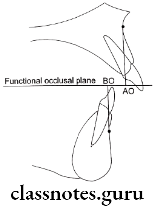
Significance Of Wits’s Apprasial: Measures relation of maxilla to mandible

Use: In nonreliable ANB cases

Obtained by:
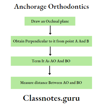
Orthodontics Cephalometrics Wit's Appraisal

Variation Of Wit’s Appraisal:

  • Males – BO ahead of AO
  • Females – BO coincides with AO
  • Skeletal Class 2 – BO behind AO
  • Skeletal Class 3 – BO ahead of AO

Orthodontics Cephalometrics Wits apprisal

Question 14. FMA.
Answer.

Cephalometric FMA:

  • FM – plane – extending from orbitale to prior
  • Mandibular plane
  • The angle formed between these two
  • Value – 25°

Orthodontics Cephalometrics Tweed Analysis

Clinical FMA:

  • The angle formed between the lower border of the mandate ble and Frankfort horizontal plane
  • Normally intersect at the occipital region

Significance Of FMA:

  • Indicates growth pattern
  • If coincides
    • At occipital – Normal
    • Beyond occipital – Horizontal
    • Anterior to occipital – Vertical

Orthodontics Cephalometrics Tweed Analysis

Orthodontics Cephalometrics Assessment of the lips 2

Question 15. Cephalogram
Answer.

Cephalogram

  • Cephalogram is a cephalometric radiography introduced by Holly Broadbent and Herbert Hofrath in 1931

Types Of Cephalogram:

  • Lateral cephalogram – provides a lateral view of the skull
  • Frontal cephalogram – provides an anteroposterior view of the skull

Cephalogram Uses:

For diagnosis:

  • Study of dental and soft tissue structures
  • Classify skeletal and dental abnormalities
  • Assess facial type
  • For treatment planning
  • For presuming the changes
  • For predicting growth-related changes
  • For research work

Question 16. Tweed’s triangle.
Answer.

Formed by:

  • Frankfort mandibular plane angle [FMPA] – 25°
    • Angle by the intersection of the FH plane and mandibular plane
  • Incisor mandibular plane angle [IMPA] – 90°
    • The angle formed by the intersection of the long axis of lower incisor and mandibular plane
  • Frankfort mandibular incisor angle [FMIA] – 65°
    • The angle formed by the long axis of lower incisor and FH plane
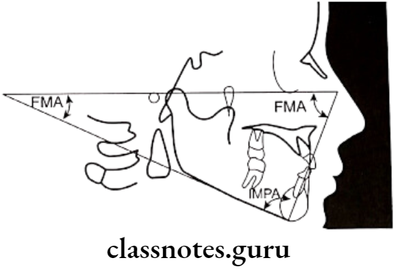
Orthodontics Cephalometrics Tweed Analysis

Cephalometrics Viva Voce

  • Y axis indicates the direction of the growth pattern of the mandible
  • Richard A. Riedel introduced the ANB angle
  • Cephalometric tracing is made on the frosted surface of an acetate tracing sheet
  • E plane is also called the aesthetic plane
  • Normal facial angle – 82-95°
  • The normal value of the Y axis in Down’s analysis – 53-66°
  • The normal value of cant of occlusion – 1.5 – 14°
  • Normal value of interincisal angle – 130-150°
  • The normal mandibular plane angle in Steiner’s analysis is 29°
  • Decreased SNB angle indicates mandibular retrognathism
  • Decreased ANB angle indicates skeletal Class 3
  • The decreased facial angle indicates skeletal class 2
  • A decreased angle of convexity indicates the prognathic profile
  • Increased AB plane angle indicates class 3
  • Increased upper incisor to NA angle indicates proclined upper incisors
  • Increased lower incisor to NB angle indicates proclined lower incisors
  • The facial angle indicates antero-posterior positioning of the mandible to the upper face
  • AB plane angle signifies the maxilla-mandibular relationship to the facial plane’
Orthodontics Lab Procedures Question And Answers

Orthodontics Lab Procedures Definitions

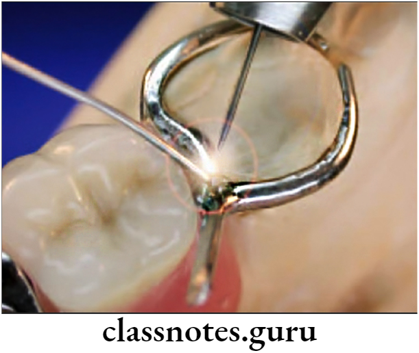
  • Soldering
    • It is a process of joining metals by the use of a filler metal that has a lower fusion temperature than that of the metals being joined.
  • Welding
    • Involves the joining of two metals without introducing a third metal.

The Records Through The Reviewing Process

Orthodontics Lab Procedures Important Notes

  • Types of solders
    • Investment soldering
    • Freehand soldering
  • Composition of flux
    • Borax glass – 55%
    • Boric acid – 35%
    • Silica – 10%
  • Gases used for soldering.
    • Gas air/oxygen torch flame
    • Hydrogen
    • Natural gas
    • Acetylene
    • Propane

Orthodontics Lab Procedures Short Essays

Question 1. Soldering and Welding.
Answer.

Soldering: It is a process of joining metals by the use of a filler metal that has a lower fusion temperature than that of the metals being joined.

Types of soldering:

Investment soldering:

Indication: Presence of large contact area between metals

  • In need of precision
    Procedure: By embedding metals in investment

Freehand soldering:

  • Indication: Common orthodontic procedures
  • Procedures: Soldering by holding metals precisely

Steps of freehand soldering:

  • Clean the surfaces
  • Assemble the parts
  • Select proper solder and flux
  • Select proper joint
  • Flux application
  • Application of solder
  • Quenching

Read And Learn More: Orthodontics Short And Long Essay Question And Answers

Indications of freehand soldering:

  • Join parts of appliances
  • Fastens attachments to bands


Orthodontics Lab Procedures Soldering In Procedures

Welding: Involves joining of two metals without introducing a third metal

Welding Procedure:

  • Selection of electrode
  • Placement of metals between 2 electrodes
  • Maintain pressure on it
  • Switch on electrode
  • Keep it for sometime

Indications of welding:

  • For banding
  • For brackets and molars tubes

Question 2. Shape Memory Alloy – Nickel titanium alloy is a shape memory alloy.
Answer.

Synonym: Nitinol (Nickel Titanium Naval Ordinance Laboratory)

Inventor: William R. Buchler

Introduced by: Andersen in 1971

Properties of shape memory alloy:

  • Super elasticity
  • Shape memory
  • High working range
  • Low stiffness

Advantages of shape memory alloy: Procedure low and more constant force on teeth

Disadvantages of shape memory alloy:

  • Cannot be bent
  • Cannot be soldered/welded

Orthodontics Lab Procedures Short Questions And Answers

Question 1. Solder and Flux.
Answer.

Solder: Alloys used as filler metal between two metals

Properties of solder:

  • Resist tarnish and corrosion
  • Low fusion temperature
  • Free-flowing
  • Similar strength as the metals to be joined
  • Colour similar to metals to be joined
  • Ex – Gold, silver, copper, zinc, tin and nickel

Flux: means flowing

  • Important for good solder joint

Uses of flux:

  • Removal of oxide coating of solder
  • Dissolves impurities
  • Prevents oxidation
  • Reduces melting point

Flux Composition:

  • Borax glass – 55%
  • Boric acid – 35%
  • Silica – 10%

Question 2. Antiflux.
Answer.

Flux:

  • Means flowing
  • Important for good solder joint

Uses of Flux:

  • Removal of oxide coating of solder
  • Dissolves impurities
  • Prevents oxidation
  • Reduces melting point

 Composition of Flux:

  • Borax glass – 55%
  • Boric acid – 35%
  • Silica – 10%

Antiflux:

  • Confines flow of molten solder
  • Ex. Lead pencil markings
    • Graphite lines
    • Iron rouge

Question 3. Gases used for soldering.
Answer.

  • Gas air/oxygen torch flame
  • Hydrogen
  • Natural gas
  • Acetylene
  • Propane

Question 4. Sensitization and stabilization of stainless steel.
Answer.

Sensitization: At 400-900 c, stainless steel resists corrosion and tarnish

  • At lower temperatures, carbon reacts with the surface granules and forms chromium iron carbide causing tarnish and corrosion
  • This can be prevented by decreasing carbon content or by cold working of stainless steel

Stabilization:

  • The process to prevent sensitization
  • For it, two/more metals are added, for example, titanium, niobium

Question 5. Irreversible Hydrocolloids (Alginate).
Answer.

Advantages of Irreversible Hydrocolloids:

  • Easy to manipulate
  • Comfortable
  • Inexpensive
  • Pleasant taste
  • Hydrophilic
  • Biocompatible

Disadvantages of Irreversible Hydrocolloids:

  • Tears easily, and require immediate pouring
  • Incompatible with epoxy resin

Composition of Irreversible Hydrocollids:

  • Potassium Alginate
  • Calcium sulfate dehydrate
  • Potassium sulfate
  • Diatomaceous earth
  • Sodium phosphate
  • Glycol
  • Pigments
  • Sodium silicofluoride

Orthodontics Lab Procedures Viva Voce

  • Gold foil filling is the best example of cold welding
  • Welding is a process of joining two metals directly under pressure
  • The space provided between the metals to be joined is 0.5 mm
  • Borax glass is the main component of dental flux
  • The oxidized layer in the metallic surfaces to be joined is removed by antireflux
  • Nickel in dental solder gives a white color to the solder
  • The copper component in solder gives the yellow color
  • Fusion temperature exceeding 150 degrees is used for brazing

Surgical Orthodontics Question And Answers

Surgical Orthodontics Important Notes

Surgical Orthodontics

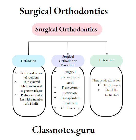
  • Pericision/circumferential suprarenal fibrotomy
    • Performed in case of rotations
    • In it, gingival fibres are incised to prevent relapse
    • Performed under LA with several 11-knife

Surgical Orthodontics Long Essays

Question 1. Classify surgical orthodontics. Discuss minor surgical orthodontic procedures.
Answer.

Surgical Orthodontic Procedure

Minor Procedure:

  • Extractions
    • Therapeutic
    • Serial
    • Carious teeth
    • Malformed teeth
    • Impacted teeth
    • Supernumerary teeth
  • Surgical uncovering of teeth
  • Frenectomy
  • Pericision
  • Transplantation of teeth
  • Corticotomy

Major procedures:

  • Orthodontic surgeries
  • Cosmetic surgeries
  • Surgical corrections of clefts
  • Surgical RME

Minor Surgical Procedures:

Extraction:

  • Therapeutic extraction
    • To gain space
    • Should be atraumatic
  • Surgical extraction
    • Removal of some deciduous teeth followed by specific permanent
    • Done during mixed dentition period
    • In severe arch-length deficient
  • Supernumerary teeth
    • It leads to malocclusion or development of cyst
    • Thus, needs to be extracted

Surgical Uncovering of Impacted Teeth:

  • Impacted teeth cause deflection of teeth
  • Common – Maxillary permanent canine
    • Cause – Arch length deficiency, Mucosal/bony barriers
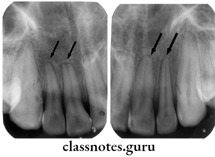
    • These barriers are removed to allow the eruption of impacted teeth

Orthodontics Surgical Orthodontics Periapical radiograph anterior view

Frenectomy:

  • Abnormal labial frenum causes mid-line diastema
  • Frenum is excised along with attached fibrous tissue
  • Undermining of the mucosa of the lip is done

Orthodontics Surgical Orthodontics Maxillary Labial Frectonomy

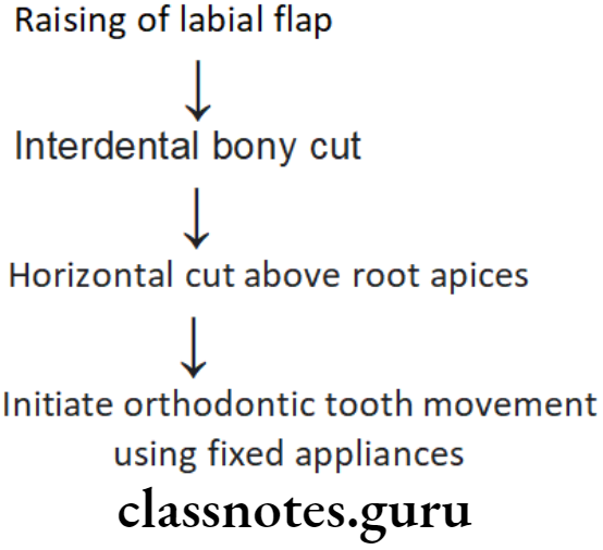
Corticotomy:

Indication of Corticotomy:

  • Proclination with spacing

The procedure of Corticotomy:

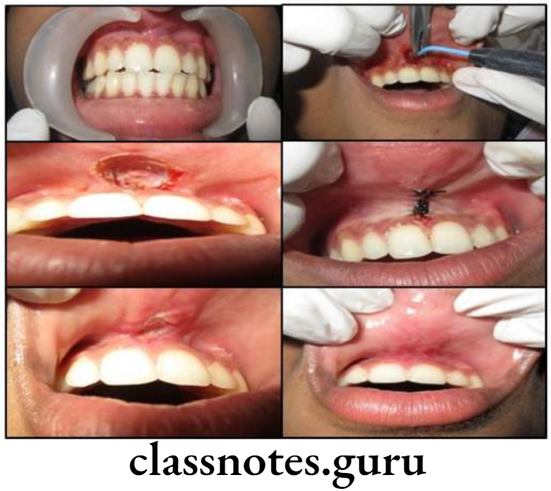
Orthodontics Surgical Orthodontics Corticotomy Procuedure

Precision:

Synonym – Circumferential Supra-crystal Fibrotomy [CSF]

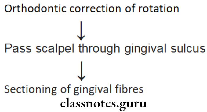
  • It is the procedure to counter the relapse tendency of stretched gingival fibres

Cause of Relapse: Failure of adaptation of transeptal and alveolar crystal group of gingival fibres to the new tooth position

The procedure of Relapse:

Orthodontics Surgical Orthodontics Pericision Procedure

Surgical Orthodontics Short Essays

Question 1. Genioplasty.
Answer.

  • Used as an adjunctive.

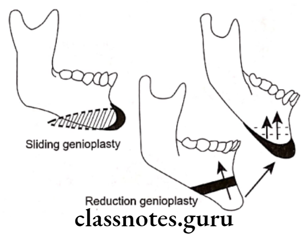
Types of Genioplasty:

Augmentation genioplasty:

  • Horizontal osteotomy cut given
  • Mobilize the segment and fix

Reduction genioplasty:

  • Horizontal osteotomy cut given
  • Setback fragment and fix it

Straightening genioplasty:

  • Horizontal osteotomy cut given
  • Shift it laterally and fix it

Read And Learn More: Orthodontics Short And Long Essay Question And Answers

Lengthening genioplasty:

  • Horizontal osteotomy cut given
  • Shift fragment inferiorly
  • Sandwich grafts in between them

Orthodontics Surgical Orthodontics Sliding and reduction genioplasty

Question 2. Orthognathic Surgery in Maxilla.
Answer.

  • Carried out along with orthodontic therapy to correct dentofacial deformities
  • Involves fracturing of facial skeletal parts and repositioning them as desired

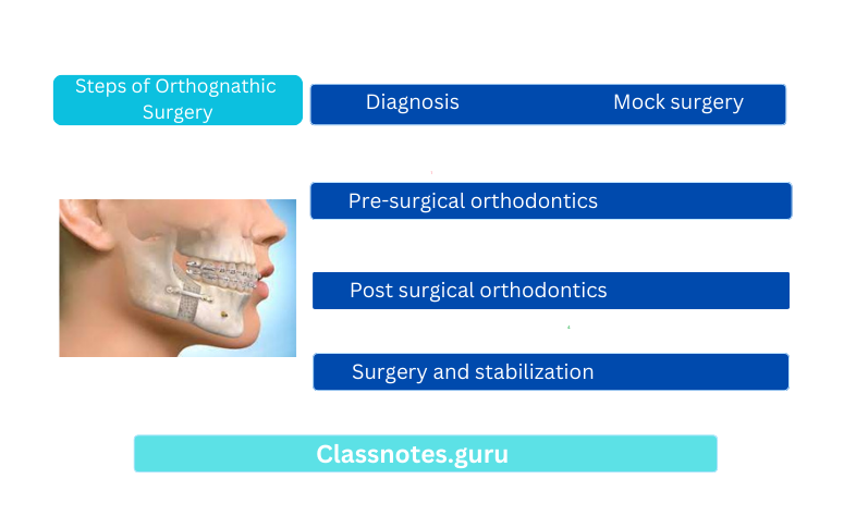
Steps of Orthognathic Surgery:

  • Diagnosis
  • Pre-surgical orthodontics
  • Mock surgery
  • Surgery and stabilization
  • Post-surgical orthodontics

Steps of Orthognathic Surgery

Orthodontics Surgical Orthodontics Orthognathic surgery

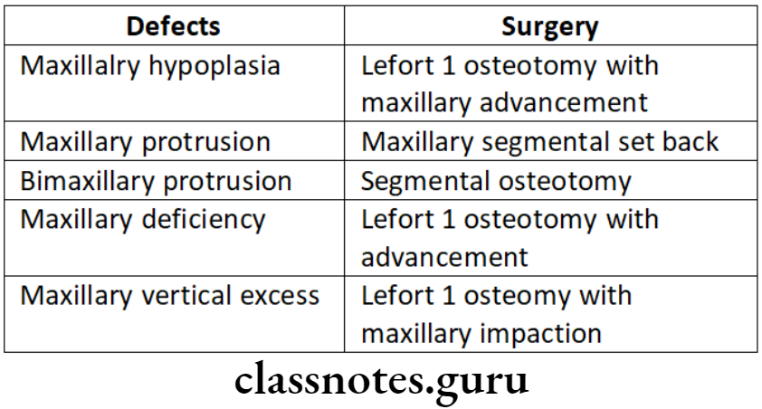
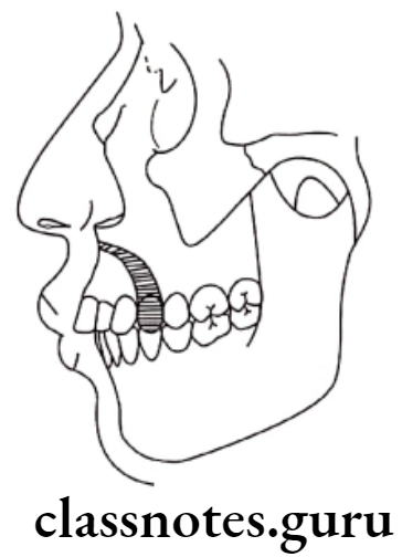
Orthodontics Surgical Orthodontics Maxillary protrusion

Orthodontics Surgical Orthodontics Class 3 skeletal pattern

Orthodontics Surgical Orthodontics Long face is usually associated with vertical maxillary

Orthodontics Surgical Orthodontics Bimaxillary protrusion require maxillary and mandibular segmental osteotomy

Surgical Orthodontics Short Questions And Answers

Question 1. Frenectomy.
Answer.

  • Abnormal labial frenum causes midline diastema
  • Frenum is excised along with attached fibrous tissue
  • Undermining of the mucosa of the lip is done

Question 2. Precision.
Answer.

  • Synonym – Circumferential Supra-crystal Fibrotomy [CSF]
    • It is the procedure to counter the relapse tendency of stretched gingival fibres
  • Cause of Relapse: Failure of adaptation of transeptal and alveolar crystal group of gingival fibres to the new tooth position

Procedure of Pericision:

Orthodontics Surgical Orthodontics Pericision Procedure

Surgical Orthodontics Viva Voce

  • Precision is done to prevent relapse of rotation of teeth.
  • Precision involves surgical lysis of the gingival.

Retention And Relapse In Orthodontics Question And Answers

Retention And Relapse Definition

  • Retention
    • Maintaining newly moved teeth in position, long enough to aid in stabilizing their correction.
  • Relapse
    • It is the loss of any correction achieved by orthodontic treatment.

Retention And Relapse

Retention And Relapse Important Notes

Types of retention:

  • Limited retention:
    • Class 1 non-extraction
    • Deep bites
    • Class 1, Class 2 Div. 1 and Div. 2 extraction
  • Natural retention:
    • Anterior cross bites
    • Serial extraction
    • Highly placed canines
    • Posterior crossbite
  • Permanent retention:
    • Midline diastemas
    • Rotation
    • Class 2 div.2 deep bites
    • Abnormal musculature
    • Cleft palate patients

Types of retainers:

  • Removable:
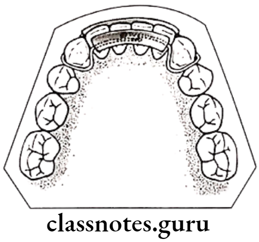
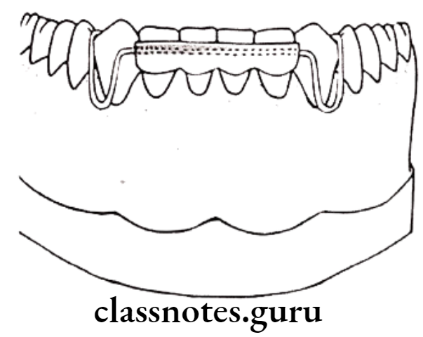
    • Hawely’s appliance
    • Begg retainers
    • Clip on retainers
    • Wrap-around
    • Kesling tooth position
    • Invisible retainers
  • Fixed:
    • Fixed appliance
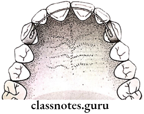
    • Banded canine to canine
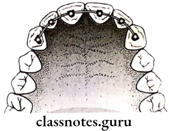
    • Bonded lingual
    • Band and spur retainer

Read And Learn More: Orthodontics Short And Long Essay Question And Answers

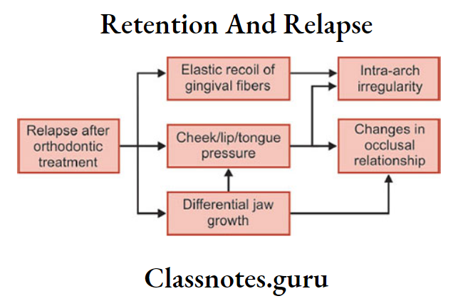
Causes of relapse:

  • Periodontal ligament traction
  • Growth related changes
  • Bone adaptation
  • Muscular forces
  • Failure to eliminate the original cause
  • Role of the third molar
  • Role of occlusion

Retention And Relapse Long Essays

Question 1. Enumerate theories of retention. Add a note on causes of relapse.
Answer.

Retention

Definition of retention: By Moyers

  • Maintaining newly moved teeth in position, long enough to aid in stabilizing their correction.

Theorems: 9 theorems by Riedel and 10th Moyers

  • Moved teeth return to their former position
  • Elimination of cause prevents relapse
  • Malocclusion should be over-corrected
  • Proper occlusion should be achieved
  • Bone and adjacent tissues should be provided time to readapt around moved teeth
  • Lower incisors must be placed upright
  • Corrections carried out during growth periods are less likely to relapse
  • Farther the teeth have been moved, the lesser the risk of relapse
  • Arch form, particularly mandibular, cannot be permanently altered by appliance therapy
  • Many treated malocclusions require permanent retaining devices

Types of retention

Limited retention:

  • Class 1 non-extraction
  • Deep bites
  • Class 1, Class 2 Div. 1 and Div. 2 extraction

Natural retention:

  • Anterior crossbite
  • Serial extraction
  • Highly placed canines
  • Posterior crossbite

Permanent retention:

  • Midline diastema
  • Rotation
  • Class 2 div.2 deep bites
  • Abnormal musculature
  • Cleft palate patients

Retainers: Passive appliances that help in maintaining and stabilizing the position of teeth long enough to permit readaptation of supporting structures

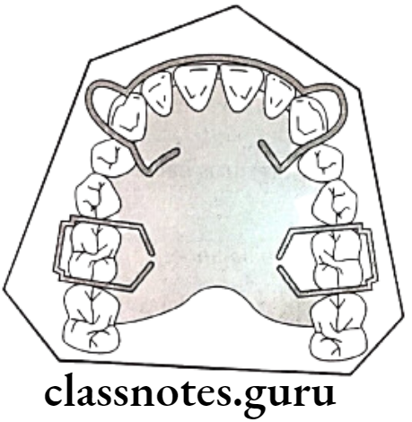
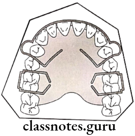
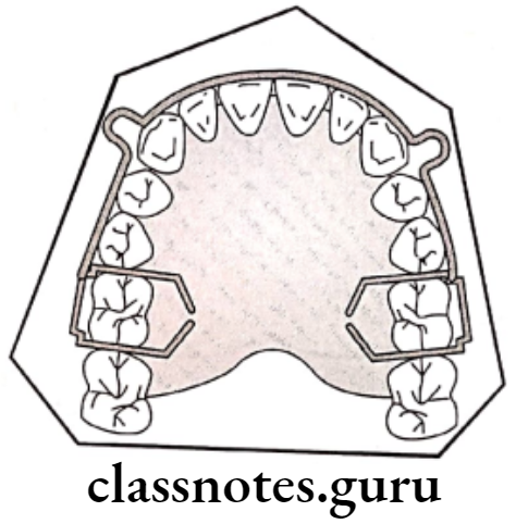
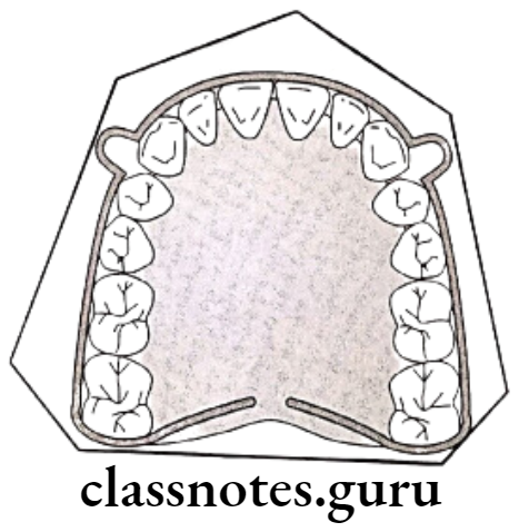
Orthodontics Retention And Relapse Hawley's retainer

Orthodontics Retention And Relapse Hawley's retainer with long labial bow

Orthodontics Retention And Relapse Hawley's retainer with labial bow

Orthodontics Retention And Relapse Begg wrap around retainer

Types of retainers

  • Removable retainers
    • Hawely’s appliance
    • Begg retainer
    • Clip on retainer
    • Wrap-around
    • Kesling tooth position
    • Invisible retainer

Orthodontics Retention And Relapse Clip on retainer labial

Orthodontics Retention And Relapse Lingual view

  • Fixed retainers
    • Fixed appliance
    • Banded canine to canine
    • Bonded lingual
    • Band and spur retainer

Orthodontics Retention And Relapse Banded canine to canine retainer

Orthodontics Retention And Relapse Bonded canine to canine retainer

Orthodontics Retention And Relapse Band and spur retainer

Relapse: It is the loss of any correction achieved by orthodontic treatment

Causes of Relapse:

  • Periodontal ligament traction:
    • Due to orthodontic tooth movement, PDL and gingival fibres are stretched
    • They readapt to the newly moved teeth
    • Different fibres require different periods for it
    • Principle fibres – 4 weeks
    • Gingival fibres – 40 weeks
    • If not provided sufficient time, leads to relapse
  • Growth-related changes:
    • Due to the continuation of abnormal growth pattern
  • Bone adaptation:
    • Normal, bony trabeculae are arranged perpendicular to the long axis of teeth
    • During orthodontic treatment, arranged parallel to the long axis of teeth
    • After treatment returns to original position
  • Muscular forces:
    • Abnormal muscle balance
  • Failure to eliminate the original cause:
    • The cause should be properly diagnosed and a treatment plan must be done accordingly
  • Role of the third molar:
    • Eruption time – 18-21 years i.e. usually after completion of orthodontic treatment
    • Produces pressure
    • Results in crowding recurrence
  • Role of occlusion:
    • Failure to eliminate habits like bruxism
    • Failure to achieve centric occlusion

Retention And Relapse Short Essays

Question 1. Define retention, and explain schools of retention.
Answer.

Retention: Maintaining newly moved teeth in position long enough to aid in stabilizing their correction – By Moyers

Schools Of Retention:

  • Occlusion School – By Kingsley
    • Proper occlusion is important as it safeguards the stability in the new position of patients
  • Apical base school:
    • By Alex Lundstorm, Mc.Cauley and Nance
      • By Alex Lundstorm – Suggest apical base as an important factor in the correction of malocclusions
      • Mc. Cauley – Added inter-canine and intermolar width should be maintained
      • Nance – Noted arch length cannot be permanently increased to a major extent.
  • Mandibular Incisor School – Grieves and Tweed
    • Stability increases if mandibular incisors are placed upright
  • Musculature School – Rojer
    • Functional muscle is necessary for post-treatment stability.

Question 2. Permanent retention.
Answer.

Conditions of Permanent Retention:

  • Midline diastema
  • Severe rotations
  • Generalized spacing
  • Patients with abnormal musculature
  • Arch expansion
  • Cleft palate patients
  • Class 2 div.2 deep bite

Appliances Used:

Retainers: Passive appliances that help in maintaining and stabilizing the position of teeth long enough to permit readaptation of supporting structures

Types of Retainers:

Removable Retainers

Removed and reinserted at patient’s will

  • Hawely’s Appliance – By Charles Hawley
    • Consists of – short labial bow, Adam’s clasps on molars

        Modifications of Hawely’s appliance:

    • Long labial bow – for space closure distal to the canine
    • Fitted labial bow – Excellent retention
    • Soldered labial bow
    • Anterior bite plane – for deep bite

       Advantages of Hawley’s appliance:

    • Simple and easy to fabricate
    • Comfortable to patient
    • Acceptable

Begg retainer – By P.R. Begg

Consists Of Begg retainer:

  • Labial wire till last erupted molar, curves around it
  • Spans palate – Acrylic portion

Advantage of Begg retainer: Eliminate risk of spacing between canine and premolar

Clip on retainer:

  • Labial wire covering incisors and canine then wrapped around same teeth lingually

Wrap around retainer:

  • Wire wrapped around all erupted teeth labially as well as lingually

Keeping tooth position – By H.D. Kesling

  • Thermoplastic rubber covering the clinical crown of upper and lower teeth
  • This leads to difficulty in speech and TMJ problems

Invisible retainers: Covers clinical crowns and part of the gingiva on a transparent thermoplastic sheet

Fixed retainers

  • Fixed appliance itself
  • Banded canine-to-canine retainer
    • Thicker wire over lingual surface soldered on canine bands
  • Bonded lingual retainers
    • Stainless steel/Etglioy wire is etched and bonded lingually over interiors
  • Band and spur
    • For rotation
    • The moved tooth is bands and spur are soldered over it.

Orthodontics Retention And Relapse Hawley's retainer

Orthodontics Retention And Relapse Clip on retainer labial

Orthodontics Retention And Relapse Lingual view

Orthodontics Retention And Relapse Banded canine to canine retainer

Orthodontics Retention And Relapse Bonded canine to canine retainer

Orthodontics Retention And Relapse Band and spur retainer

Retention And Relapse Short Questions And Answers

Question 1. Define retention.
Answer.

By Moyers

  • Maintaining newly moved teeth in position, long enough to aid in stabilizing their correction

Question 2. Define relapse.
Answer.

  • It is the loss of any correction achieved by orthodontic treatment

Question 3. Causes of relapse.
Answer.

  • Periodontal ligament traction
  • Due to orthodontic tooth movement, PDL and gingival fibres are stretched
  • They readapt to the newly moved teeth
  • Different fibres require different periods for it
  • Principle fibres – 4 weeks
  • Gingival fibres – 40 weeks
  • If not provided sufficient time, leads to relapse

Growth-related changes:

  • Due to the continuation of abnormal growth pattern

Bone adaptation:

  • Normal, bony trabeculae are arranged perpendicular to the long axis of teeth
  • During orthodontic treatment, arranged parallel to the long axis of teeth
  • After treatment returns to the original position

Muscular forces:

  • Abnormal muscle balance

Failure to eliminate the original cause:

  • The cause should be properly diagnosed and a treatment plan must be done accordingly

Role of the third molar:

  • Eruption time – 18-21 years i.e. usually after completion of orthodontic treatment
  • Procedures pressure
  • Results in crowding recurrence

Role of occlusion:

  • Failure to eliminate habits like bruxism
  • Failure to achieve centric occlusion

Question 4. Conditions where retention is not required/Natural retention.
Answer.

  • Anterior crossbite
  • Serial extraction
  • Highly placed canines
  • Posterior crossbite

Question 5. Permanent retention.
Answer.

  • Midline diastema
  • Rotation
  • Class 2 div.2 deep bites
  • Abnormal musculature
  • Left palate patients

Retention And Relapse Viva Voce

  • According to Alex Lundstorm, the apical base is the key to retention
  • According to Grieves and Tweed, mandibular incisors are a key factor in retention
  • Full-time retention after comprehensive orthodontic therapy is needed for 4-5 months
  • Upper anterior crossbite correction with no adequate overbite requires retention of 3-6 months
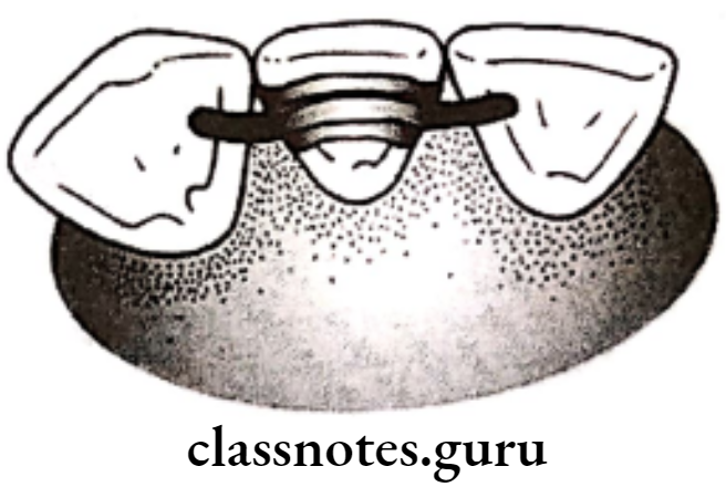
  • Opening of premolar space is a drawback of standard Hawley retainer
  • Prevention of wedging effect on extraction site by Hawley retainer can be attempted by long labial bow
  • Relapse is the loss of any correction achieved by orthodontic treatment

Mandibular Retroguathism Question And Answers

Miscellaneous Short Essays

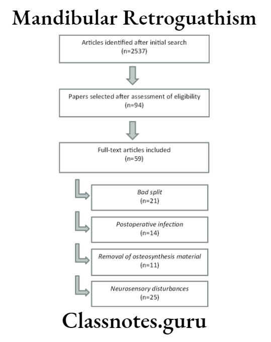
Mandibular Retroguathism

Question 1. Mandibular retroguathism.
Answer.

  • Refers to more backward placement of jaw.

Features of retrognathism:

  • Facial profile – convex
  • Facial divergence – Posterior divergent
  • Anteroposterior relation – Class 2
  • Mento labial sulcus – Deep
  • Hyperactive mentalis activity
  • Reduced nasolabial angle

Treatment of retrognathism:

  • In growing patients – Myofunctional therapy Ex. Activator, FR2
  • In non-growing patients
    • Orthodontic camouflage – Extraction of 1st premolars
    • Mandibular advancement

Read And Learn More: Orthodontics Short And Long Essay Question And Answers

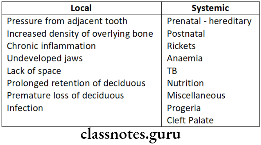
Question 2. Causes of Canine impaction.
Answer.

Orthodontics Miscellaneous Causes of Canine impaction

Question 3. Mechanism of Bone Growth.
Answer.

Bone Deposition and resorption

  • Together with bone deposition and resorption is called bone remodeling

Effects of bone deposition and resorption:

  • Change in size
  • Change in shape
  • Change in proportion
  • Change in bone relationship with adjacent structures

Cortical Drift

  • Movement of bone occurs towards bone deposition called cortical drift
  • If bone deposition and resorption are equal
  • Thickness of bone remains, constant
  • If bone deposition is more than resorption
  • The thickness of bone is more toward the deposition

Displacement

  • Movement of whole bone as a unit

Primary displacement:

  • Displacement of bone as a result of its own growth

Secondary displacement:

  • Displacement of bone as a result of growth of adjacent bone

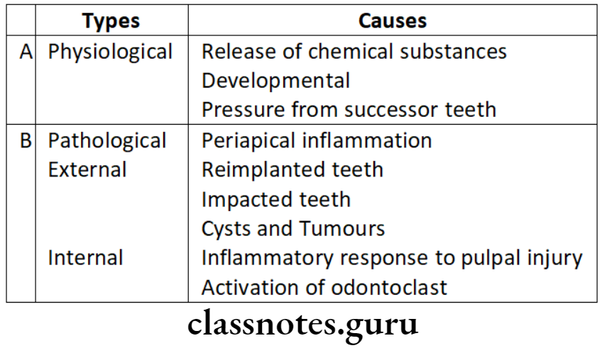
Question 4. Causes of Root resorption.
Answer.

Orthodontics Miscellaneous Causes of root resorption

Question 5. Envelope of Discrepancy.
Answer.

  • It helps in treatment planning
  • The choice of treatment depends on the movement of the teeth required
  • By orthodontic (fixed mechanotherapy) the tooth can be moved only at a specific distance
  • If the movement of teeth beyond this limit is required, orthopedic/functional appliances can be used
  • Beyond this limit, the treatment of choice is orthognathic surgery

Orthodontics Miscellaneous Fixed Mechanotheraphy

Question 6. Self-correcting Anomalies/Transient Malocclusions.
Answer.

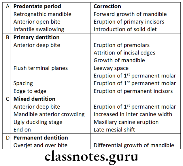
Orthodontics Miscellaneous Anomalies

Miscellaneous Short Questions And Answers

Question 1. Apertognathia.
Answer.

It is a condition in which there is space between upper and lower teeth when some teeth are in contact at one/more points

Orthodontics Miscellaneous Apertognathia

Question 2. Growth site and Growth center.
Answer.

Growth site:

  • These are growth fields that have a special significance in the growth of a particular bone
  • Posses intrinsic growing potential
    Example. Condyle, maxillary tuberosity

Growth Centres:

  • These are growth sites that control the overall growth of bone
  • Have growth potential
    Example. Epiphyseal plates of long bones

Question 3. Cranial Base Flexure.
Answer.

  • During the embryonic phase, the cranial base is flexed between the pituitary fossa and sphenoccipital junction
  • It is accompanied by developing brain stem

Cranial Base Flexure Results

  • Downward placement of foramen magnum
  • Aids in increased neurocranial capacity
  • Downward displacement of the face
    Age: 10th week of IU life, Angle – 65th

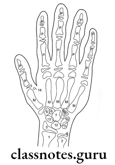
Question 4. Carpal Index.
Answer.

  • One of the skeletal maturity indicator
  • Used as a part of hand wrist
  • Carpals – consist of eight small bones arranged in

Proximal Row

  • Scaphoid
  • Triquetral
  • Lunate
  • Pisiform

Distal row

  • Trapezium
  • Trapezoid
  • capitate
  • Humate
  • These bones show specific patterns of appearance, ossification, and union
  • These are compared with standards

Orthodontics Miscellaneous Anatomy of hand wrist

Question 5. Torquing Auxillaries.
Answer.

Torquing Auxillarie Springs:

  • Made of stainless steel /Ni – titanium
  • Force exerted is directly proportional to the diameter and modulus of elasticity of the material of the wire
  • Stainless steel exerts greater force

Question 6. Sterilization in Orthodontics.
Answer.

Definition: Defined as the destruction of all life forms

Types of Instruments:

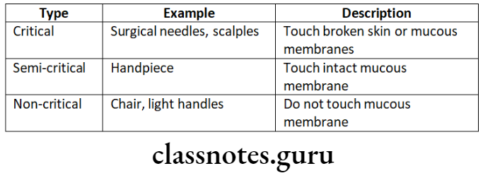
  • Critical – Penetrate the mucosa
  • Semi-critical – Touches mucosa but does not penetrate
  • Least critical – Surfaces touched during treatment

Instruments Requiring Sterilization:

  • Mirrors
  • Explorers
  • Banding and bonding instruments
  • Bands
  • Pillers
  • Ligature directors

Question 7. Growth Trends.
Answer.

By overlapping consequent cephalograms Tweed, designed a pattern of growth called “Growth trends”

Groups

Type A

  • Simultaneous growth of maxilla and mandible
  • 25% of case
  • ANB angle unchanged

Type A subdivision

  • Protruding maxilla
  • ANB angle increased

Type B

  • Increase in maxillary growth

Type B subdivision

  • ANB angle large
  • Unfavorable

Type C

  • Increased growth of the mandible
  • Decreased ANB angle

Type C Subdivision

  • Mandibular incisors touches lingual surfaces of maxillary incisors

Question 8. Growth Curve.
Answer.

  • Indicates the degree of difference between two growing individuals in all four planes including the time factor
  • As everyone does not have the same growth pattern, deviation from normal growth pattern cannot be diagnosed
  • Thus the growth of such individuals is compared with a standard growth chart

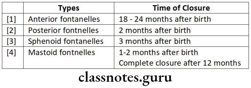
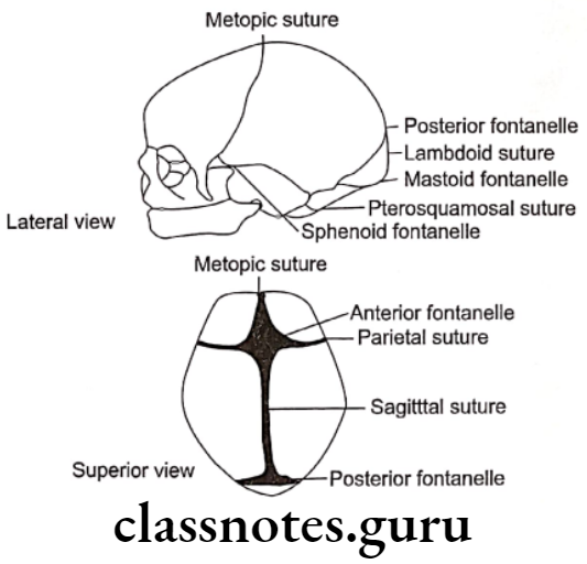
Question 9. Fontanelles.
Answer.

  • They bridge the gap between bones that limit them.
  • Made up of durameter, primitive periosteum and aponeurosis

Orthodontics Miscellaneous Fontanelles

Frontanelles Importance:

  • Indicates brain development
  • A depressed level indicates dehydration
  • Increased level indicates increased intracranial pressure

Orthodontics Miscellaneous Fontanelles and sutures in neonatal skull

Question 10. Safety valve mechanism.
Answer.

  • Increase in inter-canine width is one of the important factor in overcoming incisal liability
  • At the age of 12, maxillary anterior prolines such that inter-canine width increase

Significance of safety value mechanism:

  • This increase in maxillary inter canine width hinders the forward growth of the mandible
  • This increase in width behaves like it holds the forwardly growing mandible

Question 11. Curve of Spee.
Answer.

  • It is antero-posterior curve of occlusion
  • It begins from the tip of lower canine to the cusp tips of bicuspids and molars upto the condyle

Significance of curve of Spee:

  • Normal value – 1.5 – 2mm
  • If the curve is extended, it forms a circle of about 4 diameter
  • It represent the axial alignment of lower teeth
  • It requires a gradual progressive increased mesial tilting of teeth towards the molar

Question 12. Anterior bite plane and its mode of action.
Answer.

Uses of anterior bite plane: For treatment of deep bite

Mode of action:

  • Consist of the flat ledge of acrylic behind the upper anterior
  • When the patient bites the mandibular incisors contact the bite plane, thus dis occlude the posterior due to which they are free to erupt

Components of anterior bite plane:

  • Adam’s clasp on molar – As a retainer
  • Labial bow – Counter any forward component of force on upper anterior

Question 13. Orbital law of canine.
Answer.

  • It is used in Simon’s classification
  • Orbital plane perpendicular to Frankfort horizontal plane is used
  • This plane is dropped down from the bony orbital margin directly under the pupil of the eye
  • According to Simon, this plane should pass through the distal third of the upper canine
  • This is known as “Simon’s law of canine “or” Orbital law of canine”

Orbital law of canine significance:

  • It is used to describe malocclusion in a sagittal plane
  • When the dental arch is farther from the orbital plane it is called protraction
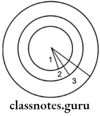
  • When the dental arch is closer then it is called retraction