Rationale Of Endodontics Question And Answers

Rationale Of Endodontics Important Notes

  • Fish Zones
    • Zone of infection
      • A zone of infection is present in the center of the lesion
      • Zone of infection is characterized by PMNs
    • Zone of contamination
      • Zone of contamination is characterized by round-cell infiltration
      • Zone of contamination demonstrates cellular destruction due to toxins from the central zone of infection
      • Because of autolysis and death of bone cells, the lacunae were empty
      • Lymphocytes are prevalent everywhere
    • Zone of irritation
      • Zone of irritation is characterized by macrophages and osteoclasts
      • The collagen framework was digested by phagocytic cells, the macrophages
      • Zone of irritation demonstrates much activity preparatory to repair
    • Zone of stimulation
      • Zone of stimulation is characterized by fibroblasts and osteoblasts
      • In this zone, the effects of toxins were mild enough to be stimulant which results in the laying down of collagen fibres around the zone of irritation
      • This new bone is built in an irregular fashion

Read And Learn More: Endodontics Question and Answers

Endodontic Therapy Principles

Rationale Of Endodontics Short Essays

Question 1. Rationale of Endodontics.
Answer.

Rationale Of Endodontics

Endodontics is a branch of dealing with the disease and injuries of the pulp. Endodotic procedures are required only when the infected organisms reach the pulp or else It can be treated by conventional methods.

Rationale Of Endodontics – Causes Of Infection:

  • Trauma
  • Latrogenic
  • Chemicals
  • Aseptic techniques
  • Pathologic wear
  • Toxins
  • Micro organisms

Endodontics Rarely Motile And Motile

    • Cyst
    • Granuloma
    • Chronic abscess
  • Only if the periapical pathologies occur that cannot be treated by conventional methods, endodontic procedures are the only solutions
  • Thus by endodontic procedures, infection is eliminated by keeping in mind the cause and healing occurs.

Rationale Of Endodontics Short Answer

Question 1. Fish Zones.

Fish Zones

Endodontics Rationale of Endodontics Fish zones

Goals Of Root Canal Treatment

Endodontics Rationale of Endodontics Healthy vascular pulp

Procedural Accidents Endodontic Question And Answers

Procedural Accidents Important Notes

  • Sodium hypochlorite extrusion into periradicular tissues causes
    • Immediate effects
      • Swelling
      • Pain
      • Ecchymosis
    • Long term effects
      • Paraesthesia
      • Scarring
      • Muscle weakness
      • Extrusion of hydrogen peroxide causing tissue emphysema

Procedural Accidents In Endodontics

Procedural Accidents Short Essays

Question 1. Management Of Perforations.
Answer.

Management Of Nonfurcal Cervical Perforations:

  • Hemorrhage control with 1:50,000 epinephrine followed by perforation repair with MTA
    Prevention:
  • Study proper anatomy before proceeding with the treatment
  • Remove all caries
  • Repeated and thorough evaluation of radiographs is done

Management Of Cervical Perforation In Furcation Area:

  • Confirm the perforation through radiograph
  • Electronic apex locator is used to differentiate a bleeding canal from perforation
  • MTA is used for sealing perforation
    Prevention:
  • Study preoperative radiographs and evaluate pulp chamber morphology
  • Access bur penetration for depth and angulation should be confirmed
  • Straight line access is obtained
  • If crown is present remove it before access preparation
  • Remove existing restorations if present

Read And Learn More: Endodontics Question and Answers

Question 2. Ledges.
Answer.

Ledges

Ledges is an internal transporation of the canal which prevents positioning of an instrument to the apex in an otherwise patent canal.

Ledges Etiology:

  • Curve canals
  • Forceful instrumentation
  • Failute to recapitutale
  • Skipping of files

Ledges Features:

  • Loss of tactile sensation
  • Loosening of instrument

Ledges Treatment:

Endodontics Procedural Accidents Ledges Treatment

Ledges Prevention:

  • Use of NiTi files
  • Avoid of skipping of instruments
  • Frequent irrigate
  • Careful preparation

Endodontics Procedural Accidents Hoe is used to plane the preparation walls

Endodontic Treatment Complications

Procedural Accidents Short Questions And Answers

Question 1. Zipping.
Answer.

Zipping Definition:

  • Zipping is defined as the apical transportation of a curved canal caused due to improper shaping technique

Zipping Features:

  • Zip formation adopts an elliptical shape at the apical endpoint.
  • Zipping is caused by using progressively larger and stiffer files to working length.
  • Zipping can result in a poorly cleaned root canal, which fails to provide a resistance form to compact gutta-percha, and leads to obturation which is vertically over-extended but internally under-filled.
  • This contributes to leakage, infection, and failure.

Question 2. Masserann Kit.
Answer.

Masserann Kit

Device used to remove broken instruments from root canal

Masserann Kit Components:

  • Flat gauge
  • Extractor
  • Stephan
  • Handle

Masserann Kit Steps:

Endodontics Procedural Accidents Masserann kit Steps

Procedural Accidents Viva Voce

  • Brasseler endo extractor kit, Masserman kit, extractor system, and separated instrument retrieval system are used in retrieval of broken instruments from root canal

Endodontic Periodontal Relationship Short Essays

Instrument Separation In Root Canal

Question 1. Endo-Perio Lesion.

Endo-Perio Lesion:

Endodontic Periodontal Relationship Endo-Perio Lesion

Obturation Of Root Canal Question And Answers

Obturation Of Root Canal Important Notes

  • Techniques of heat compaction of gutta percha
    • Vertical compaction
    • Injectable gutta percha
    • Thermo-mechanical compaction
    • Core carrier technique
  • Composition of gurra percha cones

Endodontics Obturation Of Root Canal Composition of gutta percha cones

  • Ideal requirements of filling material
    • Easy to introduce and easy to remove
    • Seal canal laterally as well as apically
    • Impervious to moisture
    • Should not shrink after insertion
    • Should be bactericidal
    • Should not stain the tooth
    • Should not irritate periapex
    • Should be sterile

Root Canal Obturation Techniques

Obturation Of Root Canal Long Essays

Question 1. Describe briefly different obturation techniques. Add a note on materials used for obturation.
Answer.

Obturation Methods:

Use Of Cold Guttapercha:

  • Lateral compaction

Use Of Chemically Softened Guttapercha:

  • Chloroform
  • Halothane
  • Eucalyptol

Read And Learn More: Endodontics Question and Answers

Use Of Heat-Softened Guttapercha:

  • Vertical compaction
  • System B
  • Sectional compaction
  • Thermoplasticized
  • Solid core
  • Mcspadden

Obturation Of Root Canal Lateral Compaction:

Endodontics Obturation Of Root Canal Lateral Compaction

Endodontics Obturation Of Root Canal Tugback with master gutta-percha cone

Endodontics Obturation Of Root Canal Compaction of guttapercha using spreader

Endodontics Obturation Of Root Canal Cut the protruding gutta percha points

Endodontics Obturation Of Root Canal Use of more accesory cones to complete obturation of the canal

Obturation Of Root Canal Advantages:

  • Reduces over filling

Obturation Of Root Canal Disadvantages:

  • Do not produce homogenous mass
  • May not fill canal irregularities

Obturation Of Root Canal Obturating Materials:

  • Plastics – GP, resilon
  • Solid/metal core – Silver points, gold
  • Cements and pastes – Hydron, MTA, Calcium phosphate

Obturation Of Root Canal Properties:

  • Easily introduced
  • Seal canal
  • Stable
  • Baceriostatic
  • Non-staining
  • Radiopaque
  • Non-irritating
  • Sterile
  • Easily removed
  • Imprevious to moisture

Obturation In Endodontics

Question 2. What are the various obturation techniques? Write in detail about thermoplasticized technique.
Answer.

Obturating Methods:

Obturating Methods Use Of Cold Guttapercha:

  • Lateral compaction

Obturating Methods Use Of Chemically Softened Guttapercha:

  • Chloroform
  • Halothane
  • Eucalyptol

Obturating Methods Use Of Heat-Softened Guttapercha:

  • Vertical compaction
  • System B
  • Sectional compaction
  • Thermoplasticized
  • Solid core
  • Mcspadden

Thermoplasticized Gutta Percha Technique

Thermoplasticized Gutta Percha Principle:

  • It comprises a pressure apparatus consisting of an insulated electrically heated syringe barrel and a seledction of needles ranging from 18-25 gauge size
  • The plunger is designed to prevent backward flow of the gutta percha

Thermoplasticized Gutta Percha Technique:

  • Canal preparation
  • Drying of canal
  • Sealer is coated on the canal walls
  • Gutta percha is electrically heated in a handheld gun that contains a chamber surrounded by a heating element
  • Selected needle is positioned at 3-5mm short of the working lenght
  • Gutta percha is gradually injected by sqeezing the trigger of the gun and the needle is gradually withdrawn as the canal gets filled apically
  • Gutta percha is then compacted using pluggers of appropriate size
  • Similarly the rest of the canal is filled

Question 3. Root canal sealers.
Answer.

Root Canal Sealers

  • Root canal sealers are necessary to fill irregularities and minor discrepancies between the canal walls and filling

Root Canal Sealers Requirements:

  • Should be tacky
  • Should create a hermetic seal
  • Should be radiopaque
  • Should be miscible
  • Should be bacteriostatic
  • Should set slowly
  • should insoluble in oral fluids
  • Should not shrink
  • Should not stain

Root Canal Sealers Functions:

  • Antimicrobial agent
  • Lubricant
  • Binding agent
  • Fills the space between material and canal walls
  • As obturationg material
  • Give radiopacity

Root Canal Sealers Classification:

Eugenol:

  • Eugenol Silver Containing:
    • Kerr sealer
      • Powder
        • Zinc oxide
        • Silver
        • Thymol iodide
      • Liquid
        • Oil of cloves
        • Canada balsam
  • Eugenol Silver Free:
    • Grossman’s sealer
    • It consists of
      • Powder
        • Zinc oxide
        • Staybellite resin
        • Bismuth subcarbonate
        • Barium sulphate
        • Sodium borate
      • Liquid
        • Eugenol

Eugenol  Medicated:

  • Iodoform

Eugenol Non-Eugenol:

  • Hydron
  • Glass ionomer
  • Diaklet
    • It is chemically polyketone
    • It consists of
      • Powder
        • Zinc oxide
        • Bismuth phosphate
      • Liquid
        • Polyvinyl resin

Eugenol Advantages:

  • Good adhesion to tooth
  • Rapid set
  • High tensile strength

Eugenol Disadvantages:

  • Tacky in texture
  • Difficult to manipulate

Gutta Percha Obturation Method

Obturation Of Root Canal Short Essays

Question 1. Ideal requirements of root canal sealers. Add a note on AH26.
Answer.

Root Canal Sealers Requirements:

  • Should be tacky
  • Should create hermetic seal
  • Should be radiopaque
  • Should be miscible
  • Should be bacteriostatic
  • Should set slowly
  • Should be insoluble in oral fluids
  • Should not shrink
  • Should not stain

AH26:

  • This is an epoxy resin containing sealer
  • It is adhesive, well tolerated by tissues and provides good seal

AH26 Disadvantages:

  • Staining of tooth structure as it contains silver
  • It is insoluble in solvents

AH26 Plus:

  • This eliminates all the disadvantages associated with AH26

Question 2. Obturating materials.
Answer.

Obturating materials Materials:

  • Plastics – GP, resilon
  • Solid/metal core – Silver points, gold
  • Cements and pastes – Hydron, MTA, Calcium phosphate

Obturating Materials Properties:

  • Easily introduced
  • Seal canal
  • Stable
  • Bacteriostatic
  • Non-staining
  • Radiopaque
  • Non-irritating
  • Sterile
  • Easy removed
  • Imprevious to moisture

Question 3. Gutta Percha.
Answer.

Gutta Percha

Endodontic filling material

Gutta Percha Forms:

  • Alpha
  • Beta
  • Amorphous

Gutta Percha Available Forms:

  • GP points
  • Auxillary points
  • Greater taper
  • Syringe
  • Medicated

Gutta Percha Advantages:

  • Compactibility
  • Inertness
  • Dimensional stable
  • Tissue tolerance
  • Radiopacity
  • Plasticity
  • Dissolve in some solvents

Gutta Percha Disadvantages:

  • Lack of rigidity
  • Lack of adhesiveness
  • Easily displaced

Warm Vertical Compaction Technique

Question 4. Grossman’s Sealer.
Answer.

Grossman’s Sealer Composition:

Grossman’s Sealer Powder:

  • Zinc oxide
  • Staybelite resin
  • Barium sulfate
  • Bismuth subcarbonate
  • Sodium borate

Grossman’s Sealer Liquid:

  • Eugenol

Grossman’s Sealer Properties:

  • Plasticity
  • Slow setting time
  • Good sealing

Grossman’s Sealer Disadvantage:

  • Require vigorous mixing
  • Setting time – 2 hours at 37°C

Grossman’s Sealer Influenced By:

  • Quality of ZnO and pH
  • Mixing
  • Humidity
  • Temperature and dryness of slab and spatula

Obturation Of Root Canal Short Questions And Answers

Question 1. Composition of Grossman’s sealer.
Answer.

Grossman’s Sealer Powder:

  • Zinc oxide
  • Staybelite resin
  • Barium sulfate
  • Bismuth subcarbonate
  • Sodium borate

Grossman’s Sealer Liquid:

  • Eugenol

Question 2. Diaket
Answer.

Diaket

  • By Schmidt in 1951

Diaket Composition:

Diaket Powder:

  • Zinc Oxide
  • Bismuth phosphate

Diaket Liquid:

  • 2,2 dihydroxy – 5,5 dichlorodiphenyl methane
  • Triethanolamine
  • B-diketone
  • Caproci acid
  • Copolymers

Diaket Advantages:

  • Good adhesion
  • Fast setting
  • Stable
  • Superior tensile

Diaket Disadvantages:

  • Toxic
  • Tacky
  • Setting is effected by phenol

Root Canal Filling Materials

Question 3. McSpadden compaction.
Answer.

Mcspadden Compaction Technique:

Endodontics Obturation Of Root Canal Mcspadden compaction Technique

McSpadden Compaction Advantages:

  • Less time
  • Easy to insert
  • 3D obturation

McSpadden Compaction Disadvantages:

  • Unable in narrow and curved canals
  • Frequent breakage
  • Overfilling
  • Shrinkage of guttapercha


Endodontics Obturation Of Root Canal Thermo-mechanical compaction of gutta percha

Question 4. AH Sealer
Answer.

AH26:

  • This is an epoxy resin containing sealer
  • It is adhesive, well tolerated by tissues, and provides a good seal

AH26 Disadvantages:

  • Staining of tooth structure as it contains silver
  • It is insoluble in solvents

AH26 Plus:

  • This eliminates all the disadvantages associated with AH26

Root Canal Filling Materials

Obturation Of Root Canal Viva Voce

  • Gutta-percha cones may be kept sterile in screw-crapped vials containing alcohol.
  • Lateral compaction is easy and quick to perform.
  • Tug back refers to the apical seal fit of the master cone.
  • Inadequate obturation is the most common cause of RCT failure.
  • Silver cones are contraindicated in filling a root canal if the tooth is to be restored with a post and care.
  • Ideally, the length of the post and core should be 2/3rd of the root canal.
  • Injectable gutta-percha is especially useful while managing canal irregularities.
  • Thermafill contains a center carrier which is grooved along 600 of their circumference. and has a coating of gutta-percha.
  • The silver cone is stiffer than gutta-percha.
  • Endorez is a visible light-curable urethane dimethacrylate resin-based sealer.

Management Of Discolored Teeth Question And Answers

Management Of Discolored Teeth Important Notes

  • Management Of Discolored Teeth Superoxol
    • It is 30% solution of hydrogen peroxide by weight and 100% by volume in pure distilled water
    • It is mixed with sodium perborate into a paste and used in walking bleach technique
  • Management Of Discolored Teeth Bleaching techniques:
    • Non vital bleaching technique
      • Thermocatalytic
        • It involves the placement of 35% hydrogen peroxide liquid into the debrided pulp chamber and acceleration of the oxidation process by heating instrument into pulp chamber.
      • Walking bleach
        • In this technique, superoxol is used
    • Vital bleaching technique
      • Power bleaching
        • 35% hydrogen peroxide is used and oxidation is acceletated by applying heat or intense light
      • Night guard bleach
        • 10-15% carbamide peroxide is applied with the help of bleaching trays
  • Management Of Discolored Teeth Composition of Mc Inns solution.

Endodontics Management of Discolored Teeth Composition of Mc Inns solution

Management Of Discolored Teeth Long Essays

Question 1. Management of discolored teeth
Answer.

Causes Of Discolored Teeth Intrinsic Stains:

  • Pre-Eruptive Causes:
    • Disease-Haematological disorders
    • Medication-tetracycline
  • Post-Eruptive Causes:
    • Pulpal changes
    • Trauma
    • Aging
    • Dental caries

Read And Learn More: Endodontics Question and Answers

Causes of Discolored Teeth Extrinsic Stains:

  • Daily Acquired Stains:
    • Plaque
    • Tobacco use
    • Poor oral hygiene
    • Gingival hemorrhage
  • Chemicals:
    • Chlorhexidine
    • Metallic stains

Bleaching Is Done To Treat Discolored Teeth

Bleaching:  Bleaching is a procedure that involves lightening the color of a tooth through the application of a chemical agent to oxidize the organic pigmentation in the tooth.

Bleaching Techniques:

  • For vital teeth
    • Home bleaching
    • In-office bleaching
    • Thermocatalytic
    • Non-thermocatalytic
    • Microabrasion
  • For non-vital teeth
    • Thermocatalytic
    • Walking bleach
    • Inside/Outside
    • Closed chamber
  • Laser-assisted bleaching

Nightguard Bleaching Indications:

  • Mild generalized
  • Age-related
  • Mild fluorosis
  • Mild tetracycline staining
  • Superficial staining
  • Stains from smoking tobacco

Bleaching Contra-Indications:

  • Teeth with insufficient enamel
  • Severe fluorosis
  • Non-complaint patients
  • Pregnancy
  • Severe tetracycline staining
  • Fractured tooth
  • Opaque white spots

Bleaching Steps:

Endodontics Management of Discolored Teeth Management of discolored teeth Steps

Bleaching Agents Used:

  • 10% carbamide peroxide
  • 15% carbamide peroxide
  • Hydrogen peroxide

Bleaching Side Effects:

  • Gingival irritation
  • Altered taste sensation
  • Tooth Sensitivity

Management Of Discolored Teeth

Management Of Discolored Teeth Short Essays

Question 1. Vital bleaching.
Answer.

In-office bleaching:

  • It uses 35% hydrogen peroxide solution

Vital Bleaching Indications:

  • Mild fluorosis
  • Mild tetracycline stains
  • In severe discoloration
  • To match the existing color of the crown that is lighter than natural teeth

Vital Bleaching Contraindications:

  • Superficial stains
  • Carious tooth structure
  • Hypersensitive teeth
  • Children with a large pulp chamber
  • Pregnant and lactating mothers
  • Exposed root surfaces

Vital Bleaching Technique:

Endodontics Management of Discolored Teeth In office beaching Technique

At-Home Bleaching Indications:

  • Superficial enamel discolorations
  • Mild yellow discolorations
  • Brown fluorosis discolorations
  • Age-related discolorations

At-Home Bleaching Contraindication:

  • Severe enamel loss
  • Hypersensitive teeth
  • Bruxism
  • Presence of caries
  • Defective coronal restorations
  • Allergy to bleaching gels

At-Home Bleaching Technique:

Endodontics Management of Discolored Teeth At office bleaching Technique

Question 2. Carbamide peroxide
Answer:

Carbamide Peroxide Definition

  • Carbamide Peroxide is also known as urea hydrogen peroxide
  • Carbamide Peroxide concentration ranges from 3-45% depending on at-home and in-office bleach
  • Commonly 10% carbamide peroxide is used

Carbamide Peroxide Mechanism Of Action

  • Carbamide Peroxide→Urea + Ammonia + carbon dioxide + 3.5% hydrogen peroxide

Teeth Discoloration Treatment

Question 3. Thermocatalytic Bleaching.
Answer.

Thermocatalytic Bleaching Light Sources Used:

  • Conventional
  • Tungsten halogen
  • Xenon plasma
  • Argon and Co2
  • Diode laser

Thermocatalytic Bleaching Indication:

  • Superficial stains
  • Mild to moderate stains

Thermocatalytic Bleaching Contraindications:

  • Extensive restoration
  • Severe discoloration
  • Extensive caries

Thermocatalytic Bleaching Advantages:

  • Comfortable to patient
  • Less time needed
  • Safe

Thermocatalytic Bleaching Disadvantages:

  • Expensive
  • Frequent
  • Dehydration of teeth
  • Rubber dam discomfort.

Thermocatalytic Bleaching Procedure:

Endodontics Management of Discolored Teeth Thermocatalytic Bleaching Procedure

Endodontics Management of Discolored Teeth Thermocatalytic technique of bleaching for vital teeth

How To Whiten Stained Teeth

Question 4. Microabrasion.
Answer.

Microabrasion

In it, a microscopic layer of enamel is eroded and abraded with solution leaving intact enamel.

Microabrasion Indications:

  • Intrinsic stains
  • Hypomineralised enamel
  • Decalcification
  • Enamel fluorosis
  • Superficial stains

Microabrasion Contra-Indications:

  • Age-related
  • Hypoplastic lesions
  • Deep stains
  • Tetracycline staining

Microabrasion Advantages:

  • Minimum discomfort
  • Easy
  • Results in shiny and smooth surface

Microabrasion Disadvantages:

  • Ineffective for deeper stains
  • Removes enamel layer

Microabrasion Steps: 

Endodontics Management of Discolored Teeth Microabrasion Steps

Question 5. Bleaching of non-vital tooth.
Answer.

Bleaching Of Non-Vital Tooth – Thermocatalytic Technique:

Endodontics Management of Discolored Teeth Thermocatalytic technique

Bleaching Of The Non-Vital Tooth – Walking Bleach technique:

Endodontics Management of Discolored Teeth Walking Bleach Technique

Bleaching Of The Non-Vital Tooth- Inside/Outside Bleaching:

Endodontics Management of Discolored Teeth Inside and Outside Bleaching

Closed Chamber:

  • Bleaching paste was applied to the tooth via a bleaching tray

Intrinsic vs. extrinsic Tooth Stains

Question 6. Walking Bleach.
Answer.

Walking Bleach Indications:

  • Discoloration of the pulp chamber
  • Moderate to severe tetracycline staining
  • Dentin discoloration

Walking Bleach Contraindications:

  • Defective enamel
  • Enamel discoloration
  • Presence of caries

Walking Bleach Steps:

Endodontics Management of Discolored Teeth Walking Bleach Technique.

Endodontics Management of Discolored Teeth Removal of coronal gutta percha using rotary instrument

Endodontics Management of Discolored Teeth Placement of protective barrier over gutta percha

Endodontics Management of Discolored Teeth Placement of bleaching mixture into pulp chamber

Endodontic Bleaching Techniques

Question 7. Microbrasion and macrobrasion.
Answer.

Microbrasion And Macrobrasio

In it a microscopic layer of enamel is eroded and abraded with solution leaving intact enamel.

Microbrasion Indications:

  • Intrinsic stains
  • Hypomineralised enamel
  • Decalcification
  • Enamel fluorosis
  • Superficial stains

Microbrasion Contra-Indications:

  • Age-related
  • Hypoplastic lesions
  • Deep stains
  • Tetracycline staining

Microbrasion Advantages:

  • Minimum discomfort
  • Easy
  • Results in shiny and smooth surface

Microabrasion Disadvantages:

  • Ineffective for deeper stains
  • Removes enamel layer

Microabrasion

  • Microabrasion involves using of dental drill and finishing bur to remove stains
  • In this composite finishing bur is used on a high speed hand piece to remove the defect
  • While working on the defect very light pressure is applied to prevent any irreversible damage to the tooth
  • The process is finished with abrasive rubber point
  • During the process air water spray is used to maintain the temperature and to keep the tooth hydrated

Management Of Discolored Teeth Short Questions And Answers

Question 1. Causes of Discoloration of tooth.
Answer.

Discoloration Of Tooth Intrinsic Stains:

  • Pre-eruptice causes:
    • Disease-Haematological disorders
    • Medication-tetracycline
  • Post-Eruptive Causes:
    • Pulpal changes
    • Trauma
    • Aging
    • Dental caries

Discoloration Of Tooth Extrinsic Stains:

  • Daily Scquired Stains:
    • Plaque
    • Tobacco use
    • Poor oral hygiene
    • Gingival haemorrhage
    • Chemicals
    • Chlorhexidine
    • Metallic stains

Management Of Discolored Teeth

Question 2. Mc-Innes Solution.
Answer.

Mc-Innes Solution Composition:

  • 30% Hydrogen peroxide – 5 parts
  • 36% hydrochloric acid – 5 parts
  • Anesthetic ether – 1 part

Mc-Innes Solution Method:

Apply the solution with a cotton pellet to the stained area and allow it to remain for 5-10 minutes

Question 3. Superoxol
Answer.

Superoxol

  • Superoxol is a 30% solution of hydrogen peroxide by weight in 100% volume of distilled water
  • Superoxol is a powerful oxidizing agent
  • Superoxol is highly irritating of the soft tissues

Question 4. Uses of laser in endodontics
Answer.

Uses Of Laser In Endodemtics

  • Diagnosis
    • Laser Doppler flowmetry – to assess blood flowmetry
    • Nd:YAG laser – for vitality test
  • Pulp capping and Pulpotomy
    • To treat exposed pulp tissues using Co2 lasers
  • Root canal treatment
    • To modify root canal walls
    • Sterilization of root canals
  • Treatment of incomplete fracture
  • Apicoectomy
  • Treatment of dentinal hypersensitivity
  • Sterilization of instruments
  • Bleaching

Irrigation And Intracanal Medicaments Question And Answers

Irrigation And Intracanal Medicaments Important Notes

  • Sodium hypochlorite
    • It acts as
      • Lubricant during instrumentation in root canal
      • Solvent for dissolution of pulp
      • Antiseptic and disinfectant by combining both protoplasm of bacterial cell and destroying it
      • Bleaching agent by releasing nascent oxygen
    • It is used in concentration varying from 0.5-75%
    • Very Potent antimicrobial agent
    • Effectively dissolves pulpal remnants
    • Disadvantages
      • Inability to remove smear layer
      • Unpleasant taste
      • Toxicity
  • Advantages of alternate use of sodium hypochlorite and hydrogen peroxide in root canal preparation
    • Effervescent reaction which pushes debris out of the canal
    • Solvent action of sodium hypochlorite on the organic debris of pulp tissue
    • Disinfecting and bleaching action of both solutions

Irrigation And Intracanal Medicaments Long Essays

Intracanal Medicaments In Endodontics

Question 1. What are various root canal irrigants? Write in detail requirements and techniques of irrigation.
Answer.

Irrigant:

Irrigant is a liquid used to lubricate the canal walls and flush out the debris and micro-organisms from the root canals

Irrigant Types:

Chemically Non-Active

  • Water:
    • Saline
    • LA
  • Chemically Active [ACDE]:
    • Alkali – Sodium Hypochlorite
    • Acids – HCl
    • Antibacterial agents – Chlorhexidine
    • Chelating agents – EDTA
    • Detergents – Sodium lauryl sulfate
    • Enzymes – Streptokinase
    • Oxidizing agents – Hydrogen peroxide
      Requirements:

      • Should have antimicrobial activity
      • Should be non-toxic and biocompatible
      • Should dissolve necrotic and vital pulp tissues
      • Should be tissue or debris solvent
      • Should serve as lubricant
      • Should have low surface tension
      • Should be non-reactive to periapical tissues
      • Should not be easily neutralized in the canal and should retain its effectiveness
      • It should be of moderate cost and ease to store

Read And Learn More: Endodontics Question and Answers

Irrigant Techniques:

Irrigant Techniques Manual

  • Syringe Irrigation With Needles:
    • It involves dispensing an irrigant into the root canal using irrigation needles of varying gauges
    • Gauge 27-30
    • Depth of insertion 2-3mm from the working length
    • Design – blunt-ended size vented needle
  • Brushes:
    • Bristles help in cleaning the uninstrumented recesses of the radicular pulp spaces
  • Manual Dynamic Agitation:
    • In this, a well-fitted greater taper gutta-percha master cone is moved up and down the instrumented canal containing irrigant in short 2-3mm strokes

Irrigant Techniques Machnine Assisted

  • Rotary brushes
    • Consist of micro brushes attached to rotary handpieces
  • Continuous irrigation during instrumentation
    • It contains an irrigant delivery unit attached to the Quantec-E-irrigation system
  • Sonic irrigation
    • involves sonic waves
  • Ultrasonic irrigation
    • Operates at frequencies of 25-30kHz
  • Pressure alternation devices
    • EndoVac
    • RinsEndo

Endodontic Irrigation Techniques

Question 2. Enumerate the various root canal irrigants. What are the functions of irrigants? Write in detail on chlorhexidine and sodium hypochlorite as intracanal irrigants.
Answer.

Intracanal Medicaments In Endodontics

Irrigant: Irrigant is a liquid used to lubricate the canal walls and flush out the debris and micro-organisms from the root canals

Irrigant Types:

Irrigant Chemically Non-Active

  • Water:
    • Saline
    • LA
  • Chemically Active [ACDE]:
    • Alkali – Sodium Hypochlorite
    • Acids – HCl
    • Antibacterial agents – Chlorhexidine
    • Chelating agents – EDTA
    • Detergents – Sodium lauryl sulfate
    • Enzymes – Streptokinase
    • Oxidizing agents – Hydrogen peroxide

Functions Of Irrigant In Endodontics

  • Remove dentinal shaving
  • Germicidal
  • Increase efficiency of instruments
  • Dissolve necrotic tissue and remove it
  • Bleaching action

Chlorhexidine:

  • Chlorhexidine is biguanide
  • Available as oral rinses
  • Used in concentration 0.2-2%

Chlorhexidine Advantages:

  • Longer antibacterial action
  • Used with sodium hypochlorite
    • When chlorhexidine comes in contact with sodium hypochlorite there is the formation of parachloroaniline which is cytotoxic
    • It interferes with the seal of the root filling
    • Flushing with normal saline is a must
  • Biocompatible

Chlorhexidine Disadvantages:

  • Does not dissolve pulp tissue

Sodium Hypochlorite Solution

  • It is a clear, pale, green-yellow liquid
  • A strong odour of chlorine
  • Easily miscible with water
  • Decomposes by light

Sodium Hypochlorite Mechanism:

Endodontics Irrigation And Intracanal Medicaments Sodium Hypochlorite Solution Mechanism

Intracanal Medicaments In Endodontics

Factors Affecting Sodium Hypochlorite Mechanism Activity:

  • Increases:
    • Volume of solution
    • Heating of solution
    • Time of contact
  • Decreases:
    • Strong time
    • EDTA

Hypochlorite Mechanism Activity Advantages:

  • Dissolve tissue
  • Antibacterial and bleaching action
  • Lubricate canal
  • Economical

Hypochlorite Mechanism Activity Disadvantages:

  • High surface tension
  • Irritate tissue
  • Irritate eyes
  • Causes inflammation of the gingiva
  • Bleaches clothes
  • Bad odour and taste
  • Corrosive to instruments

Intracanal Medicaments In Endodontics

Hypochlorite Mechanism Activity Combined With:

  • Calcium hydroxide
  • EDTA
  • Chlorhexidine

Irrigation And Intracanal Medicaments Short Essays

Question 1. Irrigating Solution.
Answer.

Irrigating Solution Properties:

  • Broad spectrum
  • Inactive endotoxin
  • Dissolve debris
  • Debride the canal
  • Good lubricant
  • Less toxic
  • Prevent/dissolve smear layer

Irrigating Solution Functions:

  • Remove dentinal shaving
  • Germicidal
  • Increase efficiency of instruments
  • Dissolve necrotic tissue and remove it
  • Bleaching action

Irrigating Solution Commonly used:

  • Chemically Non-Active:
    • Water:
    • Saline
    • LA
  • Chemically active [ACDE]:
    • Alkali – Sodium Hypochlorite
    • Acids – HCl
    • Antibacterial agents – Chlorhexidine
    • Chelating agents – EDTA
    • Detergents – Sodium lauryl sulfate
    • Enzymes – Streptokinase
    • Oxidizing agents – Hydrogen peroxide

Root Canal Irrigation Solutions

Question 2. Ideal requirements of intracanal medicaments. Describe in detail about medicaments.
Answer.

Intracanal Medicaments Functions:

  • Destroy remaining bacteria
  • Useful in treatment of apical periodontitis

Intracanal Medicaments Properties:

  • Germicidal
  • Non-irritant
  • Stable
  • Low surface tension
  • Should not induce immune response
  • Should not interfere with repair
  • Should not stain tooth

Intracanal Medicaments Chemicals Used:

  • Essential oils – Eugenol
  • Phenolic compounds – Phenol, Aldehydes
  • Calcium hydroxide
  • Chlorhexidine gluconate
  • Corrticosteroid antibiotic
  • Antibiotic
  • Halogens – Sodium hypochlorite, Iodine

Question 3. Calcium Hydroxide.
Answer.

Calcium Hydroxide

Intracanal medicament

Calcium Hydroxide Forms:

  • Paste form
  • Powder form

Calcium Hydroxide Indication:

  • Weeping canal
  • Treatment of Phoenix abscess
  • Resorption
  • Apexification
  • Pulp capping
  • Decrease post-operative pain
  • As sealer
  • For periapical lesion

Calcium Hydroxide Disadvantages:

  • Difficult to remove
  • Decreases setting time

Calcium Hydroxide Functions:

  • Inhibits root resorption
  • Stimulates periapical healing
  • Encourage mineralization

Question 4. Composition and uses of mineral trioxide aggregate.
Answer.

Mineral Trioxide Aggregate Composition:

  • Tricalcium silicate
  • Tricalcium aluminium oxide
  • Silicate oxide
  • Bismuth oxide
  • Calcium
  • Phosphate
  • Water

Mineral Trioxide Aggregate Uses:

  • As root end filling material
  • Used in Pulpotomy

Sodium Hypochlorite In Endodontics

Irrigation And Intracanal Medicaments Short Questions And Answers

Question 1. Hydrogen Peroxide.
Answer.

Hydrogen Peroxide

Hydrogen Peroxide is clear, odourless liquid

Hydrogen Peroxide Mechanism:

Endodontics Irrigation And Intracanal Medicaments Hydrogen Peroxide.

Use: As an irrigating solution

Combine with: 5.2% NaOCl

Question 2. Eugenol.
Answer.

Eugenol

Eugenol is an intracanal medicament

Eugenol Effects:

Eugenol Low Dose [Beneficial]:

  • Inhibits PG synthesis
  • Inhibits nerve activity
  • Inhibits chemotaxis

Eugenol High Dose [Harmful]:

  • Induces cell death
  • Inhibits cell respiration

Eugenol Uses:

  • Intracanal medicament
  • Root canal sealer
  • Temporary sealant

Calcium Hydroxide Intracanal Medicament

Question 3. RC Prep.
Answer.

RC Prep Composition:

  • EDTA
  • Urea peroxide
  • Carbowax

RC Prep Properties:

  • Lubricant
  • Cleaning agent
  • Antibacterial

RC Prep Uses:

  • Allows deeper penetration of medicament into the dentin

Question 4. Ledermix.
Answer.

Ledermix Composition:

  • Gluco corticosteroid
  • Triamcinolone
  • Dimethyl chlortetracycline

Ledermix Uses:

  • Initial dressing agent
  • Reduces incidence of pain
  • Provide rapid relief
  • Useful in cases of infection and inflammation

Question 5. PBSC.
Answer.

PBS

Endodontics Irrigation And Intracanal Medicaments PBSC

PBSC Route Of Administration:

PBSC Form-Paste Form:

  • Injected into root canals
  • Impregnated on paper points

Advance – Nystatin Replaces Caprylate

PBSC Disadvantage:

  • The patient may have an allergy to penicillin

Calcium Hydroxide Intracanal Medicament

Question 6. Schilder’s Objectives for cleaning and shaping of Canal.
Answer.

Schilder’s Objectives For Cleaning And Shaping Of Cana

  • Root canal preparation should develop continuously tapering cone
  • Making preparations in multiple planes to introduce the concept of flow
  • Making the canal narrower apically and wider coronally
  • Avoid transportation of foramen
  • Keep the apical opening as small as possible

Endodontics Irrigation And Intracanal Medicaments Diagrammatic representation of objectives of canal preparation

Irrigation And Intracanal Medicaments Viva Voce

  • EDTA is used at a concentration of 15%
  • MTAD is a Mixture of Tetracycline, Acid [citric acid], and Detergent

Instrumentation Of Endodontics Question And Answers

Instrumentation Of Endodontics Important Notes

  • Classification of instruments
    • Grossman’s classification
      • Exploring instruments – smooth broaches, endodontic explorer
      • Debridement – barbed broaches
      • Shaping instruments – reamers and files
      • Obturating instruments – pluggers, spreaders, lentulospirals
    • ISO grouping of instruments
      • Group 1 – hand use only
      • Group 2 – made to be attached to a handpiece
      • Group 3
        • Engine given latch type
        • Rotary canal instruments
      • Group 4 – root canal points
  • Standardization of instruments
    • By Ingle le Vine
      • Numbering from 10 – 100
        • Advance by 5 up to 60
        • Advance by 10 up to 100
      • Diameter
        • D at tip – 1/10 of a mm
        • Ex. For instrument No.25 D1 = 0.25mm
        • D2 at the end of shaft
      • Length of working portion
        • 16mm from D1 to D2
      • Taper of 0.02 mm per mm
      • Angle of the tip 75 ± 15 degrees
      • Color coding
  • H files
    • Made from a round blank to produce spiral flutes
    • They show higher cutting efficiency but is more prone to fracture
    • The angle between cutting edge and the long axis of the instrumentis about 60-65°
    • They are designed primarily for a linear filling motion
    • Due to positive rake angle they cut in one direction only

Read And Learn More: Endodontics Question and Answers

Endodontic Instruments Names

  • K flex files
    • Cross section is rhombus or diamond shaped
    • They have better cutting ability and flexibility
    • The instrument is fabricated of V-4 steel
  • Unifiles
    • Mc-Spadden modified the traditional H files
    • The blade present a S shape or double helix design
    • It has two continuous cutting edges
    • It can be used for cutting in both filling and reaming action
    • They are less subject to fracture
    • They are stiff in coronal and middle thirds but bends in apical 1/3rd
  • Nickel – titanium files
    • Composition
      • Nickel – 54%
      • Titanium – 44%
      • Cobalt – 0.2%
      • Boran – added to improve surface hardness
    • At high temperature, it exists a body centered cubic lattice referred to as autestentic phase – a stronger phase
    • On cooling, this phase transfers to close packed hexagonal, weak phase known as martensitic phase
    • Two unique features of it are: shapre memory and super elasticity
    • The main disadvantage of it is its cutting efficiency is only 60%

Instrumentation In Endodontics

  • Flexogates
    • Derivative of Gates Glidden drills
    • Used to enlarge the apical region of the canal
    • Less likely to cause apical transporation
    • Less fracture resistant
    • Non cutting guiding tip
  • Giromatic and Racer handpiece
    • Giromatic
      • It is commonly used reciprocating handpiece
      • It accepts only latch-type instrument
      • In this type the quarter turn motion is delivered 300 times per minute
      • It uses a barder broach or reamer through 90° reciprocating arc at a speed of 1000 cycles/min
    • Racer handpiece
      • It uses a standard file and oscillates the file in root canal
      • These instruments can be used for opening root canals but should not be used for root canal preparation
  • Peaso reamer
    • They are more often used in preparing coronal portion of the root canal for past and core
    • Instrument fractures always near the shank
  • Ultrasonic and sonic instruments
    • Used for cleaning and shaping of root canals
    • Ultrasonic instruments contains a magnetostrictive hand piece which hold a K file or diamond file
    • Ultrasonic and sonic instruments is operated at 2000 – 25000/section
    • Ultrasonic and sonic instruments uses sodium hypochlorite as irrigant
    • Sonic instruments operate at 1500 – 6500 cycles/min
    • Ultrasonic and sonic instruments uses water as irrigant
  • Colour coding

Endodontics Instrumentation Colour coding

  • Lentulospirals
    • They are used for the placement of sealer, cement, and calcium hydroxide
    • Lentulospirals must be operated clockwise in the handpiece and started or stopped outside the root canal
    • If started in the canal it may cut into the wall of the root canal and break

Endodontic Instruments Names

  • Modifications of H file
    • Safety hed stroem
    • Hyflex file
    • Unifiles
    • S file
  • Differences between reamer and file

Endodontic Instruments

Endodontics Instrumentation Reamer and File

  • Gates Glidden drill
    • Uses
      • To remove lingual shoulder during access cavity preparation
      • To enlarge root canal orifices
      • For shaping cervical third of root canal in step back preparation

Endodontics Instrumentation Gates Glidden drill

Endodontic Instruments List

Instrumentation of Endodontics Long Essays

Question 1. Classify Endodontic Instruments. Describe standardization and sterilization of them.
Answer.

Grossman’s Classification:

  • Exploring Instruments – Smooth broach
  • Extripating Instruments – Barked broach
  • Cleaning and Shaping Instrument – Files and Reamers
  • Obturating Instruments – Pluggers and Spreaders

ISO And FDI Classification

  • Group 1 – Hand operated Instruments
  • Group 2 – Engine driven Instruments
    • Parts
    • Cutting head
    • Latch type of attachment
  • Group 3 – Fabricated from single piece of metal Ex. Gates Glidden drill
  • Group 4 – Usually materials Ex. GP points, paper points

Standardization: By Ingle le Vine

  • Numbering from 10-100
    • Advance by 5 up to 60
    • Advance by 10 up to 100
  • Diameter
    • D at tip – 1/10 of a mm
    • Ex. For instrument No.25 D1 = 0.25mm
    • D2 at the end of shaft
  • Length of working portion
    • 16mm from D1 to D2
  • Taper of 0.02 mm per mm
  • Angle of the tip 75 ± 15 degrees
  • Color coding

Endodontics Instrumentation Diagrammatic representation of an endodontic instrument

Endodontics Instrumentation Standardization of Instrument and Colour

Endodontic Instruments

Sterilization:

Endodontics Instrumentation Sterilization

Instrumentation of Endodontics Short Essays

Question 1. Endosonic Instruments.
Answer.

Introduced By Richman

Activation:

Activation

Endodontic Instruments Advantages

  • Cleans the canal
  • Enhances the action of NaOCl

Endodontic Instruments Types:

  • Magnetostrictive
  • Piezoelectric

Endodontic Instruments Mechanism:

  • Navigation
  • Acoustic streaming

Endodontic Instruments Names

Endodontic Instruments Uses:

  • Access enhancement
  • Orifice location
  • Irrigation
  • Sealer placement
  • Guttapercha obturation
  • MTA placement
  • Endodontic retreatment.

Endodontics Instrumentation Acoustic streaming

Root Canal Instrumentation Techniques

Question 2. Obturating Instruments.
Answer.

  • Obturating Instruments Spreaders:
    • Use: For lateral compaction
    • Material Used: Stainless steel/Nickel titanium
    • Types:
      • Hand – Do not have standardized size and shape
      • Finger – Standardized and color-coded to match the size of GP points
  • Obturating Instruments Pluggers:
    • Use: For vertical compaction, For sectional method
    • Material Use To Pack: Calcium hydroxide/MTA

Endodontics Instrumentation Spreader and plugger tips

  • Lentulospiral:

Question 3. Broaches.
Answer.

Broaches Types:

  • Smooth broaches
  • Barbed broaches

Broaches Broaches:

  • Short handles instruments
  • Made from round steel wires

Broaches Uses:

  • Extripation of pulp tissues
  • Removal of dressing
  • Loosen necrotic debris
  • Pathfinder [smooth broach]

Broaches Method Of Use:

Endodontics Methods Of Use

Broaches Precaution:

  • Should not be forced apically into canal

Endodontics Instrumentation Barbed broach

Question 4. H-files.
Answer.

H-Files Manufacture:

  • Round steel wire, machined to produce spiral flutes

H-File Advantage:

  • Higher cutting efficiency
  • Push debris coronally

H-files Manner Of Use:

  • Pull motion, cut dentin when the instrument is withdrawn

H-Files Features:

  • Positive Rake angle
  • Distance between the flutes

H-Files Disadvantages:

  • Aggressive
  • Lack flexibility
  • Tend to fracture

H-Files Use:

  • For flaring coronal half of preparation
  • To machnie straight canals

H-Files Modifications:

  • Unifiles
  • Helifile
  • S-file

Endodontics Instrumentation Diagrammatic view of Hedstrom file

Rotary Vs Hand Files In Endodontics

Question 5. NiTi Files.
Answer.

NiTi Files

Known as Nitinol [NiTi Navol Ordinance Laboratory]

NiTi Files Types And Composition:

  • 55 NiTinol [55% Ni, 45% Ti]
  • 60 NiTinol [60% Ni, 40% Ti]

NiTi Files Advantages:

  • Shape memory
  • Super elasticity
  • Softer
  • Corrosion resistance
  • Resiliency is good

NiTi Files Disadvantage:

  • Poor cutting efficiency
  • Do not show signs of fatigue
  • Poor resistance to fracture

Instrumentation of Endodontics Short Answers

Question 1. Lentulospirals.
Answer.

Lentulospirals

  • Lentulospirals is a obturating instrument
  • Have latch-type attachment

Lentulospirals Used:

  • To coat the walls of the root canal for sealer

Lentulospirals Significance:

  • By sealing the canal walls, space between the canal wall and the material is avoided

Instrument Used Along With It: Giromatichand piece.

Endodontics Instrumentation Lentulo spiral

Question 2. Peaso – Reamer.
Answer.

Peaso – Reamer

  • Peaso – Reamer is a latch type of rotatory instruments
  • Have cutting sides
  • Made up of stainless steel

Peaso – Reamer Used For:

  • For parallel post-preparation
  • To remove gutta-percha from the canal

Peaso – Reamer Precaution:

  • Peaso-Reamer does not follow canal curvature
  • Peaso – Reamer is not flexible
  • Should be used with low speed to prevent over-instrumentation and perforation

Peaso – Reamer Sizes:

Endodontics Instrumentation Peaso Reamer Sizes

Endodontics Instrumentation Commercially available instrument holders and gauzes

Endodontic File Systems

Question 3. Gates glidden Drill.
Answer.

Gates Glidden Drill Description:

  • Head – flame shaped with safe tip
  • Culting end – Resembles American football
  • Shank – long, elliptical shaped

Gates Glidden Drill Used Along With:

  • Latch attachment, slow speed handpiece

Gates Glidden Drill Uses:

  • Enlarging orifice in coronal 3rd of tooth
  • To prepare post-space
  • To remove guttapercha/instrument

Gates Glidden Drill Sizes:

Endodontics Instrumentation Gates glidden drill sizes

Endodontics Instrumentation Due of safety design

Question 4. Files and Reamer
Or
Cross section of files and reamers.
Answer.

Files And Reamer

  • These are cleaning and shaping instrument
  • Difference

Endodontics Instrumentation Cross section of files and reamers

Endodontics Instrumentation Triangular blank and lesser number of flutes in reamer

Endodontic File Systems

Question 5. Sterilization of instruments.
Answer.

Sterilization Of Instruments

Endodontics Instrumentation Sterilization

Instrumentation Of Endodontics Viva Voce

  • Broaches break easily
  • Broaches should not be inserted unless the root canal is enlarged to a size of 20 or 25 reamer or file
  • File contains more number of flutes and is four sided instrument
  • Giromatic and Racer are contra angled engine driven instruments
  • The instrument should be used with a 1/4 – 1/2 turn and withdrawn with a pull stroke
  • Gates Glidden drill and Peaso reamer are power driven instruments
  • Over instrumentation causes forcing of debris into periapocal area
  • Instrumentation short of apex causes ledging of canal
  • Instrumentation with large instruments causes enlarging of canal
  • Reamers are used with pushing-rotating motion
  • Files are used with rasping or pulling motion
  • H file is used to finish the instrumentation of the coronal third of the root canal
  • Endodontic explorer are most commonly used diagnostic and exploring instruments for idenfication of canal orifices

Endodontic Microbiology Question And Answers

Endodontic Microbiology Of Endodontics Important Notes

  • Persistent positive cultures occurs due to
    • Leakage between appointments due to faulty coronal temporary restorations
    • Undetected perforation
    • Contamination of canal

Endodontic Microbiology

Endodontic Microbiology Of Endodontics Short Essays

Question 1. Endodontic microbiology. Add a note on cultural techniques.
Answer.

Endodontic Microbiology:

  • Generally, all bacteria that inhabit the oral cavity can invade the pulp space during and after pulp necrosis to participate in the infection of the canal and to enter the periapical tissues leading to periapical periodontitis.

Anaerobic gram-negative bacteria

Read And Learn More: Endodontics Question and Answers

  • Facultative gram-negative bacteria
    • Neisseria
    • Capnocytophaga
    • Haemophilus
  • Anaerobic gram positive bacteria
    • Actinomyces
    • Eubacterium
  • Facultative gram positive bacteria
    • Enterococcus
    • Streptococcus
    • Lactobacillus

Bacteria In Root Canal Infections

Culture Techniques:

  • Cell Culture – Growth of cells occur, but does not organize into tissues
  • Tissue Culture – Growth of tissue, part/whole organ occurs
  • Anaerobic culture – In absence of oxygen
  • Pure culture – Contains single cell species
  • Primary Culture – Tissue sample taken directly from animals
  • Secondary Culture – Derive from primary
  • Plate Culture – Culture grown on agar media
  • Streak Culture – Media inoculated by drawing infected wire
  • Suspension Culture – Cells are suspended in media
  • Siant Culture – Culture made on titled tube

Clinical Diagnostic Methods In Endodontics Question And Answers

Clinical Diagnostic Methods Important Notes

  • Clinical Diagnostic Methods Grades of mobility
    • Grade 1 – noticeable/barely discernable movement of the teeth within the sockets
    • Grade 2 – lateral/horizontal mobility within a range of 1mm or less
    • Grade 3 – movement greater than 1 mm or when the tooth can be depressed into the sockets
  • Clinical Diagnostic Methods Thermal tests
    • According to Grossman, a response to cold reflects a vital pulp regardless of whether it is normal or abnormal
    • A heat test does not confirm vitality
    • An abnormal response to a heat test however indicates the presence of a pulpal or periapical disorder requiring endodontic treatment
    • Thermal test should be first performed on the teeth to be used as controls
    • The tests are performed by placing the stimuli on the incisor-labial or the occlusal-buccal surface
    • According to Cohen, the temperature for performing heat tests is 65.5°C or 150F
    • The temperature of dry ice used in the cold test is -78°Clinical
    • The painful response which subsides when the stimuli are removed from the tooth indicates reversible pulpitis
    • Painful response which doesn’t subside on removal of stimuli indicates irreversible pulpitis
  • Clinical Diagnostic Methods Clinical Diagnostic Methods Electric pulp testing
    • The electrolytes applied to it are Nicholas-colloidal graphite, Grossman toothpaste
    • The best localization or placement of electrolytes during this test are incisal 2/3rd of labial surfaces of maxillary premolars and molars
    • During EPT gives should not be used
    • To eliminate biased decisions, EPT is performed first on the control tooth
    • EPT cannot be used on patients having a cardiac pacemaker
    • EPT is not useful for recently erupted teeth with immature apex
    • EPT does not test the vitality of the pulp as it depends on the nerve supply to the pulp whereas pulp vitality depends on the blood supply
    • The two main varieties of pulp tester available are bi-polar and mono-polar
  • Clinical Diagnostic Methods Xeroradiography
    • It uses a rigid aluminium/selenium-coated photoreceptor plate
    • The plate is electrically charged placed in a waterproof electric cassette positioned in the mouth and exposed to X-rays at a lower level of radiation
    • Advantages:
      • Produces sharper, clearer and finer details of the image
      • Reduces patient radiation dose
      • Pronounced edge enhancement effect
  • Clinical Diagnostic Methods Pulse oximetry
    • It is a non-invasive oxygen saturation monitor
    • Detects pulp vitality by testing the integrity of blood supply to pulp
    • It uses red and infrared wavelengths
  • Clinical Diagnostic Methods Clinical Diagnostic Methods Laser Doppler flowmetry
    • A non-invasive method to measure the blood flow
    • A better and more reliable method for determining the pulp vitality the electric pulp testing method
  • Radiovisiography
    • It contains 3 components
    • Radio – consists of a hypersensitive intra=oral sensor and a conventional x-ray unit
    • Visio – consists of a video monitor and display processing unit
    • Graphy – It is high-resolution video printer that instantly provides a hard copy of the screen image

Pulp Vitality Tests In Endodontics 

Clinical Diagnostic Methods Long Essays

Question 1. Discuss various diagnostic methods in Endodontics
Answer:

Endodontics Diagnosis:

  • It is defined as the utilization of scientific knowledge to identify a diseased process and differentiate it from another disease process.

Read And Learn More: Endodontics Question and Answers

Diagnostic Process In Endodontics :

Consists of 4 steps

Step 1:

  • Chief Complaint:
    • Reason for which patient has come to the dentist
    • Recorded inpatient’s own words
  • Medical/Dental History:
    • To know the patient’s attitude towards treatment:

Diagnostic Tests In Endodontics

Endodontics Investigation:

Step 2:

  • Provisional Diagnosis:
    • Based on the patient’s chief complaint

Step 3:

Differential Diagnosis In Endodontics:

  • Having similar signs and symptoms

Endodontic Diagnosis

Step 4:

Final Diagnosis In Endodontics

Examination:

  • Extraoral:
    • Physical appearance
    • Examination of swelling
    • Palpation of salivary glands
    • Palpation of TMJ
    • Palpation of lymph nodes
  • Intraoral:
    • Soft tissue examination
    • Hard tissue examination
  • Radiograph:
    • Features seen in periapical lesions
      • Loss of lamina dura
      • Aetiology of necrosis
    • Interpretations
      • Black/Grey area
      • Decay, pulp, Abscess, Cyst
      • White area
      • Enamel, restoration
      • While line
      • Lamina dura

Clinical Diagnostic Methods In Endodontics

Diagnostic Methods In Endodontics Uses:

  • For diagnosis
  • For examining the extent of caries
  • For calcification, necrosis, obstruction
  • For periodontal lesions
  • For examining perforation
  • For determining the working length

Diagnostic Methods In Endodontics – Vitality Test:

  • Thermal test:

Endodontics Clinical Diagnostic Methods Thermal Test

  • Electrical Pulp Testing:
    Use: To suggest the vitality of tooth
    Limitation: Does not give any information about the vascular supply:

    • Instrument Used:
      • Battery operated
      • Graduation form 0-10
      • Deliver direct current of high-frequency
    • Technique:

Endodontics Technique

    • Precaution:
      • Recording should be compared with normal adjacent and contralateral tooth
  • Test Cavity:
    • It is done only when other methods fail.

Diagnostic Methods In Endodontics Procedure:

Diagnostic Methods in Endodontics

  • Anaesthesia Testing:
    • Useful when the patient is unable to localized pain.
    • Procedure:
      • Anaesthesia is the most posterior tooth in the suspected quadrant

Endodontics Anaesthesia testing

  • Bite Test:
    • Useful if the patient complaints of pain on occlusion
    • Causes Of Pain:
      • Endo-period lesion
      • Crack in tooth
    • Material Used:
      • Cotton swab
      • Toothpick
      • Orangewood stick
      • Tooth sloth.

Procedure:

Endodontics Bite Test

Endodontic Diagnosis Techniques

Clinical Diagnostic Methods Short Essays

Question 1. RVG [Radio-Visiography].
Answer.

Diagnostic Methods In Endodontics – Radio:

  • Sensitive intraoral sensor
  • It transmits information via a fiberoptic bundle to CCD

Diagnostic Methods In Endodontics – Visio:

  • Video monitor
  • Display processing unit
  • It magnifies the images
  • It is memorised by the computer

Diagnostic Methods In Endodontics – Graphy:

  • Consists of a high-resolution video printer.

Diagnostic Methods In Endodontics Advantages:

  • Reduction of radiation
  • Production of instantaneous images
  • Control of contrast
  • Elimination of film
  • Magnifies images
  • Storage of information
  • Infection control
  • Time saver

Endodontic Diagnosis

Diagnostic Methods In Endodontics Disadvantages:

  • Expensive
  • Soft tissue imaging is not possible
  • Bulky

Question 3. Xero Radiography.
Answer.

Xero Radiography

  • Does not require films/dark rooms
  • Imaging was recorded on an aluminium plate coated with selenium.

Xero Radiography Procedure:

Endodontics Xero Radiography

Xero Radiography Advantages:

  • Edge enhancement
  • Improves visualization
  • Both positive and negative images are possible

Xero Radiography Disadvantages:

  • Discomfort to patient
  • Variation in exposure time
  • Development must be done within 15 minutes

Diagnostic Tools In Endodontics 

Question 4. Thermal Test/Cold Test.
Answer.

Thermal Test/Cold Test

Endodontics Clinical Diagnostic Methods Thermal test and cold test

Question 5. Electric pulp Tester.
Answer.

Electric Pulp Tester Use – To suggest the vitality of the tooth

Electric Pulp Tester Limitation:

  • Does not give any information about the vascular supply

Electric Pulp Tester Instrument Used:

  • Battery operated
  • Graduation from 0 – 10
  • Deliver direct current of high-frequency

Electric Pulp Tester Precaution:

Recording should be compared with normal adjacent and contralateral tooth

Electric Pulp Tester Various Tester:

  • Burton vitallometer [But not battery operated]
  • Dialogue
  • Pelton crame compact
  • Neotest ADP

Electric Pulp Tester Interpretation:

  • Slight response – Inflamed pulp
  • No response – Pulpal necrosis

Endodontic Diagnosis

Electric pulp Tester False Responses:

  • Acute alveolar abscess
  • Contact with gingival tissue
  • In multirooted tooth
  • Recently traumatized tooth
  • Recently erupted tooth
  • Patients with high pain threshold
  • Calcified canal
  • Partial necrosis of pulp
  • Premedication
  • Poor battery
  • Extensive restoration

Endodontics Clinical Diagnostic Methods Battery operated pulp tester

Question 6. Pulp Vitality Test.
Answer.

Pulp Vitality Test

  • Thermal test:

Endodontics Clinical Diagnostic Methods Thermal Test

  • Electrical Pulp Testing:
    Use: To suggest the vitality of the tooth
    Limitation: Does not give any information about the vascular supply

    • Instrument Used:
      • Battery operated
      • Graduation from 0 – 10
      • Deliver direct current of high-frequency
    • Precaution:
      • Recording should be compared with normal adjacent and contralateral tooth

Question 7. Diagnostic aids in endodontics
Answer.

Diagnostic Aids In Endodontics – Examination:

  • Extraoral:
    • Physical appearance
    • Examination of swelling
    • Palpation of salivary glands
    • Palpation of TMJ
    • Palpation of lymphnodes
  • Intraoral:
    • Soft tissue examination
    • Hard tissue examination
  • Radiograph:
    • Features seen in periapical lesions
      • Loss of lamina dura
      • Aetiology of necrosis
    • Interpretations
      • Black/Grey area
      • Decay, Pulp, Abscess, Cyst
      • White area
      • Enamel, restoration
      • White line
      • Lamina dura

Diagnostic Aids In Endodontics Uses:

  • For diagnosis
  • For examining, necrosis, obstruction
  • For calcification, necrosis, obstruction
  • For periodontal lesion
  • For examining perforation
  • For determining the working length

Endodontic Diagnostic Procedures

Clinical Diagnostic Methods Short Answers

Question 1. Test Cavity.
Answer.

Test Cavity

It is done only when other methods fail.

Question 2. Anaesthesia testing.
Answer.

Anaesthesia Testing

Useful when the patient is unable to localized pain.

Anaesthesia Testing Procedure:

  • Anaesthesia is the most posterior tooth in the suspected quadrant.

Clinical Diagnostic Methods Viva Voce

  • Tenderness on percussion of a tooth indicates extension of pulpal disease or infection into the periapical area
  • Electric pulp testers most frequently employ low-frequency current
  • Electric pulp testing measures the sensory nerve response to irritants of electricity
  • The mobility test is the ability to move a tooth between two fingers
  • Electric pulp testing is done to differentiate between vital and non-vital pulp
  • A violent response to heat and instant relief to cold is indicative of reversible pulpitis
  • Percussion tests evaluate the status of the periodontium surrounding teeth

Endodontics Miscellaneous Question And Answers

Miscellaneous Important Notes

  • Uses of implant
    • Periodontally involved teeth requiring stabilization
    • Transverse root fracture involving loss of apical fragment
    • Pathologic resorption of root apex
    • Pulpless tooth
    • Internal resorption

Miscellaneous Short Essays

Question 1. Apexification.
Answer.

Apexification

Treatment of non-vital immature tooth to induce root end closure by suitable medicament.

Apexification Procedure:

Endodontics Miscellaneous Apexification Procedure

Question 2. Role of Radiographs in Endodontics.
Answer.

Role Of Radiographs In Endodontics

  • Assess root form and length
  • Assess the dimension of root canals
  • Assess the progression of caries
  • Assess the presence of any obstructions, resorptions
  • Assess lamina dura
  • Periapicaltrabeculae pattern
  • Root fracture
  • Evaluate cases

Read And Learn More: Endodontics Question and Answers

Radiographs in Endodontics Limitations:

  • 2-dimensional representation
  • Necrosis cannot be determined
  • Difficult to differentiate periapical lesions
  • Overlapping of anatomic landmarks

Endodontics Miscellaneous Questions

Miscellaneous Short Questions And Answers

Question 1. Apexogenesis.
Answer.

Apexogenesis

Involves the treatment of traumatized vital teeth to retain the vitality of the tooth.

Apexogenesis Procedure:

Endodontics Miscellaneous Apexogenesis Procedure

Question 2. Silver Points.
Answer.

Silver Points Uses:

  • Narrow, curved, and tortuous canals
  • For by-passing ledges

Silver Points Available:

  • Size – 60-65
  • Standardized sizes

Silver Points Advantages:

  • Flexible
  • Can be pre-curved
  • Easy to place

Silver Points Disadvantages:

  • Noncompressible
  • Does not reach irregularities of canals
  • Difficult to remove
  • Get corroded

Question 3. Glutaraldehyde.
Answer.

Glutaraldehyde

  • Toxic, irritant, allergenic
  • High-level disinfectant
  • Use – For disinfection of heat-sensitive material
  • Activation – By addition of Sodium bicarbonate
  • Remains potent for 14 days
  • Manner – 2% glutaraldehyde
  • Immersion for 20 min – for disinfections
  • Immersion for 6 – 10 hours for sterilization

Endodontics Viva Questions

Question 4. Sodium perborate.
Answer.

Sodium perborate

  • Constituent of bleaching gel
  • Available form – Monohydrate
    • Trihydrate
    • Tetrahydrate
  • Composition – 95% perborate + 10% oxygen

Question 5. Weeping Canals.
Answer.

Weeping Canals

  • After root canal treatment in some cases, reddish exudates appear with periapical radiolucency

Weeping Canals Feature: Asymptomatic/Tender on percussion

  • When opened in the next appointment, exudates stop by again reappear in next appointment
  • This is called the “Weeping Canal”

Weeping Canals Treatment 1:

Endodontics Miscellaneous Weeping Canals Treatment

Reason: The pH of the periapical area is acidic which turns to basic by Calcium hydroxide.

Question 6. Bicuspidization/Hemisection.
Answer.

Bicuspidization/Hemisection

Section of the root along with the respective crown portion

Bicuspidization Indications:

  • Periodontal disease
  • Severe bone loss
  • Untreatable roots
  • Root fracture

Bicuspidization Contra-Indications:

  • Fused roots
  • Lack of Osseous support
  • Uncooperative patients

Endodontic Mcqs With Answers

Question 7. Incision and Drainage.
Answer.

Incision And Drainage Indication:

  • Purulent/Hemorrhage exudates

Incision And Drainage Protocol:

  • Intraoral and localized swelling
    • Only incision and drainage
  • Intraoral, diffuse swelling
  • Incision and drainage
  • Followed by systemic antibiotics

Incision And Drainage Hard And Diffuse:

  • Allow to become soft and localized
  • Incision and drainage done thereafter
  • Horizontal incision is given on the dependent part by blade No.11 or 12

Endodontics Miscellaneous Frequency of canals found at different levels of root canals

Question 8. Endodontic Implants.
Answer.

Endodontic Implants Indications:

  • Periodontally weak teeth
  • Root fracture
  • Short root
  • Root resorption

Endodontic Implants Contraindications:

  • Curved canal
  • Proximity to anatomic structures
  • Insufficient tooth support.

Endodontic Implants Procedure:

Endodontic Quiz Questions

Endodontics Miscellaneous Endodontic Implants Procedure

Question 9. Immediate root resection.
Answer.

Immediate Root Resection Indication:

  • Multirooted teeth.

Immediate Root Resection Contraindications:

  • Fused roots
  • Proximity to vital structures
  • Uncooperative

Immediate Root Resection Procedure:

Endodontics Miscellaneous Immediate root resection Procedure

Common Endodontic Questions

Question 10. Management of separated instruments.
Answer.

Management Of separated instrument

  • Diagnose radiographically

Management Of Separated Instrument Retrieval System:

  • Masserian kit
  • Endo extractor
    • It consists of a wedge that holds the instrument and removes it
  • Instrument retrieval system [IRS]

Endodontics Miscellaneous Retrieval System

Diseases Of Pulp Question And Answers

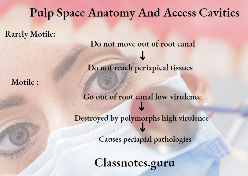
Diseases Of Pulp Important Notes

  • Diseases Of Pulp Formation Of Chloesterol Crystals
    • Crystals are formed from chloesterol released by
      • Disintegrating erthrocytes of stagnant blood vessels within the lesion
      • Lymphocytes, plasma cells and macrophages
      • The circulating plasma lipids
  • Diseases Of Pulp Phoenix Abscess
    • It is an acute inflammatory reaction superimposed on an existing chronic lesion such as cyst or granuloma
    • The exaceberation of a chronic lesion is most commonly associated with the initiation of root canal therapy in a completely asymptomatic tooth
    • Radiograph shows well-defined periradicular lesion
  • Features Of Hyperalgesia
    • Spontaneous pain
    • Allodynia
    • Increased response to painful stimuli
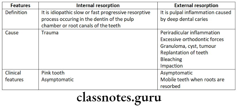
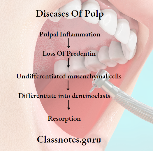
  • Hyperalgesia Internal Resorption
    • The cause is not known
      • Clinical Features:
        • Mostly asymptomatic
        • Pink spot if lesion occur in crown
        • Radiographically there is alteration or enlargement in the appearance of the pulp chamber or the wall of the root canal due to clastic activity
    • Hyperalgesia Treatment:
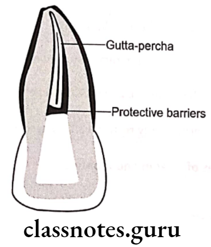
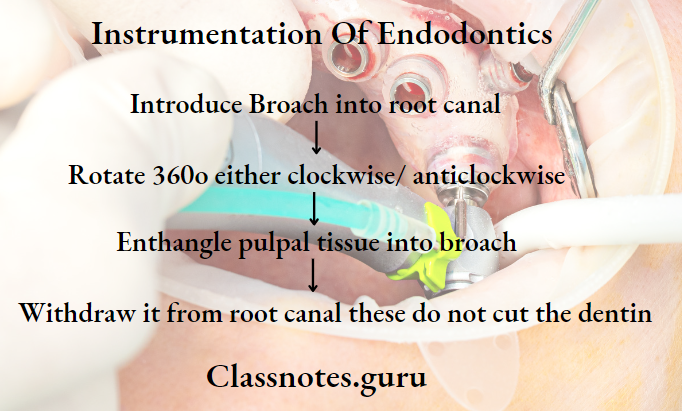
      • Extirpation by pulp stops resorption
      • Obturation of the defect requires effort and plasticized gutta percha
      • It root is perforated, the MTA should be used to repait the defect surgically
      • When repair has been completed the canal with its defect is obturated with plasticized gutta percha
  • Inflammation induces
    • Vasodilation
    • Increased capillary pressure
    • Increased vascular permeability
  • Chronic apical abscess
    • Asymptomatic or only slightly symptomatic if the sinus tract is obstructed
    • Sinus tract allows continuous drainage of pus forming in the periapical lesion through the oral mucosa
    • Most commonly associated with apical radiolucency
  • Cracked Tooth Syndrome
    • Diagnostic tests for cracked tooth syndrome:
      • Visual examination aide by use of transillumination
      • Use of methylene blue dye
      • Biting test with the use of rubber wheels, cotton tip applicator, moist cotton rolls and tooth sloth
      • Cold test and EPT
      • Radiographs
      • Ultrasound
  • Cracked Tooth Syndrome Treatment:
    • Stabilize the tooth with orthodontic band and wait for 2 weeks
      • If symptoms subsides, proceed for full coverage crown
      • If symptoms persiste, procedd for RCT and then crown
        • Extraction if tooth has poor prognosis
  • Limitations of direct pulp capping in primary teeth
    • Internal resorption
    • Calcification
    • Chronic pulp infection
    • Necrosis
    • Interradicular involvement

Diseases Of Dental Pulp

Diseases Of Pulp Long Essays

Question 1. Describe about etiology, clinical features and treatment of acute apical abscess.
Answer.

Acute Apical Abscess:

Acute Apical Abscess can be defined as a localized acute or chronic suppurative infection in the periapical region of a tooth

Read And Learn More: Endodontics Question and Answers

Acute Apical Abscess Etiology:

  • Extension of pulpal infection into periapical tissue
  • Fracture of tooth with pulp exposure
  • Accidental perforation of the apical foramen during root canal treatment
  • Extension of periodontal infection into the periapical tissues
  • Anachoretic infection of the periapical tissues

Acute Apical Abscess Clinical Features:

  • Acute abscess produces severe pain in the affected tooth
  • There will be localized swelling and an erythematous change in the overlying mucosa
  • The pain aggrevates during percussion and when pressure is applied
  • Acute Apical Abscess causes extrusion of the tooth from its socket
  • The associated tooth in non-vital and sometimes it can be mobile also
  • The affected area of the jaw may be tendered on palpation
  • Application of heat intensifies pain
  • Pus discharging sinus often develops
  • Chronic periapical abscess often produces dull pain

Acute Apical Abscess Complications:

  • Space infections
  • Septicaemia
  • Ludwig’s angina
  • Cavernous sinus thrombosis
  • Osteomyelitis

Acute Apical Abscess Radiographic Features:

  • Widening of periodontal ligament space
  • There is loss of lamina dura
  • Area of diffuse periapical rarefaction is seen
  • Margins vary from well defined to poor defined
  • In advanced cases, the trabeculae are destroyed
  • Radiolucency may involve adjacent tooth
  • Osteitis can occur at the side of root
  • Maxillary posterior teeth may lead to destruction of a portion of the antral floor
  • Roots of the affected teeth may show resorption

Diseases Of Pulp And Periapical Tissues

Acute Apical Abscess Management:

  • Emergency opening of the pulp chamber through passing file into the preiapical region
  • Through and through drain is placed in the abscess and irrigated with 1:1 mix of 3% H2O2 and normal saline solution
  • Antibiotics
    • Pencillin 500mg QID for 5 days
  • Endodontic treatment
    • Root canal treatment or extraction of offending tooth as required, is carried out in 24-48 hours.
  • Warm saline mouth rinse

Pulp Diseases In Endodontics

Diseases Of Pulp Short Essays

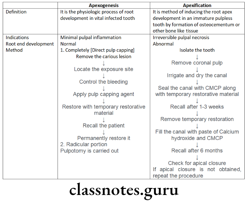
Question 1. Apexogenesis and apexification.
Answer.

Apexogenesis and apexification

Endodontics Diseases Of Pulp Apexogenesis And Apexification

Question 2. Pulpotomy.
Answer.

Pulpotomy Definition

Pulpotomy is complete removal of coronal part of dental pulp followed by placement of suitable dressing or medicament which promotes healing and preserves tooth vitality

Pulpotomy Technique

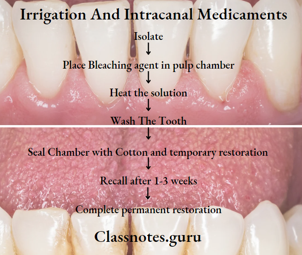
Anaesthesize and isolate the tooth

  • Remove the carious lesion
  • Entire roof of coronal pulp is removed
  • With the help of spoon excavator cut out the coronal pulp
  • Irrigate the pulp chamber
  • Control the bleeding with the help of moist cotton pellet
  • Dry the pulp chamber
  • Now place a cotton moistened with 1.5 concentration of Buckley’s formocreson solution into the canal
  • Keep it for 5 minutes
  • Now remove it and dry the canal
  • Restore the tooth with thick paste of zinc oxide eugenol
  • Place base of zinc polycarboxylate cement over it
  • Finally, permanently restored with stainless steel crown

Pulpitis Symptoms And Treatment

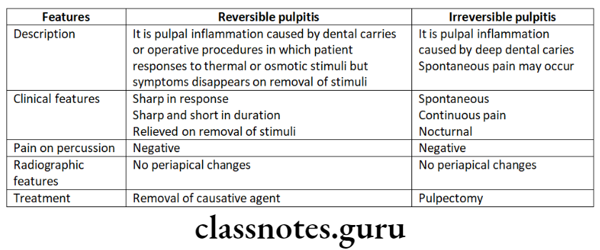
Question 3. Acute Reversible Pulpitis.
Answer.

Acute Reversible Pulpitis

Acute Reversible Pulpitis is an acute inflammatory response to noxious stimuli

Diseases Of Pulp And Periapical Tissues

Acute Reversible Pulpitis Etiology:

  • Trauma
  • Thermal injury
  • Chemical stimulus
  • Deep restoration

Acute Reversible Pulpitis Symptoms:

  • Sharp sudden pain on stimulus
  • Pain relieves on removal of stimuli

Acute Reversible Pulpitis Diagnosis:

  • Clinically – caries
  • Traumatic occlusion
  • Percussion test – Negative
  • Radiograph – Normal PDL and lamina dura
  • Vitality – Early response

Acute Reversible Pulpitis Treatment:

  • No endodontic treatment required
  • Sedative dressing placed
  • Desensitize the tooth
  • Use of cavity varnish

Endodontics Diseases Of Pulp Insertion of deep restoration causing pulp inflammation

Question 4. Phoenix Abscess.
Answer.

Phoenix Abscess

Phoenix Abscess is an acute inflammatory reaction superimposed on existing chronic lesions

Phoenix Abscess Etiology:

  • Influx of necrotic products

Phoenix Abscess Symptoms:

  • Tender tooth
  • Elevation of a tooth from socket
  • Tender soft tissue

Phoenix Abscess Diagnosis:

  • Pulp test – Negative
  • Radiograph – radiolucency at the apex

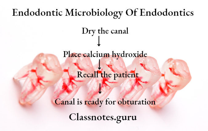
Phoenix Abscess Treatment:

  • Drainage
  • RCT

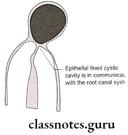
Question 5. Radicular Cyst.
Answer.

Radicular Cyst

Inflammatory cyst

Radicular Cyst Etiology:

  • Caries
  • Trauma
  • Developmental defects

Radicular Cyst Symptoms:

  • Asymptomatic
  • Gradual swelling
  • Palatal expansion
  • Non-vital tooth

Radicular Cyst Radiograph:

  • Round, pear/ovoid radiolucency
  • Narrow radiopaque margin

Radicular Cyst Treatment:

  • Endodontic treatment
  • Apicoectomy
  • Extraction
  • Enucleation

Endodontics Diseases Of Pulp Cyst formation in periapical area

Diseases Of Pulp Short Questions And Answers

Question 1. Reversible and irreversible pulpitis – differences
Answer.

Reversible And Irreversible Pulpitis

Endodontics Diseases Of Pulp Reversible and Irreversible Pulpitis

Necrosis Of Dental Pulp

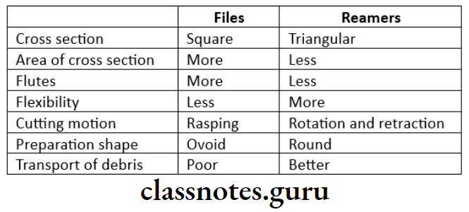
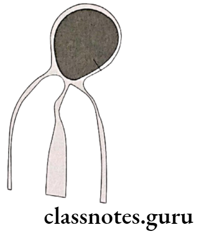
Question 2. Differences between internal and external resorption.
Answer.

Difference Between Internal And External Resorption

Endodontics Diseases Of Pulp Internal and External resorption

Question 3. Root resorption.
Answer.

Root Resorption

Resorption of teeth can be defined as chronic progressive damage or loss of tooth structure due to the action of odontoclasts

Root resorption Classification

  • Physiological:
    • Resorption of roots of deciduous teeth
  • Pathological:
    • External resorption
    • Internal resorption

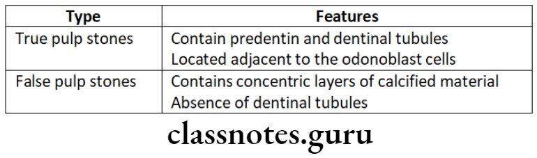
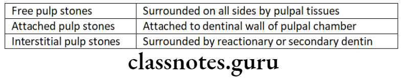
Question 4. Pulp stones.
Answer.

Pulp Stones

Deposition of calcified mass within the dental pulp for no apparent reason is called pulp calcification

Pulp Stones Types:

  • Depending On The Microscopic Structure:

Endodontics Diseases Of Pulp Depending on microscopic structure

  • Depending upon location:

Endodontics Diseases Of Pulp Depending upon location

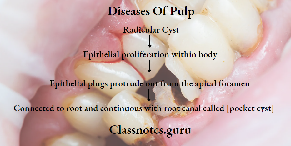
Question 5. Pocket Cyst.
Answer.

Pocket Cyst

Pocket Cyst is the variation of the radicular cyst.

Pocket Cyst Athogenesis:

Endodontics Diseases Of Pulp Pocket Cyst Pathogenesis

Endodontics Diseases Of Pulp Pocket or bay cyst

Reversible Vs Irreversible Pulpitis

Question 6. Pink tooth.
Answer.

Pink Tooth Etiology:

  • Trauma, Chronic pulpitis:

Pink Tooth Pathogenesis:

Endodontics Diseases Of Pulp Pink tooth Pathogenesis

Pink Tooth Symptoms:

  • Asymptomic
  • Appears pink due to inflamed pulp

Pink Tooth Treatment:

  • Pulp Extirpation

Endodontics Diseases Of Pulp Pink tooth treatment

Question 7. Pulpal Diseases Classification.
Answer.

Pulpitis:

  • Acute
  • Chronic

Pulpal Degeneration:

  • Calcific
  • Other

Necrosis:

Question 8. Classification of Periradicular Diseases.
Answer.

  • Acute peroradiculae diseases.
    • Acute alveolar abscess
    • Acute apical periodontitis
  • Chronic periradicular disease.
    • Chronic alveolar abscess
    • Granuloma
    • Cyst
  • Condensing osteitis
  • External root resorption
  • Diseases with non-endodontic origin

Question 9. Irreversible Pulpitis.
Answer.

Irreversible Pulpitis

Irreversible Pulpitis is a persistent inflammatory condition of pulp.

Irreversible Pulpitis Classification:

  • Acute
  • Chronic

Irreversible Pulpitis Etiology:

  • Bacteria
  • Injury to pulp

Irreversible Pulpitis Features:

  • Sharp piercing, shooting, continuous pain that remains even after removal of stimulus
  • Referred pain
  • Difficult to localize
  • D/D – Reversible Pulpitis

Irreversible Pulpitis Treatment:

  • Pulpectomy

Diseases Of Dental Pulp

Question 10. Causes of Pulpal diseases.
Answer.

Causes Of Pulpal Diseases

  • Pulpal Diseases Physical:

    • Mechanical:
      • Trauma
      • Pathologic wear
      • Crack tooth syndrome
      • Barodontalgia
      • Thermal injury
      • Electrical injury
  • Pulpal Diseases Chemical:
    • Phosphoric acid
    • Acrylic monomer
  • Pulpal Diseases Bacterial:
    • Toxins
    • Anachoresis

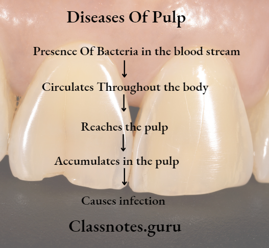
Question 11. Anachoresis.
Answer.

Anachoresis

Endodontics Diseases Of Pulp Anachoresis

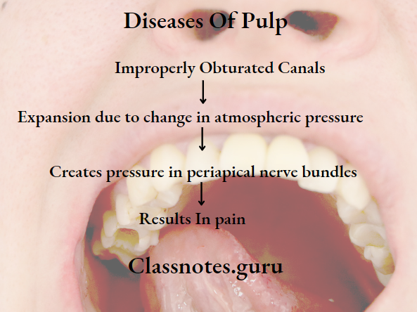
Question 12. Aerodontalgia.
Answer.

Aerodontalgia

  • Dental pain occuring due to change in altitude

Aerodontalgia Features:

  • Asymptomatic at ground level
  • Pain occurs after few hours of reaching high altitude or into deep sea

Aerodontalgia Reason:

Endodontics Diseases Of Pulp Aerodontalgia Reason

Question 13. Reversible Pulpitis.
Answer.

Reversible Pulpitis

Reversible Pulpitis is an acute inflammatory response to noxious stimuli

Reversible Pulpitis Etiology:

  • Trauma
  • Thermal injury
  • Chemical stimulus
  • Deep restoration

Reversible Pulpitis Symptoms:

  • Sharp sudden pain on stimulus
  • Pain relieves on removal of stimuli

Reversible Pulpitis Diagnosis:

  • Clinically – caries
  • Traumatic occlusion
  • Percussion test – Negative
  • Radiograph – Normal PDL and lamina dura
  • Vitality – Early response

Reversible Pulpitis Treatment:

  • No endodontic treatment is required
  • Sedative dressing placed
  • Desensitize the tooth
  • Use of cavity varnish

Pulp Diseases In Endodontics

Question 14. Acute alveolar abscess
Answer.

Acute Alveolar Abscess

  • Acute alveolar Abscess is a localized collection of pus in the alveolar bone at the root apex of the tooth following the death of pulp with the extension of the infection through the apical foramen into periarticular tissue

Acute Alveolar Abscess Etiology:

  • Invasion of bacteria from necrotic pulpal tissue
  • Trauma, chemical or mechanical
  • Chemical or mechanical treatment causing periapical tissue irritation during root canal treatment

Acute Alveolar Abscess Clinical Features:

  • Fever, increased WBC count
  • Tooth is non-vital
  • Rapid onset of pain
  • Slight tenderness to intense throbbing pain
  • Marked pain to biting
  • Swelling
  • Mobility

Acute Alveolar Abscess Treatment:

  • Drainage of abscess
  • Incision and drainage
  • Extraction

Diseases Of Pulp Viva Voce

  • In acute apical abscess, pain is due to the build-up of pressure periodically due to the accumulation of pus
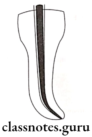
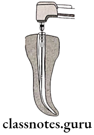
  • Internal resorption is also known as Pink tooth of mummery
  • The sinus tract prevents exacerbation of the lesion by providing continuous drainage of the periradicular lesion
  • Anachoresis refers to the attraction or fixation of blood-borne bacteria in areas of inflammation
  • Internal resorption is due to asymptomatic and chronic irreversible pulpitis
  • Abnormally exaggerated subjective response to painful stimuli is called hyperpathia
  • Allodynia is spontaneous pain
  • Dysasthesia pain arises from the pulp and periradicular tissues
  • The hallmark sign of neuropathic pain is hyperalgesia
  • Enamel cracks can be better disclosed by using a dye or by transilluminating the tooth with fiber-optic
  • Chronic pulpitis is difficult to diagnose because the patient does not have any subjective symptoms
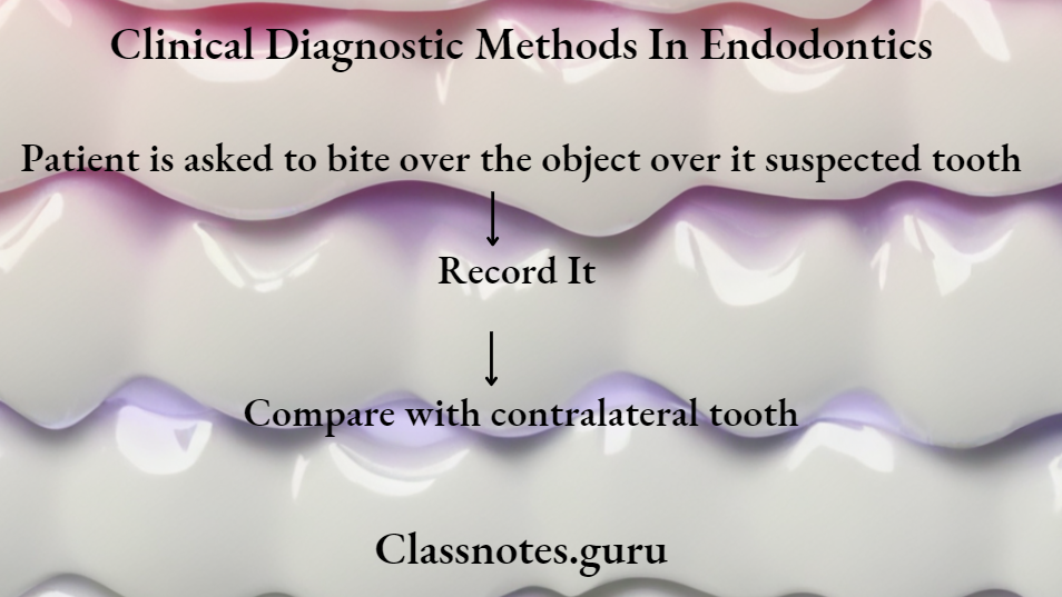
  • Diagnosis of cracked tooth syndrome is made by biting on a solid object and releasing pressure