White Blood Cell Disorders Question And Answers

Disorders Of White Blood Cells Important Notes

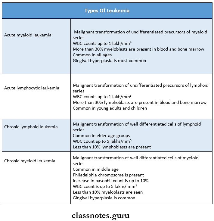
1. Types of leukemia

Disorders Of White Blood Cells Types Of Leukemia

2. Syndromes associated with leukemia

  • Down’s syndrome
  • Bloom’s syndrome
  • Klinefilter’s syndrome
  • Wiskott-Aldrictis syndrome
  • Fanconi’s anemia

3. Infectious mononucleosis or glandular fever

  • Caused by Epstein Barr virus
  • Characterized by atypical leukocytes consisting of
    • Oval or horseshoe nuclei
    • Irregular nuclear chromatin
    • Basophilic foamy or vacuolated cytoplasm

Read And Learn More: Pathology Question And Answers

  • Diagnostic tests
    • Monospot test
    • Paul Bunnel test
      • The normal titer of agglutinin and hemolysin in human blood against sheep blood cells does not exceed 1:8
      •  In a positive Paul Bunnel test, the titer may rise to 1:4096

4. Agranulocytosis

  • Mostly occurs due to the ingestion of drugs like
    • Amidopyrine
    • Barbiturates
    • Chloramphenicol
    • Quinine
    • Sulfonamides
  • Features
    • Presence of infection in the oral cavity, GIT, genitourinary tract, respiratory tract, and skin
    • Oral manifestation
    • Necrotizing ulcerations of the oral mucosa, pharynx, tonsils
    • Rapid destruction of supporting tissues of the teeth

5. Cyclic neutropenia

  • It is characterized by periodic cyclic diminution of leukocytes
  • Cycle commonly occurs at every 3 weeks
  • Loss of alveolar bone around the teeth is an important oral manifestation

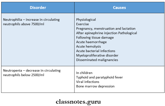
6. Disorders of neutrophil count

Disorders Of White Blood Cells Disorders Of Neutrophil Count

7. Leukaemoid reactions

  • It is defined as a reactive excessive leucocytosis in the peripheral blood resembling that of leukemia
  • It may be myeloid or lymphoid
  • The myeloid type is a more common form
  • Myeloid leukemoid reactions are seen in
    • Infections like staphylococcal pneumonia, TB, meningitis, diphtheria, sepsis, endocarditis
    • Intoxication
    • Malignant diseases like multiple myeloma
    • Severe hemorrhage and hemolysis
  • Lymphoid leukemoid reaction are seen in
    • Infections like infectious mononucleosis, cytomegalovirus infection, pertussis
    • Malignant diseases rarely produce it

8. Most commonly affected bones in multiple myeloma are:

  • Skull
  • Spine
  • Ribs
  • Pelvis

Disorders Of White Blood Cells Long Essays

Question 1. Classify leukemia. Describe the clinical picture, blood, and bone marrow findings in acute leukemia.
Answer:

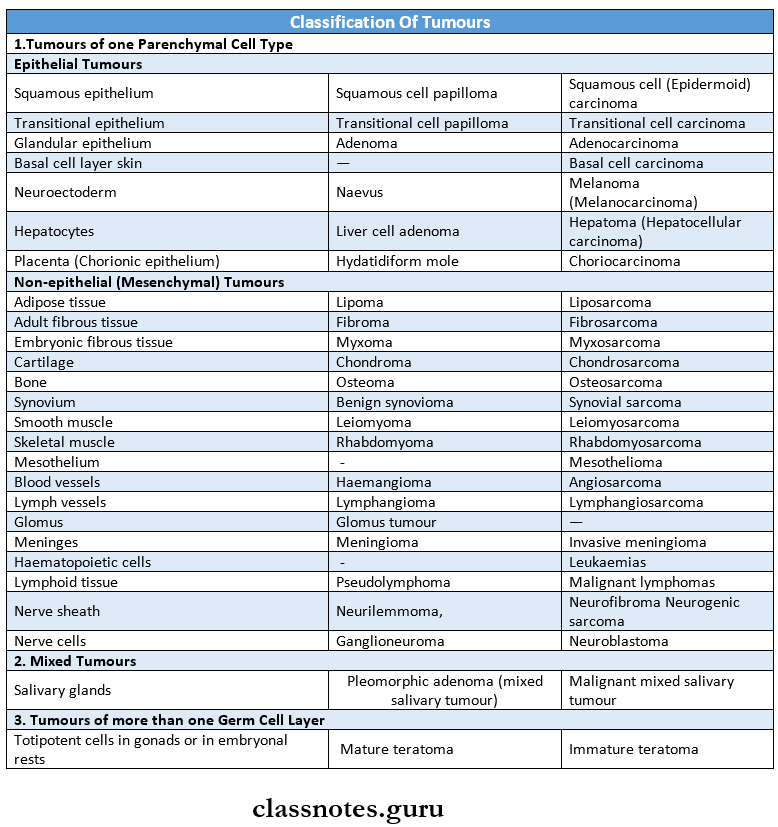
Leukemia Classification:

1. Based on cell types predominantly involved.

  • Myeloid
  • Lymphoid.

2. Based on the natural history of the disease:

  • Acute
  • Chronic.

WHO classification of myeloid neoplasm:

1. Myeloproliferative Diseases:

  • Chronic myeloid leukaemia (CML], {Ph chromo¬some t(9;22) (q34;ll), BCR/ABL-positive}
  • Chronic neutrophilic leukemia
  • Chronic eosinophilic leukemia/ hypereosinophilic syndrome
  • Chronic idiopathic myelofibrosis
  • Polycythaemia vera (PV]
  • Essential thrombocythaemia (ET)
  • Chronic myeloproliferative disease, unclassifiable

2. Myelodysplastic/Myeloproliferative Diseases: Chronic myelomonocytic leukemia (CMML)

3. Myelodysplastic Syndrome (MDS):

  • Refractory anemia (RA)
  • Refractory anemia with ring sideroblasts (RARS)
  • Refractory cytopenia with multilineage dysplasia (RCMD)
  • RCMD with ringed sideroblasts (RCMD-RS)
  • Refractory anemia with excess blasts [RAEB-1)
  • RAEB-2
  • Myelodysplastic syndrome unclassified (MDS-U)
  • MDS with isolated del 5q

4. Acute Myeloid Leukaemia (AML):

  • AML with recurrent cytogenetic abnormalities
    • AML with t(8;21)(q22;q22)
    • AML with abnormal bone marrow eosinophils {inv(16][pl3q22]}
    • Acute promyelocytic leukaemia {tC15;17](q22;ql2)}
    • AML with llq23 abnormalities (MLL)
  • AML with multilineage dysplasia
    • With prior MDS
    • Without prior MDS
  • AML and MDS, therapy-related
    • Alkylating agent-related
    • Topoisomerase type 2 inhibitor-related
    • Other types
  • AML, not otherwise categorized
    • AML, minimally differentiated
    • AML without maturation
    • AML with maturation
    • Acute myelomonocytic leukemia (AMML)
    • Acute monoblastic and monocytic leukemia
    • Acute erythroid leukemia
    • Acute megakaryocytic leukemia
    • Acute basophilic leukemia
    • Acute panmyelosis with myelofibrosis
    • Myeloid sarcoma

5. Acute Biphenotypic Leukaemia

Acute Leukemia: It is a leukemia characterized by predominant undifferentiated leucocytes precursors

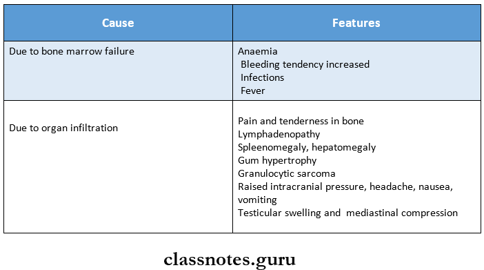
Acute Leukemia Clinical Features:

Disorders Of White Blood Cells Acte Leukemia Cilinal Features

Acute Leukemia Blood Picture:

  • Moderate reticulocytosis
  • Normochromic anemia
  • Increased platelet count
  • WBCs count is more than 100000 per microlitre

Acute Leukemia Bone Marrow Examination:

  • Cellularity- Bone marrow is hypercellular
  • It shows predominantly myeloblasts and premyelo¬cytes
  • Leukemic cells are demonstrated by Romanowsky stains
  • Erythropoiesis shows
    • Reduced erythropoietic cells
    • Dyserythropoiesis,
    • Megaloblastic features
    • Ring sideroblasts
    • Megakaryocytes

Question 2. Define leukemia. Describe the etiology, clinical features, blood, and bone marrow picture of chronic myeloid leukemia.
Answer:

Leukemia: leukemia are a group of disorders arising from the malignant transformation of hematopoietic cells leading to an increased number of white blood cells in blood and/or bone marrow.

Chronic myeloid leukemia:

Chronic myeloid leukemia Etiology: It is a myeloproliferative disorder.

  • Occurs as a result of the malignant transformation of plea- potent stem cells leading to the accumulation of a large number of immature leukocytes in the blood.
  • Radiation exposure and genetic factors have been implicated in the development of CML

Chronic myeloid leukemia Clinical features:

  • Onset is usually slow, initial symptoms are often non-specific example: weakness, pallor dyspnoea, and tachycardia.
  • Symptoms due to hypermetabolism such as weight loss, anorexia, and night sweats.
  • Splenomegaly is almost always present and is frequently massive. In some patients, it may be associated with acute pain due to splenic infarction.
  • Bleeding tendencies such as bruising, epistaxis, menorrhagia, and hematomas may occur.
  • Visual disturbance, neurologic manifestations.
  • Juvenile CML is more often associated with lymph node enlargement than splenomegaly.

Peripheral blood picture of CML:

  • Leucocyte count is elevated often > 1,00,000 cells/pl.
  • Circulating cells are predominantly neutrophils, metamyelocytes, and myelocytes but basophils and eosinophils are also prominent.
  • A typical finding is an increased number of platelets (thrombocytosis).
  • Anaemia is usually of moderate degree and is normocytic, normochromic in type. Normablasts may be present occasionally.
  • A small portion of myeloblasts usually < 5% are seen.

Bone marrow examination:

  • Cellularity – Hyper cellular with total/partial re-placement of fat spaces by proliferating myeloid cells.
  • Myeloid cells -Myeloblasts are only slightly increased.
  • Erythropoiesis -Normoblasts but there is a reduction in erythropoietic cells.
  • Megakaryocytes – Conspicuous but are usually smaller in size than normal.
  • Increase in number of phagocytes.

Disorders Of White Blood Cells Short Essays

Question 1. Leukocytosis
Answer:

Leukocytosis is an increase in the number of white cells & is common in a variety of reactive inflammatory states caused by microbial and non-microbial stimuli.

Leukocytosis Causes: Leukocytosisare relatively non-specific and can be classified on the basis of particular white cell series affected as follows:

Causes of Leukocytosis: Leukocytosis are relatively non-specific and can be classified on the basis of particular white cells series affected as follows:

1. Neutrophilic leucocytosis.

  • Cute bacterial infections especially those caused by pyogenic organisms.
  • Sterile inflammation caused by tissue necrosis (myocardial infarction, burns)

2. Eosinophylic leucocytosis (eosinophilia)

  • Allergic disorders such as asthma, hay fever
  • Allergic skin diseases, for example., pemphigus, dermatitis herpetiform.
  • Parasitic infestations
  • Drug reactions
  • Certain malignancies, for example., Hodgkin’s disease and some non-hodking’s lymphomas.
  • Collagen vascular disorders and some vasculitis.

3. Basophilic leucocytosis (basophilia): Rare often indicates CML.

4. Monocytosis.

  • Chronic infections, ie.g., tuberculosis
  • Bacterial endocarditis
  • Rickettsiosis and malaria
  • Collagen vascular diseases, for example., systemic lupus erythematosus (SLE)
  • Inflammatory bowel diseases, for example., ulcerative coli¬tis

5. Lymphocytosis: Usually accompanies monocytosis in many disorders associated with it.

  • Chronic immunologic stimulation, for example., tuberculo¬sis, brucellosis and
  • Viral infections, for example., hepatitis A, cytomegalovirus, Epstein-Barr virus, and bordetella pertussis infections.

Question 2. Causes of neutrophilic leukocytosis
Answer:

  • Acute bacterial infections especially those caused by pyogenic organisms.
  • Sterile inflammation caused by tissue necrosis (myocardial infarction, burns)

Question 3. Agranulocytosis
Answer:

  • The term agranulocytosis is used to describe a state of severe neutropenia.
  • A reduction in the number of granulocytes in the blood is known as neutropenia.

Agranulocytosis Causes: Neutropenia can be caused by the following reasons.

  • Drug-induced: Anti-cancer, antibiotics, anticonvulsants, antithyroid drugs
  • Hematological disease: Aplastic anemia, acute leukemia, etc.
  • Infections: Malaria, TB, typhoid,
  • Autoimmune: Systemic lupus erythematosus
  • Congenital: Cyclic neutropenia.

Agranulocytosis Clinical features:

  • Initially patient develops malaise, chills, and fever, with subsequent marked weakness and fatiguability.
  • Massive growth of microorganisms due to inability to produce leukocyte response.
  • Infections are usually present as ulcerating, necrotizing lesions of the gingiva, the floor of the mouth, buccal mucosa, and other sites within the oral cavity known as agranulo¬cytic angina.
  • Lymphadenopathy and hepatosplenomegaly may be present.

Agranulocytosis Treatment:

  • Patients with neutropenia and fever must be sent to the hospital.
  • In severe infections, neutrophil transfusion must be done.
  • Broad-spectrum antibiotics and antifungal drugs are to be given.

Question 4. Leukemoid reaction
Answer:

  • Leukaemoid reaction is defined as the reactive excessive leucocytosis in peripheral blood resembling that of leukemia in the subject who does not have leukemia.
  • Leukaemoid reaction may be myeloid or lymphoid.

Myeloid leukemoid reaction:

  • The majority of leukemoid reactions involve the granulocyte series.
  • It may occur in infection, intoxication, malignant diseases, severe hemorrhages, and severe hemolysis.

Lymphoid leukemoid reaction: It is found in conditions such as infectious mononucleosis, whooping cough, chicken pox, measles, and tuberculosis.

Agranulocytosis Lab diagnosis:

  • Leukocytosis
  • Infective cases may show toxic granulation and Dohle bodies in the cytoplasm of neutrophils.

Question 5. Peripheral blood picture of acute lymphatic leukemia
Answer: Acute lymphatic leukemia shows

1. Anaemia:

  • Normochromic
  • Shows few nucleated cells

2. Thrombocytopenia: Platelet count decreases to 100000 cells

3. White blood cells:

  • WBC count is variable
  • It may exceed 100000 cells or may be less than that
  • Leukocytes appear as blasts cells

Disorders Of White Blood Cells Short Questions And Answers

Question 1. Eosinophilia
Answer:

  • An increase in the number of eosinophilic leukocytes is referred to as eosinophilia.
  • The causes of eosinophilia are as follows:
  • Allergic disorders: Bronchial asthma, urticarial, drug hypersensitivity
  • Parasitic infestations: Trichinosis, echinococcosis, intestinal parasitism.
  • Skin diseases: Pemphigus, dermatitis herpetiformis, erythema parasitism.
  • Certain malignancies: Hodgkin’s diseases and some non-Hodgkin’s lymphomas.
  • Pulmonary infiltration which eosinophilia syndrome
  • Irradiation.
  • Miscellaneous disorders: Sarcoidosis, rheumatoid arthritis, polyarteritisnodosa.

Question 2. Blood picture in chronic myeloid leukemia
Answer:

  • Leucocyte count is elevated often > 1,00,000 cells/Bll.
  • Circulating cells are predominantly neutrophils, metamyelocytes, and myelocytes but basophils and eosinophils are also prominent
  • The typical finding is an increased number of platelets (thrombocytosis).
  • Anaemia is usually of moderate degree and is normocytic, normochromic in type. Normablasts may be present occasionally.
  • A small portion of myeloblasts usually < 5% are seen.

Question 3. Peripheral smear layer findings in acute leu¬kemia
Answer:

1. Anaemia:

  • Always present generally severe, progressive, and normochromic.
    • A moderate reticulocytosisupto 5% and a few nucleated red cells may be present.

2. Thrombocytopenia:

  • Platelet count < 50,000/pl
    • When the platelet count is below 20,000/01 serious spontaneous haemorrhagic episodes develop.
    • Acute promyelocytic leukemia may be associated with a serious coagulation abnormality called dis-seminated intravascular coagulation.

3. White blood cells:

  • In advanced cases, WBC > 1,00,000/pl
    • The majority of leucocytes in the peripheral blood are blasts and there is often neutropenia due to mar¬row infiltration by leukemic cells.

Question 4. Burkitt’s lymphoma
Answer:

  • Burkitt’s lymphoma is a rare type of non-Hodgkin’s lymphoma (NHL).
  • Epstein-Barr virus (EBV) and HIV are associated with the development of Burkitt’s lymphoma.
  • This is the most rapidly progressive tumor first described in African children.
  • Most patients present with lymphadenopathy and abdominal mass.
  • It has a tendency to metastasize to the CNS.
  • Treatment should be initiated promptly with intensive chemotherapy. Prophylactic chemotherapy is also given. 70-80% of patients may be cured.

Question 5. Multiple myeloma
Answer: Multiple myeloma is a malignant disease arising from the neoplastic transformation of plasma cells of mono-clonal origin

Multiple Myeloma Clinical Features:

  • Age- in older age
  • Anaemia
  • Bone pain
  • Infections
  • Pathological fractures
  • Renal failure
  • Spinal cord compression

Multiple Myeloma Investigations:

  • Bence- Jones proteins in urine
  • Reversal of serum albumin/globulin ratio
  • Increase in total serum proteins

Multiple Myeloma Treatment:

  • Radiotherapy
  • Biphosphonates
  • Autologous peripheral cell transplantation
  • Administration of thalidomide and proteasome inhibi¬tors
  • Chemotherapy includes:
    • Melphalan
    • Cyclophosphamide
    • Doxorubicin
    • Dexamethasone

Diseases Of Cardiovascular Disorders Important Notes

1. Clinical patterns of angina

  • Stable or typical angina
  • Prinzmetal’s variant angina
  • Unstable or crescendo angina

2. Forms of Creatine phosphokinase

  • CK-MM-Derived from skeletal muscle
  • CK-BB-Derived from the brain and lungs
  • CK-MB-Derived from cardiac muscle

3. TypesofischaemicheartdiseaseJHD

  • Stable angina
  • Unstable angina
  • Chronic IHD
  • Myocardial infarction
  • Sudden ischaemic death

Diseases Of Cardiovascular Disorders Short Question And Answers

Question 1. Hyperlipidemia
Answer:

  • Hyperlipidaemia is a major risk factor for atherosclerosis
  • It occurs due to
    • Diabetes mellitus
    • Myxoedema
    • Nephritic syndrome
    • Von Gierke’s disease
    • Xanthomatosis
    • Familial hypercholesterolemia
  • Dietary regulation and administration of cholesterol-lowering drugs have beneficial effects on reducing the risk of ischaemic heart diseases

Question 2. Atheromatous plaques
Answer: A fully developed atherosclerotic lesion is called atheromatous plaque

Atheromatous plaques Appearance:

1. Gross appearance

  • Appear as white to the yellowish-white lesion
  • Size-1-2 cm in diameter
  • Appears raised on the surface

2. Cut surface

  • Shows luminal surface as a firm, white fibrous cap
  • The central core is composed of yellow to yellow-white soft material called atheroma

3. Microscopic

  • The fibrous cap is covered by endothelium
  • It is composed of smooth muscles, dense connective tissue, and extracellular matrix
  • The cellular area contains macrophages, foam cells, lymphocytes and smooth muscle cells
  • The deeper central soft core consists of extracellular lipid material, cholesterol cleft, Fibrin, neurotic debris, and lipid-laden foam cells

Specific Systemic Disorder Question And Answers

Oral Medicine Specific Systemic Disorder Important Notes

1. Multinucleated Giant Cells Are Seen In

  • Cherubism
  • Hyperparathyroidism ‘
  • Aneurysmal bone cyst
  • Giant cell granuloma
  • Osteoclastoma
  • Osteosarcoma
  • Herpes
  • Leprosy
  • Eosinophilic granuloma

2. Specific Systemic Russell’s Bodies

  • They are immature plasma cells
  • Seen in
    • Chronic inflammatory disease
    • Multiple myeloma
    • Periapical granuloma

3. Specific Systemic Infectious Mononucleosis

  • Caused by Epstein Burr virus
  • Transmitted through oropharyngeal secretion through deep kissing
  • Oral excretion of the virus may continue for as long as 18 months following the onset of the disease

Oral Medicine Specific Systemic Disorder Short Essays

Question 1. Infective endocarditis.

Answer:

Infective Endocarditis

  • Infective Endocarditis is a serious disorder which is the most common bacterial origin

Infective Endocarditis Pathogenesis:

Specific Systemic Disorder

Infective Endocarditis Predisposing Factors:

  • Rheumatic/congenital heart disease
  • Recent surgical correction of the congenital valvular defect within 6 months
  • Hypertrophic cardiomyopathy
  • Surgical trauma

Infective Endocarditis Clinical Features:

  • Age – middle age group

Read And Learn More: Oral Medicine Question and Answers

Infective Endocarditis  Features:

  • Progressive weakness
  • Loss of weight
  • Dyspnea
  • Anorexia
  • Muscular and joint aches and pains
  • Low-grade fever
  • Petechiae hemorrhage in the conjunctiva and oral mucosa

Infective Endocarditis Management:

  • 20,000,000 units of penicillin in combination with gentamicin for 2 weeks
  • Early removal of infected valve with a sterile replacement

Infective Endocarditis Prevention:

  • Proper history should be taken from the patient
  • Administer the prophylactic antibiotic therapy before dental treatment
  • Consult the physician
  • Make the patient rinse with an antibacterial mouthwash
  • Use of a traumatic dental procedure

Question 2. Oral manifestations of renal diseases.

Answer:

Oral Manifestations Of Renal Diseases

  • Patients – may complaints of ammonic taste and smell due to a high concentration of urea in saliva
  • Xerostomia due to dehydration and mouth breathing
  • Oral mucosa is reddened and covered with thick exudates and a pseudomembrane
  • Stomatitis appears as frank ulceration with a red coat
  • Low caries index
  • Enamel hypoplasia
  • Pulpal narrowing and calcification
  • Severe tooth erosion
  • Loss of lamina dura

Question 3. Renal Osteodystrophy.

Answer:

Renal Osteodystrophy Clinical Features:

  • Muscle cramps are more common
  • Sensory neuropathy may cause paresthesia
  • Motor neuropathy may present as foot drop
  • Autonomic neuropathy may cause delayed gastric emptying, diarrhea, and postural hypertension
  • There may be hyperprolactinemia and hyperparathyroidism
  • There is a loss of libido and sexual functions
  • Growth retardation occurs
  • Bone fractures occur more frequently
  • CVS – Hypertension
    • Atherosclerosis
  • Skeletal – Gradual softening and bowing of bone

Radiographic Features:

  • Thinning of the bony cortex
  • Loss of lamina dura
  • The thickness of the mandibular cortex is reduced
  • Increase in medullary space

Renal Osteodystrophy Management:

  • Vitamin D supplement
  • A diet with high phosphate content is advised

Oral Medicine Specific Systemic Disorder Short Answers

Question 1. Bronchial Asthma.

Answer:

Bronchial Asthma

Bronchial Asthma is a spontaneously reversible spasmodic contraction of the smooth muscles of the bronchi resulting in bronchiolar narrowing

Bronchial Asthma Types:

  • Extrinsic asthma
  • Intrinsic asthma
  • Mixed asthma
  • Status asthmaticus

Bronchial Asthma Clinical Features:

  • Age and Sex – Common in young boys
  • Features:
    • The sensation of chest fullness
    • Increased heart rate
    • Dehydration
    • Wheezing, coughing, shortness of breath
    • Extreme fatigue
    • Severe hypoxia
    • Cyanosis

Bronchial Asthma Management:

  • Terbutaline-like drugs to prevent bronchial smooth muscle constriction
  • Xanthine derivatives like aminophylline
  • Corticosteroid like hydrocortisone
  • Emergency management
  • Inhalation of a solution containing 0.1 mg isoproterenol or 1:1000 epinephrine by nebulizer or
  • Injection of 0.1 ml of 1:1000 epinephrine

Question 2. Vasovagal Syncope.

Answer:

Vasovagal Syncope Features:

  • Nausea, vomiting
  • Rapid heart rate
  • Decreased blood pressure
  • Papillary dilation
  • Hyperpnea
  • Coldness in hand and feet
  • Loss of consciousness
  • Dizziness
  • Pallor
  • Weakness and sweating

Vasovagal Syncope Management:

  • The patient should be made to lie down in a supine position with legs raised
  • Loosen the tight clothing
  • 100% oxygen is administered
  • An ammonia ampule is crushed and held under the patient’s nose
  • After complete recovery, the patient should be slowly brought to semi reclined position.

Question 3. Brown’s tumor

Answer:

Brown’s Tumor

Brown’s Tumor is an endocrine disorder occurring due to an excess of circulating parathyroid hormone

Brown’s Tumor Clinical Features:

  • Age and Sex – Common in middle-aged women
  • Classic triad:
    • Kidney stones
    • Bone resorption
    • Duodenal ulcers
  • Renal symptoms:
    • Renal calculi
    • Hematuria
    • Back pain
    • Psychological symptoms – Emotionally unstable
    • GIT symptoms – Anorexia, nausea, vomiting
  • Skeletal:
    • Bone pain a Pathologic fractures
    • Bone deformities
    • Hypercalcemia
  • Generalized symptoms:
  • Muscle weakness
  • Fatigue
  • Weight loss
  • Insomnia
  • Headache
  • Polydipsia and polyuria

Brown’s Tumor Oral Manifestations:

  • Brown tumor – Intraoral/Extraoral swelling appears

Brown’s Tumor Teeth:

  • Gradual loosening
  • Drifting and loss of teeth
  • Malocclusion

Brown’s Tumor Management:

  • Surgical – Hyperplastic tissue is removed
  • Vitamin D – Oral administration of vitamin D
  • Parathyroidectomy
  • Restriction of dietary phosphate, phosphate binding agent, and aluminum salts

Oral Medicine Specific Systemic Disorder Viva Voce

  1. The palate is the most common oral site for Kaposi’s sarcoma
  2. Cafe au lait pigmentations are found in neurofibromatosis
  3. Amoxicillin is the drug of choice for rheumatic heart disease
  4. Epileptic seizures found in children are called petit mal
  5. The tonic phase of epilepsy is associated with cyanosis
  6. Reiter’s disease is caused by chlamydia trachomatis

Disorders Of Platelets Question And Answers

Disorders Of Platelets Important Notes

1. Partial thromboplastin time

  • Used to measure the intrinsic factors as well as factors common to both intrinsic and extrinsic pathways
  • The normal range is – 30-40 sec
  • Causes of prolonged partial thromboplastin time are
  • Parenteral administration of heparin
    • DIC
    • Liver disease
    • Circulating anticoagulants

2. Thrombin time

  • It is a semi-quantitative test for fibrinogen deficiency
  • Normal range – 20-30 sec
  • Common causes of prolonged thrombin time are
    • Presence of heparin
    • DIC

3. Bleeding time

  • Normal range – 3-8 min
  • Prolonged in
    • Thrombocytopenia
    • Von Willebrand disease
    • Vascular abnormalities
    • Disorders of platelet functions

4. Clotting time

  • Normal range – 4-9 min
  • Prolonged in
    • Hemophilia
    • Vitamin K deficiency
    • Liver diseases
    • Anticoagulant administration

5. Von Willebrand disease

  • It occurs due to qualitative defect in Von Willebrand factor in blood
  • It is characterized by
    • Normal platelet count
    • Prolonged bleeding time
    • Defective platelet aggregation
    • Reduced factor 8 activity

6. DIC (Disseminated Intravascular Coagulation)

  • It is characterized by systemic activation of the blood coagulation system
  • Results in the generation and deposition of fibrin leading to Microvascular thrombi in various organs
  • Severe bleeding complications occur
  • It is seen in promyelocytic leukaemia

Disorders Of Platelets

Disorders Of Platelets Long Essays

Question 1. What is disseminated intravascular coagulation? Describe its etiopathogenesis.
Answer:

Disseminated Intravascular Coagulation: Disseminated intravascular coagulation is a complex thrombo-hemorrhagic disorder occurring as a secondary complication in some systemic diseases

Disseminated Intravascular Coagulation Etiopathogenesis: Pathogenesis of DIC includes the following events

Read And Learn More: Pathology Question And Answers

1. Activation of coagulation: Etiological factors like massive tissue injury, presence of infections, and endothelial damage cause activation of coagulation by the release of tissue factor

2. Thrombotic phase:

  • Endothelial damage from various thrombogenic stimuli causes:
    • Generalized platelet aggregation
    • Platelet adhesion
    • Deposition of small thrombi and emboli throughout microvasculature

3. Consumption phase: Consumption of coagulation factors and platelets occurs

4. Secondary fibrinolysis:

  • The fibrinolytic system is secondary activated
  • This causes the breakdown of fibrin resulting in the formation of FDPs in circulation

Disorders Of Platelets Intravascular Coagulation

Disorders Of Platelets Short Essays

Question 1. Bleeding time
Answer:

  • Bleeding time (BT] is defined as the time lapse between skin puncture and the arrest of bleeding.
  • BT is the time from the onset of bleeding to the stoppage of bleeding. The bleeding stops due to the formation of a temporary haemostatic plug.
  • Procedure: Under aseptic conditions, give a deep finger prick and note the time. Then remove the blood drop every 15 s with a clotting paper (along the edges] at a different spot till the bleeding stops. Count the number of spots and calculate BT as follows:
  • BT = Initial time + (number of spots – 1] x 15 s

Bleeding Time Indications:

  • It is a useful screening test in patients with a history of prolonged bleeding.
  • In patients with bleeding disorders before any surgical procedures.

Bleeding Time Interpretation: An abnormal BT is usually the result of

  • Abnormalities in the structure or ability of capillary blood vessels to contract or
  • Abnormalities in the number or functional integrity of the platelets.

Question 2. Thrombocytopenia
Answer: Thrombocytopenia is defined as a reduction in the peripheral blood platelet count below the lower limit of normal i. e., below 1,50,000/microlitre

  • Platelet counts in the range of 20,000 – 50,000 cells/microlitre are associated with an increased risk of post-traumatic bleeding.
  • Spontaneous bleeding is evident when the count falls below 20,000 cells/microlitre

Thrombocytopenia Causes:

1. Impaired platelet production:

  • Generalised bone marrow failure.
  • Aplastic anaemia, leukaemia, megaloblastic anaemia, marrow infiltration.
  • Selective suppression of platelet production.
    • Drugs (Anticancer, cytotoxic, alcohol], Infection (HIV, measles].

2. Accelerated platelet destruction:

  • Immunologic destruction.
    • Autoimmune – ITP, SLE
    • Drug associated – Heparin, sulfa compounds.
    • Infections – HIV, cytomegalovirus infections.
  • Non – immunologic destruction,
    • DIC
    • Gaint haemangiomas
    • TTP

3. Splenic Sequestration: Splenomegaly.

4. Dilutional loss: Massive transfusion of old stored blood to bleeding patients.

Thrombocytopenia Clinical features:

  • Usual manifestations are petechial haemorrhage, purpura, easy bruising, epistaxis, mucosal bleeding such as menorrhagia in women, nasal bleeding, bleeding from gums, and haematuria.
  • Intracranial haemorrhage is rare.
  • Splenomegaly and hepatomegaly may also occur.

Thrombocytopenia Investigations:

  • Low platelet counts,
  • Prolonged bleeding times,
  • Normal coagulation profile
  • Abnormal long clot retraction time.

Thrombocytopenia Treatment:

  • Platelet transfusion.
  • Treatment of the underlying cause.

Question 3. Hemophilia
Answer:

  • Haemophilia is an X-linked recessive disorder characterised by the deficiency of factor 8 (Haemophilia A] classic haemophilia]
  • Whereas inherited deficiency of factor 9 (Christmas factor/plasma thromboplastin component] produces Christmas disease/haemophilia B (Christmas disease].

Hemophilia A: Pathogenesis: It is caused by quantitative reduction of factor 8 in 90% of cases while 10% have normal/increased levels of factor 8 but reduced activity.

Hemophilia A Clinical features:

  • In severe cases -Bleeding is spontaneous.
  • In mild disease – Bleeding is rarely spontaneous
  • Bleeding occurs mainly in joints (Haemarthrosis), muscles (haematoma), and viscera/in retroperitoneum but can involve any organ system.
  • Spontaneous intracranial haemorrhage and oro-pharyngeal bleeding are rare, but when they occur they are the most feared complications.

Hemophilia A Treatment: Factor 8 replacement therapy, consisting of factor 8 concentrates/plasma cryoprecipitates.

Haemophilia B:

  • Haemophilia B is rarer than haemophilia A.
  • Inheritance patterns and clinical features of factor 9 are indistinguishable from those of classic haemophilia but accurate laboratory diagnosis is critical since haemophilia B requires treatment with different plasma fractions.

Hemophilia B: Treatment: infusion of either fresh frozen plasma/plasma enriched with factor 9

Question 4. Von Willebrand’s disease
Answer:

Von Willebrand’s Disease: This is the most common hereditary coagulation disorder occurring due to qualitative/quantitative defects in Von Willebrand’s factor.

  • Von Willebrand factor is a multimeric plasma glycoprotein synthesized by megakaryocytes and endothelial cells. It serves two major functions as follows.
  • Acts as a carrier protein for factor 8.
  • Helps in the adhesion of platelets to subendothelial collagen.

Von Willebrand’s Disease Clinical features:

  • Spontaneous bleeding from mucous membranes and excessive bleeding from wounds.
  • Gastrointestinal bleeding.
  • Epistaxis, menorrhagia, and superficial bruises are present

Von Willebrand’s Disease Lab investigations:

  • Prolonged bleeding time,
  • Normal platelets count,
  • Bleeding time increased,
  • Reduced vWF levels, factor 8 activity reduced.

Von Willebrand’s Disease Treatment:

  • Factor 8 and vWF concentrate transfusion,
  • The bleeding episodes can be managed by giving vasopressin which increases the VWF levels.
  • Persistent bleeding is treated with factor 8 concentrate.
  • Cryoprecipitate transfusion
  • Antifibrinolytic agent
  • Example: Tanexamie acid is useful in conjunctive therapy during dental procedures.

Question 5. Pancytopenia
Answer: Pancytopenia means the occurrence of anaemia, leukopenia and thrombocytopenia together.

The causes of pancytopenia are as follows:

1. Aplastic anaemia

2. Pancytopenia with normal or increased marrow cellularity.

  • Myelodysplastic syndromes
  • Hypersplenism
  • megaloblastic anaemia

3. Paroxysmal nocturnal haemoglobinuria

4. Bone marrow infiltrations.

  • Haematologic malignancies (leukaemias, lympho-mas, myelomas)
  • Nonhaematologic metastatic malignancies
  • Storage diseases
  • Osteopetrosis.

Question 6. Idiopathic Thrombocytopenic Purpura.
Answer: It is characterized by the immunologic destruction of platelets and normal or increased megakaryocytes in bone marrow

Idiopathic Thrombocytopenic Purpura Types:

  1. Acute
  2. Chronic

Idiopathic Thrombocytopenic Purpura Clinical Features:

  • Usual manifestations
  • Petechial haemorrhage
  • Purpura
  • Easy bruising
    • Epistaxis
    • Mucosal bleeding such as menorrhagia in women
    • Nasal bleeding
    • Bleeding from gums
    • Hematuria
  • Intracranial haemorrhage is rare
  • Splenomegaly and hepatomegaly may also occur

Idiopathic Thrombocytopenic Purpura Lab Diagnosis:

  • Diagnosis can be made from the following findings
    • Thrombocytopenia
    • Microangiopathic haemolytic anaemia
    • Leucocytosis
    • Bone marrow aspiration shows increased megakaryocytes
    • Examination of biopsy shows typical microthrombi in arterioles, capillaries and venules

Disorders Of Platelets Short Question And Answers

Question 1. Prothrombin time
Answer:

  • Prothrombin measures the extrinsic system factor 7 as well as factors in the common pathway
  • In it, tissue thromboplastin and calcium are added to the test

Normal Time: 10-14 seconds

Prolonged In:

  • Administration of oral Anticoagulants
  • Liver disease
  • Vitamin K deficiency
  • Disseminated intravascular coagulation

Question 2. Screening tests for bleeding disorders
Answer:

Disorders Of Platelets Screening tests for bleeding disorders

Red Blood Cell Disorders Question And Answers

Disorders Of Red Blood Cells Important Notes

1. Types of Anaemia

Disorders Of Red Blood Cells Types Of Anaemia

2. Morphological types of anaemia

Disorders Of Red Blood Cells Morphological Types Of Anaemia

3. Triad of Plummer-Vinson syndrome

  • Iron deficiency anaemia
  • Koilonychia or spoon-shaped nails
  • Hysteric dysphagia

4. Disorders Of Red Blood Cells Terms

Disorders Of Red Blood Cells Disorders Of Red Blood Cells Terms

5. Thalassaemia

  • α-thalassaemia is due to deficient synthesis of α chain
  • β-thalassaemia is due to deficient synthesis of β chain
  • heterozygous form of thalassaemia is called thalassaemia minor
  • The homozygous form of thalassaemia is called thalassaemia major

Thalassaemia Features:

    • Occurs within the first two years of life
    • Siblings are commonly affected
    • Present as
      • Hypochromic and microcytic anaemia
      • Prominent cheekbones
      • Depression of nasal bridge
      • Prominent premaxilla
      • Protrusion of maxillary anterior teeth

Read And Learn More: Pathology Question And Answers

  • Laboratory findings
    • Target cells, safety-pin cells and normoblasts are seen
    • Poikilocytosis, Anisocytosis present
    • Elevated serum bilirubin level
    • Bone marrow shows large immature RBC
  • Radiographic findings
    • Crew-cut or hair-on-end appearance on the surface of the skull
    • Salt and pepper appearance of trabeculae pattern of maxilla and mandible

6. Hormones essential for RBC production

  • Erythropoietin
  • Androgens
  • Thyroxine

7. Metals essential for RBC production

  • Iron
  • Cobalt
  • Manganese

8. Vitamins essential for RBC production

  • Vitamin B12
  • Vitamin C
  • Vitamin B6
  • Vitamin E
  • Vitamin B2

9. Types of haemoglobin

  • Adult haemoglobin – HbA
    • Consists of 2 alpha chains and 2 beta chains
  • Fetal haemoglobin – HbF
    • Consists of 2 alpha chains and 2 gamma chains

Disorders Of Red Blood Cells Long Essays

Question 1. Define and classify anaemia. Describe labora¬tory diagnosis and Clinical features of iron deficiency Anaemia.
Answer:

Anaemia: Anaemia is defined as haemoglobin concentration in blood below the lower limit of the normal range for the age and sex of the individual.

In an adult male 13 g/dl and for females 11.5 g/dl is taken as the lower lift of the normal haemoglobin range.

Classification of anaemias:

  1. Pathophysiologic:
  • Anaemia due to increased blood loss
    • Acute post-haemorrhagic anaemia
    • Chronic blood loss
  • Anaemias due to impaired red cell production
    • Cytoplasmic maturation defects
      • Deficient haem synthesis: Iron deficiency anaemia
      • Deficient globin synthesis: Thalassaemic syndromes
    • Nuclear maturation defects
      • Vitamin B12 and/or folic acid deficiency: Megaloblastic anaemia
    • Defects in stem cell proliferation and differentiation
      • Aplastic anaemia
      • Pure red cell aplasia
    • Anaemia of chronic disorders
    • Bone marrow infiltration
    • Congenital anaemia
  • Anaemias due to increased red cell destruction [Haemolytic anaemias).
    • Extrinsic (extracorpuscular) red cell abnormalities
    • Intrinsic (intracorpuscular) red cell abnormalities

2. Morphologic:

  1. Microcytic, hypochromic
  2. Normocytic, normochromic
  3. Macrocytic, normochromic

Iron deficiency anaemia:

  • Commonest nutritional deficiency disorder.
  • Iron is mainly available from a diet rich in meat, liver, beans and green vegetables.
  • Milk is a poor source of iron.
  • The daily iron requirement is 1 mg in males and 2 mg in females.
  • Absorption of iron is facilitated by the presence of acid in the stomach and vit. C, while antacids and calcium phosphates decrease it.

Anaemia Laboratory diagnosis:

  • Serum iron and ferritin are low.
  • Total iron binding capacity is increased
  • Transferrin saturation is below 16%.
  • Stool examination for parasites and occult blood is useful.
  • Endoscopic and radiographic examination of the GI tract is needed to detect the source of bleeding.

Haematological findings: Examination of peripheral blood picture.

  • Size: Microcytic anisocytosis
  • Chromicity: Anisochromia is present.
  • Shape: Poikilocytosis is often present, pear-shaped tailed variety of RBC, elliptical form common.
  • Reticulocytes: Present, either normal / reduced
  • Osmotic fragility: Slightly decreased
  • ESR: Seldom elevated
  • Absolute value: MCV, MCH and MCHC are reduced.

Bone marrow findings:

  • Marrow cellularity – increased due to erythroid hy¬poplasia micronormoblast
  • Marrow iron-reduce reticuloendothelial iron stores and absence of siderotic iron granules from developing normoblasts.

Anaemia Clinical Features:

1. Anaemia:

  • Usual symptoms are weakness, fatigue, dyspnoea on exertion, palpitations, pallor of skin, mucous membranes and sclera.
  • Older patients may develop angina and congestive cardiac failure.
  • Women-menorrhagia is a common symptom.

2. Epithelial tissue changes:

  • Nails (koilonychia or spoon-shaped nails)
  • Tongue (Atrophic glossitis)
  • Mouth (angular stomatitis)

Question 2. Describe in detail clinical fea¬tures, peripheral blood smear and bone marrow findings in megaloblastic anaemia.
Answer:

Megaloblastic anaemia Clinical Features:

  • Aneamia,
  • Glossitis,
  • Neurological manifestations –
  • Numbness,
  • paraesthesia,
  • weakness,
  • Ataxia, dimihinshed reflexes.
  • Others- mild jaundice,
  • Angular stomatitis, purpura, malabsorption, Anorexia.

1. Blood picture:

  • Haemoglobin concentration- falls.
    • MCV and MCH increases
    • MCHC decreases/remains normal,
  • Reticulocyte count – is low
  • Red blood cells – blood smear demonstrates aniso- cytosis, poikilocytes and presence of macroscale-cytes
  • Leucocytes – total WBC count is less than c Thrombocytes – giant platelets are present.

2. Bone marrow findings:

  • Marrow cellularity – hypercellular bone marrow with decreased myeloid: erythroid ratio,
  • Erythropoiesis – erythroid hyperplasia is due to characteristic megaloblastic erythropoiesis.
    • Megaloblasts are abnormal, large, nucleated erythroid precursors, having nuclear cytoplas¬mic asynchrony. Nuclei are large, having fine re¬ticular and open chromatin,
    • Abnormal mitosis may be seen in megaloblasts.
  • Marrow iron – increase in the number and size of the iron granules in erythroid precursors. Iron in reticulum cells is increased.

Disorders Of Red Blood Cells Short Essays

Question 1. Sings and Symptoms of Anaemia
Answer:

Signs and symptoms of anaemia :

  1. CNS: Faintness, giddiness, headache, tinnitus, drowsiness, numbness, and tingling sensations of hands and feet.
  2. Cardiovascular system: Hyperdynamic circulation may be present with tachycardia, collapsing pulse, cardiomegaly, and dyspnoea, in elderly with congestive heart failure.
  3. Gastrointestinal system: Nausea, constipation, weight loss, anorexia, flatulence.
  4. Renal system: Mild proteinuria.
  5. Ocular: Retinal haemorrhages if there is associated vascular disease.
  6. Reproductive system: Menstrual disturbances such as menorrhoea and menorrhagia, loss of libido.
  7. General: Pallor (of mucous membranes, conjunctiva, skin), tiredness, weakness, easy fatiguability, leth¬argy, headache).

Disorders Of Red Blood Cells  Short Question And Answers

Question 1. ESR
Answer: If a sample of blood with an anticoagulant is allowed to stand in a vertical tube, the RBC settles down due to gravity as compared to plasma with a clear supernatant layer of plasma. The rate at which the cells settle down is called ESR.

Method of Estimation of ESR:

  • There are two standard methods as follows:
  1. Wintrobe’s and Landsberg’s method: ESR is meas-ured in undiluted blood in a haematocrit tube.
  2. Westergren’s method: ESR is measured in venous blood diluted accurately with 31.3 g/L trisodium cit¬rate in the proportion of 1:4 (1 volume of 3.8% so¬dium citrate and 4 volume of blood).
  • The conditions were ESR is markedly raised are as follows:
  1. Infective: Tuberculosis, kala-azar, in most of chronic infections.
  2. Inflammation: Rheumatoid arthritis, rheumatic fever, other connective tissue disorders.
  3. Neoplastic: Multiple myeloma, lymphoma, parapro- anaemia.
  4. Miscellaneous: Aplastic anaemia, autoimmune disorders, mixed connective tissue disorders.

Question 2. Thalassemia
Answer:

  • Thalasemia are hereditary disorder characterized by quantitative abnormalities of globin chain synthesis, i.e., reduce production of globin chains.
  • Reduction in alpha chain synthesis leads to alpha thalassemia and that of the beta chain leads to beta thalassemia.
  • Beta thalassemia major (Cooley’s anaemia) is a severe disease and manifests in childhood. This condition is transfusion dependent and generally fatal by 30 years of age.
  • Beta thalassemia minor is a mild disease, nontransfusion dependent and patients can live full normal lives.

Thalassemia Clinical features:

  • The important clinical manifestations are anaemia, re-current mild jaundice and hepatosplenomegaly.
  • The family history may be positive.
  • Expansion of bones.
  • Iron overload due to repeated blood transfusion.

Thalassemia Laboratory findings:

Tests such as

  • Haemoglobin electrophoresis,
  • Decreased osmotic fragility,
  • Amniocentesis and chorionic villi biopsy are performed for prenatal diagnosis in inherited conditions like thalassemia.

Thalassemia Treatment:

  • Supportive treatment in the form of packed cell transfusion may be needed. However, repeated transfusion may lead to iron overload which in turn may require treatment with chelating agents, deferoxamine. Folic acid, 5 mg daily is given to meet the increased demand.
  • Bone marrow transplantation is curative in thalassemia.

Question 4. Aplastic anaemia
Answer:

Aplastic anaemia is characterized by

  • Anaemia
  • Leukopenia
  • Thrombocytopenia
  • Hypocellular bone marrow

Aplastic anaemia  Etiology:

  • Idiopathic
  • Secondary to drugs, viruses, pregnancy
  • Hereditary

Aplastic anaemia  Clinical Features:

  • Anaemia
  • Excessive tendency to bleed
  • Easy bruising
  • Epistaxis
  • Gum bleeding
  • Heavy menstrual flow
  • Petechiae
  • Predisposition to infections

Aplastic anaemia  Investigations:

  • Blood smear- shows normocytic, microcytic anaemia, decreased granulocytes and platelet count
  • Chromosomal studies for inherited disorders

Aplastic anaemia  Treatment:

  • Bone marrow transplantation
  • Immunosuppressive therapy
  • Packed red cell transfusions
  • Granulocytes transfusions

Question 5. Pernicious anaemia
Answer:

  • Also known as Addisonian megaloblastic anaemia.
  • Pathogenesis – an autoimmune reaction against gastric parietal cells that causes atrophy of gastric mucosa.
  • About 2 – 3% of cases of pernicious anaemia develop carcinoma of the stomach.

Pernicious anaemia Lab Diagnosis:

  • Hyper gastrinemia,
  • Biochemical tests reveal serum bilirubin,
  • LDH, Hb, ferritin and iron.
  • Hematologic findings in blood and bone marrow are the same as those of megaloblastic anaemia.

Pernicious anaemia Treatment:

  • Parenteral Vit. B12 replacement therapy
  • Symptomatic and supportive therapy such as physiotherapy for neurologic deficits and occasionally blood transfusion.

Question 6. PCV
Answer: Packed cell volume is defined as the ratio of the volume of RBCs to that of whole blood and is expressed as a percentage Also called as haematocrit.

Methods of estimation of PCV:

  1. Macro (Wintrobe’s) method: Blood is centrifuged for half an hour at 3000 rpm.
  2. Micro haematocrit method: Using a capillary tube, blood is centrifuged at 12,5000 rpm.
  3. Electronic method: Employ automated counters, where the derivation of RBC count, PCV, and MCV are closely interrelated.

Three layers are observed after centrifugation.

  1. Plasma layer: Top most, clay yellow/straw coloured.
  2. Buffy coat: Middle layer of WBC and platelets about 1 mm thick.
  3. Column of RBC: Bottom layer.
  • Increased buffy coat: Severe degree of leucocyte- sis/leukaemia.
  • Colour of plasma layer:
    • Red colour – Haemolysis,
    • Yellow colour – jaundice.
  • Diseases causing raised PCV:
    • Polycythaemia,
    • Burns,
    • Shock
    • Dehydration due to severe vomiting, diarrhoea,
    • Profuse sweating.
  • Diseases causing low PCV:
    • Anaemia,
    • Pregnancy.

Question 7. Sickle cell anaemia
Answer:

  • Sickle cell haemoglobin is produced due to substitution of valine for glutamine at the 6th position of a beta-globin chain of normal adult haemoglobin. (HbA).
  • In heterozygous form – 40% of Hb is HbS,
  • In homozygous – 100% of Hb is HbS (Individual suffers from sickle cell anaemia).

Sickle cell anaemia Clinical features:

  • Severe haemolytic anaemia.
  • Impaired growth and development, increased susceptibility to infectious.
  • The distorted shape of sickle cells blocks blood circulation and causes repeated vaso-occlusive episodes.
  • Oral findings – generalised osteoporosis, loss of tribe- collation of jawbones.
  • Radiographic findings – hair on end appearance OR skull, tower-shaped skull, generalised osteoporosis, enlarged medullary cavities.

Sickle cell anaemia Lab findings:

  • Anaemia, a positive sickling test,
  • Sickle cells and target cells are seen on blood film.

Question 8. Megaloblastic anaemia
Answer:

  • Megaloblastic anaemias are disorders caused by impaired DNA synthesis and are characterised by a distinc¬tive abnormally in the haematopoietic precursors in the bone marrow in which the maturation of the nucleus is delayed relative to that of the cytoplasm.
  • Defective DNA synthesis due to deficiency of Vit Biz/folic acid (folate)

Megaloblastic anaemia Treatment: Hydroxycobalamin IM injection 1000 micro¬gram for 3 weeks, oral folic acid tablets daily for 4 months.

Question 9. Hereditary spherocytosis
Answer:

  • Hereditary spherocytosis is a common type of hereditary haemolytic anaemia in which red cell membrane is abnormal.
  • It is inherited as an autosomal dominant disorder.
  • Pathogenesis: Deficiency in the structural protein of the red cell membrane spectrin.
  • Clinical features: Anaemia, jaundice, splenomegaly and pigmented gall stones.
  • Laboratory findings.
    • Anaemia and reticulocytosis
    • Rlnnd film shows mirrosnherorvtps.
    • Osmotic fragility
    • Negative direct Coomb’s test
    • Spontaneous auto haemolysis.

Question 10. Lab diagnosis of anaemia
Answer: Laboratory diagnosis of anaemia includes

  • Peripheral blood smear
    • It shows
      • Variations in size of RBCs- microcytic or macrocytic
      • Variations in the shape of RBCs- poikilocytosis
      • Spherocytosis- spindle-shaped RBCs
      • Nucleated RBCs
      • Inadequate haemoglobin formation
      • Presence of Howell-Jolly bodies
      • Irregularly contracted red cells

Haemoglobin content: Haemoglobin content decreases in anaemia

  • Red cell indices are used like MCV, MCH, MCHC
  • ESR estimation
  • Bone marrow aspiration
  • Leucocyte and platelet count
  • Reticulocyte count

Question 11. Hemosiderin
Answer: Hemosiderin is formed by aggregates of ferritin

Hemosiderin Microscopic Appearance:

  • Appears as golden yellow to brown granular pigment
  • Seen within the mononuclear phagocytes of the bone marrow, spleen and liver

Hemosiderin Pathological Conditions:

  • Excessive hemosiderin occurs in
    • In the presence of excessive breakdown of red cells
    • During systemic overload of iron
    • Thalassaemia
    • Sideroblastic anaemia
    • Alcoholic cirrhosis
    • Multiple blood transfusions

Hemosiderin Effects:

  • Black eye
  • Brown induration in lungs
  • Parenchymatous deposition of hemosiderin
  • Reticuloendothelial deposition

Question 12. Pancytopenia.
Answer: It is the simultaneous presence of anaemia, leucopenia and thrombocytopenia.

Pancytopenia Causes:

  • Aplastic anaemia,
  • Paroxysmal nocturnal haemoglobinuria
  • Megaloblastic anaemia.
  • Bone marrow infiltrations.

Question 13. Beta thalassemia major.
Answer: It is stated with either a complete absence of beta chain synthesis or only a small amount of beta chains are formed

Beta thalassemia major Clinical Features:

  • Cooley’s anaemia
  • Bony expansion
  • Hepatosplenomegaly
  • Retarded growth
  • Delayed puberty
  • Diabetes Mellitus
  • Damage to the liver and heart

Pancytopenia Treatment:

  • Repeated blood transfusion
  • Use of folic acid supplements
  • Chelation therapy
  • Bone marrow transplantation

Tumor Pathology Question And Answers

Common Specific Tumours Important Notes

1. Basal cell carcinoma

  • It is locally invasive
  • It rarely metastasize
  • It is a slow growing tumour of middle age
  • It occurs exclusively on hairy skin
  • Common site: above the line of the tragus of the ear to the corner of the mouth

2. Squamous cell carcinoma

  • It may arise from any parts of the skin and mucous membrane lined by squamous epithelium
  • Predisposing factors are
    • Solar keratosis
    • Chronic ulcers
    • Draining sinus
    • Osteomyelitis
    • Chewing betel nuts
    • Tobacco
    • Radiation

3. Teratoma: They are complex tumours composed of tissues derived from more than one of the three germ layers – endoderm, mesoderm and ectoderm

4. Hamartoma: It is a mass of disorganised but mature cells indigenous to the particular site

5. Various tumours:

Common Specific Tumours Various Tumours

Common Specific Tumours Short Essays

Question 1. Teratoma
Answer:

  • Teratomas are complex tumours composed of tissues derived from more than one of the three germ cell layers
  • They are commonly seen in the gonads of males and females

Teratoma Types:

  • They are classified into three types
    • Mature teratoma
    • Immature teratoma
    • Teratoma with malignant proliferation

Read And Learn More: Pathology Question And Answers

Teratoma Morphology:

  • Gross appearance
    • They appear as large, grey-white masses
    • It enlarges the involved testis
  • Cut surface:
    • Shows grey-white solid areas, cystic and honey-combed areas
    • Foci of cartilage and bone are also seen
  • Microscopic features:

Common Specific Tumours TeratomaMicroscopic features

Question 2. Basal cell carcinoma
Answer:

  • Also called as rodent ulcer. Most common tumour.
  • If is a locally invasive, slow-growing tumour of middle-aged individuals which rarely metastasizes.

Basal cell carcinoma Etiology:

  • Prolonged exposure to strong sunlight
  • UV rays
  • Arsenic is used in skin ointments.
  • Dysregulation of the PTCH pathway
  • Inherited effects of PTCH gene causing BCC, gorlin syndrome.

Basal cell carcinoma Clinical features:

  • The majority of lesions occur on the face, usually abore a line joining lobe of the ear and the angle of the mouth.
  • Common sites are the inner and outer canthus of the eye, the eyelids bridge of the nose and around nasolabial fold.
  • Most common pattern is a nodule-ulcerative lesion a slow-growing small nodule that undergoes central with pearly, rolled margins.
  • Tumour enlarges in size by burrowing and by destroying the tissues locally like a rodent and hence the name”rodent ulcer.

Basal cell carcinoma Microscopic examination: A most common pattern is solid basal cell carcinoma in which the dermis contains irregular masses of basaloid cells having the characteristic peripheral palisaded appearance of the nuclei.

Basal cell carcinoma Treatment:

  • Tumours are usually treated with complete local excision.
  • BCC responds well to radiation. Radiation is indicated in elderly patients with extensive lesions, does – 4000 – 6000 gy units.

Question 3. Microscopic picture of osteosarcoma
Answer:

  • Microscopic examination of osteosarcoma shows following features
    • Sarcoma cells
      • The tumour cells are anaplastic mesenchymal stromal cells
      • They show marked pleomorphism and polymorphism
      • Cells may be spindles, round, oval, polygonal or bizarre tumour giant cells
      • They show hyperchromatism and atypical mitosis
    • Osteogenesis
      • The anaplastic sarcoma cells form an osteoid matrix and bone directly
      • It lies interspersed between the areas of tumour cells

Question 4. Microscopic picture of Osteoclastoma
Answer:

  • Microscopic examination of osteoclastoma shows
    • A large number of osteoclast-like giant cells which are regularly scattered throughout the stroma
    • Giant cells may contain as many as 100 benign nuclei and are similar to normal osteoclast
    • Stromal cells are mononuclear cells and are the tumour cells
    • They are uniform, plump, spindle-shaped or round to oval in shape
    • They may have varying degrees of atypia and mitosis

Question 5. Fibroma
Answer: Fibroma are uncommon tumours of soft tissues

Fibroma Types:

Based on microscopic appearance, fibroma are of three types

Common Specific Tumours Fibroma Types

Common Specific Tumours Short Question And Answers

Question 1. Malignant melanoma
Answer: Malignant melanoma is a tumour arising from pigment-forming cells i.e., melanoblast which are desired from the neural crest.

Malignant Melanoma Etiology: Unknown but there is the role of excessive exposure of white skin sunlight.

Common site:

  • Oral and anagenital mucosa,
  • Oesophagus,
  • Conjuctiva,
  • Orbit,
  • Leptomeninges.

Malignant melanoma Clinical features:

  • Flat/slightly elevated nerves which has variegated pigmentation, and irregular borders and have undergone secondary changes of ulceration, bleeding and increase in size.
  • Depending upon the clinical course and prognosis, cutaneous malignant melanomas are of 4 types.
    • Lentigo maligna melanoma
    • Superficial spreading melanoma
    • Acral lentiginous melanoma
    • Nodular melanoma.
  • Malignant melanoma Spread: Metastatic spread in very common via lymphatics.
  • Malignant melanoma Treatment: Surgery – main modality.
    Palliative and supportive – other modalities.

Question 2. Carcinoma in situ
Answer: When the cytological features of malignancy are present but the malignant cells are confined to epithelium without invasion across the basement membrane, it is called as carcinoma in situ/intra epithelial neoplasia.

Common sites:

    • Uterine cervix at the junction of ecto and endocervix
    • Banen’s disease of the skin.
    • Actinic/solar keratosis.
    • Oral leukoplakia
    • Interlobular and intraductal carcinoma of the breast.
  • The area involved may be single and small/multifocal.
  • It may return to normal/may develop into invasive cancer.
  • In cervical cancer, there is sequential transformation from squamous metaplasia, to epithelial dysplasia, to carcinoma in situ and eventually in invasive cancer.

Question 3. Premalignant lesions
Answer:

Premalignant lesions Definition: Pre-malignant lesions is defined as morphologically altered tissue in which cancer is more likely to occur than its apparently normal counterparts

Premalignant lesions Examples:

  • Leukoplakia
  • Erythroplakia
  • Mucosal changes associated with smoking habits
  • Carcinoma in situ
  • Bowen’s disease
  • Actinic keratosis

Question 4. Sequestrum
Answer:

  • Sequestrum is a fragment of dead tissue, usually bone, that has separated from healthy tissue as a result of injury or disease
  • It is avascular

Sequestrum Types:

  • Primary sequestrum
    • A piece of dead bone that completely separates from sound bone during the process of necrosis
  • Secondary sequestrum
    • A piece of dead bone that is partially separated from sound bone during the process of necrosis but may be pushed back into position

Question 5. Osteoporosis
Answer: Osteoporosis is a common clinical syndrome involving multiple bones in which there is quantitative reduction of bone tissue mass

Osteoporosis Features:

  • Common in elder people
  • Increased risk of fractures occurs
  • Causes pain and deformity

Osteoporosis Types:

  • Primary osteoporosis
    • Occurs without any underlying disease or medication
    • Occurs due to the following factors
      • Genetic factors
      • Reduced physical activity
      • Deficiency of sex hormones
      • Combined deficiency of calcitonin and estrogen
  • Secondary osteoporosis
    • Occurs due to underlying diseases like
      • Chronic anaemia
      • Acromegaly
      • Hepatic disease
      • Hyperparathyroidism

Question 6. Osteomyelitis
Answer:

Osteomyelitis Definition: An infection of bone that results in inflammation of bone involving the periosteum and haversian system is called osteomyelitis

Osteomyelitis Etiology:

  • Odontogenic infections
  • Traumatic injury
  • Periostitis
  • Hematogenous spread

Predisposing Factors:

  • Conditions reducing host defences
    • Diabetes
    • Malnutrition
    • Leukaemia
    • Alcoholism
  • Conditions compromising vascularity
    • Radiation
    • Paget’s disease
    • Fibrous disease
    • Malignancy

Question 7. Pyogenic Osteomyelitis
Answer:

  • Pyogenic osteomyelitis is usually caused by bacterial infections
  • It occurs most commonly in the long bones of infants and young children

Pyogenic Osteomyelitis Etiological Agents:

  • Staphylococcus aureus
  • E. coli
  • Pseudomonas
  • Klebsiella
  • Anaerobes

Pyogenic Osteomyelitis Features:

  • Painful and tender limb
  • Fever
  • Malaise
  • Leucocytosis
  • Draining sinus tract

Pyogenic Osteomyelitis Complications:

  • Septicaemia
  • Acute bacterial arthritis
  • Pathologic fractures
  • Development of squamous cell carcinoma
  • Secondary amyloidosis
  • Vertebral osteomyelitis

Question 8. Ewing’s sarcoma.
Answer: It is a malignant small round cell tumour

Ewing’s sarcoma Variants:

  • Classic Ewing’s sarcoma
  • Soft tissue Ewing’s sarcoma
  • Primitive neuroectodermal tumour

Ewing’s Sarcoma Clinical Features:

  • Age – Between 5-20 years
  • Sex – Common in females
  • Site-
  • Shafts and metaphysis of long bones
  • Flat bones such as the pelvis and scapula
  • Presentation
    • Pain, swelling, and tenderness of the affected area
    • Fever
    • Leucocytosis
    • Elevated ESR

Pathology Neoplasia Short And Long Essay Question And Answers

Neoplasia Important Notes

1. Exfoliative cytology or Pap smear

  • It involves the study of cells that spontaneously shed off from epithelial surfaces into body cavities or body fluids
  • It is based on the principle that malignant cells are incohesive and loose
  • Thus they shed off into the lumen
  • The cells are obtained by scraping, brushing or washing mucosal surfaces

2. Metastasis

  • It is used to distinguish benign and malignant tumours
  • Malignant tumours can metastasize
  • Carcinomas metastasize through lymphatics while sarcomas through blood vessels

3. Oncogenic viruses

  • These are viruses which induce carcinogenesis
  • They may contain either DNA or RNA

Examples:

Neoplasia Oncogenic viruses

Neoplasia Long Essays

Question 1. Define neoplasia. Give the difference between benign and malignant tumours. Add a note on the paraneoplastic syndrome.
Answer:

Neoplasia Definition:

Neoplasia is defined as a mass of tissue formed as a result of the abnormal, excessive, uncoordinated, autonomous and purposeless proliferation of cells even after cessation of stimulus for growth which causes it.

Differences between benign and malignant tumours:

Neoplasia Differences Between Benign and Malignant Tumours.

Paraneoplastic Syndrome:

Para neoplastic syndromes are a group of conditions developing in patients with advanced cancer which are neither explained by direct and distant spread of the turn-over nor by the usual hormone elaboration by the tissue of origin of the tumour.

  • They occur in 10% -15% of patients with cancer.
  • Various clinical syndromes included in INS are.
    • Endocrine syndrome.
      • Hypercalcemia
      • Cushing’s syndrome
      • Polycythemia
      • Flypoglycacmia.

Read And Learn More: Pathology Question And Answers

    • Neuromyopathic syndromes
    • Haematologic and vascular syndrome
    • Gastrointestinal syndrome
    • Renal syndrome
    • Cutaneous syndromes
    • Amyloidosis.

Question 2. Define neoplasia. Classify tumours. Discuss the mode of spread of malignant tumours.
Answer:

Classification of Tumours:

Neoplasia Classification Of Tumours

Spread of Tumours: It is by 2 ways.

1. Local invasion/direct spread:

  • Benign tumours:
    • Form encapsulated/circumscribed masses
    • These that expand and push aside the surrounding normal tissues without actually invading, in- filtrating/metastasising.
  • Malignant tumours: They also enlarge by expansion.
    • These tumours invade via the route of least resistance
    • Often cancers extend through tissue spaces, via lymphatics, blood vessels, and perineural spaces and may penetrate bone.
    • More commonly, tumours invade thin-walled capillaries and veins than thick-walled arteries.

2. Metastasis/Distant spread:

  • Metastasis is defined as the spread of tumours by inva¬sion in such a way that discontinuous secondary tumour mass/masses are formed at the site of lodgement.
  • Benign tumours do not metastasise while all malignant tumours with a few exceptions like gliomas of the CNS and basal cell carcinoma of skin can me¬tastasis.

Routes of Metasis:

1. Lymphatic spread: In general, carcinomas metastasize by lymphatic route

Involvement of lymph nodes by malignant cells may be of two forms.

Neoplasia Involvement Of Lymph Nodes By Mailgnant Cells

2. Haemategenous spread:

  • Sarcomas spread through hematogenous spread
  • The common site for blood-borne metastasis are
    • Lung,
    • Breast,
    • Thyroid,
    • kidney,
    • Liberate and
    • Ovary.
  • Spread:

Neoplasia Haemategenous Spread

Various other routes:

Neoplasia Various Other Routes

Question 3. Define tumour. Write briefly about carcino¬gens. Describe gross and microscopic features of squamous cell carcinoma.
Answer:

  • Tumour: Tumour is defined as a mass of tissue formed as a result of abnormal, excessive, uncoordinated, autonomous and purposeless proliferation of cells
  • Carcinogens: The agents which can induce tumours are called carcinogens

Types of Carcinogens:

Neoplasia Types Of Carcinogens

Squamous Cell Carcinoma Features:

  • Age: common in older individuals
  • Sites involved
    • Lower lip
    • Lateral tongue
    • The floor of the mouth
    • Soft palate
    • Gingiva
    • Alveolar ridge
    • Buccal mucosa
  • Presentation
    • Initially asymptomatic lesion
    • Resembles leukoplakia
    • Appears as a white or red nodule or fissure over the mucosa
    • The advanced lesion appears as rapidly enlarging exo-phytic growth or ulcer or tumour-like mass
    • The ulcer has persistent induration around the periph¬ery with elevated and everted margins
    • May predispose candidal infections
    • May be a secondary infected

Squamous Cell Carcinoma Morphology:

Gross appearance:

  • It appears as nodular or lucrative growth
  • Shows fungating and polypoid mass without ulceration
  • Margin- elevated and indurated

Cut section

It shows grey-white endophytic as well as exophytic tumours

Microscopic appearance

  • It is characterised by malignant cells
  • These cells show variable degrees of differentiation
  • Cells invade through the basement membrane into the dermis

Arrangement

  • Cells are arranged in concentric layers called epithelial pearls
  • They contain keratin material in the centre of the cell masses
  • Cells are separated by lymphocytes

Question 4. Discuss about injury caused by ionizing radiation.
Answer:

  • Ionizing radiation like X-rays, alpha, beta and gamma rays can cause cancer
  • Cancer caused by them are
    • Cancer of
      • Thyroid
      • Skin
      • Breast
      • Ovary
      • Uterus
      • Lung
      • Myeloma
      • Salivary glands
      • Leukaemia

Ionizing radiation Mechanism:

  • It causes DNA damage by one of the following mechanism
    • Direct damage to cellular DNA
    • Dislodges ions from water and other molecules of the cell and result in the formation of highly reactive free radicals that causes damage

Effects of Radiation:

  • Chromosomal breakage
  • Translocation
  • Point mutation

Factors effecting it

  • Type of radiation
  • Dose
  • Dose rate
  • Frequency
  • Host factors
    • Age
    • Individual susceptibility
    • Immune competence
    • Hormonal influences
    • Type of cells irradiated

Question 5. Define carcinogenesis. Discuss in detail the chemical carcinogenesis.
Answer:

Definition

  • Carcinogenesis means the mechanism of induction of tumours
  • Agents that induce tumours are called carcinogens

Chemical carcinogenesis:

  • Chemical carcinogens have highly reactive electrophile groups that directly damage DNA leading to mutations and eventually cancer.
  • Depending upon the mode of action, they are classified as:
    1. Initiator carcinogens.
    2. Promoter carcinogens

1. Initiator carcinogens: They can initiate the process of neoplastic transformation.

  • Direct-acting carcinogens: They do not require metabolic activation.
    • Alkylating agents: Anticancer drugs (Cyclophosphamide, busulfan, melphalan, nitrosourea),
    • β-propionolactone and episodes.
  • Indirect-acting agents (Procarcinogens): They require metabolic activation.
    • Polycyclic and heterocyclic aromatic hydrocarbons: Benzathracenes, benzopyrene.
    • Aromaticamaines, amides and azo dyesmaphthylamine ([3-naphthylamine), Ben-zidine and azo dyes like 2 – acctylaminofluorene, dimethyl amino azo benzene (butter yellow)
    • Naturally occurring products: Chemical derived from plants and microbial sources i.e., aflatoxin Bl,
    • Others: Nitrosamines/Nitrosamides in gastric carcinoma, insecticides, fungicides etc.

2. Promoter carcinogens:

  • It promotes further clonal proliferation and expression of initiated cells
  • Examples Phorbol esters, phenols, hormones like estrogen

Question 6. Define neoplasia. Classify oncogenic viruses and explain the role of viruses in carcinogenesis.
Answer:

Neoplasia Definition:

Neoplasia is defined as a mass of tissue formed as a result of abnormal, excessive, uncoordinated, autono¬mous and purposeless proliferation of cells even after cessation of stimulus for growth which causes it

Oncogenic Viruses:

  • They are associated with neoplasms
  • Based on nucleic acid content, oncogenic viruses are divided into 2 groups:
  1. DNA viruses
  2. RNA viruses

Role Of Virus In Carcinogenesis:

Neoplasia Mode Of DNA virus

Neoplasia Mode Of RNA virus

Neoplasia Short Essays

Question 1. Characteristics of malignancy
Answer:

1. Rate of growth:

  • Malignant tumour cells have increased mitotic rates and slower death rates i.e., cancer cells do not follow normal control in the cell cycle and are immortal.
  • Also, the rate of growth is directly proportional to the degree of differentiation.

2. Clinical and gross features:

  • Clinically, malignant tumours grow rapidly, ulcerate on the surface, invade locally into deep tissues, may spread to distant sites, produce weight loss, anorexia and anaemia.
  • Grossly, irregular in shape, poorly circumscribed and extending into adjacent tissues secondary changes like haemorrhage, infarction and ulceration are seen more often.

3. Microscopic features:

  • Malignant tumours have a poor resemblance to origin.
  • Basal polarity is lost
  • Pleomorphism is present hyperchromatism and abnormal mitotic figures are seen.
  • The nucleocytoplasmic ratio is increased anisonucleo- sis is generally present
  • Tumour giant cells are present with nuclear atypia.
  • The function may be retained/lost/abnormal.

4. Local invasion:

  • Tumours invade via routes of least resistance eventually most cancers recognize no anatomic boundaries.
  • Cancers extend through tissue space, permeate lymphatics, blood vessels, and perineural spaces and may penetrate the bone by going through nutrient foramina.

5. Metastasis/Distance spread:

  • Lymphatic spread – In general carcinomas metastasize by the lymphatic route.
  • Hematogenous spread – Common route for sarcomas.
  • Spread along body cavities and natural passages – Routes are trans coelom, epithelial lined surfaces, CSF, and implantation.

Question 3. Staging of tumours
Answer:

  • Staging of cancer is determined by surgical exploration or imaging and is based on the size, local and regional lymph node spread and distant metastasis.
  • Staging is a system to determine the prognosis and choice of treatment of malignant cancer.
  • Important systems of staging which currently in use are as follows:
    • TNM system (T-primary tumour, N-regional lymph node involvement, M-metastases]
    • ATC (American Joint Committee) System.
  • Both systems take into account the following criteria;
    • Size of the primary tumour
    • Nodal involvement
    • Metastasis

TNM Staging: For each of the 3 components T, N and M, numbers are added to indicate the extent of involvement as under:

  • T0 to T4: In sites to the largest and most expensive primary tumour
  • N0 to N3: NO nodal involvement to widespread lymph node involvement.
  • M0 to M2: NO metastasis to disseminated haematoge- nous metastasis.

AJC system: Cancers are divided into stages 0 to 4 and take into account all the 3 components i.e., size, nodal spread and distant metastasis.

Neoplasia Short Question And Answers

Question 1. Oncogenes
Answer:

  • Mutant versions of proto-oncogenes that function autonomously without a requirement for normal growth-promoting signals are known as oncogenes.
  • A normal gene/proto-oncogene is converted / acti¬vated to an oncogene by.
    • Change in the structure of the gene
    • Change in regulations of gene expression.
  • Oncogenes are activated by.
    • Point mutation and deletion
    • Chromosomal translocation
    • Gene amplification.

Question 2. Oncogenic viruses
(or)
Virus-related human tumours and examples.
Answer:

  • Oncogenic viruses are associated with neoplasms.
  • Based on nucleic acid content, oncogenic viruses are divided into 2 groups.
    • DNA viruses
    • KNA viruses.

DNA oncogenic viruses: DNA oncogenic viruses have direct access to the host cell nucleus and are incorporated to the genome of the host cell DNA.

Classified into 5 groups.

  1. Pap ova virus: Responsible for skin warts (squamous cell papillomae and invasive cervical cancer.
  2. Herpes virus: Epstein- ban virus (EBV] Burkitt’s lymphoma and human herpes virus 8 kaposi’s sar¬coma.
  3. Adenovirus: Causes respiratory tract infections and pharyngitis
  4. Poxvines: They cause moluscum contagiosum
  5. Hepadna virus: Hepatitis B virus.

Question 3. Lab diagnosis of cancer
Answer:

Neoplasia Lab Diagnosis Of Cancer

Question 4. Burkitt’s lymphoma
Answer:

  • It is a distinctive type of B-cell lymphoma caused by Epstein-Barr virus [EBV] infection.
  • 3 sub-groups of Burkitt’s lymphoma are:
  1. African endemic
  2. Sporadic
  3. Immunodeficiency associated.

Burkitt’s lymphoma Etiology – EBV infection and immune suppression.

Burkitt’s Lymphoma Features:

  • The disease affects children and adolescents
  • Involves extranodal sites, particularly the jaw, gas- tro intestinal tract and gonads.

Histological appearances: Tightly packed lymphoblasts interspersed with phagocytic macrophages which impart a starry-sky appearance in histological sections.

Question 5. Etiology of oral cancer
Answer:

Neoplasia Etiology Of Oral Cancer

Question 6. Ionizing radiation
Answer: Ionizing radiation like X-rays, alpha, beta and gamma rays can cause cancer

Ionizing radiation Mechanism:

  • It causes DNA damage by one of the following mechanism
    • Direct damage to cellular DNA
    • Dislodges ions from water and other molecules of the cell and result in the formation of highly reactive free radicals that causes damage

Effects of Radiation:

  • Chromosomal breakage
  • Translocation
  • Point mutation

Question 7. Tumour markers
Answer:

  • Tumour markers are biochemical assays of products elaborated by the tumour cells in blood/other body flu¬ids.
  • These methods clack sensitivity as well as specificity and can be used.
    • As an adjacent to pathologic diagnosis arrived at by other methods and not for primary diagnosis of can¬cer.
    • Can be used as prognostic and therapeutic purposes
  • Tumour markers include:
    • Oncofetal antigens
      • Alpha foetoprotein
      • Carcinoembryonic antigen.
    • Enzymes
      • Prostrate acid phosphatase
      • Lactic dehydrogenase
    • Hormones
      • Human chronic gonadotropin
      • Calcitonin
      • Ectopic hormone production
      • Cancer-associated proteins.

General Microbiology Historical Introduction Question And Answers

Historical Introduction Important Notes

1. Scientists And Their Contribution

Historical Introduction Scientists And Their Contribution

Read And Learn More: Microbiology Question and Answers

2. Staining

Historical Introduction Staining

Historical Introduction Short Answers

Question 1. Koch’s postulates/Robert Koch?
Answer:

. Koch’s Postulates/Robert Koch

  • Robert Koch is known as the father of medical microbiology.
  • His contributions to microbiology are:

1. Robert Koch Introduced

  • Methods of obtaining bacteria in pure cultures using solid media.
  • Staining techniques for bacteria
  • Methods for isolation of pure strains of bacteria.

2. Robert Koch Discovered

  • Anthrax bacillus
  • Tubercle bacillus
  • Cholera vibrios.

3. Robert Koch Proposed Koch’s Postulates

  • Koch’s Postulates:
    • According to Koch’s postulates, a micro-organism can be accepted as the causative agent of an infectious disease only if
      • The organism is constantly associated with the lesions of the disease
      • If it is possible to isolate the organism in pure cultures from the lesions of the disease.
      • Inoculation of such pure culture into suitable laboratory animals will reproduce the lesions of the disease.
      • If it is possible to re-isolate the bacterium in pure culture from the lesions produced in the experimental animals.

4. He Demonstrated Koch’s Phenomenon Through His Observation Of Guinea Pigs

  • It Is As Follows
    • Koch’s Phenomenon:
      • Robert Koch observed that guinea pigs already infected with tubercle bacillus responded with an exaggerated inflammatory response when injected with the tubercle bacillus (or) its protein.
      • This hypersensitivity reaction is known as Koch’s phenomenon.

Question 2. Louis Pasteur.
Answer:

Louis Pasteur

  • He was a trained chemist in France.
  • He is known as the father of microbiology.

General Microbiology Microbiology

Important Contributions In Microbiology:

  • He developed methods and techniques of bacteriology.
  • Introduced sterilization techniques.
  • Developed steam sterilizer, autoclave, and hot air oven
  • Developed live attenuated vaccine for prophylactic use
  • He studied
    • Chicken cholera
    • Anthrax
    • Hydrophobia
  • He also coined the term vaccine.
  • He proved that all forms of life even microbes arose only from their like and not de novo.
  • He proposed a microbial theory of fermentation to explain the role of microorganisms in disease production
  • He controlled the diseases of silkworm


Morphology And Physiology Of Bacteria Question And Answers

Morphology And Physiology Of Bacteria Important Notes

1. Bacteria And Their Arrangement

Morphology And Physiology Of Bacteria- Bacteria And Their Arrangement

Read And Learn More: Microbiology Question and Answers

2. Contents Of Bacterial Cell

  • Cell
  • Cytoplasmic membrane
  • Cytoplasm
  • Nucleus
  • Capsule
  • Flagella
  • Fimbriae
  • Spore

3. Organism And Shape Of Its Spores

Morphology And Physiology Of Bacteria Organism And Shape Of Its Spores

Morphology And Physiology Of Bacteria Long Essays

Question 1. Classify bacteria depending on their shape. 
Answer:

Morphology And Physiology Of Bacteria

Depending Upon The Shape, Bacteria Are Classified Into:

Morphology And Physiology Of Bacteria Bacteria Classified

Morphology And Physiology Of Bacteria Shapes and different arrangements of bacteria

Morphology And Physiology Of Bacteria Short Essays

Question 1. Bacterial Spore
Answer:

Bacterial Spore

  • Spores are highly resistant resting stages of the bacteria formed in unfavorable environmental conditions.
  • Sporulation is not a method of reproduction.

Morphology Of Spore

Morphology And Physiology Of Bacteria Structure of bacterial spore

Spores consist of the following structures Cell

Morphology And Physiology Of Bacteria Spores Consists

  • Some of the spores contain exosporium
  • It is the loose outer covering

Morphology And Physiology Of Bacteria

Bacterial Spore Shape And Position:

  • Bacterial Spore Position
    • Spores may be
      • Central
      • Subterminal (or)
      • Terminal
  • Bacterial Spore Shape:
    • They may be
      • Spherical
      • Oval
        • Depending on the diameter, they may be
      • Bulging
      • Non-bulging.

Morphology And Physiology Of Bacteria Types of bacterial spores

Bacterial Spore Properties:

  • Resistance
    • Spores are extremely resistant to ordinary boiling, disinfectants, and heating.
  • Used for Sterilization
    • Spores of some species of bacteria act as indicators for proper sterilization
  • Gemination
    • Gemination is the process of spore conversion into vegetative cells under favorable conditions.

Bacterial Spore Demonstration:

Morphology And Physiology Of Bacteria Demonstration

Spore Forming Bacteria are:

Morphology And Physiology Of Bacteria Spore Forming Bacteria

Question 2. Gram staining/Gram stain?
Answer:

Gram Staining/Gram Stain

  • Gram stain is the most widely used stain in bacteriology.
  • Gram staining is the essential procedure used in the identification of bacteria and is frequently the only method required to study their morphology.

Gram Staining Method:

  • Gram staining involves four basic steps.
    • The primary staining with a pararosaniline dye such as crystal violet, methyl violet (or) gentian violet for one minute
    • Application of gram’s iodine [dilute solution of io¬dine) over the slide for one minute.
    • Decolorization with an organic solvent such as ethanol, acetone (or) aniline for 10-30 seconds
    • Counterstaining with a dye of contrasting color such as carbon fuchsin, safranin, or neutral red for 30 seconds

Differentiation on gram staining: It is called a differential stain because it differentiates between gram-positive and gram-negative bacteria

Morphology And Physiology Of Bacteria Differentiation on gram staining

Gram Staining Mechanism:

Morphology And Physiology Of Bacteria

1. The exact mechanism is not understood.

2. It may be attributed to the following:

  • Permeability of bacterial cell wall and cytoplasmic membrane:
    • The protoplasm of the Gram-positive cells is more acidic than that of Gram-negative cell
    • Due to this, it retains the basic dyes strongly
    • Now when iodine is added to it, the acidic nature of the protoplasm increases
    • As a result, iodine combines with the dye and forms a dye-iodine complex and fixes the dye in the bacterial cell
    • The Cytoplasmic membrane of Gram-positive cells is less permeable to this complex
    • Thus the dye-iodine complex gets trapped within the cell
    • In contrast, the Gram-negative cell wall has increased permeability
    • This leads to the outflow of the complex during decolorization
  • The integrity of the cell wall
    • If the cell wall is damaged, the Gram-positive bacteria becomes Gram-negative

Question 3. Acid Fast stain/Ziehl Neelsan Stain?
Answer:

Acid Fast stain/Ziehl Neelsan Stain

  • The acid-fast stain was discovered by Ehrlich and subsequently modified by Ziehl and Neelsen.
  • Some organism retains carbol fuchsin even when colorized with acid. Such organisms are called acid-fast organisms.
  • Example: Mycobacteria

Acid-fast Stain Method:

  • Pour carbol fuchsin satin on a slide containing a fixed smear
  • Gently heat the underside of the slide till it produces steam
  • Leave carbol fuchsin over the slide for 5-10 minutes along with intermittent heating
  • Don’t allow the slide to dry out for it adds stain to it and reheat
  • Wash the slide in tap water
  • Decolorize the smear with 20% sulphuric acid and wash it With water
  • Repeat the procedure till the pink/ red color stops coming out
  • Counter-stain the smear with 2% methylene blue for 1¬2 minute
  • Wash it with water and air dry it
  • Observe under microscope

Acid-fast Stain Microscopic Examination: Acid-fast bacilli appear red in the blue background of pushing cells and epithelial cells

Acid-fast Stain Principle:

  • Acid fastness depends on
  • The high content of lipids, fatty acids, and higher alcohols found in the cell wall of Mycobacterium
  • The integrity of the cell wall

Question 4. Name four staining techniques in microbiology.
Answer:

Commonly Used Staining Techniques In Microbiology Are As Follows.

Morphology And Physiology Of Bacteria Four staining techniques in microbiology

Question 5. Bacterial cell wall
Answer:

Bacterial Cell Wall

  • The cell wall is a tough and rigid structure
  • It surrounds the bacteria like a shell

Bacterial Cell Wall Functions:

  • Accounts for the shape of the cell
  • It takes part in cell division
  • Protects the cell against osmotic damage
  • Provide rigidity
  • It possesses a target site for antibiotics, lysosomes, and bacteriophages

Morphology And Physiology Of Bacteria

Bacterial Cell Wall Structure:

  • The rigid part of the cell wall is peptidoglycan
  • It has the following components

1. Lipoprotein Layer It connects the peptidoglycan to the outer membrane

2. Outer Membrane

  • It contains certain proteins named outer merm brane proteins
  • These are target sites for bacteriocins

3. Lipo-polysacharride .

  • This layer consists of lipid A to which polysaccha¬ride is attached
  • The toxicity is associated with lipid A
  • The polysaccharide determines a major surface an¬tigen to 0 antigen
  • It contains endotoxin in gram-negative bacteria
  • It is composed of 3 regions as follows
    • Region 1 – polysaccharide portion (0 antigen specificity)
    • Region 2 – core polysaccharide
    • Region 3 – Lipid A portion (responsible for tox¬icity)

4. Periplasmic Space

  • It is the space between the inner and outer membrane
  • It contains various binding proteins

5. Peptidoglycan

Morphology And Physiology Of Bacteria Clinical Microbiology

Morphology And Physiology Of Bacteria Short Question And Answers

Question 1. Name capsulated bacteria
Answer:

Name Capsulated Bacteria

  • Streptococcus pneumonia
  • Klebsiella sp.
  • Bacillus antacids
  • Cryptococcus neoformans. (a fungus)

Question 2. Fimbriae
Answer:

Fimbriae

  • Fimbriae is also called pili
  • These are hair-like appendages projecting from the cell surface as straight filaments.

Fimbriae Types:

  • Common pili
  • Sex of F (fertility) pili
  • Col 1 (colicin) pili

Fimbriae Functions:

  • Adhesion
  • Transfer of genetic material

Morphology And Physiology Of Bacteria

Fimbriae Demonstration:

  • Fimbriae are demonstrated by
    • Electron microscopy
    • Haemagglutination test

Question 3. Flagella
Answer:

Flagella: Flagella are cytoplasmic appendages protruding through a cell wall.

Flagella Structure:

  • They are thread-like structures
  • Size:
    • Length – 5-20 micrometers
    • Diameter- 0.01-0.02 micrometer

Flagella Parts:

  • It is composed of three parts
    • Filament
      • It lies external to the cell
      • It is connected to the hook at the cell surface ‘250
    • Hook
      • The hook-basal body is embedded in the cell envelope
    • Basal body.
      • It contains outer and inner rings

Flagella Composition:

  • The flagella is made up of flagellin, a protein
  • Specific flagellar antibodies are produced in high titers

Flagella Functions:

  • These are organs of locomotion.
  • Flagellar antibodies are used for serodiagnosis

Flagella Types:

Morphology And Physiology Of Bacteria Flagella Types

Morphology And Physiology Of Bacteria Arrangement of flagella

Question 4. Differences between Gram-positive and Gram-negative cell walls:
Answer:

Differences Between Gram-Positive And Gram-Negative Cell Walls

Morphology And Physiology Of Bacteria Differences between Gram positive and Gram negative cell wall

Morphology And Physiology Of Bacteria Viva Voce

  1. Gram-positive bacteria appear violet and Gram-negative bacteria appear red on staining
  2. In acid-fast staining, a positive reaction gives a red color while a negative reaction gives a blue color
  3. All cocci except Neisseria are Gram-positive
  4. Flagella is a locomotory organ
  5. The nucleus of bacteria consists of plasmids or episomes
  6. Bacteria without cell walls are called Mycoplasma

General Microbiology Infection Question And Answers

Infection Important Notes

1. Types Of Infection

Infection Types of infection

Read And Learn More: Microbiology Question and Answers

2. Sources Of Infection

Infection Sources Of Infection

3. Exotoxin

  • Generally formed by Gram-positive bacteria
  • Heat labile, highly antigenic, highly specific
  • Inactivated by formalin
  • Converts toxins into toxoids

4. Endotoxin

  • Produced by Gram-negative bacteria
  • Heat stable, less potent, nonspecific
  • Cannot be toxoided

Infection Short Essays

Question 1. Modes of transmission of infection.
Answer:

Modes of Transmission Of Infection

1. Contact:

  • It may be direct (or) indirect
  • Infections spread by direct contact are sexually transmitted diseases such as syphilis, AIDS, and gonorrhea.
  • A contagious disease is a term used for the disease acquired by direct contact
  • Indirect contact may be through the agency of fomites which are inanimate objects such as clothing, toys, etc.

2. Inhalation:

  • Respiratory infections such as the common cold, influenza, and T.B. are acquired by inhalation.
  • These organisms are shed into the environment by patients in secretions from the nose (or) throat during sneezing, coughing (or) speaking.

3. Inoculation: In some instances, pathogens may be directly inoculated into the tissues of the host For Example: Tetanus spores in deep wounds, and rabies virus directly by bite of a rabid animal.

4. Ingestion: Intestinal infections like cholera, dysentery, food poisoning, and most of parasitic infections are acquired by ingestion of food (or) drink contaminated by pathogens.

5. Vectors:

  • Vectors are anthropods or other invertebrate hosts Examples: are flies, ticks, and mosquitoes.
  • Transmission by vector may be either mechanical or biological.

6. Transplacental Transmission: Some pathogens can cross the placental barrier and infect the fetus in utero. This is known as vertical transmission.

Transplacental Transmission Example. Teratogenic infections like rubella.

7. Iatrogenic And Laboratory Infections:

  • If meticulous care in asepsis is lacking certain infections like AIDS and hepatitis B may sometimes be transmitted during procedures such as injections, lumbar punctures, blood transfusions, and surgery. These are known as iatrogenic infections.
  • Laboratory personnel handling infectious material are at risk of infection transmission.

General Microbiology Infection 2 Types Of Microbiology Infections

Question 2. Sources of infection?
Answer:

Sources Of Infection

Sources of infection may be endogenous (or) exogenous sources.

1. Endogenous Sources: Organisms of normal flora are usually non-pathogenic but occasionally they behave as pathogens outside their habitat.

Example: Virtdians streptococci (normal flora of mouth) can cause infective endocarditis.

2. Exogenous Sources:

  • Most of the infections are exogenous In origin.
  • Exogenous sources include.
    • Human cases and carriers.
      • Humans themselves are the most common source of infection.
    • Animals.

Infection Exogenous Sources Animals

    • Insects
      • Blood-sucking insects such as mosquitoes, ticks mites, and flies may transmit several pathogens to man.
      • Anopheles mosquito – malaria.
    • Soil and water
      • Spores of tetanus and gas gangrene bacilli remain viable in the soil for decades and serve as a source of infection.
    • Food:
      • Contaminated food acts as a source of infection in cases of food poisoning, diarrhea, and dysentery.
      • Example: Food poisoning by staphylococcus.

Question 3. Differences between Exotoxin and endotoxin.
Answer:

Differences Between Exotoxin And Endotoxin

Infection Differences between Exotoxin and Endotoxin

Question 4. Nosocomial or hospital-acquired infections?
Answer:

Nosocomial Or Hospital-Acquired Infections

  • Cross-infection acquired in hospitals is called hospital-acquired hospital-associated or nosocomial infection.
  • They may manifest during their stay in the hospital or sometimes after the patient is discharged.
  • They may spread through droplets infection, dust, skin scales, in animate sources.
  • Direct spread through contact and indirectly through equipment and materials.

Nosocomial Infections And Causative Organisms

Infection Nosacomial Infections and Causative Organisms

Prevention or Control of Hospital-Acquired Infections:

  • Proper sterilization and disinfection of the inanimate objects.
  • Control of route of transmission by regular washings of hands, and disinfection of equipment.
  • Isolation of an infectious patient
  • Administration of antibiotics and antiseptics to the carrier staff (or) source patient
  • Vaccination to susceptible hosts.

Bacterial Genetics

Bacterial Genetics Important Notes

1. Some Important Terms

Bacterial Genetics Some Important Terms

Read And Learn More: Microbiology Question and Answers

2. Types Of Drug Resistance

  • Mutational drug resistance
  • Transferable drug resistance

Bacterial Genetics Short Essays

Question 1. Methods Of Genetic Transfer
Answer:

Gene Transfer In Bacteria Occurs By Following Methods

  • Genetic Transformation
    • It is the transfer of genetic information through free or naked DNA
    • Factors influencing the transformation are
      • The physical state of donor DNA
      • Competency of the recipient cell
      • The fate of DNA upon entering the cell
    • It occurs naturally in Bacillus subtilis, Streptococcus pneumonia, Hemophilus influenza, Neis erria
    • It can be induced in the laboratory
  • Genetic Transfer Transduction
    • It is the transmission of a portion of DNA from one bacterium to another by a bacteriophage
    • A phage particle may carry a segment of host DNA besides its own nucleic acid
    • When this infects another bacterium DNA transfer is affected and the recipient cell acquires new characteristics coded by the donor DNA
    • It is not confined to transfer to chromosomal DNA
    • but can transfer episomes and plasmids
  • Genetic Transfer Significance
    • Widespread mechanism
    • Used for genetic mapping
    • Used In genetic engineering In the treatment of some Inborn errors of metabolism
  • Lysogenic Conversion
    • In this method, a new genetic material Is Incorporated Into the bacterial chromosome known as prophage
    • This carries genetic Information to a bacterium which may code for new characteristics
    • The bacteria possessing this prophage is known as a lysogenic bacterium
  • Genetic Transfer Conjugation
    • The transfer of genetic information from one bacterium to another through matting is called conjugation
    • It takes place between a male cell that consists of F plasmid and a female cell which lacks F plasmid
    • The donor DNA combines with the recipient DNA resulting in genetic recombination

Transfer Factor

  • Transfer factor is a low molecular weight substance resistant to trypsin but gets inactivated at 56°C for 30 minutes.
  • It plays a role in the transfer of cell-mediated immune response in man.
  • It is not antigenic.
  • The transferred cell-mediated immune response is systemic and not local.
  • The mechanism of action is not known. However, it may stimulate the release of lymphokines from sensitized T lymphocytes.

Transfer Factor Uses:

  • Used in T cell deficiency (Wiskott Aldrich syndrome) patients.
  • Treatment of malignant melanoma and other type of cancer.
  • Used in the treatment of tuberculosis and lepromatous leprosy.

DNA Probes

DNA probes are radiolabelled or chromogenically labeled pieces of single-stranded DNA which can be used for the detection of homologous DNA by hybridization

DNA Probes Advantages

  • The high degree of specificity
  • Able to detect minute amounts of complementary DNA

DNA Probes Use

  • In clinical microbiology
  • Direct detection of microbes in specimens
  • To detect microbes which are either difficult or impossible to culture
    • Identification of culture isolates
    • Strain identification
    • To identify toxins, virulence factors
    • Identification of resistant markers

Bacterial Genetics The Classification Of Bacterial Replicons

Plasmids

  • Plasmids are double-stranded circular DNA molecules present in the cytoplasm of bacteria
  • They are capable of autonomous replication
  • They are important vectors in genetic engineering
  • They are able to transfer genes from one bacterium to another
  • Two members of the same group of plasmids cannot occur in the same cells
  • They confer properties like toxigenicity and drug resistance

Bacterial Genetics Viva Voce

  1. Restriction endonucleases are enzymes that cleave double-stranded DNA
  2. Southern blotting identifies DNA fragments
  3. Northern blotting analyses RNA
  4. Western blotting identifies antigens
  5. Pathogenicity is the ability of microbial species to produce disease
  6. Virulence is degree of Pathogenicity of microbe