Drugs Used In Periodontal Therapy

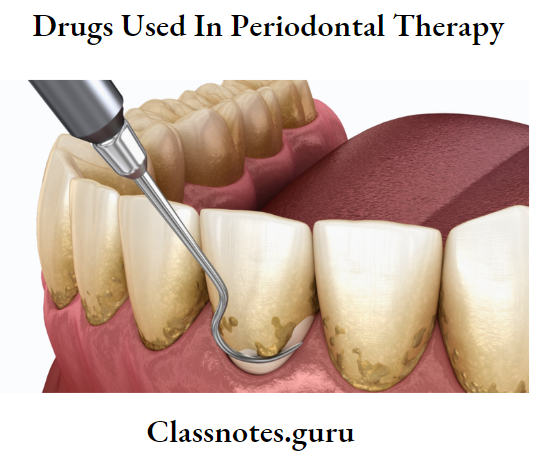
Drugs Used In Periodontal Therapy Short Essays

Question 1. Why antibiotics are not routinely used in periodontal therapy?
Answer:

Periodontal therapy:

Drugs Used In Periodontal Therapy Perodontal therpy image

Periodontal Therapy Uses Of Antibiotics:

  • Reduce/eliminate bacteria
  • Retards bone loss
  • Reduce/Eliminate the need for surgery
  • Useful in aggressive periodontitis

Read And Learn More: Periodontics Question and Answers

periodontal therapy Not Used Routinely:

  • Despite the above use, systemic administration is not recommended routinely as
    • It produces systemic effects
    • Disturbs the functioning of various systems of the body, such as GIT
    • Certain drugs are contraindicated in certain conditions like pregnancy
    • Besides, this systemic administration is useless unless there is plaque and calculus removal.
    • The presence of a thick band of calculus prevents the penetration of the drug into the site
  • Thus, it is used only as an adjunctive.

Question 2. Tetracycline in periodontics
Answer:

  • Tetracycline are widely used drugs in the treatment of periodontal diseases

Tetracycline in Periodontics Clinical Use:

  • It is used as an adjunct in the treatment of localized aggressive periodontitis
  • A contains is a frequent microorganism associated with localized aggressive periodontitis and is tissue invasive, Therefore mechanical removal of calculus and plaque from root surfaces may not eliminate this bacterium from periodontal tissues
  • Systemic tetracycline in conjunction with scaling and root planning can
    • Eliminate tissue bacteria
    • Arrest bone loss
    • Suppresses A.a. comitans
    • Allows mechanical removal of root surface deposits and elimination of pathogenic bacteria from within tissues

Tetracycline in periodontics Actions:

  • Has the ability to concentrate in periodontal tissues
  • Inhibits growth of A.a. contains
  • Exerts anti-collagenase effect
  • Inhibits tissue destruction
  • Aids in bone Regeneration

Tetracycline in periodontics Dose:

  • 250 mg Qid

Tetracycline in Periodontics Side Effects:

  • GI disturbances
  • Photosensitivity
  • Hypersensitivity
  • Increased blood urea nitrogen
  • Dizziness, headache
  • Blood dysplasias
  • Tooth discoloration in children

Question 3. LDD (Local Drug Delivery).
Answer:

LDD Advantages:

  • Greater concentrations of drug at the site
  • Slow release of drug
  • Direct effect on the area
  • Reduced systemic effects

LDD Contraindications:

  • Allergic to drug
  • Children below 10 years
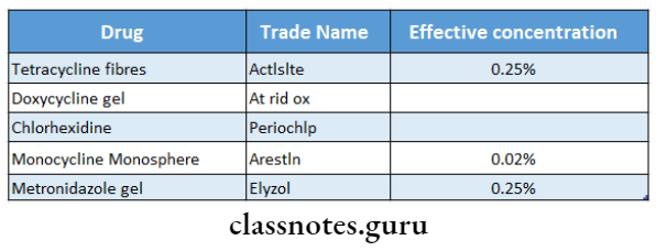
LDD Drugs Used:

Drugs Used In Periodontal Therapy Drugs used

Question4. Methods of Delivery.
Answer:

1. Keye’s technique:

  • Apply slurry of sodium bicarbonate and hydrogen peroxide over tooth brush
  • Tooth brushing

Limitation: Does not reach periodontal pocket

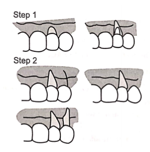
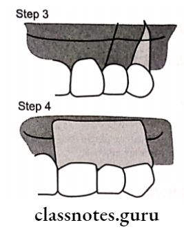
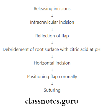
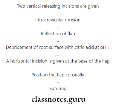
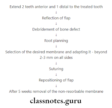
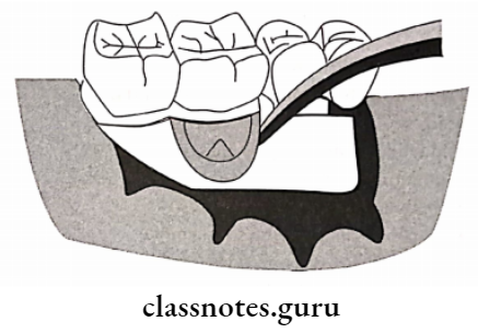
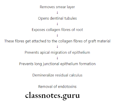
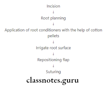
2. Root Bio-modification:

  • Application of root conditioner during surgery

Methods of Delivery Effects:

  • Prevents long junctional epithelium
  • Improves healing

Methods of Delivery Agents Used:

  • Tetracycline
  • Citric acid pH1
  • Fibronectin

3. Irrigation:

Methods of Delivery Types:

  • Home Irrigation
    • Supra gingival
    • Subgingival
    • Marginal
  • Professional Irrigation
    • It delivers medicament into the periodontal pockets via irrigation devices

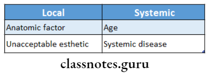
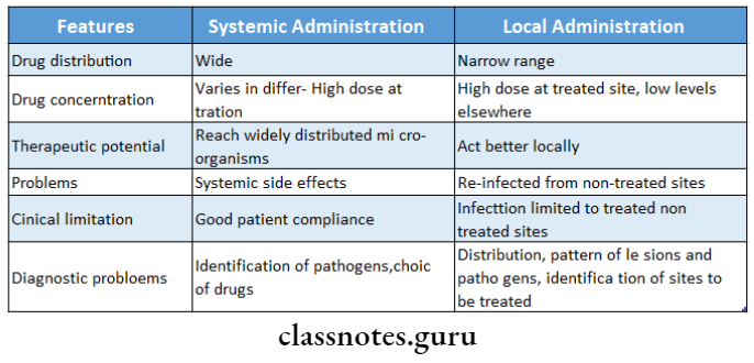
Question 5. Compare local and systemic drug delivery systems.
Answer:

Drugs Used In Periodontal Therapy Compare local and systemic drug dellivery system

Question 6. Metronidazole in periodontal therapy
Answer:

  • Metronidazole is a nitroimidazole compound used to treat protozoal infections

Metronidazole  Spectrum Of Activity:

  • Effective against
    • A. contains
    • P. gingivalis
    • P. intermedia

Metronidazole Uses In Periodontics:

  • To treat
    • Gingivitis
    • Acute necrotizing ulcerative gingivitis
    • Chronic periodontitis
    • Aggressive periodontitis
  • A single dose of metronidazole appears in both serum and GCF
  • When administered systemically, it reduces the growth of anaerobic flora
  • Used as a supplement to rigorous scaling and root planning Subgingival use
    • A dental gel containing metronidazole benzoate is used
    • It gets converted into an active substance by esterases in GCF

Adverse Effects:

  • GIT effects
    • Nausea, anorexia, abdominal pain, metallic taste in the mouth, looseness of stool
    • Headache, stomatitis, glossitis, dryness of mouth, furry tongue, dizziness, rashes, neutropenia, insomnia
    • Prolonged use causes peripheral neuropathy High doses cause convulsions

Drugs Used In Periodontal Therapy Short Question And Answers

Question 1. Advantages of LDD
Answer:

  • Greater concentration of drug at the site
  • Slow release of drug
  • Direct effect on the area
  • Reduced systemic effects

Question 2. Periochip
Answer:

  • It is a small chip composed of a biodegradable hydrosol-lazed gelatin matrix cross-linked with glyceraldehyde
  • It also contains glycerin and water
  • 2.5 mg of chlorhexidine is incorporated into it
  • It slowly releases chlorhexidine and maintains drug concentration in gingival crevicular fluid for at least 7 days
  • Size of chip: 4*5*0.35 mm

Question 3. Keye’s technique
Answer:

  • It refers to the application of a slurry of sodium bicarbonate and hydrogen peroxide over the toothbrush
  • Tooth brushing of it is done

Keye’s technique Limitation:

It does not reach the periodontal pocket

Question 4. Activity
Answer:

  • Among tetracycline-releasing devices, the most widely. It should be selective and effective against micro- used is activity periodontal fiber
  • It is a monolithic thread of a biologically inert, non-than retard resorbable plastic copolymer containing 25% tetracycline hydrochloride powder
  • The fiber is packed into a periodontal pocket secured with a thin layer of cyanoacrylate adhesive and left in place for 7–12 days
  • Due to the continuous delivery of tetracycline, a local concentration of active drug in excess of 1000 mg/l can be. Maintained throughout the period

Activity Effects:

  • Decreases pocket depth
  • Increases attachment levels
  • Decreases bleeding tendency

Question 5. Define antiseptic and antibiotics
Answer:

Antiseptic:

  • Antiseptic is an agent that destroys microorganisms and can be used on living tissues

Antibiotics:

  • An antibiotic is a chemical substance produced by microorganisms that have the capacity to inhibit the growth or kill another organism in a dilute solution

Question 6. Arestin
Answer:

  • Ares tin is a locally delivered, sustained-release form of minocycline microsphere
  • It is used for subgingival placement as an adjunct to scaling and root planning
  • 2% minocycline is encapsulated into bioresorbable mi- mi-microspheres in a gel carrier

Ares tin Effects:

  • Increase in clinical attachment level in patients with pockets of 6 mm or greater
  • Reduction in probing depth
  • It should destroy microorganisms rather

Question 7. Properties of ideal antibiotics
Answer:

  • It should be selective and effective against microorganisms without injuring the host
  • It should destroy microorganisms rather than retard their growth
  • It should not become ineffective as a result of bacterial resistance
  • It should not be inactivated by enzymes, plasma pro- teens or body fluids
  • It should quickly reach bactericidal levels in the entire body and be maintained for long periods
  • It should have minimal side effects

Drugs Used In Periodontal Therapy Viva Voce

  1. Metronidazole belongs to nitroimidazole
  2. The minimum effective concentration of tetracycline needed in GCF is 2-4 μg/m
  3. The mechanism of action of metronidazole is to disrupt bacterial DNA synthesis
  4. The mechanism of action of penicillin is it inhibits bacterial cell wall production
  5. Penicillin is bactericidal
  6. Pseudomembranous colitis with diarrhea or cramping is a side effect of clindamycin
  7. All strains of A.a.comitans are susceptible to ciprofloxacin
  8. The mechanism of action of erythromycin is it inhibits protein synthesis by binding to the 50S ribosomal subunit
  9. Atridox is used for subgingival delivery of doxycycline

Occlusal Evaluation And Therapy In The Management Of Periodontal Disease Short Essay Question And Answers

Occlusal Evaluation And Therapy In The Management Of Periodontal Disease Long Essays

Question 1. Enumerate occlusal evaluation procedures.
Answer:

Clinical Occlusal Evaluation Procedures:

1. TMJ screening examination

  • TMJ screening evaluation includes
    1. Interincisal opening
      • Interincisal distance is recorded in millimeters
    2. Opening/closing pathway
      • Any deviations from the midline path are noted
    3. Temporomandibular joint sounds
      • Clicking or crepitus is noted
    4. Temporomandibular joint tenderness
      • Bilateral palpation over condyles is examined
    5. Muscle tenderness
      • Masseter, pterygoid and temporal muscles are examined

Read And Learn More: Periodontics Question and Answers

2. Intraoral evaluation of occlusion

  • It includes
    • Identification of occlusion in maximum inter-occupation or intercuspal position
      • The patient is asked to close into the maximum inter-cuspal position
      • The presence or absence of contacts is examined
    • Excursive movement
      • The patient is asked to move into right and left excur- sions and observe tooth contact patterns
    • Initial contact in centric relation
      • Guide patient’s mandible in centric relation
      • Record any deflection present
    • Tooth mobility
      • Mobility of the tooth is recorded
    • Attrition
      • It is defined as wear caused by tooth-to-tooth contact
      • Significant attrition of teeth indicates bruxism

3. Role of articulated casts

  • Identifies occlusal contacts that can deflect mandible, deflect mobile teeth or cause trauma to teeth
  • Localizes wear facets, trial occlusal adjustment

Occlusal Evaluation And Therapy In The Management Of Periodontal Disease

Question 2. Describe the steps of occlusal adjustments.
Answer:

Occlusal adjustments also called occlusal equilibration or coroplast is the selective reshaping of occlusal surfaces with the goal of establishing a stable, non-traumatic occlusion

Occlusal Adjustments Steps:

1. Removal of retrusive prematurities

  • Remove retrusive prematurities and eliminate the deflective shift from retruded contact position to in a tra-occlusal position
  • Retrusive prematurities are located on the mesial inclines of maxillary cusps and distal inclines of mandibular cusps

2. Adjustments of the intercuspal position

  • Adjustment of intra-occlusal position relieves su- pra contacts and achieves occlusal stability
  • It can be achieved by reducing in size of the cusp and deepening the fossa

3. Test for excessive contact on incisive teeth in intra- cuspal position

  • Contact relationship may be tested with Mylar strips and checking for fremitus
  • Supracontacts are marked and reduced

4. Remove posterior protrusive supra contacts

  • Obtain bilateral protrusive movement
  • Prematurities are corrected by grinding maxillary teeth

5. Correct prematurities on the balancing side

  • Prematurities on the balancing side are corrected next
  • It is present on the inner inclines of mandibular buccal cusps and the inner inclines of maxillary lingual cusps of the first and second molars

6. Reduce supra-contacts on the working side

  • Reduce supra contacts on laterotrusive side
  • They are reduced by reducing inclines of buccal up-per and lingual lower cusps (BULL)

7. Elimination of undesirable gross occlusal features

  • Extruded teeth, plunger cusp, uneven marginal ridges of adjacent teeth, rotated or malposed teeth, occlusal wear facets, etc are corrected

8. Recheck the occlusal contact relationship in all positions

  • Recheck occlusal contact relationship in all positions

9. Finishing and polishing

  • Adjusted surfaces of the teeth are smoothened and polished

Occlusal Evaluation And Therapy In The Management Of Periodontal Disease Short Essays

Question 1. Indications and steps of coroplast
Answer:

Coroplast Indications:

  • Trauma-occlusal trauma
  • TMJ problems
  • After the elimination of gingival and infra bony pockets

Coroplast Steps:

  1. Removal of retrusive prematurities
  2. Adjustments of the intercuspal position
  3. Test for excessive contact on incisive teeth in intra- cuspal position
  4. Remove posterior protrusive supra contacts
  5. Correct prematurities on the balancing side
  6. Reduce supra contacts on the working side
  7. Elimination of undesirable gross occlusal features
  8. Recheck the occlusal contact relationship in all positions
  9. Finishing and polishing

Question 2. Effects of orthodontic treatment on periodontal tissues
Answer:

Effects Of Orthodontic Treatment On Perio- Dental Tissues:

1. Iatrogenic effects associated with orthodontic treatment

  • Orthodontic treatment may cause injuries to the teeth and periodontium in most of the cases
  • Usually, these changes are reversible, and regeneration and repair of the tooth structures and period-dental tissues can occur while in some cases the changes may result in irreparable damage

2. Root resorption

  • During orthodontic therapy some amount of root resorption is unavoidable

3. Effects of orthodontic bands on the periodontium

  • Gingivitis and gingival hyperplasia are short-term effects
  • Long-term effects are loss of attachment, root resorption, or no effects

4. Effects of orthodontics on dentition with normal height of attachment apparatus

  • Orthodontic forces cause no damage to the supra- Time of treatment: alveolar connective tissue

Question 3. The rationale for orthodontic tooth movement in periodontal therapy
Answer:

Rationale For Orthodontic Treatment:

1. Reducing plaque retention:

  • Crowded teeth and mesially inclined teeth create plaque accumulation sites that are difficult to clean
  • Crowding creates enlarged contact surfaces and al-tiered embrasure spaces that are displaced apically

2. Improving gingival and osseous form:

  • There is an interrelation between the position of the tooth, the shape of the gingiva, and the bone that surrounds it
  • Orthodontic treatment may improve the shape of the periodontium and reduces the need for bone surgery

3. Facilitating prosthetic replacements:

  • The uprighting of tilted abutment teeth may be im- important for a better-contoured crown which will benefit the surrounding periodontal condition

4. Improving esthetics:

  • Correction of pathologic tooth migration and di-asthma between anterior teeth results in improved esthetics

Occlusal Evaluation And Therapy In The Management Of Periodontal Disease Short Answers

Question 1. Coronoplasty.
Answer:

The procedure of selective reshaping of the occlusal surface with the goal of establishing a stable – nontraumatic occlusion.

It Is an Invasive Procedure:

Coronoplasty Goals:

  • Reshaping crown surface
  • Elimination of supra contacts
  • Creation of stable occlusion

Coronoplasty Indications:

  • Trauma-occlusal trauma
  • TMJ problems

Coronoplasty Time of treatment:

  • After the elimination of gingival inflammation and periodontal pockets

Question 2. Forced eruption
Answer:

  • Forced eruption applies to procedures that involve orthodontic movement with gentle forces
    The purpose is the coronal shift of the bone at the base of infrabony defects, thus reducing the depth of the de-fact
  • The elongated tooth thereafter can be reduced in height by grinding and elimination of the infra-bony pocket Forced eruption can also be done to manage teeth that have fractures to make possible the restoration of the tooth
  • To erupt the tooth forcefully, either the adjacent teeth must be bracketed and a wire placed or a wire must be bonded directly to the adjacent teeth and an elastic trac- tion applied from the wire to the tooth

Occlusal Evaluation And Therapy In The Management Of Periodontal Disease Viva Voce

  1. The location of bands and brackets determines the outcome of orthodontic therapy
  2. Orthodontic brackets on the posterior teeth are positioned relative to the marginal ridges and cusps
  3. Orthodontic brackets on the anterior teeth are positioned relative to the incisal edges
  4. The tooth should erupt 4 mm orthodontically for the purpose of restoration if a tooth fracture extends to the level of alveolar bone
  5. To avoid relapse and intrusion of an orthodontically erupted tooth 6 months time period is necessary for stabilization


Periodontics Maintenance Phase Short Essay Question And Answers

Maintenance Phase Short Essays

Question 1. SPT (Supportive Periodontal Treatment).
Answer:

SPT Rationale:

  • Prevent/minimize the recurrence of disease
  • Control etiological factors of disease

SPT Goals By Aap:

  • Prevent/minimize recurrence and progression of disease
  • Prevent/reduce the incidence of tooth loss
  • Increase the probability and treating in a timely manner.

Read And Learn More: Periodontics Question and Answers

SPT Objectives:

  • Preservation of bone support
  • Maintenance of attachment
  • Oral hygiene maintenance

SPT Procedure:

Maintenance Phase Procedure

Maintenance Phase Short Question And Answers

Question 1. Describe in short the important factors causing periodontal disease
Answer:

Causes Of Recurrence Of Periodontal Diseases:

  • Inadequate or insufficient treatment that has failed to remove all the potential factors favoring plaque accumulation
  • Incomplete calculus removal in areas of difficult access is a common source of problems
  • Inadequate restorations placed after the periodontal treatment was completed
  • Failure of the patient to return for periodic check-ups
  • Presence of some systemic diseases that may affect host resistance to previously acceptable levels of plaque

Splint In Periodontal Therapy Short And Long Essay Question And Answers

Splint In Periodontal Therapy Definition

Periodontal Splint

  • A splint is an appliance used for maintaining or stabilizing mobile teeth to their functional position

Splint In Periodontal Therapy Important Notes

1. Classification of splints

  • According to the Period of stabilization
    1. Temporary<6 months
    2. Provisional – for months up to several years
    3. Permanent – Indefinite
  • According to the type of material
    1. Bonded composite
    2. Braided wire
    3. A-splints
  • According to the location
    1. Intra-oral
    2. Extra-oral

Read And Learn More: Periodontics Question and Answers

2. Requisites of splints

  • Easy to prepare
  • Economical
  • Rigid, durable, and stable
  • Easy to remove and replace
  • Self-cleansing
  • Easy to maintain
  • Esthetically accepted
  • Non-irritating to adjacent tissues

Splint In Periodontal Therapy Long Essays

Question 1. Define periodontal splint. Give indications and contraindications for splinting of teeth.
Answer:

Periodontal Splint Definition:

  • A splint is an appliance used for maintaining or stabilizing mobile teeth to their functional position

Periodontal Splint Indications:

  • Stabilize mobile teeth to improve patient comfort and to provide stability
  • Stabilize moderate to advanced tooth mobility
  • Stabilize teeth in secondary occlusal trauma
  • Stabilize teeth following acute trauma
  • Stabilize teeth when increased tooth mobility interferes with normal masticatory function
  • Stabilize teeth following orthodontic movement
  • Prevent tipping or drifting of teeth
  • Create adequate stability
  • Prevent extrusion of unopposed tooth

Periodontal Splint Contraindications:

  • Presence of periodontal inflammation
  • Presence of an insufficient number of nonmobile teeth to stabilize mobile teeth
  • Presence of inadequate oral hygiene
  • Absence of prior occlusal adjustment

Question 2. Define periodontal splint. Discuss their role as ad adjuncts in periodontal therapy.
Answer:

Periodontal Therapy Definition:

  • Splint is an appliance used for maintaining or stabilizing mobile teeth to their functional position

Periodontal Therapy Role Of Splinting In Periodontal Diseases

  1. Rest
    • Immobilization permits undisturbed healing
  2. Redirection of forces
    • Splinting leads to the redirection of forces in the axial direction over all the teeth
  3. Redistribution of forces
    • Distributes forces over a number of teeth and thereby prevents excessive forces on mobile teeth
  4. Restoration of functional stability
    • Restores functional occlusion
    • Stabilizes the remaining mobile abutment teeth
  5. Maintenance of arch integrity
    • Proximal contacts are restored
    • Prevents food impaction and further breakdown of Loop wire is tightened periodontal tissues
  6. Psychologic well being
    • Restores feeling of dentition, comfort, and esthetics

Question 3. Define periodontal splinting. Mention the ideal requisites of a periodontal splint and write in detail about the splinting procedure.
Answer:

Periodontal Splinting Definition:

  • A splint is an appliance used for maintaining or stabilizing mobile teeth to their functional position

Periodontal Splinting Requisites:

  • Easy to prepare
  • Economical
  • Rigid, durable, and stable
  • Easy to remove and replace
  • Self-cleansing
  • Easy to maintain
  • Esthetically accepted
  • Non-irritating to adjacent tissues

Periodontal Splinting Procedure:

Splint In Periodontal Therapy Procedure

Splint In Periodontal Therapy Short Essays

Question 1. Splints.
Answer:

Splints Definition:

  • An appliance is used for maintaining or stabilizing mobile teeth to their functional position.

Splints Objectives

  • Provide rest
  • By redirecting forces
  • Preserve arch integrity
  • Restores function
  • Stabilizes mobile teeth
  • Prevent eruption of unopposed teeth

Splint In Periodontal Therapy Short Question and Answers

Question 1. Permanent splinting
Answer:

  • Permanent splints are indicated in dentitions that cannot maintain final stability after periodontal and restorative treatment
  • They can be either fixed or removable
  • Permanent splints utilizing cast restoration may be placed as part of a restorative phase of therapy

Pulpo Periodontal Problems Short Essay Question And Answers

Pulpoperiodontal Problems Short Essays

Question 1. Endo-perio lesion.
Answer:

Pulpoperiodontal Problems Endo-perio lesion

Read And Learn More: Periodontics Question and Answers

Pulpoperiodontal Problems Possible pathways for spread of infection between pulp and periodontal tissues

Originally endodontic problem with fistulization from the apex and along the root to the gingiva. Pulpal infection can also spread through accessory canals to the gingiva or to the furcation (primary endo lesion)

Pulpoperiodontal Problems Possible pathways for spread of infection between pulp and periodontal tissues.

A periodontal pocket can infect the pulp through a lateral canal and this in turn can result in a periapical lesion (primary periosecondary endo lesion)

Pulpoperiodontal Problems Possible pathways for spread of infection between pulp and periodontal tissues..

A periodontal pocket can deepen to the apex and second- dary involve the pulp (primary percolation)

Pulpoperiodontal Problems Possible pathways for spread of infection between pulp and periodontal tissues...

A long-standing periapical lesion draining through the periodontal ligament can become a secondary complication, leading to a “retrograde periodontitis” (primary endosecon- diary percolation)

Pulpoperiodontal Problems Possible pathways for spread of infection between pulp and periodontal tissuess.

Pulpoperiodontal Problems Viva Voce

  1. The most common cause of the pulpal disease is dental caries
  2. The organisms being cultured predominantly from the infected root canals are Gram-negative anaerobes
  3. If the inflammatory lesion of the pulp cannot be resolved even after the elimination of the source of trauma then it is described as irreversible pulpitis
  4. Sensation from the core of the pulp is initiated by unmyelinated C fibers
  5. While treating combined periodontal and endodontic lesions, endodontic lesions should be treated first
  6. A dental abscess with a draining sinus tract indicates a lesion of endodontic origin

Furcation Involvement

Furcation Involvement Definition

Furcation Involvement

  • Furcation involvement refers to the invasion of the bifurcation and trifurcation of multirooted teeth by periodontal disease.

Root Resection

  • Root Resection is the surgical removal of all or a portion of the root before or after endodontic treatment.

Hemisection

  • One root with its corresponding crown portion is cut and removed

Read And Learn More: Periodontics Question and Answers

Bicuspidisation

  • The molar is cut without the removal of any part of the crown or root

Furcation Involvement Important Notes

1. Types of furcation

Furcation Involvement Types of furcation

Furcation Involvement Long Essays

Question 1. Define and classify furcation. Enumerate treatment of grade III furcation areas.
Answer:

Furcation Definition:

  • Furcation involvement refers to the invasion of the bifurcation and trifurcation of multirooted teeth by periodontal disease.

Classification of furcation:

1. According to Glickman:

  • Grade 1
    • Early/Incipient lesion
    • Suprabony pocket
    • Slight bone loss
    • No radiographic changes
  • Grade 2
    • Bone destruction on one/more aspects
    • But the portion of the bone is intact
    • Cul-de-sac lesion
    • A radiograph may or may not reveal the involvement
  • Grade 3
    • Complete inter-radicular bone loss
    • No soft tissue loss
    • Radiograph shows the radiolucent area between roots
  • Grade 4
    • Inter-radicular bone loss
    • Gingival recession
    • Radiolucent area radiographically

Furcation Involvement Glickman's classification of furcation involvement

Furcation Involvement Glickman's classification of furcation involvement.

2. According to Tarnow & Fletcher:

  • Subgroup A: Vertical bone loss upto 1/3rd of inter-radicular height (0-3 mm)
  • Subgroup B: Vertical bone loss upto 2/3rd of inter-radicular height (4-7 mm)
  • Subgroup C: Vertical bone loss beyond apical third (>7 mm)

Furcation Involvement Vertical classifi cation of furcation involvement

Furcation Treatment:

Objectives:

  • Facilitate maintenance
  • Prevent further attachment loss
  • Obliterate furcation defects

Furcation Treatment Options:

1. For grade 1

  • Scaling and root planning
    • Grade I can be accessible by scaling and root planning
  • Curettage
    • Done by Gracey curettes, Quetin furcation cu- rettes
  • Gingivectomy
    • It is the excision of the soft tissue wall

2. For grade 2

  1. Traditional methods
    • Scaling and root planning- inaccessible areas
    • Curettage
    • Osteoplasty
    • It involves shaping of bone to prevent plaque accumulation
    • Hemisection
    • It is the surgical removal of the root with the associated part of the crown
  2. Regenerative procedures
    • GTR (Guided Tissue Regeneration)
    • It involves the placement of membrane to prevent
    • formation of long junctional epithelium
    • Coronally repositioned flap
    • Root conditioners

3. For Grade 3

  • Tunneling
    • Done in mandibular 1 molar due to better ac- accessibility
  • It transforms Grades 2 to Grades 3 and 4
  • Has increased risk of root caries
  • So not used nowadays
  • GTR
  • Hemisection

4. For grade 4

  • Extraction is done

Furcation Involvement Short Essays

Question 1. Root Resection.
Answer:

Root Resection Definition:

  • Root Resection is the surgical removal of all or a portion of the root before or after endodontic treatment.

Furcation Involvement Root separation and resections.

Root Resection Indication:

  • Gingival recession
  • Class 2 or 3 furcation
  • Severe bone loss

Root Resection Contraindications:

1. Local:

  • Poor oral hygiene
  • Fused roots
  • Endodontically untreated roots.

2. Systemic:

  • Systemic diseases

Furcation Involvement Root separation and resection

Root Resection Technique:

Furcation Involvement Technique

Furcation Involvement Short Question And Answers

Question 1. Grade 3 furcation involvement
Answer:

Grade 3 Furcation Involvement Features:

  • Complete inter-radicular bone loss
  • No soft tissue loss
  • Radiograph shows the radiolucent area between roots

Grade 3 Furcation Involvement Treatment:

  • Tunneling
    • Done in mandibular I molar due to better accessibility
    • It transforms Grades 2 to Grade 3 and 4
    • Has increased risk of root caries
    • So not used nowadays
  • GTR
    • It involves the placement of membrane to prevent for- motion of long junctional epithelium
  • Hemisection
    • It is the surgical removal of the root with the associated part of the crown

Question 2. Tunneling
Answer:

  • Done in mandibular 1 molar due to better accessibility
  • It transforms Grades 2 to Grades 3 and 4
  • Has increased risk of root caries
  • So not used nowadays

Question 3. Hemisection
Answer:

  • It is the surgical removal of the root with the associated part of the crown
  • Commonly performed on mandibular molars with buc- cal or lingual class 2 or 3 furcation

Hemisection Technique:

Furcation Involvement Hemisection

Furcation Involvement Viva Voce

  1. The most commonly performed root resection is disto- the buccal root of the maxillary first molar
  2. Furcation is most commonly involved in mandibular molars
  3. Least commonly involved in maxillary 1st premolars
  4. The prognosis is poor if the furcation areas are in- involved in maxillary 1st premolars
  5. Hemisection and bicuspidisation are suitable for mandibular molars

Pregnancy Gingivitis Short Essay Question And Answers

Pregnancy Gingivitis Short Essays

Question 1. Gingival Changes in Pregnancy.
Answer:

  • Time after first trimester

Gingival Changes in Pregnancy Changes:

Pregnancy Gingivitis Changes

Read And Learn More: Periodontics Question and Answers

Gingival Changes in Pregnancy Cause:

  • Aggravation of previous inflammation
  • Altered tissue metabolism

Gingival Changes in Pregnancy Gingiva:

  • Color Bright red/magenta
  • Consistency Soft and friable
  • Bleeding on probing – Positive
  • Size – Increased
  • Surface texture – Stippling lost
  • Shape – Mushroom like flattened spherical

Pregnancy Gingivitis Viva Voce

  1. The microorganism associated with pregnancy gingivitis is P.intermedia
  2. A pregnancy tumour is a non-neoplastic gingival enlargement


Periodontal Restorative Short Essay Question And Answers

Periodontal Restorative

Periodontal Restorative Short Question And Answers

Question 1. Biologic Width.
Answer:

Biologic Width Definition:

  • The soft tissue attachment to the tooth between the base of the gingival sulcus and the crest of the alveolar bone is called biologic width.

Biologic Width Significance:

  • Avoid its invasion during restorative procedures to prevent attachment loss

Biologic Width Calculated As:

  • Biologic width = Junctional Epithelium + Connective tissue attachment

Read And Learn More: Periodontics Question and Answers

  • 0.97mm + 1.07 mm = 2.04 mm

Periodontal Restorative Biologic width

Periodontal Restorative Viva Voce

  1. Biological width is defined as the physiologic dimensions of junctional epithelium and connective tissue attachment
  2. The biologic width is the distance between the coronal end of the junctional epithelium to the alveolar crest
  3. Placement of the supragingival margin of restoration has the least impact on the periodontium
  4. To correct the violation of biological width after rapid orthodontic extrusion of a tooth suprarenal fibrotomy is per- formed every 1 week

Mucogingival Surgery Short And Long Essay Question And Answers

Mucogingival Surgery Definition

Mucogingival Surgery: Surgical procedures are performed to correct/eliminate anatomic, developmental, or traumatic deformities of the gingiva or alveolar mucosa.

Mucogingival Surgery Important Notes

1. Mucogingival surgeries

  • Techniques of increasing the width of attached gingiva
    • Free gingival graft
    • Apically displaced flap
  • Techniques for coverage of denuded roots
    • Laterally displaced pedicle graft
    • Coronally displaced flap
    • Free gingival graft
  • Techniques to deepen the vestibule
    • Free autogenous grafts
  • Techniques for removal of a frenum

2. Undisplaced flap

  • It surgically removes the pocket
  • It does not increase the width of the attached gingiva, instead, it decreases the width
  • It is essentially an excisional procedure of the gingiva

3. Disadvantages of full-thickness flap

  • Loss of facial bone height
  • Not preferred in cases of fenestrations and dehiscence

Read And Learn More: Periodontics Question and Answers

Mucogingival Surgery Long Essays

Question 1. Define periodontal plastic surgery. Describe procedures available to cover denuded root surfaces.
Answer:

Periodontal plastic surgery Definition:

  • Surgical procedures are performed to correct/eliminate anatomic, developmental, or traumatic deformities of the gingiva or alveolar mucosa.

Periodontal Plastic Surgery Conventional Procedures

1. Laterally displaced flap:

Recipient site:

Mucogingival Surgery Recipient site

Periodontal plastic surgery  Donor site:

Mucogingival Surgery Donor site

  • Transfer the flap to the recipient site
  • Suturing of flap
  • Placement of periodontal pack

Mucogingival Surgery Localized recession

Mucogingival Surgery Generalized recession

2. Double papilla flap:

By Wainberg:

Mucogingival Surgery Various steps in laterally position flap

Mucogingival Surgery Various steps in laterally position flap.

Dissecting both facial and lingual papilla

3. Coronally-repositioned flap:

Mucogingival Surgery Various steps in laterally position flap.

Mucogingival Surgery The transposed flaps are sutures to obtain a single flap

Regenerative Procedures:

Mucogingival Surgery Regenerative procedures

Mucogingival Surgery Short Essays

Question 1. Free gingival graft
Answer:

A free gingival graft is used to create a widened zone of attached gingiva

Mucogingival Surgery Various steps in claaic techique

2. Variant techniques:

  1. Accordion technique
    • Achieved by giving alternate incisions on opposite sides of the graft
  2. Strip technique
    • Consists of 2 or 3 strips of tissue to cover the entire length of the recipient site
  3. Connective tissue technique

Question 2. GTR.
Answer:

After the flap surgery, epithelium from the excised margin. No need for donor site may proliferate apically

1. Classical technique of GTR:

Mucogingival Surgery Classical technique

  • This results in the formation of long junctional epithelium
  • Thus this should be prevented
  • To prevent this, a membrane is placed between healing connective tissue and cementum
  • Such a membrane is GTR (Guided Tissue Regeneration)

Types of GTR:

  • Degradable- Collagen, Guidor membrane
  • Non-degradable- Millipore, Teflon membrane

GTR Indications:

  • Esthetic demand
  • In single tooth with wide deep localized recession
  • In the presence of root sensitivity
  • In recession associated with class V restorations

GTR Advantages:

  • No need for a donor site
  • Highly esthetic
  • Disadvantages:
  • Technique sensitive
  • Expensive

GTR Technique:

  • The incision along with releasing incisions extending

Mucogingival Surgery Technique

Mucogingival Surgery Membrane adptation

Mucogingival Surgery Various steps in coronally positioned flp

Question 3. Coronally displaced flap
Answer:

Coronally displaced flap Indications:

  • Esthetic coverage of exposed roots
  • Gingival recession

Coronally displaced flap Advantages:

  • Treatment of multiple areas
  • Adjacent teeth are safe
  • The high degree of success
  • It does not increase the existing problem

Coronally displaced flap Disadvantages:

  • No need for two surgical procedures

Coronally displaced flap Technique:

Mucogingival Surgery Coronally displaced flap

Mucogingival Surgery Short Question And Answers

Question 1. Frenectomy.
Answer:

Frenectomy is the complete removal of the frenum including its at-attachment to the underlying bone

Frenectomy Technique:

Mucogingival Surgery Technique..

Question 2. Mucogingival Problems.
Answer:

  • Deep pockets
  • Recession
  • High frenal attachment
  • Inadequate width of attached gingiva

Question 3. Indications of Mucogingival surgery.
Answer:

  • Augmentation of the edentulous ridge
  • Prevention of Residual Ridge Resorption (R)
  • Crown – lengthening
  • Esthetic purpose

Question 4. Frenectomy and frenotomy
Answer:

Frenectomy:

  • Frenectomy is the complete removal of the frenum including attachment to the underlying bone

Frenotomy:

  • Frenectomy is the relocation of the frenum usually in a more apical position

Frenectomy Indications:

  • Esthetic purposes
  • Deepening of vestibule in mandibular anterior area

Frenectomy Technique:

Mucogingival Surgery Frenotomy

Question 5. High frenal attachment
Answer:

High frenal attachment is a condition where the frenum is. attached too close to marginal gingiva.

High frenal attachment Etiology:

  • Genetic
  • Gingival recession

High frenal attachment Effects:

  • Tension on frenum
  • Plaque accumulation
  • Inhibit proper placement of toothbrush
  • Poor oral hygiene
  • Deep maxillary anterior vestibule
  • Esthetic problems

Question 6. Vestibuloplasty.
Answer:

  • It is a procedure for vestibule extension
  • As described by Edlan and Mejchar

High frenal attachment Technique:

Two vertical incisions are given from the junction of marginal and attached gingiva to approx 12 mm from the alveolar margin into the vestibule

Mucogingival Surgery Vestibuloplasty

High frenal attachment Types:

  1. Labial vestibuloplasty
  2. Lingual vestibuloplasty

Question 7. Objectives of mucogingival surgery
Answer:

  • Widening of attached gingiva
  • Coverage of denuded roots
  • Removal of an aberrant frenum
  • Creation of some vestibular depth
  • As an adjunct to routine pocket elimination procedure

Mucogingival Surgery Viva Voce

  1. Surgical removal of the frenum is indicated when tension on the frenum may tend to open the sulcus
  2. The ideal thickness of a free gingival autograft is 1-1.5 mm
  3. Revascularization of a free gingival autograft starts from 2nd or 3rd day
  4. The central portion of the free gingival margin is last to vascularize
  5. Healing of free gingival autograft of intermediate thickness of 0.75 mm is completed by 10.5 weeks
  6. After 24 weeks free gingival autograft placed on de- nude bone shrink by 25%


Osseous Surgery Short Essay Question And Answers

Osseous Surgery Definition

Respective Osseous Surgery

  • Respective Osseous Surgery is defined as a procedure by which changes in the alveolar bone can be accomplished to rid it of deformities induced by the periodontal disease process or other related factors such as exostosis and tooth supra eruption.

Osteoplasty

  • Osteoplasty is the reshaping of bone without removing tooth-supporting bone

Ostectomy

  • Ostectomy is the removal of tooth-supporting bone

Read And Learn More: Periodontics Question and Answers

Osseous Surgery Definition Important Notes

1. Types of grafts

Osseous Surgery Types of grafts

2. Indications for resection osseous surgery

  • Interdental craters
  • One wall osseous defects
  • Wide shallow two-wall defects
  • Exostosis
  • Supra erupted teeth

3. Guided tissue regeneration, GTR

  • It is based on the assumption that only the periodontal ligament cells have the potential for regeneration of the attachment apparatus of the tooth
  • The technique consists of placing barriers of different types to cover the bone and periodontal ligament to prevent the migration of epithelium

4. Bioglass

  • It is an alloplastic or non-graft material
  • It consists of sodium and calcium salts, phosphates and silicon dioxide

5. Types of membranes of GTR

Osseous Surgery Types of membrranes of GTR

6. Steps in osseous resection surgery

  • Vertical grooving
  • Radicular blending
  • Flattening interproximal bone
  • Gradualizing marginal bone

Osseous Surgery Long Essay

Question 1. Define Respective Osseous Surgery. Mention its indication, contraindications and steps involved in it.
Answer:

Respective Osseous Surgery Definition:

  • Respective Osseous Surgery is defined as a procedure by which changes in the alveolar bone can be accomplished to rid it of deformities in- duced by the periodontal disease process or other related as exostosis and tooth supra eruption.

Respective Osseous Surgery Indications:

  • One wall defect
  • Thick bony margins

Respective Osseous Surgery  Shallow Crater:

Contraindication:

Osseous Surgery Contraindication

Respective Osseous Surgery  Orally:

  • Improper oral hygiene
  • High DMFT
  • Dentinal Hypersensitivity
  • Advanced Periodontitis

Respective Osseous Surgery Steps:

  1. Vertical Grooving
  2. Radicular blending
  3. Flattening of interproximal bone

4. Gradualizing marginal bone

Osseous Surgery Gradualizing marginal bone

Following these steps

Osseous Surgery Steps in osseous resective surgery

Question 2. Classify bone replacement grafts and describe the merits and demerits of each type
Answer:

Classification of replacement grafts:

Replacement grafts Autogenous bone grafts:

1. Bone from intraoral sites:

  • Osseous coagulum
  • Bone blend
  • Intraoral cancellous bone marrow transplants
  • Bone swaging

2. Bone from extraoral sites:

  • Iliac autograft

Replacement grafts Allografts:

  • Undecalcified freeze-dried bone allograft
  • Demineralized freeze-dried bone allograft

replacement grafts Xenografts:

Non-bone graft materials:

  • Sclera
  • Cartilage
  • Plaster of Paris
  • Plastic materials
  • Calcium phosphate biomaterials
  • Bioactive glass
  • Coral derived materials
  • Bioactive glass

Osseous Surgery Materials and Merits and Demerits

Question 3. Define osseous surgery. Discuss various osseous grafting procedures.
Answer:

 Osseous grafting Definition:

  •  Osseous Grafting is defined as a procedure by which changes in the alveolar bone can be accomplished to rid it of deformities induced by the periodontal disease process or other related factors such as exostosis and tooth supra eruption

 Osseous grafting Procedures:

1. Osseous coagulum:

  • Donor Sites:
  • Lingual ridge
  • Exostoses
  • Edentulous ridges
  • Bone distal to terminal tooth

 Osseous grafting Technique:

Bone is removed from the donor site by carbide bur
Osseous Surgery Osseous grafting procedures.

2. Bone blend:

Osseous Surgery Bone blend

3. Cancellous bone marrow transplants:

Osseous Surgery Bone Swaging

4. Bone swaging:

  • Bone from an edentulous area adjacent to the defect is pushed into contact with the root surface without fracturing the bone at its base

5. Allografts:

Osseous Surgery Allografys

Question 4. Describe in brief various reconstructive Osseous techniques.
Answer:

 Osseous Techniques Reconstructive Osseous Surgery:

 Osseous techniques Non-Graft associated new attachment.

1. Removal of Junctional/Pocket Epithelium through the following

  • Curettage
  • Chemical agents
    • Sodium sulfide
    • Phenol
    • Camphor
    • Antiform
  • Ultrasonic methods- Lacks tactile sensation
  • Surgical methods
    • ENAP
    • Gingivectomy
    • Widman flap
    • Coronally displaced flap

2. Prevention of Epithelial Migration.

  1. GTR
    • Degradable – Collagen
    • Non-degradable – Millipore
  2. Clot stabilization, wound protection and space creation by
    • Grafts
    • Membranes
  3. Coronally displaced flap
  4. Root conditioners
    • Citric acid
    • Fibronectin
    • Tetracycline

3. Growth Factors (GF)

  • Regulates events in wound healing
  • Example: Platelet-derived GF
    • Insulin-like GF

4. Enamel Matrix Proteins

  • Favours periodontal regeneration

 Osseous techniques Graft-associated new attachment

1. Allografts:

  • Tissue transfer between individuals of the same species

2. Xenografts:

  • Grafts from different species
  • Causes severe allergic reactions

3. Alloplasts:

  • For restoration of the periodontium
  • Example: Sclera, dura, Cartilage

 Osseous techniques Combined techniques

  • Combination of both graft and non-graft-associated methods

Osseous Surgery Short Essays

Question 1. GTR (Guided Tissue Regeneration).
Answer:

  • After the flap surgery, epithelium from the excised margin may proliferate apically.
  • This results in the formation of long Junctional epithelium
  • Thus this should be prevented
  • To prevent this, a membrane is placed between healing connective tissue and cementum
  • Such a membrane is (GTR)

GTR Types:

  • Degradable – Collagen, Guidor membrane
  • Nondegradable – Millipore, Teflon membrane

GTR Procedure:

Osseous Surgery Procedure

Osseous Surgery Flap is positioned coronally and sutured

Osseous Surgery Membrane adaptation

Question 3. Root conditioners/Root Biomodification effects.
Answer:

Root Conditioners Effects:

Osseous Surgery Root conditoners

Root Conditioners Examples:

  • Citric acid at pH 1
  • Fibronectin
  • Tetracycline

Root Conditioners Procedure:

Osseous Surgery Examples

Question 3. Non-bone graft materials
Answer:

Non-Bone Graft Materials:

  1. Sclera
  2. Cartilage
  3. Plaster of Paris
    • Calcium sulphate
  4. Plastic materials
    • Hard tissue replacement
  5. Calcium phosphate biomaterials
    • Hydroxyapatite
    • Tricalcium phosphate
    • Calcitite
  6. Bioactive glass-consists of
    • Sodium and calcium
    • Phosphates
    • Silicon dioxide
  7. Coral derived materials
    • Natural coral material
    • Coral derived porous hydroxyapatite

Question 4. Define osteotomy and osteoplasty. Explain how they differ from each other.
Answer:

Ostectomy:

  • It is a procedure of radicular and inter-radicular removal of supporting bone to eliminate osseous deformities
  • It is done by a technique which involves the removal of supporting bone to produce a positive gingival and osseous architecture

Ostectomy Indications:

  • To reduce hemisepta-like defects and shallow intrabony defects
  • To correct reverse architecture induces by periodontal disease
  • Moderate to advanced furcation involvements
  • Elimination of interdental craters
  • Intrabony defects not amenable to regeneration

Ostectomy Contraindications:

  • Anatomic limitations like prominent external oblique ridge or zygomatic arch, etc
  • Esthetic limitations like high smile line anteriorly
  • Availability of effective alternative treatment
  • Areas of insufficient remaining attachment

Ostectomy Advantages:

  • Pocket elimination
  • Establishment of physiologic gingival and osseous architecture
  • Establishment of favourable prosthetic environment Loss of attachment
  • Increased root sensitivity
  • Esthetic compromise

Ostectomy Osteoplasty:

  • It is described by Friedman in 1955
  • It is a procedure of reshaping the alveolar process to achieve a more physiological form without the removal of supporting bone

Ostectomy Indications:

  • Removal of buccal and lingual bony ledges or tori
  • Pocket elimination
  • Shallow intrabony defects
  • Shallow craters
  • Thick interproximal areas
  • Incipient furcation defects that do not necessitate re-moving supporting bone
  • Buttressing bone formation or lipping

Question 5. Allograft
Answer:

Allografts are tissue transfer between individuals of the same species but with different genetic composition

Allograft Commercial products

  • FDBA (Freeze Dried Bone Allograft)-osteoconductive
  • DFDBA (Demineralized Freeze-Dried Bone Allograft)

Allograft Disadvantages:

  • Foreign to the patient
  • Provokes immune response

Osseous Surgery Short Question And Answers

Question 1. Types of Osseous Surgery.
Answer:

1. Depending on the relative position of interdental to radicular bone

  • Positive Architecture
    • Radicular bone apical to interdental bone.
  • Negative Architecture
    • Interdental bone apical to radicular bone.
  • Flat Architecture
    • Interdental & radicular bone reduced up to the same height.
  • Ideal
    • Interproximal bone is coronal to that of facial/lingual surfaces.

2. Depending on the thoroughness of the techniques

  • Definitive osseous reshaping
    • Further reshaping would not improve the overall result.
  • Compromise osseous reshaping
    • Requires osteotomy procedures.

3. Additive-Restores bone to original levels

  • Subtractive Restores bone to the level existing at the time of surgery or apical to it.

Question 2. Instruments of Osseous Surgery.
Answer:

Osseous Surgery Hand Instruments:

  • Ronguers Freidman & Blumental
  • Interproximal files – Schluger & Sugarman
  • Back action chisels
  • Oschsenbein chisels

Osseous Surgery Rotary:

  • Carbide round burs
  • Slow-speed handpiece
  • Diamond burs

Question 3. Growth Factors.
Answer:

Growth Factors are polypeptide molecules released by the cells in osteoinductive the inflamed area

Growth Factors Secreted

  • Macrophages
  • Endothelial cells
  • Fibroblasts
  • Platelets

Growth Factors Effects:

  • Regulates events in wound healing
  • Promotes proliferation of fibroblasts
  • Favours bone formation

Growth Factors Examples:

  • Platelet-derived growth factors (PDGF)
  • Insulin-like growth factor (IGF)
  • Fibroblast growth factor (FGF)

Question 4. Enamel Matrix Proteins.
Answer:

Namely, amelogenin is secreted by Hertwigs Epithelial root sheath during tooth development and induces accel-cellular cementum formation.

Matrix Proteins Actions:

  • Enhance periodontal regeneration
  • Promote bone cell attachment
  • Increases proliferation of more immature bone cells
  • It is not osteoinductive, but it is osteopromotive in that it stimulates bone formation when combined with demineralized freeze-dried bone allograft.

Question 5. Osseous Coagulum.
Answer:

Osseous Coagulum Technique:

Osseous Surgery Osseous coagulum

Osseous Coagulum  Donor Site:

  • Lingual ridge on mandible
  • Exostosis or tori
  • Edentulous ridges
  • Bone distal to the terminal tooth.

Osseous Coagulum  Advantages:

  • Better resorbed
  • Provides additional surface area

Osseous Coagulum  Disadvantages:

  • Low predictability
  • Poor visibility

Question 6. Bone Swaging.
Answer:

Bone Swaging Requirement:

  • An edentulous area adjacent to the defect

Bone Swaging Procedure:

  • Push the bone of the edentulous area into the defect
  • Avoid fracture of basal bone

Bone Swaging Disadvantage:

  • Complicated procedure
  • Limited use

Question 7. Positive architecture
Answer:

When Radicular bone is present apical to interdental bone it is called positive architecture

Question 8. Define root planning.
Answer:

Root planning is a process by which residual embedded calculus and a portion of cementum are removed from the roots to produce a smooth, hard and clean surface

Question 9. Reversed architecture.
Answer:

  • The loss of interdental bone produces these defects including the facial and lingual plates without concomitant loss of radicular bone, thereby reversing normal architecture
  • Normally interproximal bone is coronal to facial and lingual plates forming a scalloped osseous margin
  • When the loss of interdental bone occurs without significant loss of radicular bone, normal scalloping gets reversed
  • It is more common in the maxilla

Question 10. Osteoplasty
Answer:

  • Osteoplasty is described by Friedman in 1955
  • Osteoplasty is a procedure of reshaping the alveolar process to achieve a more physiological form without the removal of supporting bone

 Osteoplasty Indications:

  • Removal of buccal and lingual bony ledges or tori
  • Pocket elimination
  • Shallow intrabony defects
  • Shallow craters
  • Thick interproximal areas
  • Incipient furcation defects that do not necessitate re-moving supporting bone
  • Buttressing bone formation or lipping

Question 11. Ostectomy
Answer:

  • Ostectomy is a procedure of radicular and inter-radicular removal of supporting bone to eliminate osseous deformities
  • Ostectomy is done by a technique which involves the removal of supporting bone to produce a positive gingival and osseous architecture

Ostectomy Indications:

  • To reduce hemisepta-like defects and shallow intrabony defects
  • To correct reverse architecture induces by periodontal disease
  • Moderate to advanced furcation involvements
  • Elimination of interdental craters
  • Intrabony defects not amenable to regeneration

Question 12. Allografts.
Answer:

Tissue transfer between individuals of the same species but with different genetic composition

Allografts Commercial Products

 Allografts Name – Effect

1. FDBA [Freeze-dried Bone allograft]

  • Osteoconductive
  • 50% bone file

2. DFDBA [Demineralized Freeze-dried bone allograft]

  • Osteoinductive

Question 13. Define osteoconduction and osteoinduction.
Answer:

Osteoconduction: It is an effect by which the matrix of the graft forms a scaffold that favours outside cells to penetrate the graft and form new bone

Osteoinduction: A process by which graft material is capable of promoting cementogenesis, osteogenesis and new periodontal ligament.

Osseous Surgery Definition Viva Voce

  1. The goal of osseous therapy is to reshape marginal bone
  2. Respective osseous surgery is performed in combination with apically displaced flap
  3. Interproximal bone normally is pyramidal in shape
  4. The level of interdental bone is the same as that of radicular bone in a flat architecture
  5. The buccolingual contour of the interdental bone in the pose- ous crater appears as that of the opposite of CEJ
  6. Gore-tex is a PTFE membrane
  7. Biobrane is a synthetic skin
  8. Emdogain is an enamel matrix protein
  9. Graft from different individuals from the same species is called allograft
  10. Enamel matrix protein is osteoconductive
  11. Boplant is xenograft
  12. The osseous coagulum is a mixture of bone dust and blood
  13. Flap best suited for grafting is the papilla preservation flap
  14. Plaster of Paris resorbs completely in 1-2