
Temporomandibular Joint Disorders Important Notes
1. Classification of TMJ disorders
- Disorders due to extrinsic factors
- Masticatory muscle disorders.
- MPDS
- Myositis.
- Problems due to trauma
- Traumatic arthritis
- Fracture
- Internal disc derangement
- Tendonitis
- Masticatory muscle disorders.
- Disorders due to intrinsic factors
- Trauma
- Dislocation
- Fracture.
- Trauma
- Internal disc displacement
- Anterior disc displacement with reduction.
- Anterior disc displacement without reduction.
- Arthritis.
- Osteoarthritis
- Rheumatoid arthitis
- Juvenile arthitis
- Infantile arthritis.
- Developmental defects.
- Agenesis
- Hypoplasia
- Hyperplasia
- Ankylosis
- Neoplasm
- Benign
- Malignant
Read And Learn More: Oral and Maxillofacial Surgery Question and Answers
2. Classification of ankylosis:
- False or true analysis
- Extra articular or intra articular
- Fibrous or bony
- Unilateral or bilateral
- Partial or complete.
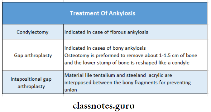
3. Treatment of ankylosis
4. Causes of trismus:
- Orofacial infection
- Trauma
- Inflammation
- Myositis
- Tetany
- Tetanus
- Neurological disorders
- Drug-induced
- Extra articular fibrosis
- Mechanical blockage
5. Eminectomy:
- It involves the excision of the articular eminence
6. Hyperplasia of condyle:
- The patient exhibits a unilateral, slowly progressive elongation of the face
- Deviation of the chin occurs away from the affected side
7. Hypoplasia of condyle:
- Facial asymmetry occurs
- Limitation of lateral excursions on one side
- Exaggeration of the antegonial notch
8. Conditions where the jaw deviates to the same side:
- Ankylosis of TMJ
- Subcondylar fractures
- Hypoplasia of condyle
9. Kaban’s protocol:
- Early surgical intervention Aggressive resection
- Ipsilateral colectomy
- Contralateral colectomy
- The lining of the glenoid fossa with temporalis fascia
- Reconstruction of ramus with osteochondral graft
- Early mobilization
- Regular follow up
10. Ankylosis features:
- Unilateral
- Face is asymmetry
- Fullness occurs on the affected side of the mandible
- Flattening on the unaffected side occurs
- Bilateral:
- Gives a typical bird-face appearance
11. Interposition arthroplasty:
Involves the creation of a gap and insertion of a barrier between the cut bony surfaces
- Advantages:
- Minimizes the risk of recurrence
- Maintains the vertical height of the ramus.
Temporomandibular Joint Disorders Long Essays
Question 1. Classify TMJ disorders. Explain in detail about anterior dislocation & its management.
Or
Describe the etiology and pathogenesis of TMJ ankylosis. Describe different surgical procedures for TMJ
Or
Describe Subluxation.
Answer:
Classification:
1. Disorders due to Extrinsic factors:
- Masticatory muscle disorders:
- MPDS
- Myositis
- Problems due to trauma:
- Traumatic arthritis
- Fracture
- Internal disc derangement
- Tendonitis
2. Disorders due to intrinsic factors:
- Trauma:
- Dislocation
- Fracture
- Internal disc displacement:
- Anterior disc displacement with reduction
- Anterior disc displacement without reduction
- Arthritis:
- Osteoarthritis
- Rheumatoid arthritis
- Juvenile arthritis
- Infantile arthritis
- Developmental defects:
- Agenesis
- Hypoplasia
- Hyperplasia
- Ankylosis:
- Neoplasm:
- Benign
- Malignant
Anterior Dislocation:
Causes of Anterior Dislocation:
- Extrinsic causes:
- Blow on the chin when the mouth is open
- Injudicious use of mouth gag
- Post-traumatic
- Intrinsic causes:
- Excessive yawning
- Vomiting
- Singing loudly
- Laughing loudly
- Opening mouth too wide
Features of Anterior Dislocation:
- Unilateral:
- Difficulty in mastication & speech
- Profuse drooling of saliva
- Deviation of the chin over the contralateral side
- The affected condyle is not palpable
- Definite depression in front of the tragus
- Bilateral:
- Pain
- Inability to close mouth
- Tense masticatory muscles
- Difficulty in speech
- Excessive salivation
- Protruding chin
- Gagging of molars
- Anterior open bite
- Difficulty in swallowing
- Hollowness in particular regions
Management of Anterior Dislocation:
- Reassure the patient
- Sedative drugs
- Pressure & massage the area
- Manipulation
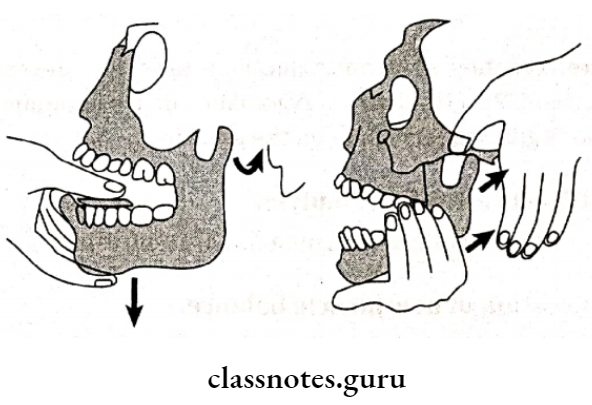
- Operator grasps the patient’s mandible
- The thumb is placed over the occlusal surfaces of the lower molars
- Fingertips are placed below the chin
- Downward pressure is placed over posteriors
- This overcomes spasms of muscles
- Backward pressure is applied which pushes the entire mandible posteriorly
- Immobilization is done
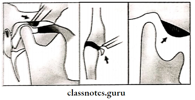
Manipulation Of Condyle:
- Capsule tightening procedure:
- Capsulorrhaphy:
- Shortening of the capsule by removing a section & suturing
- Placement of vertical incision & tightening it
- Reinforcement of capsule by stretching a strip of temporal fascia & suturing
- Capsulorrhaphy:
- Creation of mechanical obstacle:
- Osteotomy on an eminence by Lindermann
- Placement of graft over eminence by Mayor
- Osteotomy on the zygomatic arch by Dautry
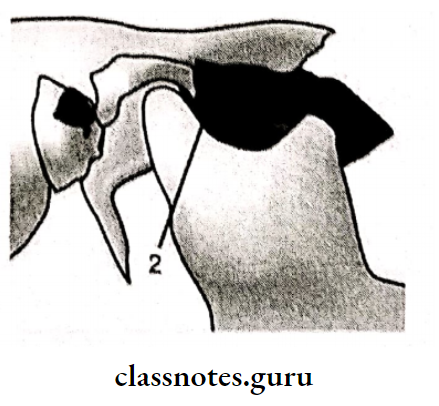
Dautry’s zygomatic arch osteotomy:
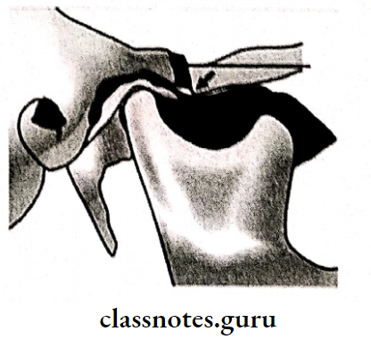
Mayor’s grafting technique on the eminence:
- Direct restrain of condyle: Temporalis fascia turned down & sutured
- Creation of new muscle balance: Temporalis tendon divided & sutured in a horizontal manner
- Removal of mechanical obstacles:
- Meniscectomy: Torn meniscus is removed
- High condylectomy: Excision of the superior portion of the condyle
- Eminectomy: Excision of the articular eminence
Question 2. Enumerate causes of inability to open the mouth. How to treat a case of bony ankylosis.
Or
Trisums causes
Answer:
It is a condition in which muscle spasm prevents the opening of the mouth
Causes of inability:
- Orofacial infection
- Trauma
- Inflammation
- Myositis
- Tetany
- Tetanus
- Neurological disorders
- Drug-induced
- Extra articular fibrosis
- Mechanical blockage
Management Of Bony Ankylosis:
1. condylectomy:
- Pre-pre-auricular incision given
- Horizontal osteotomy cut given over condylar neck Condylar head is separated
- Smoothened the remaining structures
- Close the wound in layers
- If required bilateral condylectomy done
- Exposure of the condylar head via a preauricular incision
- Sectioning of the condylar head
- Breaking the fibrous adhesions
- Condylectomy complete
- Suturing the capsule
- Final skin suturing
2. Gap arthroplasty:
- Two horizontal cuts are given
- Removal of bony wedge between glenoid fossa & ramus
3. Interposition arthroplasty:
- Creation of gap
- Insertion of barrier(autogenous or alloplastic)
Kaban’s Protocol:
- Early surgical intervention
- Aggressive resection
- Ipsilateral colectomy
- Contralateral colectomy
- The lining of the glenoid fossa with temporalis fascia
- Reconstruction of ramus with costochondral graft
- Early mobilization
- Regular follow up
Question 3. Define ankylosis of TMJ. Mention etiology, clinical features
Or
Define & classify ankylosis of TMJ. Write on etiology, clinical features
Or
Classify the Ankylosis of the Temporo-Mandibular Joint. Discuss the etiology of the Temporo-Mandibular Joint.
Or
Etiology and clinical features of TMJ ankylosis and Pathogenesis
Answer:
Definition:
Ankylosis means ” Stiff joint”
Etiology of ankylosis of TMJ:
- Trauma, Congenital
- Infections -Osteomyelitis
- Inflammation, Osteoarthritis
- Rare causes, Measles
- Systemic diseases, Typhoid
- Other causes, Prolonged trismus
Clinical Features of ankylosis of TMJ:
- Unilateral:
- Deviation of the chin on the affected side
- The fullness of the face on the affected side
- Flatness on the unaffected side
- Crossbite
- Angle’s classic malocclusion
- Condylar movements absent on the affected side
- Bilateral:
- Inability to open mouth
- Neck chin angle reduced
- Class II malocclusion
- Protusive upper incisors
- Multiple carious teeth
Pathogenesis of ankylosis of TMJ:
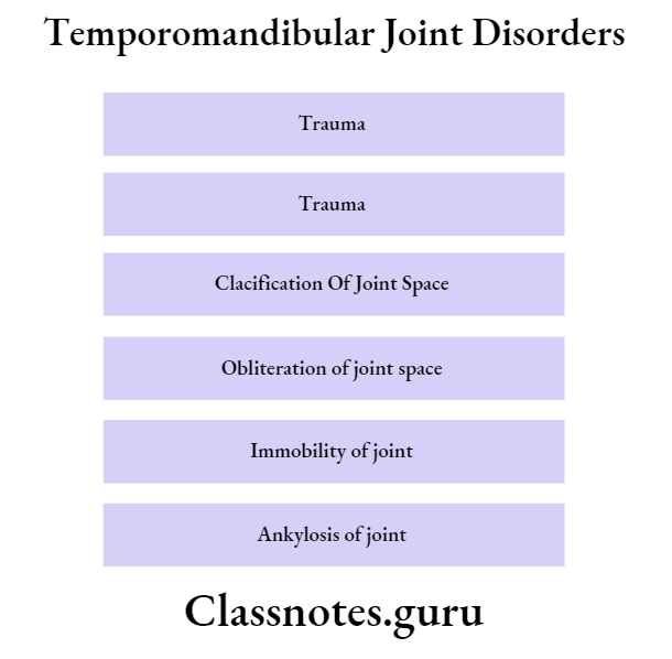
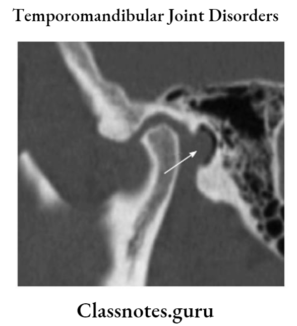
Question 4. Diagnosis of Bilateral Ankylosis in an 8-year-old boy
Answer:
Diagnosis:
- Radiographic features:
- Complete obliteration of joint space
- Normal TMJ anatomy is distorted
- Deformed condylar head
- Elongation of the coronoid process
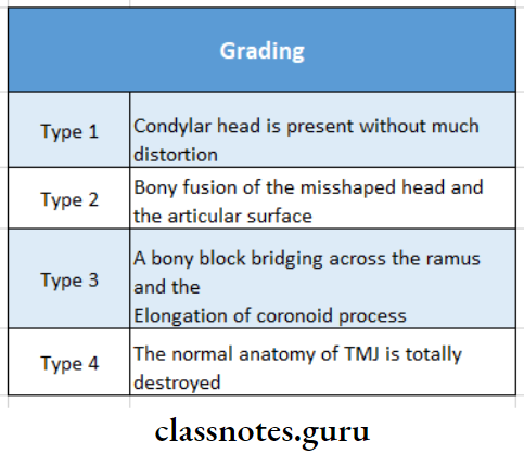
Grading:
Temporomandibular Joint Disorders Short Essays
Question 1. Pathogenesis and Treatment
Answer:
Pathogenesis:
Treatment:
- May resolve on its own
- Manipulation of the jaw by jaw stretcher
Question 2. Internal derangement of TMJ.
Answer:
Definition of TMJ:
It is the anteromedial displacement of the interarticular disc associated with the posterosuperior displacement of the condyle in the closed jaw position
Features of TMJ:
- Pain on biting
- Clicking sound over the joint
- Deviation of mandible
- Restricted mouth opening due to pain
Management of TMJ:
- Anterior repositioning appliances
- Placed on occlusal surfaces
- Supportive therapy
- NSAIDs to relieve pain
- Heat application
- Occlusal correction
Question 3. Pain dysfunction syndrome/ MPDS.
Answer:
Pain dysfunction syndrome
- It is a disorder characterized by facial pain limited to the mandibular function, muscle tenderness, joint sounds, absence of significant organic & pathologic changes in TMJ
- It may be due to functional derangement of dental articulation, psychological state of mind, or physiological state of joint
- Coined by Laskin
Etiology of Pain Dysfunction Syndrome:
- Extrinsic factors:
- Occlusal disharmony
- Trauma
- Environmental influences
- Habits
- Intrinsic factors:
- Internal derangement of TMJ
- Anterior locking of disc
- Trauma
Features of Pain dysfunction syndrome:
- Unilateral preauricular pain
- Dull constant sound
- Muscle tenderness
- Clicking noise
- Altered jaw function
- Absence of radiographic changes
- Absence of tenderness in ext. auditory meatus
Management of Pain dysfunction syndrome:
- Reassurance
- Soft diet
- Occlusal correction: 7 ‘R’s
- Remove-extract the tooth
- Reshape grind the occlusal surface
- Reposition orthodontically treated
- Restore conservative treatment Replaceby prosthesis
- Reconstruct TMJ surgery
- Regulate control habits
- Isometric exercises
- Opening & closing of mouth 10 times a day
- Medicaments
- Aspirin: 0.3-0.6 gm/4 hourly
- NSAIDS: for 14-21 days
- Pentazocine: 50 mg/ 2-3 times a day
- Heat application
- It increases circulation
- Diathermy
- Causes heat transmission to deeper tissues
- LA injections
- 2% lignocaine into trigger points
- Steroid injection
- As anti-inflammatory
- Anti-anxiety drugs
- Diazepam-2-5 mg * 10 days
- Tens
- Acupuncture
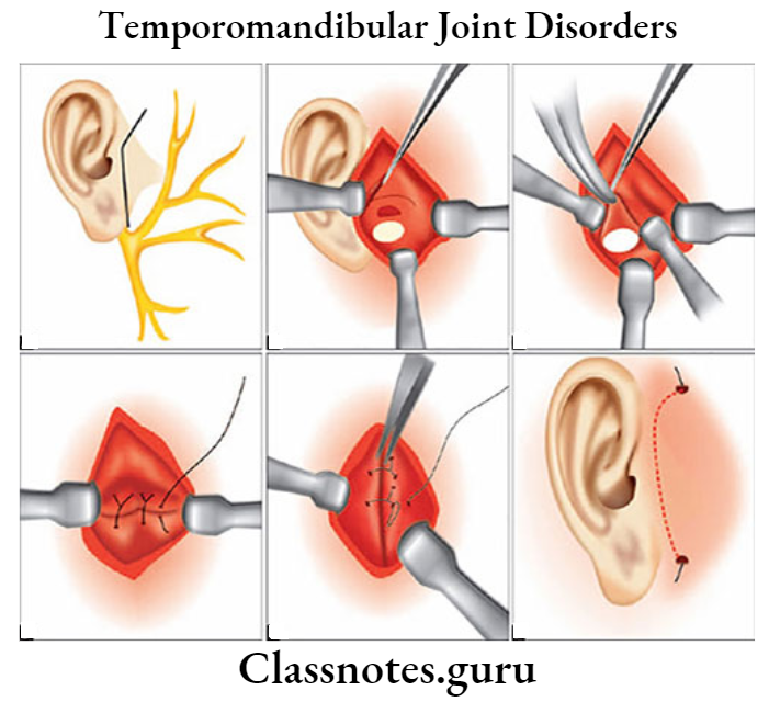
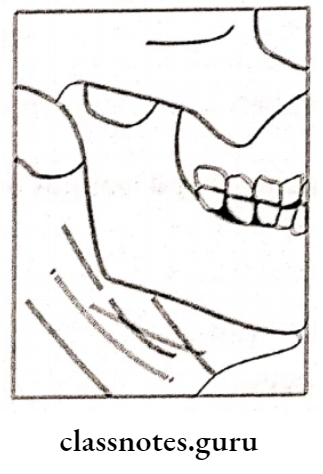
Question 4. Preauricular approach to TMJ.
Answer:
Preauricular approach to TMJ
Basic & standard approach to TMJ
Technique of TmJ:
- Shaving of the area
- Mark incision from the helix of the ear to the upper border of the tragus
- The depth of penetration of the incision should be upto superficial layer of the temporalis fascia
- Exposure of condyle, thus advantageous
- Initial incision in the preauricular fold
- Oblique incision through the superficial layer of temporalis fascia. The periosteal elevator is then inserted below the temporalis muscle to expose the lateral portion of the zygomatic arch
- Cut in the capsule to enter the TMJ space and incision through the lateral attachment of the disc, entering the inferior joint space
- After surgery, suturing of the capsule
- Suturing the wound in layers
- Final skin subcuticular suturing
Question 5. Risdon’s approach.
Answer:
Risdon’s approach
- Site Of Incision: 1 cm below the angle of the mandible
- Extent: Forward, parallel to the lower border of the mandible
- Site Seen: Neck of condyle & ramus
Disadvantages of Risdon’s approach:
- Poor access to the condylar head
- Procedures involving the articular portion of the head & meniscus cannot be performed
Question 6. Frey’s Syndrome:
Answer:
Frey’s Syndrome
This is auriculotemporal nerve syndrome
Causes of Frey’s Syndrome:
- Iatrogenic causes followed by parotidectomy
Features of Frey’s Syndrome:
- Pain in auriculotemporal nerve distribution
- Gustatory sweating
- Flushing on the affected side
Diagnosis of Frey’s Syndrome:
- Positive starch iodine test
Treatment of Frey’s Syndrome:
- Topical application of anticholinergic
- Radiation therapy
- Surgical procedures
- Skin excision
- Nerve section
- Tympanic neurectomy
Temporomandibular Joint Disorders Short Answers
Question 1. Arthroscopy.
Answer:
Arthroscopy
- Means looking into the joint
- Oral And Maxillofacial Surgery
Indications of Arthroscopy:
- Disc derangement
- Arthrosis Arthritis
- Injuries to TMJ
- Perforation of the disc
Contraindications of Arthroscopy:
- Infection
- Ankylosis
Components of Arthroscopy:
- Arthroscope
- Fibreoptic light cables
- Eye lens
The procedure of Arthroscopy:
- Anesthetized
- Palpate the joint
- Mark a point at the 12 mm anterior to the tragus
- Mark another point 1-2 mm below it
- Cutaneous incision made
- Introduction of the trocar into the capsule
- Continuous irrigation carried throughout the procedure
Question 2. Barrel bandage.
Answer:
Barrel bandage
- Used for ankylosis management
- The bandage is used to restrict the movement of the joint
- The patient is kept on a soft diet
- Restrict wide opening of the mouth while yarning, laughing
- If required, support the mandible while such activities
Question 3. Interposition arthroplasty.
Answer:
Interposition arthroplasty
- Used for the management of ankylosis
- Horizontal osteotomy cut is made
- Between two cuts, graft material is added
Various grafts are:
- Autografts:
- Cartilaginous graft
- Temporalis fascia
- Temporalis muscle
- Heterogenous graft:
- Pig bladder
- Alloplasts:
- Stainless steel
- Titanium
- Zirconium
- Tantalum
Question 4. Eminectomy.
Answer:
Eminectomy
Excision of the articular eminence
Steps of Eminectomy:
- Anesthetized
- Undermine & turn skin & subcutaneous flap upward
- A small horizontal incision was given over the zygomatic arch
- T incision is given a horizontal portion over the arch & vertical portion over the apex of the eminence.
- Periosteum reflected
- Expose eminence
- A series of bur holes are created
- Burs are connected
- Eminence is sectioned & separated
- Smoothened the base of eminence Irrigate the area
- Suture
Question 5. Ligaments of TMJ:
Answer:
Ligaments of TMJ
- Temporomandibular ligament
- It stabilizes TMJ
- It extends downward & backward from the articular eminence to the external & posterior sides of the condylar neck
- Stylomandibular ligament
- Extends from the styloid process to the mandibular angle
- Sphenomandibular ligament
- Arises from the spine of the sphenoid & is inserted into the lingual of the mandible
- It is a remnant of Meckel’s cartilage
Temporomandibular Joint Disorders Viva Voce
- The submandibular incision is given about 1 cm below the angle of the mandible
- Hemarthrosis is the extravasation of blood into joint space due to trauma
- Intraarticular injection of hydrocortisone reduces the inflammatory process within the joint
- The preauricular approach is an ideal surgical approach to TMJ ankylosis
- The interposition of temporal muscle and fascia in the treatment of ankylosis is done to prevent ankylosis
- Dautery procedure is a treatment modality for TMJ dislocation
- Bird face appearance is a feature of bilateral ankylosis
- A hypertonic saline para capsular injection is used for conservative management of TMJ subluxation and dislocation
- MPDS is the most common disorder causing pain in the masticatory apparatus along with TMJ