
Physical And Chemical Injuries Of The Oral Cavity Important Notes
- Smear Layer
- Smear Layer is an amorphous microlayer deposited on the prepared tooth surface
- Consists of inorganic enamel and dentin debris, organic pulp materially dentinal fluid, bacteria, and saliva
- Thickness -1-5 mm
- Smear Layer Functions
- Forms physical barrier
- Reduces permeability of dentin
- Prevents exit of dentinal fluid
- Acts barrier against micro-organisms
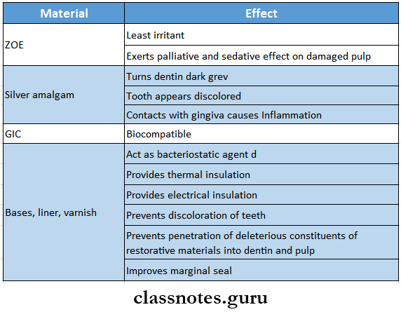
- Effect Of Restorative Materials
- Cracked Tooth Syndrome
- Cracked Tooth is characterized by sharp pain on chewing without any obvious reason
- Cracked Tooth Causes
- Attrition
- Bruxism
- Trauma
- Accidental biting on a hard object
- Presence of large restoration
- Improper endodontic treatment
- Cracked Tooth Treatment
- Stabilization by stainless steel band/crown
- Symptoms Of Sodium Hypochlorite Accidents
- Hemolysis
- Ulceration
- Facial nerve weakness
- Necrosis
- Inhibits neutrophil migration
- Damages endothelial cells and fibroblasts
- Drugs Causing Gingival Hyperplasia
- Dilantin sodium
- Cyclosporine
- Nifedipine
Physical And Chemical Injuries Of The Oral Cavity Short Question And Answers
Question 1. Describe the predisposing factors and clinical features of osteoradionecrosis.
Answer:
Osteoradionecrosis:
- Osteoradionecrosis is a radiation-induced pathologic process characterized by chronic and painful infection and necrosis is accompanied by late sequestration and sometimes permanent deformity
- This is one of the most serious complications of radiation to the head and neck
Osteoradionecrosis Predisposing Factors:
- Irradiation of an area of previous surgery before adequate healing had taken place
- Irradiation of lesion near the bone
- Poor oral hygiene
- Continued use of irritants
- Poor patient cooperation in managing irradiated tissue
- Surgery in the irradiated area
- Failure to prevent trauma to the irradiated area
Osteoradionecrosis Clinical Features:
- Nonhealing dead bone
- The bone becomes hypovascular, hypocellular, and hypomineralised
- Mandible is more effected than maxilla
- Necrosis of bone
- Infection of tissues
- Sequestration of bone
- Bone deformity
Osteoradionecrosis Treatment:
- Debridement of necrotic tissue should be done along with removal of the sequestrum
- Administration of intravenous antibiotics and hyperbaric oxygen therapy
- Maintenance of oral hygiene
- Chemotherapy- Bleomycin, Cisplatin, 5- Fluorouracil
Read And Learn More: Oral Pathology Questions and Answers
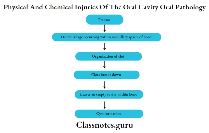
Question 2. Traumatic bone cyst
Answer:
Traumatic Bone Cyst
- Traumatic bone cyst is a pseudo cyst
- Traumatic Bone Cyst is not lined by epithelium
Traumatic Bone Cyst Etiopathogenesis:
Traumatic Bone Cyst Clinical Features:
- Age- between 10-20 years
- Sex- Females are commonly affected
- Site- in mandible, above the mandibular canal
- Painful, bony hard swelling of the jaw
- Paraesthesia of lip
- Expansion of the cortical plates
- Displacement of regional teeth
Traumatic Bone Cyst Radiographic Features:
- Smooth radiolucent area
- Size- centimeter in diameter
- Cysts involving roots of teeth have a scalloped appearance
- Intact lamina dura
- The cystic margin is well-demarcated
Traumatic Bone Cyst Histological Features:
- Absence of epithelial lining
- The cystic cavity is surrounded by loose vascular connective tissue wall
- Connective tissue stroma is made up of fibrous tissue, areas of hemorrhage, hemosiderin pigmentation, and bone resorption
- Presence of red blood cells, or giant cells adhering to the bone surface
Traumatic Bone Cyst Treatment:
- Surgical exploration
Question 3. Effects of radiation on oral tissues
Answer:
Effects Of Radiation On Oral Tissues
- Oral mucous membrane
- Mucositis
- Desquamation of epithelial layer
- Infection of the oral cavity
- Candidiasis
- Atrophic mucosa
- Ulceration
- Radiation necrosis
- Taste buds
- Degeneration
- Loss of taste sensation
- Salivary glands
- Xerostomia
- Loss of salivary secretion
- Difficult and painful swallowing
- Decreased buffering capacity
- Susceptibility to radiation caries
- Teeth
- Retards growth of teeth
- Inhibits cellular differentiation
- Premature eruption
- Retard root formation
- Pibroatrophy of pulp
- Radiation caries
- Bone
- Osteonecrosis
- Hypocellularity
- Hypoxia
- Hypovascuiarity
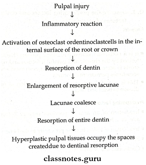
Question 4. Internal resorption
Answer:
Internal Resorption
- Internal resorption refers to the resorption process that starts internally within the tooth itself and the dentin is gradually resorbed from the pulpal side toward the periphery
Internal Resorption Etiopathogenesis
Internal Resorption Clinical Features:
- Can involve the crown portion or root portion of teeth
- Any tooth can be Involved
- Initially asymptomatic
- Later there appearance of pink pink-lined area on the crown of the tooth
- Also known as the pink tooth of mummery
- Affected tooth Is vital
Internal Resorption Radiographic Features:
- involved tooth shows the round or ovoid radiolucent area in the central portion of the tooth
- Well well-defined, spherical-shaped, radiolucent area occurs in dentin
- The external outline of the tooth is intact
Internal Resorption Types:
- internal inflammatory resorption
- internal replacement resorption
Internal Resorption Treatment:
- Extirpation of pulp
- Endodontic treatment
- Extraction
Question 5. Clinical and histologic features of nicotine stomatitis
Answer:
Nicotine Stomatitis
Nicotine stomatitis or smoker’s keratosis is tobacco-related keratosis of the oral mucosa
Nicotine Stomatitis Clinical Features:
- Affects both the hard and soft palate
- Hyperkeratosis of the epithelium
- Inflammatory swelling of palatal mucous glands
- Palatal mucosa becomes red initially and later becomes white
- On the surface, there is the presence of red dot-like areas surrounded by elevated white keratotic rings
- The surface is rough, fissured, or wrinkled
Nicotine Stomatitis Histopathological Features:
- Hyperorthokeratosis and acanthosis of surface epithelium
- Inflamed and swollen palatal mucous glands
- Squamous metaplasia of ductal epithelium
- Atrophic changes of minor salivary glands
- Moderate inflammatory cell infiltration
Question 6. Tooth ankylosis
Answer:
Tooth Ankylosis
Tooth ankylosis is a fusion between the tooth and bone
Tooth Ankylosis Etiology:
- Partial root resorption
- Traumatic injury to teeth
- Periapical inflammation
Tooth Ankylosis Clinical Features:
- Lack of mobility of tooth
- If a deciduous tooth is affected, they do not exfoliate and become submerged
- Pulpal infection
- Dull, muffled sound on percussion of tooth
Tooth Ankylosis Radiographic Features:
- Loss of periodontal ligament
- Mild sclerosis of the bone
- Blending of bone with tooth root
Question 7. Plumbism
Answer:
Plumbism
Plumbism or lead poisoning is an occupational hazard
Plumbism Etiology:
- Inhalation of lead vapor or dust in adults
- Ingestion while chewing on wood painted with lead-containing paint- in infants
Plumbism Clinical Features:
- Nausea, vomiting, constipation
- Peripheral neuritis
- Wrist drop or foot drop
- Encephalitis
- Hypochromic anemia
- Basophilic stippling of RBC
- Grey bluish-black line on marginal gingiva
- Ulcerative stomatitis
- Excessive salivation
- Metallic taste
- Swelling of salivary glands
Plumbism Treatment: Treatment of oral lesions
Question 8. Amalgam tattoo
Answer:
Amalgam Tattoo
Amalgam tattoo of oral mucous membrane is a relatively common finding in dental practice
Amalgam Tattoo Etiology:
- Therapeutic use of silver nitrate
- Condensation of amalgam in gingiva during restoration work
- From broken pieces introduced into the socket during extraction of the restored tooth
- Retrograde amalgam filling
Amalgam Tattoo Clinical Features:
- Permanent bluish-grey pigmentation of skin and mucosa
- Black or olive brown granules present in gingival areas
- Differential Diagnosis:
- Malignant melanoma
Question 9. Pink disease or Acrodynia
Answer:
Pink Disease Or Acrodynia
Pink Disease Or Acrodynia is also known as Swift’s disease
Pink Disease Or Acrodynia Etiology:
- Mercury toxicity
Pink Disease Or Acrodynia Clinical Features:
- Age- infants below the age of 2 years
- The raw beef appearance ofthe skin
- The patient has a cold, clammy feeling
- Presence of pruritic maculopapular rash
- Extreme irritability
- Photophobia with lacrimation
- Muscular weakness
- Tachycardia, hypertension
- Insomnia
- Gastrointestinal upset
- Stomatitis
- Profuse salivation
- Painful gingiva
- Bruxism
- Loosening and premature shedding of teeth
Question 10. Bruxism
Answer:
Bruxism Definition: Habitual grinding of teeth either during sleep in the night or during daytime
Bruxism Etiology:
- Local factors-hereditary
- Psychologic factor- emotional upset
- Occupational causes- carpenters
Bruxism Clinical Features:
- Attrition of teeth
- Loosening and drifting of teeth
- Gingival recession
- Hypertrophy of masticatory muscles
- Trismus
- Facial pain
- Sensitivity of teeth
Bruxism Treatment:
- Removal of irritating factors
- Use of occlusal splints
Physical And Chemical Injuries Of The Oral Cavity Viva Voce
- Demineralization of dentin widens tubules
- Peritubular dentin demineralizes quicker than Intertubular matrix
- A concussion is produced by an injury that is not strong enough to cause serious, visible damage to the tooth and the periodontal structures
- Abrasion is the wearing away of tooth substance due to mechanical means
- Subluxation is the abnormal loosening of the tooth without displacement due to sudden trauma
- An avulsion is the discoloration of a tooth from its socket due to traumatic injury
- Fusion between tooth and bone is termed ankylosis
- The most common physical injury of the bone is fractured
- The linea alba is a white line seen on the buccal mucosa extending from the commissures posteriorly at the level of the occlusal plane
- Sialolithiasis is common in the submandibular salivary gland
- Rhinoliths are calcareous concentrations occurring in the nasal cavity
- Radiation causes osteoradionecrosis in bone
- Local application of aspirin causes blanching and sloughing of the epithelium of oral mucosa
- Pink’s disease or acrodynia occurs due to mercury toxicity
- Green stains frequently seen on children’s teeth are caused by chromogenic bacteria.