
Orthodontic Diagnosis Important Notes
- Cephalic index
- The shape of the head can be evaluated based on a cephalic index of the head formulated by Martin and Saller
- Cephalic index = posterior facial height / anterior facial height
- Facial form
- Types of lips
- Facial divergence
- Nasolabial angle
- It is an angle formed between the lower border nose and a line connecting the nose and upper lips
- The normal value is 110 degrees
- Reduced in patients with proclined maxillary anterior or prognathic maxillary
- Increased in patients with retrognathic maxilla or reclined maxillary anterior
- Path of closure of mandible
- Backward in class 2 division 2
- Forward in pseudo-class 3
- Lateral in unilateral crossbites
Orthodontic Diagnosis Long Essays
Question 1. Discuss in detail the various diagnostic aids used in orthodontics.
Answer.
Diagnosis:
- It involves the collection of pertinent data in a systemic manner to help identify the nature and cause of the problem
Read And Learn More: Orthodontics Short And Long Essay Question And Answers
Diagnosis Aids:
- Comprehensive orthodontic diagnosis is established by the use of certain clinical implements called diagnostic aids
Types Of Diagnosis Aids:
- Essential Diagnostic aids
- Case history
- Clinical examination
- Study models
- Certain radiographs
- Facial radiographs
- Supplemental diagnostic aids
- Specialized radiographs
- Electromyographs
- Hand wrist radiographs
- Endocrine tests
- Estimation of basal metabolic rates
- Diagnostic set-up
- Occlusograms
- Essential Diagnostic aids:
- Aids that are important for all cases
- Involves
Case History involves:
- Personal details:
- Name – For maintaining a record
- Addressing patient
- For communication
- Age – for treatment planning
- Growth period – Myofunctional appliances
- After cessation of growth-surgical treatment
- Sex – growth spurts differ in both sex
- Address – for further correspondence
- Name – For maintaining a record
- Chief complaints: Help to identify the expectations of the patient
- Medical history: epilepsy, diabetes, and blood dyscrasias complicate the treatment
- Dental history: to know the attitude of the patient toward treatment
- Pre-natal history: for knowing the condition of the mother during pregnancy
- Forcep delivery leads to cross-bite
- Post-natal history: to know milestones of development
- Family history: some occlusions like clefts are hereditary
Clinical Examination:
General:
- Height
- Weight
- Gait
- Posture
- Body build
Extra-Oral:
- Head shape
- Facial form
- Facial Profile
- Facial symmetry
- Facial divergence
- Anteroposterior relationship
- Facial proportions
- Lips
- Nose
- Nasolabial angle
Intra-Oral:
- Tongue
- Palate
- Gingiva
- Frenal attachment
- Tonsils
- Adenoids
- Dentition


Functional Examination:
- Postural rest position
- Path of closure
- TMJ
- Swallowing
- Speech
- Respiration
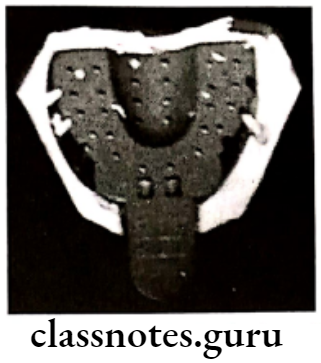
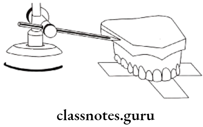
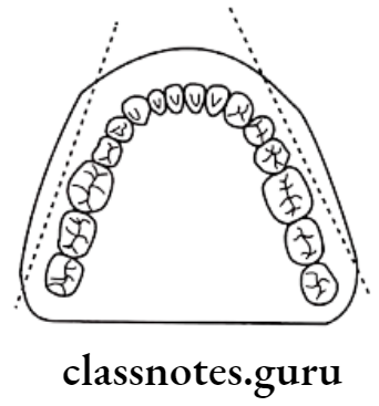
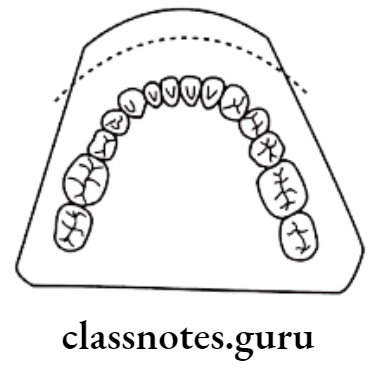
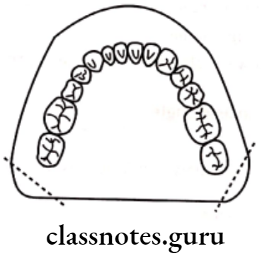
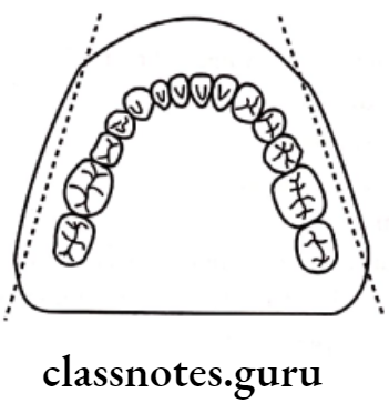
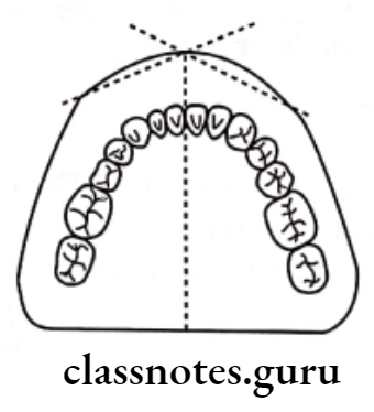
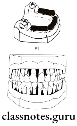
Study models:
- Provide a 3D view of the situation
Uses Of Orthodontics:
- Studies occlusion from all aspects
- Enables accurate measurement
- Treatment planning
- Assess the severity of malocclusion
- Motivate the patient
- For mock surgery
- For transparency records
Steps in Fabrication:
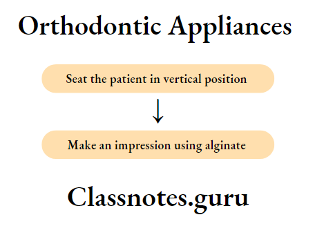
- Impression making – By alginate
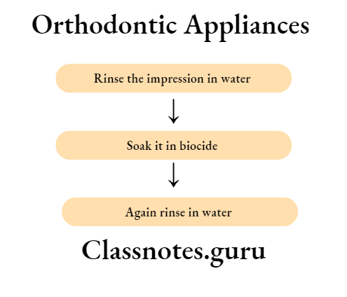
- Disinfecting it – By Biocide
- Casting the impression – By model stone
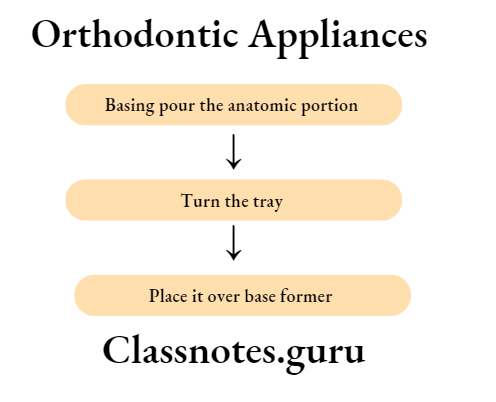
- Basing and trimming the cast
- Finishing and Polishing – By fine-grained sandpaper
Radiographs:
- Commonly used radiographs are
Cephalometric: For skeletal malocclusion
- IOPA – to know the bone condition
- Occlusion radiograph – for arch length
Facial Photographs:
- To compare pre and post-treatment conditions
Supplemental Diagnostic Aids
- Require only in specialized cases
- Includes – electromyography
- Hand wrist radiograph
- Endocrine test
- Estimation of BMR (Basal Metabolic Rate)
- Diagnostic set up
- Occlusograms
- MRI, CT scan
Question 2. What are various diagnostic aids in orthodontics? Write in detail about clinical examination and case history records.
Answer.
Diagnostic Aids:
Essential Diagnostic aids:
- Case history
- Clinical examination
- Study models
- Certain radiographs
- Facial photographs
Supplemental diagnostic aids:
- Specialized radiographs
- Electromyographs
- Hand wrist radiographs
- Endocrine tests
- Estimation of basal metabolic rates
- Diagnostic set-up
- Occlusograms
Case History involves:
- Personal details:
- Name – For maintaining a record
- Addressing patient
- For communication
- Age – for treatment planning
- Growth period – Myofunctional appliances
- After cessation of growth-surgical treatment
- Sex – growth spurts differ in both sex
- Address – for further correspondence
- Name – For maintaining a record
- Chief complaints: Help to identify the expectations of the patient
- Medical history: Epilepsy, diabetes, and blood dyscrasias complicate the treatment
- Dental history: To know the attitude of the patient toward treatment
- Pre-natal history: For knowing the condition of the mother during pregnancy
- Forcep delivery leads to cross-bite
- Post-natal history: To know milestones of development
- Family history: Some occlusions like clefts are hereditary
Clinical Examination:
General:
- Height
- Provide information on physical growth and maturation of the patient that may influence oro-facial development
- Weight
- Provide information on the physical growth and maturation of the patient that may influence oro-facial development
- Gait
- Gait abnormality is associated with neuromuscular problems
- Posture
- Abnormal posture can predispose to malocclusion
- Body build
- Types:
- Aesthetic
- Thin physique
- Narrow dental arches
- Plethoric
- Obese persons
- Large, square dental arches
- Athletic
- Normal built
- Normal-size dental arches
- Aesthetic
- Types:
Extra-Oral:
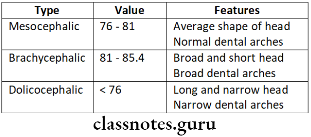
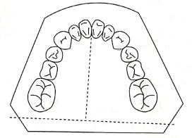
- Head shape
- Types:
- Mesocephalic
- The average shape of the head
- Normal dental arches
- Dolichocephalic
- Long and narrow head
- Narrow dental arches
- Brachycephalic
- Broad and short head
- Broad dental arches
- Mesocephalic
- Types:
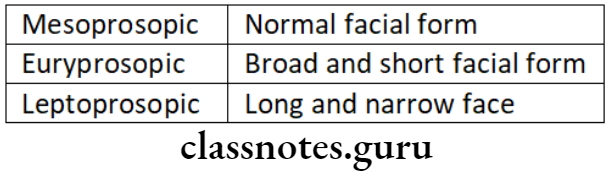
Facial form:
- Types:
- Mesoprosopic
- Average face from
- Euryprosopic
- Broad and short facial form
- Leptoprosopic
- Long and narrow facial form
- Mesoprosopic
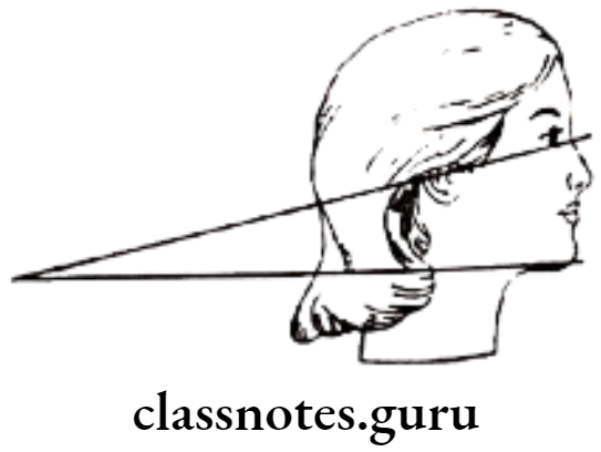
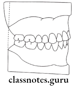
Facial profile:
- Examined by viewing the patient from the side
- Diagnoses gross deviations in the maxillo-mandibular relationship
Types:
- Straight
- Convex
- Concave
Facial symmetry:
- Determines disproportions of the face in transverse and vertical planes
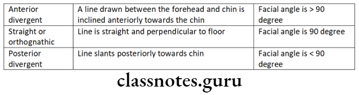
Facial divergence:
- It is an anterior or posterior inclination of the lower face relative to the forehead
Types Of Facial Divergence:
- Anterior divergence
- Posterior divergence
- Straight or orthognathic
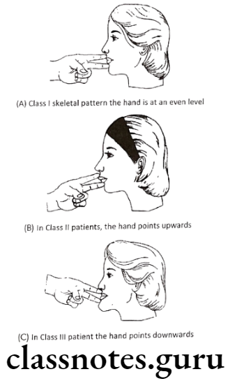
- Anteroposterior relationship
- This can be assessed by the following
- Seat the patient in an upright position
- Ask to occlude gently
- Place index and middle finger at soft tissue points A and B respectively
- In class 1 hand is at an even level
- In class 2 Index finger is anterior to the middle finger
- In class 3 Middle finger is ahead of the forefinger
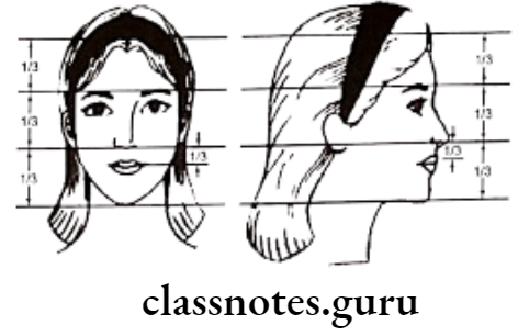
Facial proportions:
- The face is divided into three equal thirds by four horizontal planes
- At the level of hairline
- Supraorbital ridge
- The base of the nose
- The inferior border of the chin
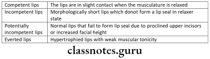
Lips:
- Types:
- Competent lips
- Incompetent lips
- Potentially incompetent lips
- Everted lips
Note:
- It contributes to the esthetics of the face
Nasolabial angle:
- It is the angle formed between the lower border of the nose and the line connecting the intersection of the nose and the upper lip
- It is normally 110 degrees
Intra-Oral:
Tongue:
- Abnormalities in the tongue disturb muscle balance and equilibrium leading to malocclusion
Palate:
- Examined for
- Palatal depth
- Presence of swelling
- Mucosal ulceration
- Presence of clefts
- Third rugae
Gingiva:
- Examined for
- Inflammation
- Recession
- Mucogingival lesions
Frenal attachment:
- Abnormal labial frenum leads to midline diastema
- Ankyloglossia leads to the narrowing of the maxillary arch
Tonsils:
- Abnormal inflamed tonsils cause alteration in tongue and jaw posture
Adenoids:
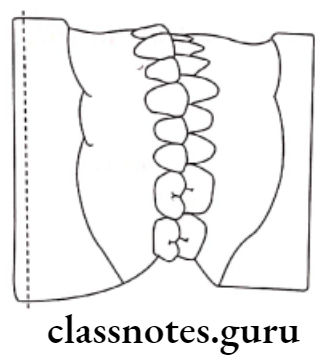
Dentition:
- Assess the following
- Number of teeth present
- Number of teeth missing
- Status of teeth-erupted or unerupted
- Presence of caries, restorations, or malformed
- Assess occlusion
- Assess overjet and overbite
- Assess individual tooth irregularities like rotations, intrusion, and extrusions
- Assess transverse relationship
- Examine the upper and lower arch separately
Question 3. Exumerate essential diagnostic aids. Describe study models in detail.
Answer.
Essential Diagnostic Aids:
- Case history
- Clinical examination
- Study models
- Certain radiographs
- Facial photographs
Study Models:
Uses Of Diagnostic Aids:
- Studies occlusion from all aspects
- Enables accurate measurement
- Treatment planning
- Assess the severity of malocclusion
- Motivate the patient
- For mock surgery
- For transparency records
Requirements Of Diagnostic Aids:
- Should accurately reproduce oral structures
- Should be pleasing to the eye
- Should accurately reproduce occlusion
- Should have a clean, smooth surface
- Should reproduce as much of the alveolar process as possible
Parts Of Diagnostic Aids:
- Anatomic portion
- Artistic portion
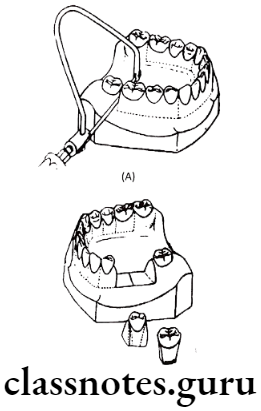
Steps:
- Impression making
Disinfecting the impression
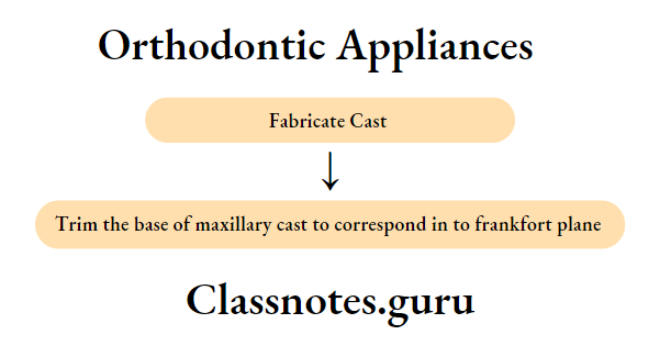
- Casting the impression – Using orthodontic stone/model stone
- Basing and trimming of cast

- Trimming
- Step 1: Trimming of mandibular base
- Step 2: The back of the mandibular model is made perpendicular to the midline
- Step 3: Both casts should occlude
- Step 4: Backs of both casts are made right-angled to base
- Step 5: Buccal cuts are made on mandibular cast
- Step 6: Anterior segments made on lower cast
- Step 7: Posterior cuts on the lower cast
- Step 8: Buccal cuts made on the upper cast
- Step 9: Anterior cuts on the upper cast
- Step 10: Posterior cuts on the upper cast

- Finishing and polishing – Done using fine-grained sandpaper
Orthodontic Diagnosis Short Essays
Question 1. Intra-oral X-rays in Orthodontics
Answer.
Types Of Intra-Oral X-rays:
Intra-Oral Periapical Radiograph:
- Techniques:
- Paralleling technique
- Bisecting angle technique
- Uses:
- View the presence/absence of teeth
- Supernumerary teeth
- Root formation
- Periapical pathology
- PDL space
- The contour of alveolar bone – to assess the abnormality
- Unerupted teeth – for tooth morphology
- Disadvantages:
- Not convenient for the entire dentition
- Gag reflex
- Uncomfortable for children
Bitewing:
- Uses:
- To assess the height and contour of bone
- To evaluate periodontal changes
- To assess interproximal calculus
Occlusal:
- Uses:
- For impacted/unerupted teeth
- For supernumerary teeth
- For foreign bodies
- To view the effects of the arch expansion procedure
Question 2. Inter-incisal angle.
Answer.
It is the angle formed between the long axis of the upper and lower incisors
- Value – 135.4°
- Range – 130 – 150.5°
Significance Of Inter-incisal Angle:
- Increased in Class 2, Division 2
- Decreased – Class 1 bimaxillary protrusion
- Class 2 Division 1
Question 3. Occlusal X-ray.
Answer.
Advantages Of Occlusal X-ray:
- Views a large segment of the dental arch
- For viewing palate and floor of mouth
- Useful in trismus
Uses Of Occlusal X-ray:
- Locate impacted teeth
- Locate fracture
- Locate supernumerary teeth
- Locate foreign bodies
- Locate effects of arch expansion procedures
- Assess the arch length
- Assess any pathology of jaws
Question 4. Overjet and Overbite.
Answer.
Overjet: It is the horizontal overlapping of maxillary and mandibular anterior
Value: 1.5 – 2mm
Significance Of Overjet: Increases in open bite cases
- Increases in anterior proclination
Overbite: It is the vertical overlapping of maxillary and mandibular anterior
Value: 1.5 – 2mm
Significance Of Overbit: Decrease – Deep bite
Increase: Open bite
Orthodontic Diagnosis Short Questions And Answers
Question 1. Facial Divergence.
Answer.
It is defined as an anterior/posterior inclination of the lower face relative to the forehead
Types Of Facial Divergence:
- Anterior divergent: A line between the forehead and chin inclines anteriorly
- Posterior divergent: A line between forehead and chin inclines posteriorly
- Straight/Orthognathic: A line between the forehead and chin is straight or perpendicular to the floor.
Question 2. Uses of Panoramic Radiograph./Orthopentogram
Answer.
- Assessing dental development
- Studying root resorption/root formation
- For Ankylosis
- Impacted teeth
- Path of eruption
- Diagnose pathology
- Status of eruption
- Unerupted teeth
Question 3. Bite Wing Radiographs.
Answer.
Records coronal part of upper and lower dentition together
Uses Of Bite Wing Radiographs:
- For proximal caries
- For interdental bone contour
- For secondary caries
- Detect overhanging restoration
- Detect periodontal changes
- Detect interproximal calculus
Question 4. Electromyography.
Answer.
Used for recording the electrical activity of muscles
Types Of Electrodes Used:
- Surface electrode – Plated superficially
- Needle electrode – Placed deeply
Useful In:
- In severe Class 2 division 1 malocclusion
- Abnormal buccinator activity
- Overclosure of jaws
- Cerebral palsy
- After orthodontic therapy
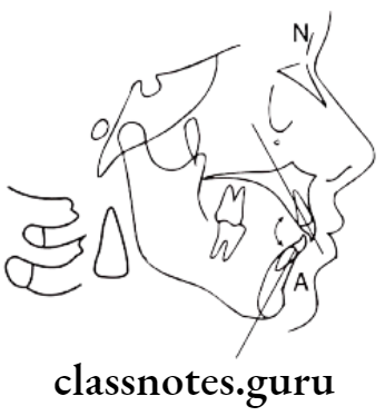
Question 5. Clinical Evaluation of Facial Profile.
Answer.
The patient is viewed from the side
Reference Lines:
- A line from forehead to point A
- A line from point A to pogonion
Types Of Profiles:
- Straight: Two lines straight line Ex. Class 1 cases
- Convex: Two lines form an angle with concavity facing tissue Ex. Class 2, Division 1
- Concave: Two lines form an angle with convexity facing tissue Ex. Class 3 malocclusion
Question 6. Importance of Family History.
Answer.
- Some malocclusions are hereditary
- These affect the treatment planning
- Thus details of it should not be neglected Ex. Class 2 malocclusion, Cleft lip and palate
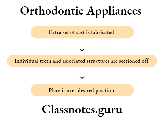
Question 7. Kesling’s diagnostic setup.
Answer.
- Proposed by H.D. Kesling
- Used for assessing the effect of treatment
Procedure Of Kesling’s Diagnostic:
Question 8. Freeway Space.
Answer.
- The position of the mandible at which muscles that are useful in opening and closing of jaws are in a state of minimal contraction is called posture rest position
- At this position, space is present between both the jaws
- This is called free-way space
Value – 3mm
Site – In canine region
Question 9. Gnathostatic Models.
Answer.
- Type of study model
- Uses: Provide 3D view of occlusion
- For treatment planning
- For assessing the outcome
Gnathostatic:
Question 10. Overjet.
Answer.
- It is a horizontal overlapping of the maxillary and mandibular anterior
- Value: 1.5 – 2mm
- Significance Of Overjet: Increases in open bite cases
- Increases in anterior proclination
Question 11. Nasolabial angle.
Answer.
- It is the angle formed between the lower border of the nose and the line connecting the intersection of the nose and the upper lip
- It is normally 110 degrees
- Reduces in
- Patients having proclined upper anterior or prognathic maxilla
- Increases in
- Patients with retrognathic maxilla or reclined maxillary anterior
Question 12. Cephalic index
Answer.
- The cephalic index was described by Martin and Saller in 1957
- It is calculated as follows
- Cephalic index = Maxillary skull width / Maxillary skull length
Interpretation Of Cephalic Index:
- Value – 76-80.9
- Indicates mesocephalic individuals
- Value – 81-85.4
- Indicates brachycephalic individuals
- Value < 75.9
- Indicates dolichocephalic individuals
- Value > 85.5
- Indicates hyperbradycephalic
Question 13. Orthodontic study models
Answer.
Uses Of Orthodontic Study Models:
- Studies occlusion from all aspects
- Enables accurate measurement
- Treatment planning
- Assess the severity of malocclusion
- Motivate the patient
- For mock surgery
- For transparency records
Requirements Of Orthodontic Study Models:
- Should accurately reproduce oral structures
- Should be pleasing to the eye
- Should accurately reproduce occlusion
- Should have a clean, smooth surface
- Should reproduce as much of the alveolar process as possible
Parts Of Orthodontic study models:
- Anatomic portion
- Artistic portion
Question 14. Importance of medical history
Answer.
- Some medical conditions contraindicate the use of orthodontic appliances
- They may require special precautionary measures to be taken before or during orthodontic therapy
- It is advisable to delay orthodontic treatment in patients suffering from epilepsy until it is controlled
- Patients with a history of blood dyscrasias may need special management if extractions are planned
- Diabetic patients can undergo orthodontic therapy if it is under control
- Patients having rheumatic fever or cardiac anomalies require antibiotic coverage for certain dental procedures
- Children who are severely handicapped either mentally or physically may require special management
- The use of aspirin may impede orthodontic tooth movement
- Patients suffering from acute, debilitating conditions should be allowed to recover before initiating orthodontic treatment
Orthodontic Diagnosis Viva Voce
- Hyperactive mental activity and abnormal buccinators activity are seen in class 2 division 1
- Overclosure of jaws is associated with accentuated temporalis muscle activity
- Normally the upper lip covers the anterior labial surface of the upper anterior except for the incisal 2-3mm while the lower lip covers the entire labial surface of the lower anterior and 2–3mm of the incisal edge of the upper anterior