
Oral Medicine Cysts Important Notes
1. Pseudo Cysts Are:
- Stafne’s cyst
- Aneurysmal bone cyst
- Hemorrhagic bone cyst
- Mucocele
2. Aneurysmal Bone Cyst
- It is a lesion of young persons
- Commonly occur in long bones and vertebral column with a history of trauma
- Characterized by excessive bleeding
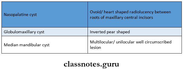
3. Cysts And Their Radiographic Features
Read And Learn More: Oral Medicine Question and Answers
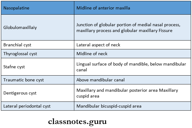
4. Cyst And Their Location
5. Cysts And Their Origin
6. Nasolabial Cyst
- Arises at the junction of the globular portion of the lateral nasal process, medial nasal process, and maxillary process
- It is a soft tissue cyst
- Has no radiographic features
7. Syndromes Associated With Dentigerous Cyst
- Cleidocranial dysplasia
- Maroteaux Lamy syndrome
8. Gorlin Goltz Syndrome
- Multiple Odontogenic keratocyst
- Basal cell carcinoma
- Bifid basal rib
- Sexual abnormalities
- Neurological and ophthalmological abnormalities
9. Rushton Bodies Are Seen In
- Periapical cyst
- Dentigerous cyst
- Gingival cyst of infants
Oral Medicine Cysts Short Essays
Question 1. Median Mandibular Cyst.
Answer:
Median Mandibular Cyst
They have been derived from epithelium remnants between the fusing mandibular process during the embryonic phase
Median Mandibular Cyst Clinical Features:
- It is a rare lesion
- Site: in the midline of the mandible
- It may cause displacement of the adjacent teeth
- The cystic swelling may be palpable buccally
- The teeth associated with the lesion are vital
- Radiographic Features:
- Well-defined small radiolucency is seen in the mid-line of the mandible
Median Mandibular Cyst Management:
- Enucleation of the cyst is done
- Care should be taken not to damage the apices of the teeth
Question 2. Gingival cyst of infants.
Answer:
Gingival Cyst Of Infants
- Gingival cysts of the infant are multiple small, nodular, keratin-filled, cystic lesions seen in the oral cavity
- Depending on their location, they are divided into:
- Cyst of the dental lamina
- These are mostly found along the alveolar ridge and are odontogenic in origin
- Epstein’s pearls
- These small cystic lesions are found along the mid-palatine raphe
- They are derived from the epithelium, entrapped along the line of fusion of the palate during embryogenesis
- Bohn’s nodules
- These are small cysts usually found along the junction of the hard and soft palate and over buccal and lingual aspects of the alveolar ridge
- They are derived from remnants of the mucosal glands
- Cyst of the dental lamina
Gingival Cyst Of Infants Clinical Features:
- They are usually multiple, asymptomatic
- They are small, discrete, white nodules developing in several parts of the oral cavity
- They may discharge the contents by fusion with the overlying alveolar mucosa
- They may undergo spontaneous regression
Gingival Cyst Of Infants Management:
- No treatment is required
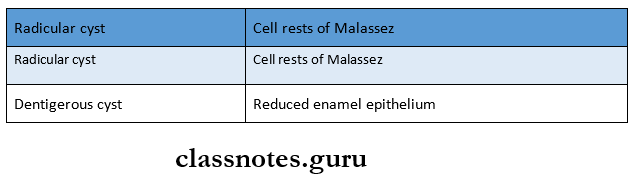
Question 3. Dentigerous cyst.
Answer:
Dentigerous Cyst Clinical Features:
- Sex: Common in males
- Age: First and 3rd decade
- Site: Mandibular 3rd molar, maxillary canines, maxillary 3rd molar
- Expansion of bone
- Facial asymmetry
- Displacement of adjacent teeth
- Resorption of adjacent teeth
Dentigerous Cyst Radiological Features:
- The unilocular, well-defined radiolucency
- Margins- sclerotic
Dentigerous Cyst Types:
- Central: covering the crown of an unerupted tooth
- Circumferential: covering the crown from all the sides
- Lateral: covering crown from the side
Dentigerous Cyst Management:
- Marsupialization- In children
- Enucleation – In adults
Question 4. Odontogenic keratocyst.
(or)
Question 4. Primordial Cyst.
Answer:
Odontogenic Keratocyst Clinical Features:
- Age: 2ndand 3rd decade
- Sex: Common in males
- Site: mandible
- Features:
- Asymptomatic
- If secondary infected, causes expansion of cortical plates
- Mobility of teeth
- Pain and tenderness of the site
Odontogenic Keratocyst Radiological Features:
- Unilocular or multilocular radiolucency
- Margins: well-defined sclerotic margins
- Expansion of cortical plates
- Soap bubble appearance
Odontogenic Keratocyst Management:
- Enucleation Of Cyst:
- Smaller single cyst through intraoral approach
- Unilocular lesions through marginal excision
- Large multilocular lesions
Resection of involved bone
↓
Reconstruction of the site
↓
Bone grafting
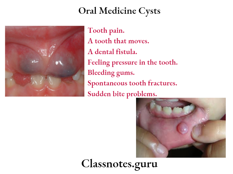
Question 5. Radicular Cyst.
Answer:
Radicular Cyst Etiology:
- Dental caries
- Fractured tooth
- Thermal/ Chemical injury to the pulp
- Iatrogenic injury to the pulp
Radicular Cyst Clinical Features:
- Sex: common in males
- Age: Young age
- Site: common in maxillary anterior
- Nonvital tooth
- Smaller cysts are asymptomatic
- Larger lesions produce slow enlarging, bony hard swelling
- Expansion and distortion of cortical plates
- Severe bone destruction
- Springiness of jawbones
- Pain is secondarily infected
- Intraoral or extraoral pus discharge
- Pathological fractures
- Formation of an abscess called “cyst abscess”
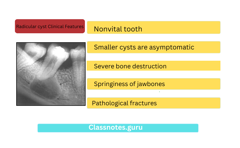
Radicular Cyst Radiological Features:
- The unilocular radiolucent area around the apex of the nonvital tooth
- Border: sclerotic
- Diameter: less than 1 cm
- Discontinuity of lamina dura
Radicular Cyst Treatment:
- Nonvital tooth
- Extraction
- RCT
- Smaller cyst
- Removed through socket
- Larger cyst
- Marsupialization
Oral Medicine Cysts Short Answers
Question 1. Residual cyst.
Answer:
Residual Cyst
- Any cyst may have an associated periapical or periodontal cyst which is asymptomatic
Residual Cyst Clinical Features:
- The patient may complain of tooth pain
- The tooth may be extracted without noticing the presence of a cyst in the region associated with the tooth
- In such cases, the cyst is known as a residual cyst
- It continues to grow even after the tooth is removed as the cystic lining is still present
- The cyst is seen in an edentulous area, in place of the extracted tooth
- Incidence is more in the maxilla than mandible
Residual Cyst Treatment:
- Enucleation
Question 2. Globulomaxillary cyst.
Answer:
Globulomaxillary Cyst
- A common type of developmental cyst
- Arises in the bone suture, between the maxilla and premaxilla
Residual Cyst Clinical Features:
- Asymptomatic
- If the secondary infection causes pain and discomfort
- Small swelling between canine and premolar
- Vital teeth
Residual Cyst Radiographic Features:
- The inverted pear-shaped radiolucent area between the roots of the upper lateral incisor and canine
- Divergence of the roots
Residual Cyst Treatment:
- Surgical excision
Oral Medicine Cysts Viva Voice
- A nasolabial cyst is a soft tissue cyst
- Stafne cyst is due to the developmental inclusion of sali-vary glandular tissue on the lingual surface of the mandible below the mandibular canal
- A nasopalatine cyst is the most common Odontogenic cyst
- Globulomaxillary cyst is found within bone
- A globulomaxillary cyst is present between the maxillary lateral incisor and cuspid
- Globulomaxillary cyst is fissural cyst
- A radicular cyst is an inflammatory cyst
- Botryoid Odontogenic cyst is a multicystic variant of lateral periodontal cyst
- Eruption cyst is a form of dentigerous cyst