
Oral Cavity And Tongue Question And Answers
Question 1. Write a short note on hard palate.
Answer:
Hard Palate
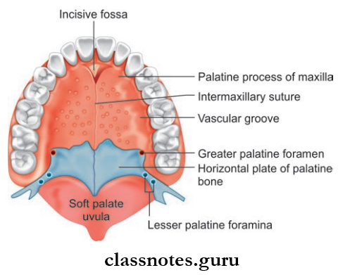
- Partition between oral and nasal cavities
- Anterior 2/3rd is formed by the palatine process of the maxilla and posterior 1/3rd by horizontal plates of palatine bones
- Anterolateral margins are continuous with alveolar arches and gums
- The posterior margin gives the attachment to the soft palate
- Superior surface forms flor of nasal cavity
- Inferior surface forms roof of oral cavity and presents the following features:
- Incisive Fossa
- Greater Palatine Foramen
- Lesser Palatine Foramen
- Posterior Nasal Spine
- Palatine Crest
- Masticatory Mucosa.
Hard Palate Blood Supply
- Arterial Supply: By greater palatine arteries
- Venous Drainage: Drains into pterygoid venous plexus and pharyngeal venous plexus.
Hard Palate Nerve Supply
- By Greater Palatine Nerve: Whole of palate except premaxilla
- Nasopalatine Nerve: Premaxilla.
Hard Palate Lymphatics: Lymph is drained into upper deep cervical nodes and to retropharyngeal lymph nodes.
Hard Palate Development
- Palate Is Developed From:
- Maxillary process, which is two in number
- Frontonasal process.
- From each maxillary process, a plate-like shelf grows medially, known as the palatal process.
- The primitive palate is formed by the fusion of medial nasal folds.
- Medial nasal folds are folds of the frontonasal process.
- Now palate proper is formed by the fusion of these three components, i.e. two palatal processes and one primitive palate.
- Each palatal process fuses with the posterior margin of the primitive palate.
- Each palatal process fuses with the other at the midline.
- Also palatal process fuses with lower free edge of the nasal septum separating nasal cavity into two.
- Most of the palate gets ossified to form hard palate and unossified posterior part forms a soft palate.
Hard Palate Applied
- Cleft Palate: Cleft Palate is a developmental anomaly due to defective fusion of various components of palate.
- Bilateral Complete Cleft Palate:
- Occurs due to complete nonfusion
- Gives rise to a Y-shaped cleft
- Associated with bilateral cleft lip.
- Unilateral Complete Cleft Palate
- Due to the complete arrest of fusion of palatine process of one side with primitive palate and nasal septum
- Associated with unilateral hair lip.
- Partial Midline Cleft: Due to complete failure of fusion of palatine process with each other.
- Cleft Of Soft Palate: Due to complete failure of fusion of palatine process with each other in the dorsal ¼ part.
- Bilateral Complete Cleft Palate:
Question 2. Write a short note on soft palate.
Answer:
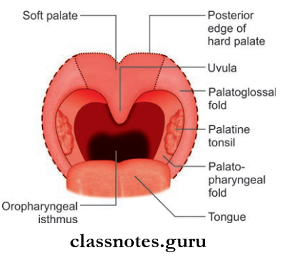
Soft Palate
- Soft Palate is a mobile muscular fold suspended from the posterior border of hard palate
- Soft Palate separates the nasopharynx from the oropharynx
Soft Palate External Features
- Consists Of Two Surfaces: Anterior and posterior
- The anterior surface is concave and marked by median raphe
- Posterior surface is convex and continuous with the floor of the nasal cavity.
- Consists Of Two Borders: Superior and inferior
- The superior border is attached to the posterior border of the hard palate
- The inferior border is free and bound to the pharyngeal isthmus.
- A small tongue-like projection hangs down from the middle and is called uvula
- From each side of base of the uvula, two curved folds of mucous membrane extend laterally and downwards
- The anterior fold is called the palatoglossal fold and contains palatoglossal muscle and merges inferiorly with side of the tongue
- The posterior fold is known as the palatopharyngeal fold which merges inferiorly with lateral wall of the pharynx.
Soft Palate Structure: It is a fold of mucous membrane containing the following parts:
- Palatine Aponeurosis: Flattened tendon of tensor veli palatini
- Levator veli palatini and palatopharyngeus above palatine aponeurosis
- Palatoglossal on inferior surface of palatine aponeurosis.
Soft Palate Blood Supply
- Arterial Supply
- Lesser palatine branch of the maxillary artery
- Ascending palatine branch of facial artery
- Branch of ascending pharyngeal artery.
- Venous: Drained by pterygoid and pharyngeal plexus of veins.
Soft Palate Lymphatics: Drains into upper deep cervical and retropharyngeal
nodes.
Soft Palate Functions
- Separates oropharynx and nasopharynx while swallowing so that food does not enter in nose
- Helps in modifiation of quality of voice
- Protects against the damage of nasal mucosa during sneezing by directing blast of air through nasal and oral cavities
- Prevents entry of sputum into nose during coughing.
Soft Palate Applied: Paralysis of soft palate in lesions of vagus nerve causes the following
- Nasal regurgitation of liquids
- Nasal twang in voice
- Flattening of the palatal arch
- Deviation of uvula to normal side.
Question 3. Write a short note on muscles of soft palate.
Answer:
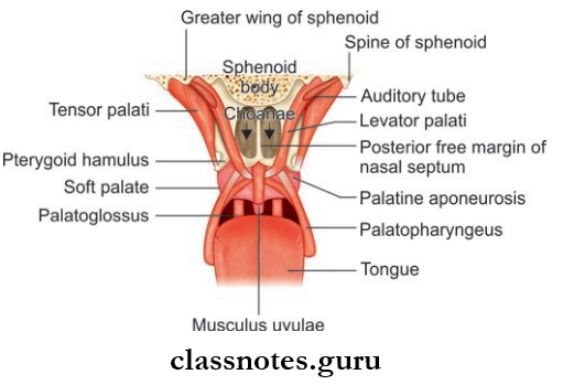
Muscles Include The Following:
- Tensor Veli Palatini
- Levator veli Palatini
- Musculus Uvulae
- Palatoglossus
- Palatopharyngeus
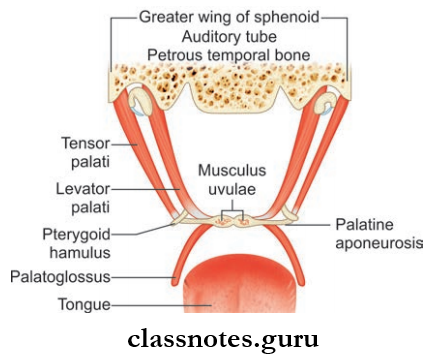
1. Tensor Veli Palatini
- Thin triangular muscle
- Tensor Veli Palatini Origin: Lateral side of Eustachian tube, adjoining part of greater wing of the sphenoid
- Tensor Veli PalatiniInsertion: Forms a tendon and expands to form palatine aponeurosis and attaches to:
- Posterior border of hard palate
- Inferior surface of hard palate behind palatine crest
- Tensor Veli Palatini Action:
- Tightening of soft palate
- Opening of Eustachian tube.
2. Levator Veli Palatini: Cylindrical muscle presents deep to tensor veli palatini
- Levator Veli PalatiniOrigin:
- Inferior aspect of Eustachian tube
- Adjoining part of inferior surface of petrous temporal bone
- Levator Veli Palatini Insertion: It spreads out and inserts to upper surface of palatine aponeurosis
- Levator Veli Palatini Action:
- Elevation of soft palate to close pharyngeal isthmus.
- Opening of Eustachian tube.
3. Musculus Uvulae: Longitudinal strip on both sides of median plane within palatine aponeurosis
- Levator Veli Palatini Origin:
- Posterior nasal spine
- Palatine aponeurosis
- Levator Veli Palatini Insertion: Mucous membrane of uvula
- Levator Veli Palatini Action: Pull uvula forwards to side of action.
4. Palatoglossus
- Palatoglossus Origin: Oral surface of palatine aponeurosis
- Palatoglossus Insertion: To side of tongue at junction of oral and\ pharyngeal parts
- Palatoglossus Action:
- Pulls the root of tongue up
- Approximation of palatoglossal arches.
5. Palatopharyngeus: Consists of two fasciculi separated by levator veli palatini.
- Palatopharyngeus Origin:
- Anterior Fasciculus: From posterior border of hard palate
- Posterior Fasciculus: Palatine aponeurosis.
- Palatopharyngeus Insertion:
- Median raphe of pharyngeal wall
- Posterior border of lamina of thyroid cartilage.
- Palatopharyngeus Action: Raise the walls of the pharynx during swallowing.
Question 4. Write a short note on Passavant’s ridge.
Answer:
Passavant’s Ridge
- The upper fibers of the palatopharyngeus muscle pass deep to the mucous membrane of the pharynx in a circular form and form a sphincter internal to the superior constrictor and form Passavant’s muscle.
- Contraction of this muscle raises a ridge called
- Passavant’s ridge on posterior wall of nasopharynx.
- When soft palate is elevated, the ridge comes in contact with soft palate and closes the pharyngeal isthmus.
Question 5. Write a short note on tongue.
Answer:
Tongue
- Mobile muscular organ in the oral cavity which is responsible for taste, speech, mastication, and deglutition
- Tongue is conical in shape and elongated posteroanteriorly and flttened dorsoventrally and is separated into right and left halves by firous septum
- Consists of root, tip, and body
- The root is attached to styloid process and soft palate above and mandible and hyoid bone below.
- The tip is the anterior free end and body is the back of tongue
- The body consists of dorsal surface and ventral surface
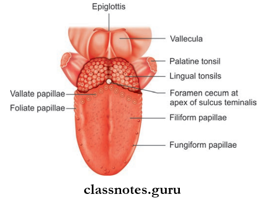
The ventral surface consists of the following features:
- Frenulum: A median fold of mucous membrane connecting tongue to the flor of mouth
- Deep Lingual Vein: Seen on either side of the frenulum
- Plica Fimbriate: A fimbriated fold of mucous membrane lateral to lingual vein
- Dorsal surface is divided into two parts by V-shaped sulcus terminals:
- Anterior 2/3rd Or Oral Part Which Presents:
- Median furrow
- Large number of papillae.
- Posterior 1/3rd or Pharyngeal Part Which Presents:
- Large number of lymphoid follicles which constitute palatine tonsil
- Large number of mucous and serous glands.
Papillae Of Tongue: These are projections of lamina propria of mucous membrane covered with epithelium. Mainly four types of papillae are seen:
- Vallate Papillae/Circumvallate Papillae
- Largest is 1–2 mm in diameter
- Arranged in a V-shape row in front of sulcus terminalis
- Resembles a truncated cone surrounded by a circular sulcus bounded by a wall
- The duct opens into sulcus and taste buds are found in the papillae and wall.
- Filiform Papillae
- Narrowest and largest in number
- Minute conical projections with pointed tips
- Located abundantly on the dorsum of tongue and is responsible for velvety appearance of tongue.
- Fungiform Papillae
- It has a rounded head and narrow base
- I present mainly at apex and margins of tongue
- Seen as discrete pink pin heads due to red rounded heads.
- Foliate Papillae
- Consists of vertical grooves and ridges near margin in front of sulcus
- Usually rudimentary in human.
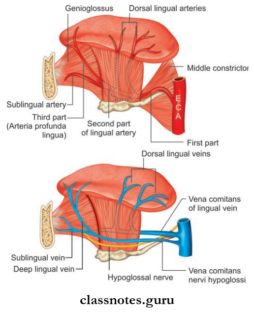
Blood Supply Of Tongue
- Arterial Supply: Mainly by lingual arteries, a tonsillar branch of the facial artery and ascending pharyngeal artery.
- Venous Drainage: Drained by deep lingual vein and venae comitantes accompanying lingual artery and hypoglossal nerve.
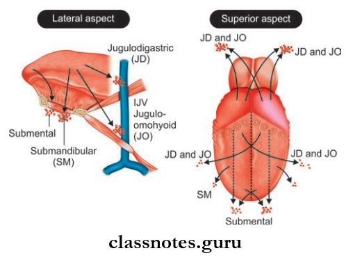
Lymphatic Drainage Of Tongue
- Apical vessels drain tip and inferior surface of tongue to submental lymph nodes
- Marginal Vessels: Drain marginal portion of anterior 2/3rd of tongue to submandibular, lower deep cervical nodes
- Lateral vessels drain anterior 2/3rd of tongue and posterior 1/3rd to upper deep cervical nodes.
Nerve Supply Of Tongue
- Motor Supply: All muscles are supplied by hypoglossal nerve except the palatoglossus muscle, which is supplied by cranial root of accessory.
- Sensory Supply
- Anterior 2/3rd
- Lingual nerve for general sensation
- Chorda tympani for special sensation.
- Posterior 1/3rd: Glossopharyngeal—both general and special sensations
- Internal laryngeal branch of superior laryngeal nerve:
- Posterior-most part—special sensation.
- Anterior 2/3rd
Development Of Tongue: Medial-most part of the fist pharyngeal arches proliferates and forms two lingual swellings.
- Lingual swellings are partially separated by a median swelling called tuberculum impar.
- Another midline swelling called hypobranchial eminence is also formed from the mesoderm of the 2nd, 3rd, and 4th arches.
- Anterior 2/3rd of the tongue is formed by the fusion of two lingual swellings with tuberculum impar.
- The cranial part of hypobranchial eminence (3rd arch) gives rise to posterior one-third of tongue.
- Posterior-most part is formed from fourth arch.
- Connective tissue is derived from mesenchyme.
- Tongue muscles are derived from occipital myotomes.
Applied Of Tongue
- Ankyloglossia or tongue tie is a condition where the frenulum extends to the tip of tongue and, as a result, the tongue becomes anchored to flor of the mouth.
- The geographical tongue is characterized by an erythematous area, devoid of papillae, surrounded by an irregular keratotic outline.
- Fissured tongue is a condition seen in cases of syphilis, vitamin B complex deficiency, or even can be congenital.
- Hairy tongue refers to a clinical condition where there is excessive formation of keratin, leading to elongation of filiform papillae which gets colored brown or black due to chromogenic bacteria and looks like hair.
- Carcinoma of the tongue usually affects the anterior 2/3rd of the tongue and is commonly seen in young men.
Question 6. Write a short note on muscles of tongue.
Answer:
Muscles Of Tongue
Consists of extrinsic and intrinsic muscles.
Intrinsic Muscles
- Intrinsic muscles are muscles located within the tongue and have no attachment outside and are responsible for changing shape of tongue
- Each half of tongue contains four intrinsic muscles, which are as follows:
- Superior longitudinal muscle
- Inferior longitudinal muscle
- Transverse muscle
- Vertical muscles
- These muscles are arranged in several planes and run in three directions—longitudinal, horizontal, and vertical
- These muscles occupy the upper part of tongue and are attached to submucous firous layer.
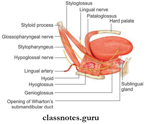
Extrinsic Muscles
- They attach tongue to the mandible, the hyoid, the styloid process and the palate
- They move the tongue as well as alter the shape
- These Include:
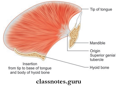
- Genioglossus
- Hyoglossus
- Styloglossus
- Palatoglossus.
Movements Of Tongue
- Alteration Of Shape Of Tongue By Intrinsic Muscles
- Superior Longitudinal:
- Shortening the tongue
- Making the dorsum concave
- Inferior Longitudinal:
- Shortening the tongue
- Making the dorsum convex
- Transverse Muscle: Narrowing and elongation
- Vertical Muscle: Broadening and flattening.
- Superior Longitudinal:
- Proper Movements
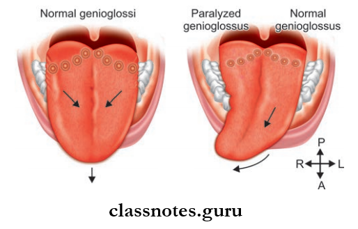
- Protrusion: Genioglossus—both muscles
- Retraction: Styloglossus—both muscles
- Depression: Hyoglossus—both muscles
- Elevation: Palatoglossus—both muscles.
Alteration Of Shape By Extrinsic Muscles
- Hyoglossus:
- Depresses sides of tongue
- Makes the dorsal surface convex.
- Styloglossus: Draws the side of the tongue upwards and downwards
- Palatoglossus: Pulls the root of tongue and approximation of palatoglossal arches.
Oral Cavity And Tongue Multiple Choice Question And Answers
Question 1. All the muscles of the tongue are supplied by the hypoglossal nerve except:
- Palatoglossus
- Styloglossus
- Hyoglossus
- Genioglossus
Answer: 1. Palatoglossus
Question 2. All of the following nerves carry taste sensations from the tongue except:
- Facial nerve
- Glossopharyngeal nerve
- Vagus nerve
- Hypoglossal nerve
Answer: 4. Hypoglossal nerve
Question 3. The most abundant papillae on the dorsum of the tongue are:
- Filiform
- Fungiform
- Foliate
- Vallate
Answer: 1. Filiform
Question 4. The pain from the tongue is referred to the ear through:
- Mandibular nerve
- Facial nerve
- Glossopharyngeal nerve
- Hypoglossal nerve
Answer: 1. Mandibular nerve
Question 5. All of the following glands pour their secretion into the vestibule of the mouth except:
- Parotid glands
- Sublingual glands
- Labial glands
- Molar glands
Answer: 2. Sublingual glands