
Blood Supply And Lymphatic Drainage Of Lower Limb Question And Answers
Arterial Supply Of Lower Limb
Question 1. Explain in detail about the femoral artery under the headings—origin, course, relations, and branches.
Answer:
The Femoral Artery
Read And Learn More: Anatomy Question And Answers
Chief artery of the lower limb.
Femoral Artery Origin
- Continuation of the external iliac artery.
- Begins behind the inguinal ligament at the level of mid inguinal point.
Femoral Artery Extend and Course
- Runs downwards and medially through the femoral triangle and adductor canal.
- At the lower end of the adductor canal, i.e. junction between the upper 2/3rd and lower 1/3rd of the thigh, it passes through the opening of the adductor magnus muscle to enter the popliteal fossa.
- From there it continues as a popliteal artery.
Femoral Artery Relations in Femoral Triangle
- Anterior: Skin, superficial fascia, deep fascia, the anterior wall of the femoral sheath.
- Posterior: Psoas major, pectineus, adductor longus muscles.
- The femoral vein lies posterior to the femoral artery
- Above the level of the apex of the femoral triangle, the femoral vein lies medial to the femoral artery.
- At the level of apex of the femoral triangle, the femoral vein lies directly behind to femoral artery.
- Below the level of the apex of the femoral triangle, the femoral vein it crosses and lies lateral to the femoral artery.
- The femoral nerve lies lateral to the upper part of the femoral artery.
- The femoral branch of the genitofemoral nerve also lies lateral to the upper part of the femoral artery.
- The Profunda femoris artery and its companion vein lie posterior to the femoral artery in its upper part, lower down they are separated by an adductor muscle.
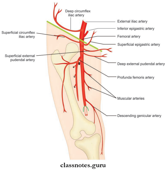
Femoral Artery Branches
- In the femoral canal
- Three Superficial Branches:
- Superficial epigastric artery
- Superficial external pudendal artery
- Superficial circumflex iliac artery.
- Three Deep Branches:
- Profunda femoris artery
- Deep external pudendal artery
- Muscular branches.
- Three Superficial Branches:
- In the adductor canal
- Muscular branches
- Descending genicular artery.
- Clinical Anatomy
- Femoral artery pulsations can be felt in the femoral triangle just below the mid-inguinal point.
- Since the femoral artery is superficial in the femoral triangle, blood can be easily withdrawn for arterial blood gas analysis.
- It is the preferred artery for coronary artery angiography and angioplasty.
- Since the femoral vein runs behind the femoral artery in the apex of the triangle any stab wounds in this area can be fatal.
Mnemonics
- Femoral artery deep branches: ‘Put My Leg Down Please’
- Profundus femoris (deep femoral artery)
- Medial circumflex femoral artery
- Lateral circumflex femoral artery
- Descending genicular arteries
- Perforating arteries
Question 2. Explain about profunda femoris artery under the headings—origin, course, termination, and branches.
Answer:
Profunda Femoris Artery Origin
- The largest branch of the femoral artery.
Profunda Femoris Artery Course
- Arises from the lateral side of the femoral artery 4 cm below the inguinal ligament in the femoral triangle.
- Gives of lateral and medial circumflex arteries.
- Descends down close to femur giving branches to muscles and terminal branches.
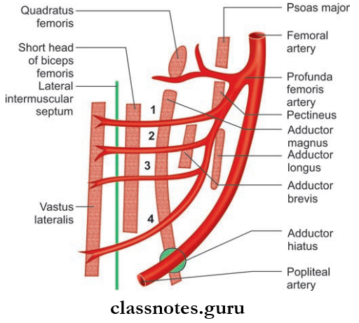
Profunda Femoris Artery Termination
- After piercing adductor magnus ends at the posterior part of the leg.
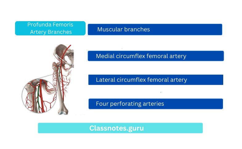
Profunda Femoris Artery Branches
- Muscular branches
- Medial circumflex femoral artery
- Lateral circumflex femoral artery
- Four perforating arteries.
Profunda Femoris Artery Clinical Anatomy
- Since it lies close to the shaft of the femur, a fracture of the femur or surgery in the area may injure the artery.
Question 3. Write a short note on the obturator artery.
Answer:
Obturator Artery
- Obturator Artery is a branch of the internal iliac artery.
- Obturator Artery accompanies the obturator nerve in the pelvis and passes through the obturator canal.
- Then, it divides into medial and lateral branches and anastomoses with medial circumflex femoral artery.
- They supply:
- Neighboring muscles
- Fat in acetabular fossa
- Head of the femur through the round ligament.
Question 4. Write a short note on the medial circumflex femoral artery.
Answer:
Medical Circumflex Femoral Artery
- Branch of profunda femoris artery
- It is divided into 2 branches:
- Ascending branch
- It anastomosis with the ascending branch of the lateral circumflex femoral artery and superior gluteal artery to form trochanteric anastomosis.
- This anastomosis gives of retinacular branches which supply the head of the femur.
- Transverse branch
- It anastomosis with the transverse branch of the lateral femoral circumflex, inferior gluteal, and fist perforating branch of profunda femoris to form a cruciate anastomosis.
- Ascending branch
- Before the terminal branches, it gives off
- Muscular branches
- Acetabular branches supply fat in the acetabular fossa
- Branch supplying head of the femur.
Medical Circumflex Femoral Artery Clinical Anatomy
Intracapsular fracture of the neck of the femur can cause avascular necrosis of the head of the femur due to damage to retinacular branches.
Question 5. Explain the popliteal artery under the headings—origin, course, termination, relation, and branches.
Answer:
Popliteal Artery
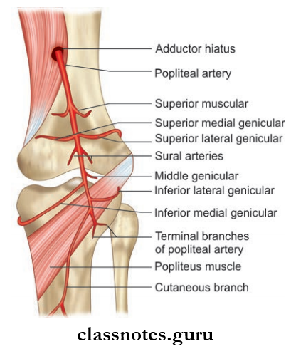
Popliteal Artery Origin
- Continuation of femoral artery.
Popliteal Artery Course
- From the adductor hiatus, it runs downwards and slightly laterally, to reach the lower border of the popliteal muscle.
Popliteal Artery Termination
- Terminates at the lower border of the popliteus into anterior and posterior tibial arteries.
Popliteal Artery Relation
- Anterior/Deep Relations:
- The popliteal surface of the femur
- Back of knee joint
- Fascia covering popliteus muscle
- Posterior/Superficial Relations:
- Popliteal vein
- Tibial nerve
- Laterally:
- The upper part of the artery related to the biceps femoris and lateral condyle of the femur
- The lower part related to plantar and the lateral head of the gastrocnemius muscle
- Medially:
- The upper part related to the biceps femoris and lateral condyle of the femur
- The lower part is related to the tibial nerve, popliteal vein, and medial head of the gastrocnemius.
Popliteal Artery Branches
- A number of large muscular branches
- Cutaneous branches
- Genicular branches—5 in number:
- Medial and lateral superior genicular branches
- Middle genicular branch
- Medial and lateral inferior genicular branches
- They form anastomosis around the knee joint.
Popliteal Artery Clinical Anatomy
- Popliteal pulsation is felt by flexing the knee and palpating the popliteal artery in the popliteal fossa, against the underlying femur. It is difficult to feel.
- Popliteal artery aneurysm is the most common arterial aneurysm.
Question 6. Write a short note on the anterior tibial artery.
Answer:
Anterior Tibial Artery Beginning
- The smaller terminal branch of the popliteal artery.
- It begins on the back of the leg at the lower border of the popliteus, opposite the tibial tuberosity.
Anterior Tibial Artery Course
- Reaches the front of the leg through an opening in the upper part of the interosseous membrane.
- Runs downward in between the muscles of the leg and continues as the dorsalis pedis artery from the point between the medial and lateral malleolus.
Anterior Tibial Artery Branches
- Muscular branches to the anterior compartment of the leg
- Cutaneous branches
- Branches to knee and ankle joints.
Question 7. Write a short note on the posterior tibial artery.
Answer:
Posterior Tibial Artery Beginning
- The larger terminal branch of the popliteal artery.
- It begins at the lower border of the popliteus, between the tibia and fibula, and deep to the gastrocnemius.
Posterior Tibial Artery Course
- It runs down through the flexor muscles to reach the level between the medial malleolus and medial tubercle of the calcaneus and passes under the flexor retinaculum.
Posterior Tibial Artery Termination
- Terminates by dividing into medial and lateral plantar arteries under the flexor retinaculum.
Posterior Tibial Artery Branches
- Muscular branches
- Articular branches to knee and ankle joints
- Nutrient artery to tibia.
Question 8. Write a short note on the dorsalis pedis artery.
Answer:
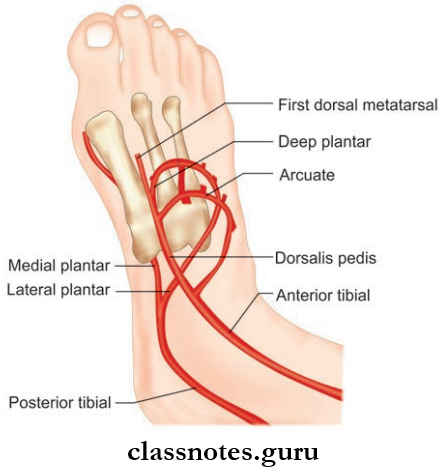
Dorsalis Pedis Artery Beginning
- Continuation of anterior tibial artery.
- Begins in front of the ankle between two malleoli as the ‘continuation of the anterior tibial artery.’
Dorsalis Pedis Artery Course
- Runs along the medial side of the dorsum of the foot from the midpoint of the medial and lateral malleoli.
- From there, it runs downwards to reach the proximal end of 1st metatarsal space.
- It eventually enters to sole to complete the plantar arch by meeting the lateral plantar artery.
Dorsalis Pedis Artery Branches
- Lateral tarsal artery
- Medial tarsal artery, both supplying tarsal bones and joints
- First dorsal metatarsal artery
- Arcuate artery → gives of 2nd, 3rd, and 4th dorsal metatarsal arteries.
Question 9. Explain in brief about the medial plantar artery, lateral plantar artery, and plantar arch.
Answer:
Medial Plantar Artery
- Division of posterior tibial artery.
- Runs along the medial border of the foot and terminates by giving of digital arteries.
Lateral Plantar Artery
- Division of posterior tibial artery
- It runs laterally between the 1st and 2nd layer of muscles of sole and continues with plantar
arch.
Plantar Arch
- It is located in between 1st and muscle layers.
- Formed by the union of continuation of dorsalis pedis artery medially and lateral plantar artery laterally.
- Gives of four plantar metatarsal arteries, each of them again gives of two digital branches.
Venous Drainage of Lower Limb

Question 10. What are the peculiarities of venous drainage of the lower limb? How it is classified?
Answer:
The Peculiarities Of Venous Drainage Of The Lower Limb
- The venous drainage of the lower limb is working against gravity.
- However, a number of factors help to make the venous return from the lower limb efficient. They are:
- Calf pump/the peripheral heart: Contraction of calf muscles squeezes the blood up along the deep veins from venous sinuses present in them.
- Accompanying arteries exert pressure on veins by arterial pulsations.
- Valves in the vein support the blood column against gravity and ensure unilateral blood flow.
- Arterial pressure and overflow from capillary bed.
- Negative intrathoracic pressure.
- Veins in the lower limb are more muscular.
- Veins of the lower limb are classified into 3 groups:
- Superficial veins
- Deep veins
- Perforating veins
- Blood is drawn from the lower limb via superficial veins and deep veins and eventually reaches the femoral vein.
- The perforator veins connect and ensure unilateral blood flow from superficial to deep veins.
Question 11. How is the superficial venous system in the lower limb is organized? Explain in detail about long and short saphenous veins under the headings—formation, course, termination, tributaries, and valves.
Answer:
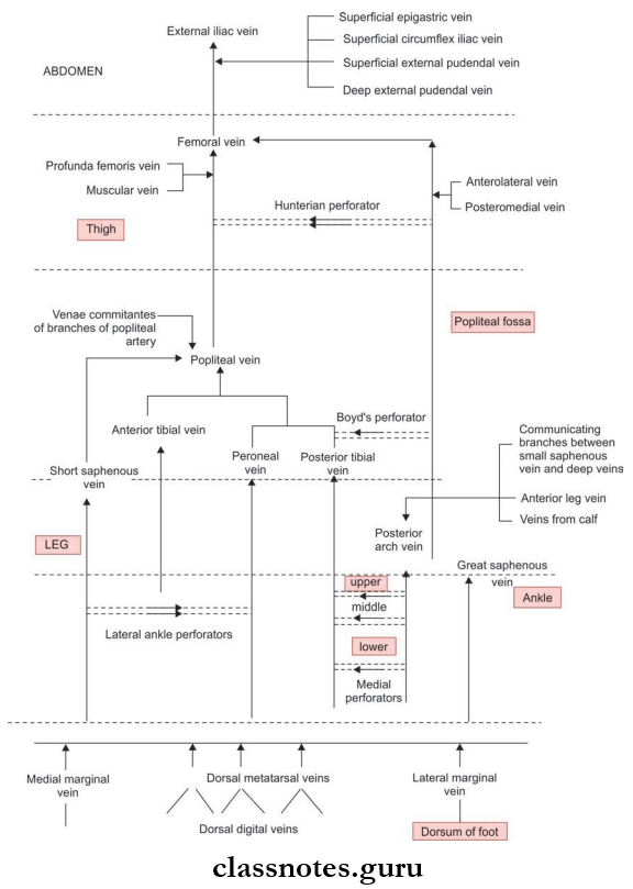
- It consists of the dorsal venous arch, a long saphenous vein, a short saphenous vein, and tributaries situated in the superficial fascia.
- Two dorsal digital veins form one dorsal metatarsal vein. Four dorsal metatarsal veins unite and form a dorsal venous arch on the dorsum of the foot over the proximal part of the metatarsal bones.
- This dorsal venous arch ascends up medially as a great saphenous via and laterally as a short saphenous vein.
- They are enriched by a number of tributaries.
- A considerable amount of blood is drained by them to deep veins directly or via perforators.
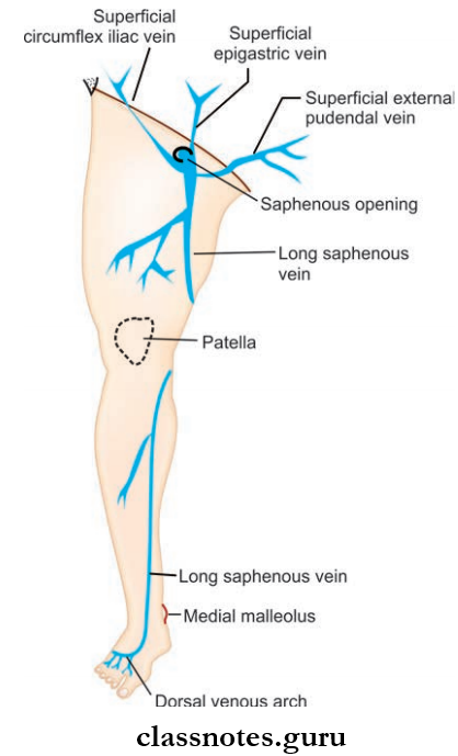
Great Saphenous Vein
- Longest vein in the body
- Easily seen in the lower limb (saphenous = easily seen).
- Formation
- By union of the medial end of the dorsal venous arch of the foot and the medial marginal vein of the foot.
- Course
- It runs upward and medially to reach the posteromedial aspect of the knee joint.
- From there it ascends to reach up to the level of saphenous opening.
- It reaches the saphenous opening by piercing the cribriform fascia and drains into a femoral vein after piercing the femoral sheath.
- Throughout its course, it receives tributaries.
- Termination
- In the femoral triangle.
- Valves
- Consists of 10–20 valves.
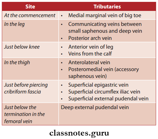
- Tributes
- Formation
Short/Small Saphenous Vein
- Formation
- Union of the lateral marginal vein with the lateral end of the dorsal venous arch.
- After formation, it ascends behind the lateral malleolus along the lateral edge of the talocalcaneal, accompanied by the sural nerve to reach the back of the leg.
- It pierces the back of leg to reach the deep fascia progresses through the head of the gastrocnemius and eventually drains to the popliteal vein after piercing the deep fascia.
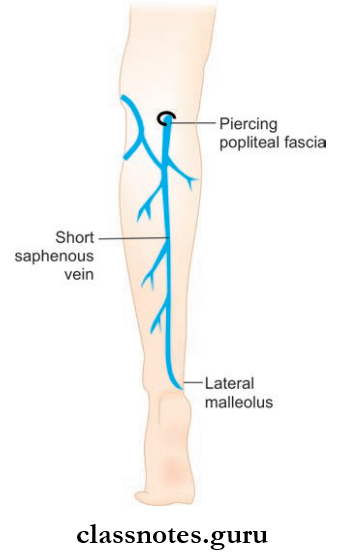
- Termination
- In to popliteal vein in the popliteal fossa.
- Valves
- It have 7–13 valves.
Question 12. Write a note on the deep veins of the lower limb. Explain separately about the femoral vein.
Answer:
Deep Veins Of Lower Limb
- The major deep veins of the lower limb are:
- A deep vein of sole → medial and lateral plantar veins
- Venae comitantes accompanying dorsalis pedis, anterior tibial, and posterior tibial arteries
- Popliteal vein
- Femoral vein
- They are located in the tight fascial compartments.
- Thy, below the knee, are arranged as a pair of venae comitantes along with arteries, but above the knee, they are almost individual.
- More valves are provided to them.
Femoral Vein
- It is the upward continuation of the popliteal vein and continues as the external iliac vein beyond the inguinal ligament.
- Course
- Begins at the lower end of the adductor canal
- Ascend up in the adductor canal and enter the femoral triangle
- Though the middle compartment in the femoral triangle, it ascends upwards and continues as an external iliac vein behind the inguinal ligament.
- Tributaries
- Great saphenous vein
- Profunda femoris vein
- Medial and lateral circumflex veins
- Deep external pudendal vein
- Direct muscular tributaries.
- Clinical Anatomy
- The femoral vein is the most commonly used vein for 4 infusions in case of peripheral circulatory failure and in infants.
- This vein is also used to insert a catheter into the right atrium and ventricle to measure pressure.
- Course
Question 13. Classify and list the perforating veins.
Answer:
Perforating Veins
- They communicate superficial veins with deep veins.
- Called perforators because they perforate deep fascia.
- They are of 2 types:
- Indirect Perforators: Connect superficial vein to deep vein only through muscular veins
- Direct Perforators: Connect superficial and deep veins directly
- Among them, five to six are important. They are:
- Adductor Canal/Hunterian Perforator: Great saphenous vein ↔ femoral vein at the lower part of adductor canal
- Knee Perforator/Boyd’s Perforator: Great saphenous vein ↔ posterior tibial vein below the knee close to tibia
- Lateral Ankle Perforator: Short saphenous vein ↔ peroneal vein at the junction of the middle and lower 1/3rd of leg
- Three Medial Ankle Perforators (Of Cockett): Great saphenous vein ↔ posterior tibial vein
- Upper medial
- Middle medial
- Lower medial.
Perforating Veins Clinical Anatomy
- Incompetency of the valves or other mechanisms helping venous return from the lower limb can cause varicose veins and deep vein thrombosis.
- In about 80% of individuals, the external iliac vein possesses a valve that prevents high back pressure by blood column on the saphenofemoral valve located in the femoral triangle. In those when the valve is absent, the chances of occurrence of varicose veins are higher.
- Calf muscles: Are known as the peripheral heart.
Question 14. Explain in detail about lymphatic drainage of the lower limb.
Answer:
Lymphatic Drainage Of The Lower Limb
The lymph nodes of the lower limb are classified into:
Superficial Lymph Nodes: Includes superficial inguinal lymph nodes
Deep lymph nodes: Includes:
- Deep inguinal lymph nodes
- Popliteal lymph nodes
- Anterior tibial lymph nodes.
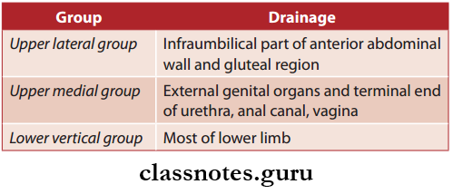
Superficial Inguinal Lymph Nodes: They are arranged in T shape into two groups:
- Upper Horizontal Group
- Have lateral set and medial set
- Lower Vertical Group.
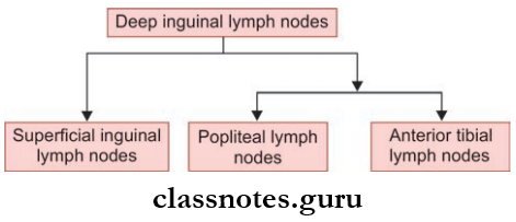
Deep Inguinal Lymph Nodes
- Four to five in number.
- Located on the medial side of the upper part of the femoral vein in the femoral triangle.
- The most proximal node of this group is known as (the gland of Cloquet or Rosenmuller) which lies in the femoral canal.
- All the lymphatics from the lower limb ultimately drain into the deep inguinal node directly or indirectly
- Only the deep part of the gluteal region and the upper aspect of the posterior part of the thigh is not drained by them.
Clinical Anatomy
- Lymphadenopathy of vertical groups of lymph nodes are seen in infection to the lower limb.
- Filariasis is characterized by lymphangitis, lymphadenitis, and lymphedema and is manifested in the lower limb fist before appearing in any other parts of the body.
Blood Supply And Lymphatic Drainage Of Lower Limb Multiple Choice Questions
Question 1. Which of the following is not a branch of the dorsal pedis artery?
- First dorsal metatarsal
- Tarsal branches
- First plantar metatarsal
- Arcuate
Answer: 3. First plantar metatarsal
Question 2. Which genicular artery pierces the fibrous capsule of the knee joint?
- Descending genicular
- Middle genicular
- Anterior tibial recurrent
- Circumflex fibular
Answer: 2. Middle genicular
Question 3. Boyd’s perforator vein is located in the:
- Saphenous ring
- Hunterian canal
- Below the knee close to the tibia
- Popliteal fossa
Answer: 3. Below the knee close to the tibia
Question 4. How many valves can be presents in the great saphenous veins?
- None
- Two to four
- Ten to twenty
- Around thirty
Answer: 3. Ten to twenty