
Temporal And Infratemporal Regions Question And Answers
Question 1. Write a short note on temporal fossa, infratemporal fossa, pterygopalatine fossa.
Answer:
- Spine of the sphenoid,
- Foramen spinosum,
- Foramen ovale,
- Lateral pterygoid plate
Temporal Fossa: Lies on the side of the skull, bounded by superior temporal line and zygomatic arch.
Temporal Fossa Boundaries
- Anterior: Zygomatic and frontal bone
- Posterior: Inferior temporal line and supramastoid crest
- Superior: Superior nuchal line
- Inferior: Zygomatic arch
- Floor Parts of the frontal, parietal, and temporal bone, temporalis muscle, and greater wing of the sphenoid
Temporal Fossa Contents
- Temporalis muscle
- Zygomatic temporal nerve and artery
- Middle temporal artery
- Deep temporal nerve and artery.
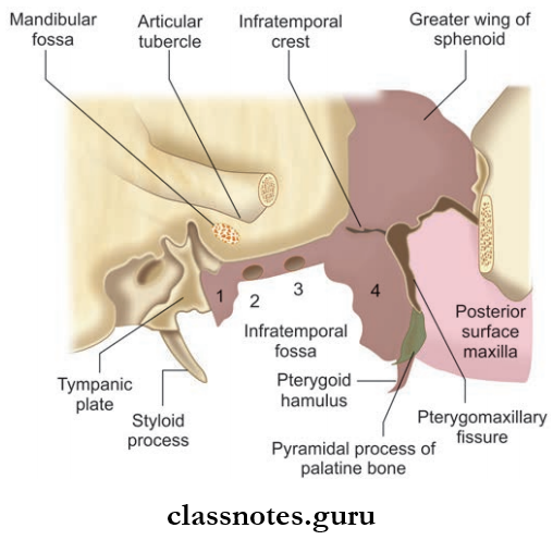
Infratemporal Fossa: Irregular space below the zygomatic arch.
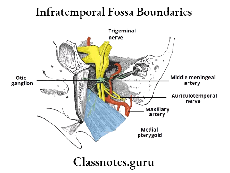
Infratemporal Fossa Boundaries
- Anterior: Posterior surface of a body of the maxilla
- Roof: Infratemporal surface of greater wing of the sphenoid
- Medial: Lateral pterygoid plate
- Lateral: Ramus of mandible
Infratemporal Fossa Contents
- Lateral pterygoid muscle
- Medial pterygoid muscle
- Mandibular nerve and maxillary nerve
- Chorda tympani nerve
- 1st and 2nd parts of maxillary artery.
Pterygopalatine Fossa: Lies in the depth of pterygomaxillary fissures.
Pterygopalatine Fossa Boundaries
- Anterior: Posterior surface of maxilla
- Posterior: Pterygoid process and greater wing of the sphenoid
- Medial: Perpendicular plate of palatine bone
- Floor: Union of anterior and posterior walls
Pterygopalatine Fossa Contents
- Maxillary nerve and its branches
- Pterygopalatine ganglion and its branches
- 3rd part of the maxillary artery and its branches.
Question 2. Write a short note on muscles of mastication.
Answer:
Muscles Of Mastication
- These are the muscle that moves the mandible during mastication and speech
- The muscles include the masseter, the temporalis, the lateral pterygoid, and the medial pterygoid.
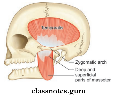
Masseter
- Masseter is quadrilateral in shape and has 2 layers
- Covers the lateral surface of ramus of the mandible.
- Masseter Origin
- Superficial layer: Anterior 2/3rd of the lower border of the zygomatic arch and zygomatic process of maxilla
- Deep layer: Deep surface of the zygomatic arch.
- Masseter Insertion
- Superficial layer: Lower part of lateral surface of ramus of the mandible
- Deep layer: Rest of the ramus of the mandible.
- Masseter Nerve supply: Masseteric nerve branch of anterior division of mandibular nerve.
- Masseter Action: Elevation of mandible to close mouth to bite.
Temporalis
- Fan-shaped muscle
- Present in the temporal fossa
- Temporalis Origin: Temporal fossa and temporal fascia
- Temporalis Insertion
- Margins and deep surface of coronoid process of mandible
- Anterior border of ramus of mandible.
- Temporalis Nerve supply: Deep temporal branches from the anterior division of mandibular nerve
- Temporalis Action: Elevates mandible, helps in side to side grinding movements, retraction of protruded mandible.
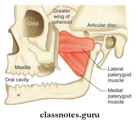
Lateral Pterygoid
- Lateral Pterygoid is a short conical-shaped muscle having upper and lower heads.
- Temporalis Origin:
- Upper Head: Arise from the infratemporal surface and crest of greater wing of sphenoid bone
- Lower Head: Lateral surface of lateral pterygoid plate
- Temporalis Insertion:
- Pterygoid fovea on the anterior surface of neck of mandible
- Anterior margin of articular disc and capsule of temporomandibular joint
- Temporalis Nerve supply: Branch from anterior division of mandibular nerve
- Temporalis Actions:
- Depresses mandible, protrusion of mandible
- Grinding movements.
Medial Pterygoid
- Medial Pterygoid is a quadrilateral muscle
- Medial Pterygoid has a small superficial and large deep head.
- Medial Pterygoid Origin:
- Superficial head: From tuberosity of maxilla and adjoining bone
- Deep head: From the medial surface of the lateral pterygoid plate and adjoining process of palatine bone.
- Medial Pterygoid Insertion: Roughened area on the medial surface of angle and ramus of mandible, behind mandibular foramen and mylohyoid groove.
- Medial Pterygoid Nerve supply: Nerve to medial pterygoid, a branch of main trunk of mandibular nerve
- Medial Pterygoid Actions:
- Elevates mandible
- Protrusion of mandible, grinding movements.
Question 3. Write a short note on the relations of medial and lateral pterygoid.
Answer:
Relations Of Medial And Lateral Pterygoid
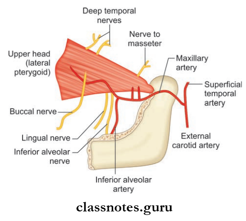
Relations Of Lateral Pterygoid: This key muscle of the region as it provides ideas about the layout of structures of the infratemporal fossa.
- Superficial
- Masseter
- Ramus of mandible
- Maxillary artery
- Tendon of temporalis.
- Deep
- Mandibular nerve
- Middle meningeal artery
- Deep head of the medial pterygoid
- Sphenomandibular ligament.
- Structures Emerging At The Upper Border
- Deep temporal nerves
- Masseteric nerve.
- Structures Emerging At Lower Border
- Lingual nerve
- Inferior alveolar nerve
- Middle meningeal artery.
- Structures Passing In Between Two Heads
- Maxillary artery
- Buccal branch of mandibular nerve.
Relations Of Medial Pterygoid
- Superficial
- The lateral pterygoid plate
- The lingual nerve
- Inferior alveolar nerve
- Maxillary artery
- Sphenomandibular ligament.
- Deep Relations
- Tensor veli palatini
- Superior constrictor of pharynx
- Styloglossus
- Stylopharyngeus.
Mnemonic: Pterygoid muscles (function)
Look at how your jaw ends up when saying first syllable of ‘Lateral’ or ‘Medial’
‘La’: Your jaw is now open, so lateral opens your mouth.
‘Me’: Your jaw is still closed, so the medial closes the mandible.
Question 4. Write a note on the temporomandibular joint.
Answer:
Temporomandibular Joint
- Bilaminar part with venous plexus,
- Posterior thick band,
- Intermediate zone,
- Anterior thick band,
- Anterior extension
- Temporomandibular Joint is a joint on each side of head between the temporal bone and mandible
- Temporomandibular Joint accounts for the movement of the mandible for speech and mastication
- Temporomandibular Joint is a synovial joint of condylar variety.
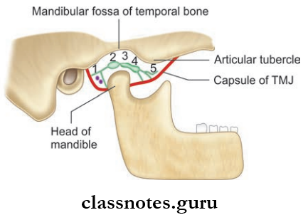
Temporomandibular Joint Articular Surfaces
- Upper articular surface is formed by the mandibular fossa and articular eminence of the temporal bone
- The lower articular surface is formed by the head of the mandible
- Both articular surfaces is covered by fibrocartilage so the joint is an atypical joint.
Temporomandibular Joint Cavity: The joint is divided into:
- Upper meniscotemporal compartment for gliding movements only
- Lower meniscomandibular compartment for gliding as well as rotational movements.
Temporomandibular Joint Articular Disc
- Temporomandibular Joint Articular Disc is an oval plate of fibrocartilage and consists of mainly collagen fibers and few cartilage cells
- Its superior surface is concavo-convex (before backward) and its inferior surface is concave
- The periphery is attached to firous capsule
- The disc consists of a thin intermediate zone and anterior and posterior branch when cut sagittaly.
- The anterior band is continuous with the tendon of the lateral pterygoid
- The posterior band is bilaminar and upper lamina is attached to the squamotympanic fissure and lower lamina is attached to back of condyle.
Temporomandibular Joint Ligaments:
- The Ligaments Include:
- Fibrous capsule
- Temperomandibular ligament
- Sphenomandibular ligament
- Stylomandibular.
- Fibrous Capsule
- It is a fibrous sac which encloses the joint cavity
- It is attached to the articular tubercle, the circumference of the articular fossa, and squamotympanic figure above and neck of the mandible below
- It is loose above the intra-articular disc and tight below the disc.
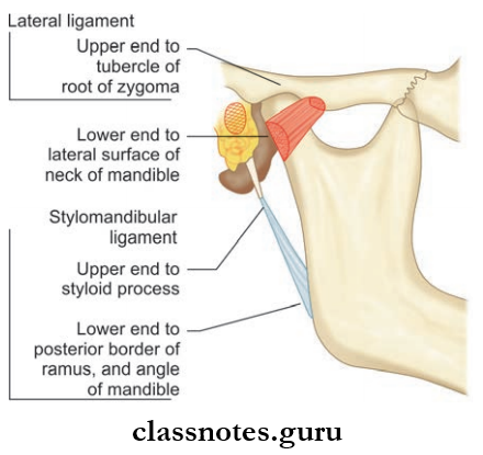
- Temporomandibular Ligament
- It is attached to the articular tubercle in the root of zygoma above and neck of the mandible below.
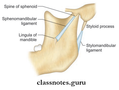
- Sphenomandibular Ligament
- It is attached to the spine of the sphenoid above and the lingula and lower margin of the mandibular foramen below
- It represents the unossified intermediate part of the sheath of Meckel’s cartilage.
- Stylomandibular Ligament
- It is an accessory ligament of temporomandibular joint
- It is attached to the lateral surface of the styloid process above and the angle and adjoining border of the ramus of the mandible below
- It is a thickening of investing layer of deep cervical fascia.
Relations Of Temporomandibular Joint
- Anterior
- Lateral pterygoid
- Masseteric nerve and artery
- Posterior
- Parotid gland
- Superficial temporal vessels
- Auriculotemporal nerve
- Lateral
- Skin and fascia
- Parotid gland
- Temporal branches of the facial nerve
- Medial
- The tympanic plate
- Spine of sphenoid
- Auriculotemporal and chorda tympani nerves
- Middle meningeal artery
- Superior
- Middle cranial fossa
- Middle meningeal vessels
- Inferior: Maxillary artery and vein
Temporomandibular Joint Nerve Supply: The auriculotemporal nerve which enters through posterior aspect of joint Masseteric nerve enters through anterior aspect of joint.
Temporomandibular Joint Blood Supply
- Arterial supply: Maxillary artery and superficial temporal artery enters the joint through posterior aspect of the capsule.
- Lymphatic Drainage: Drain into preauricular nodes, parotid nodes, and upper deep cervical nodes.
Temporomandibular Joint Stability
- More stable when mouth closed
- Forward movement of the condyle is discouraged by articular eminence and contraction of posterior fibers of the temporalis muscle
- Backward movement is prevented by lateral ligament and contraction of the lateral pterygoid muscle.
Temporomandibular Joint Movements: The movements occurring at the joint involve two basic movements.
- Gliding Movements
- Rotational Movements.
1. Gliding Movements: occurs between disc and articular eminence with disc and condyle moving forward and backward, down or up
2. The Rotational Movements occur between the disc and condyle
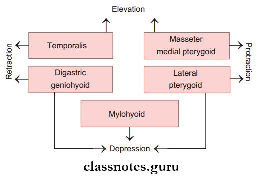
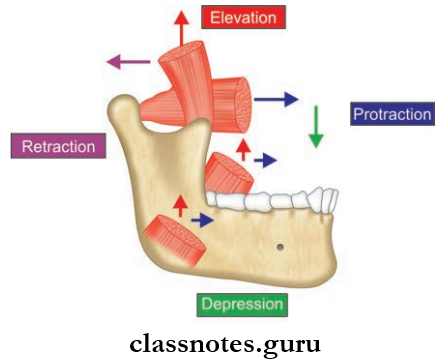
The Movements Include:
- Protraction: Done by lateral and medial pterygoid and masseter
- Retraction: Done by posterior fibers of the temporalis
Elevation: By medial pterygoid, masseter, and vertical fibers of temporalis - Depression: Caused by the lateral pterygoid
- Lateral movements: To the left side by the right lateral pterygoid and vice versa.
Temporomandibular Joint Applied
- When the articular disc is detached from the fibrous capsule, it is known as the derangement of the joint.
- Temporomandibular causes painful movements and clicking sounds while opening and closing the mouth.
- In case of surgeries of the temporomandibular joint, the facial nerve, auriculotemporal nerve, and mandibular division of the trigeminal nerve are preserved with care.
Temporal And Infratemporal Regions Multiple Choice Questions And Answers
Question 1. Which of the following muscles opens the mouth?
- Temporalis
- Lateral pterygoid
- Medial pterygoid
- Masseter
Answer: 2. Lateral pterygoid
Question 2. The temporomandibular joint is commonly dislocated:
- Medially
- Laterally
- Anteriorly
- Posteriorly
Answer: 3. Anteriorly
Question 3. The nerve to the medial pterygoid supplies all of the muscles except:
- Tensor palate
- Medial pterygoid
- Lateral pterygoid
- Tensor tympani
Answer: 3. Lateral pterygoid
Question 4. All of the structures occupy infratemporal fossa except:
- Mandibular nerve
- Chorda tympani nerve
- Pterygoid venous plexus
- Masseter muscle
Answer: 4. Masseter muscle
Question 5. Select the incorrect statement regarding otic ganglion:
- It is functionally related to the mandibular nerve
- It is topographically related to the mandibular nerve
- It is 2–3 mm in size
- It is located lateral to the tensor palate muscle
Answer: 1. It is functionally related to the mandibular nerve