
Ideal Radiographs Important Notes
- kVp controls the wavelength and penetration power of X-rays.
- Whenever kVp is increased, X-rays of shorter wavelength and high penetration power are produced. They are called hard X-rays.
- Whenever kVp is decreased, X-rays of longer wavelength and the least penetrating power are produced. They are called soft X-rays.
Characteristics of an ideal radiograph essay
Ideal Radiographs Long Essays
Question 1. Ideal radiograph.
Answer.
Ideal radiograph Definition:
- An ideal radiograph provides a great deal of information, the image exhibits proper density & contrast, has sharp outlines & is of the same shape & size as the object being radiographed
Ideal radiograph Characteristics:
- Visual characteristics:
- Density:
- It is the overall blackness or darkness of a dental radiograph
- If the density is too dark, the film will appear too dark
- As a result, images cannot be visualized properly
- A radiograph with correct density enables the radiographer to view black areas, white areas & gray areas
- Density:
Factors Affecting The Density:
First Degree Factors:
- Milliampere
- An increase in milliampere results in increased density of the film
- Exposure time
- If the exposure time is increased, then film density is increased
- Kilovoltage peak [kVp]
- If kVp increases, then film density increases
- Density varies directly to the square of the relative kVp
D ∝ [kVp]2
- Source film distance
- Density varies inversely to the square of the source film distance
Density = [kVp]2 x mA x s/[S-F distance]2
- Density varies inversely to the square of the source film distance
Ideal radiograph long essay
Read And Learn More: Oral Radiology Question and Answers
Second Degree Factors:
- Subject thickness
- Density decreases in patients with increased subject thickness
- Developmental conditions
- Overdevelopment of film leads to dark films
- Type of films
- High-speed films change the density
- Screens
- Screens require fewer mAs
- Grids
- Grids require more mAs
- Amount of filtration used
- Reduction in the use of filtration increases the density
- Fog
- Results in darkening of film
- Contrast:
- It is the difference in the degree of blackness between adjacent areas on a dental radiograph
- Dental radiographs with very dark areas & with very light areas are said to have ‘high contrast’
- Depends On The Following:
- Quality of film
- Film processing
- Subject thickness
- kVp
- Exposure time
- Geometric characteristics:
- Sharpness:
- It is capable of reproducing even the smallest details of the object on a radiograph
- Sharpness:
Factors:
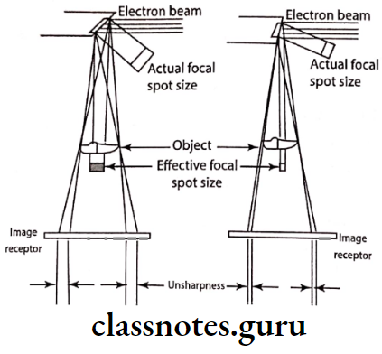
- Geometric unsharpness:
- Size of the focal spot
- Object film distance
- Target film distance
- Motion unsharpness
- Patient
- Tube
- Film
- Film unsharpness
- Grain size
- Emulsion
- Film thickness
- Fog unsharpness
- Intensifying screens unsharpness
Essay on ideal radiographic image
- Magnification:
- It refers to the image that appears larger than the actual size of the object
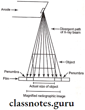
Factors:
- Target film distance:
- It is determined by the length of the position indicating device [PID]
- The longer the PID, the more parallel X-rays, therefore less magnification
- Object film distance
- Less the object film distance, less the magnification
- Use of intensifying screens
- It increases the film object distance & thus creates a magnification
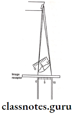
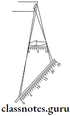
- Distortion:
- It is a variation of the actual size & shape of the object
- Increasing the vertical angulation leads to the shortening of the image
- Decreasing the vertical angulation leads to the elongation of the image
- Anatomical accuracy of radiographic image:
- Labial & lingual CEJ should superimposed
- Buccal & lingual cusps should superimposed
- The buccal portion should superimposed over the lingual portion of the alveolar bone
- No superimposition of zygoma
- Adequate coverage of the anatomic region of interest:
- Proper alignment of the film must be present
- The proper film should be selected
- Proper technique should be selected
Ideal radiograph features long answer
Ideal Radiographs Short Essays
Question 1. Density
Answer.
Density
- It is the overall blackness or darkness of a dental radiograph
- If the density is too dark, the film will appear too dark
- As a result, images cannot be visualized properly
- A radiograph with correct density enables the radiographer to view black areas, white areas & gray areas
Factors Affecting The Density:
First Degree Factors:
- Milliampere
- An increase in milliampere results in increased density of the film
- Exposure time
- If the exposure time is increased, then film density is increased
- Kilovoltage peak [kVp]
- If kVp increases, then film density increases
- Density varies directly to the square of the relative kVp
D ∝ [kVp]2
- Source film distance
- Density varies inversely to the square of the source film distance
Density = [kVp]2 x mA x s/[S-F distance]2
- Density varies inversely to the square of the source film distance
Second Degree Factors:
- Subject thickness
- Density decreases in patients with increased subject thickness
- Developmental conditions
- Overdevelopment of film leads to dark films
- Type of films
- High-speed films change the density
- Screens
- Screens require fewer mAs
- Grids
- Grids require more mAs
- Amount of filtration used
- Reduction in the use of filtration increases the density
- Fog
- Results in darkening of film
Question 2. Target film distance.
Answer.
Target film distance
- This is determined in the intraoral machine by the length of the position indicating device [PID]
- The longer the PID, the more parallel X-rays from the middle of the beam strike the object rather than the diverging rays from the periphery of the beam
- Therefore there is less magnification
- The shorter the PID, the fewer parallel X-rays from the middle of the beam strike the object and more of the diverging rays from the periphery of the beam strike the object
- Therefore, there is more magnification
Properties of a good radiograph essay
Ideal Radiographs Short Answers
Question 1. Density and Contrast.
Answer.
Density and Contrast
- When contrast is altered, is also changed
- However, when the density is altered by itself, there is no change in contrast
- This is because
- Change in kVp produces a change in contrast and density
- Change in mA alone does not change the contrast
- Thus if there is a change in contrast, density also changes
- mA is a prime factor in controlling density, but not a controlling factor for contrast
- Therefore a change in mA will produce a change in density but not in contrast
Question 2. Define ideal radiograph.
Answer.
Ideal radiograph
An ideal radiograph provides a great deal of information, the image exhibits proper density & contrast, has sharp outlines, and is of the same shape & size as the object being radiographed
Radiograph quality criteria essay
Question 3. Contrast.
Answer.
Contrast
- It is the difference in the degree of blackness between adjacent areas on a dental radiograph
- Dental radiographs with very dark areas & with very light areas are said to have ‘high contrast’
Depends On the Following:
- Quality of film
- Film processing
- Subject thickness
- kVp
- Exposure time
Ideal radiographic image parameters essay
Ideal Radiographs Viva Voce
- Density is in direct proportion to milliamperage and kilo voltage and is inversely proportional to focal spot [target] film distance
- Exposure time is inversely proportional to milliamperage and kVp. It is directly proportional to the square of the focal spot film distance
- The useful range of density for a dental X-ray is 0.3 – 2. Density increases with an increase in film fog
- Magnification = Target – film distance/Object – film distance