
Endodontics Of Anatomy Of Pulp Cavity And Its Access Opening Definitions
- Anatomic Apex:
- The Anatomic Apex is the tip or end of the root determined morphologically
- Radiographic Apex:
- Radiographic Apex is the tip or end of the root determined radiographically
- Apical Foramen:
- It is the main apical opening of the root canal
- Frequently located away from the anatomic or radiographic apex
- Apical Constriction:
- Apical Constriction is the apical portion of the root canal having the narrowest diameter
Endodontics Of Anatomy Of Pulp Cavity And Its Access Opening Important Notes
- Endodontic Anatomy
Read And Learn More: Endodontics Question and Answers
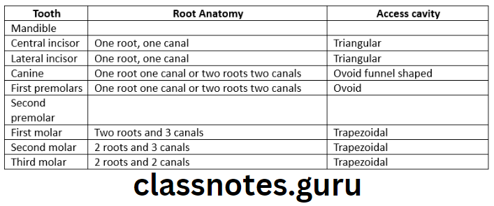
- Access cavity of mandibular first molar
- Mesiobuccal orifice is under mesiobuccal cusp
- mandibular first molar is gained access from mesiobucco apical direction
- Mesiolingual orifice is below the central groove
- mandibular first molar is explained from distobuccal direction
- The distal orifice is present in the center of tooth buccolingually
- mandibular first molar is explained from mesial direction
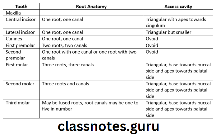
- Access cavity of maxillary first molar
- The orifice of mesiobuccal canal is gained access from distopalatal direction
- The distobuccal canal is gained access from mesiolingual direction
- The palatal root is gained access from buccal direction
Pulp Space Anatomy
Anatomy Of Pulp Cavity And Its Access Opening Short Essays
Question 1. Access cavity preparation in mandibular permanent first molar.
Answer.
Mandibular Permanent First Molar Anatomy:
- Average tooth length – 21.9mm
- Pulp chamber
- Roof is often rectangular in shape
- Mesial wall is straight
- Distal wall is round
- Buccal and lingual walls converge to meet mesial and distal walls
- Roof has four pulp horns – mesiobuccal, mesiolingual, distobuccal and distolingual
- Three distinct orifices are present in the pulpal floor – mesiobuccal, mesiolingual and distal
- Root and root canals
- Two well-determined roots are present – Mesial and distal
- Mesial root curves distally and distal root is straight
- Mesial root has two canals
- Distal root has one canal
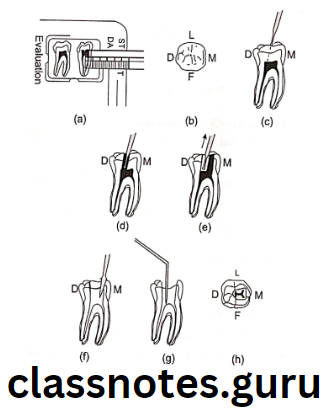
Mandibular Permanent First Molar Access Opening:
- Enamel and dentin are pentrated in the central fossa with the bur angled towards the distal root
- Bur is penetrated until pulp chamber is reached
- A drop of the bur into the pulp chamber is left
- Remove the bulk of the roof of the pulp chamber is felt
- A trapered-cylinder bur is used to remove it
- Walls of access cavity are refined with diamond bur
- Access opening is usually trapezoidal with round corners or rectangular
- The access opening extends towards the mesiobuccal cusp to uncover the mesiobuccal canal, lingually slightly beyond the central groove and distally slightly beyond the buccal groove
Anatomy Of Pulp Cavity And Its Access Opening Short Answers
Question 1. Apical foramen.
Answer.
Apical Foramen
- Apical foramen is an aperture at or near the apex of a root through which th eblood vessels and nerves of the pulp enter or leave the pulp cavity
- In young, incompletely developed teeth, the apical foramen is funnel shaped with wider portion extending outwards
- Mouth of the funnel is filled with periodontal tissue
- As root developes apical foramen becomes narrower
Apical Foramen Variations:
- Apical foramen is not always most constricted portion of the root canal
- Apical constrictions are found 0.5-1 mm away from the root apex
- Apical foramen is not always located in the center of the root apex
Access Cavity Preparation In Endodontics
Question 2. Nerve fibres of pulp.
Answer.
Nerve Fibres Of Pulp
80% of the nerves of the pulp are C fibres and rest are A delta fibres
- C Fibres:
- They are unmyelinated and fine sensory afferent fibres
- Diameter 0.3-1.2 micrometer
- Conduction is slow – 0.4 – 2m/s
- Distributed through out the pulp tissues
- Experienced as a dull, poorly localized and lingering pain
- Conduct throbbing and aching pain associated with pulp tissue damage
- A Delta Fibres:
- They are myelinated axons
- Conduction is fast 6-30m/s
- Diameter 2-5 micrometer
- Present at the pulpal periphery and inner dentin
- Interpreted as short, well-localized, sharp and pricking pain
- Associated with dental pain
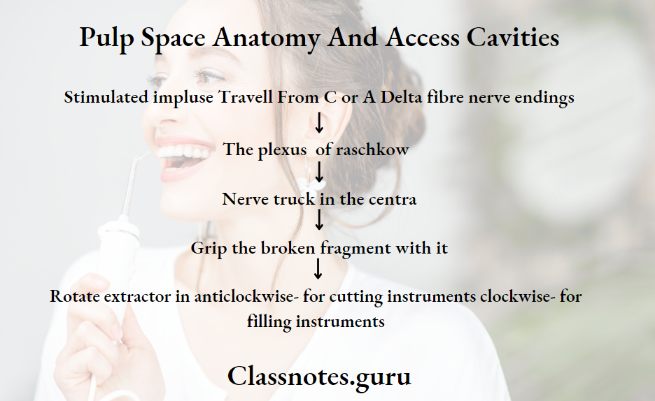
Question 3. Pain pathway.
Answer.
Pain Pathway
Root Canal Access Opening Techniques
Anatomy Of Pulp Cavity And Its Access Opening Viva Voce
- The mesiobuccal canal of the maxillary first molar is the most difficult to prepare
- The access cavity of a mandibular first molar is usually triangular
- Bifurcations and trifurcations are most common in mandibular 1st premolar
- The cervical cross-section of the maxillary 1st premolar is elliptical or kideny-shaped
- Accessory canals are common in the apical third of the root
- Among anterior teeth accessory canals are common in the mandibular central incisor
- Among posteriors accessory canals are common in the mandibular first molar
- Mandibular 1st premolar contains a prominent buccal cusp and a smaller lingual cusp that gives the crown a lingual tilt of 30°
- Among single-rooted teeth bifurcated roots are commonly seen in mandibular 1st premolars followed by incisors and canines
- In the maxillary molar, the mesiobuccal root has the greatest distal curvature and is the narrowest of all three canals
- The pulp chamber of the maxillary 1st molar is the largest in the dental arch