
Diseases Of The Skin Important Notes
- Features Of Hereditary Ectodermal Dysplasia
- Hyperhidrosis
- Hypotrichosis
- Hypodontia
- Lichen Planus
- Lichen Planus is a relatively common dermatological disorder occurring on skin and oral mucous membranes and refers to lace-like patterns produced by symbolic algae and fungal colonies on the surface of rocks in nature
- Lichen Planus Histopathology
- Hyperortho or hyper para keratinization
- Thickening of granular cell layer
- Acanthosis
- Intercellular edema of the Spinus cell layer
- The sawtooth appearance of recipes
- Presence of Civatte bodies
- Apoptotic keratinocytes
- Wickham’s Striae
- Wickham’s Striae is tiny white elevated dots present at the intersection of white lines
- Seen in lichen planus
- Psoriasis
- Small, sharply delineated dry papules each covered by delicate silvery scale
- Auspitz’s sign is present
- Extensor surfaces of extremities are affected
- Psoriasis Histopathology
- Absence of stratum granulosum
- Elongation and clubbing of rete pegs
- Intraepithelial microabscess formation called Monro abscess
- Histopathology Of Pemphigus
- Formation of vesicles or bullae within the epithelium
- Supra basilar split
- Loss of intercellular bridges
- Acanlholysls
- Disruption of prickle cells
- Presence of Tzanck cells
- Auspltz’s Sign
- Seen in psoriasis
- If the deep scales are removed, one or more tiny bleeding points are disclosed
- Tzanck Test – Used For
- Pemphigus
- Herpes simplex
- Ehler Donlos Syndrome
- Ehler-Donlos Syndrome Features
- Hyperelasl icity of skin
- Hyperextension joints
- Excessive bruising
- The patient is known as Rubber Man
- Scarred area of skin appears as crumpled cigarette papers – Gorlin’s sign
- Hypermobility of joints
- Retarded wound healing
- Ehler-Donlos Syndrome Features
- Crest Syndrome
- Associated with scleroderma
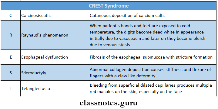
- Crest Syndrome It Consists Of:
- C – Calcinosis cutis
- R – Raynaud’s phenomenon
- E – Esophageal dysfunction
- S – Scleroductyly
- T – Scleroductyly
Diseases Of The Skin Long Essays
Question 1. Classify vesiculobullous lesions. Write In detail about the types, clinical features, pathogenesis, and histopathology of pemphigoid.
Answer:
Vesiculobullous Lesions Classification:
- Hereditary
- Epidermolysis bullosa
- Familial benign chronic pemphigus
- Dyskeratosis congenita
- Viral
- Primary herpetic gingivostomatitis
- Secondary herpetic gingivostomatitis
- Chickenpox
- Herpes zoster virus
- Measles
- Infectious mononucleosis
- AIDS
- Herpangina
- Mucocutaneous
- Pemphigus Vulgaris
- Pemphigus vegetans
- Bullous mucous membrane pemphigoid
- Lichen planus
- Miscellaneous
- Oral submucous fibrosis
- Hyperacidity
- Constipation
- Impetigo
- Erythema multiforme
Read And Learn More: Oral Pathology Questions and Answers
Pemphigoid: Pemphigoid is a group of relatively uncommon autoimmune vesiculobullous lesions characterized histologically by the subepithelial bullae formation in the basement membrane zone of the skin and epithelium
Vesiculobullous Lesions Types:
- Cicatricial pemphigoid
- Bullous pemphigoid
Vesiculobullous Lesions Clinical Features:
1. Cicatricial Pemphigoid/ Benign Mucous Membrane Pemphigoid
- Common in females
- Age group-40-50 years
- Sites Involved
- Conjunctiva
- The skin around the genitalia and near body orifices
- Mucosal surfaces of
- Nose
- Larynx
- Pharynx
- Esophagus
- Vulva
- Vagina
- Penis
- Anus
- Causes obliteration of palpebral fissure
Vesiculobullous Lesions Oral Manifestations:
- Mild erosion or desquamation of the gingival tissue
- Thick-walled vesiculobullous lesions appear
- They rupture leaving a raw, eroded, bleeding surface
- Desquamative gingivitis
- Pain
- Positive Nikolsky’s sign
- Irritation from denture, calculus, and plaque
- Secondary infection
- Sore throat
- Dysphagia
2. Bullous Pemphigoid
- Affects elderly people
- Skin lesions occur over the trunk and limbs
- Lesions persist for several weeks to several months
- Vesicles and bullae are thick-walled
- It leaves raw, eroded areas that heal rapidly
Vesiculobullous Lesions Pathogenesis:
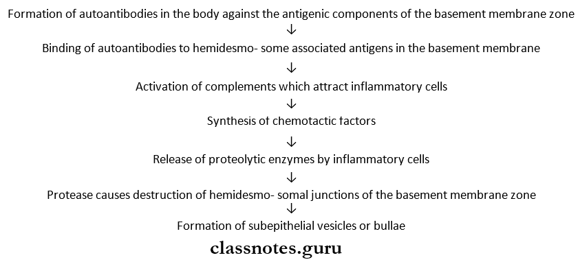
Vesiculobullous Lesions Histopathology:
- Extracellular edema and vacuolation occur in the basement membrane zone
- There is the formation of subepithelial vesicles or bullae
- The subepithelial bullae cause separation of the full-thickness epithelium from the underlying lamina propria
- The epithelium forms the roof of intact bullae
- Absence of acantholysis and epithelial degeneration
- Presence of polymorphonuclear neutrophils within the vesicular fluid
- Subepithelial connective tissue shows inflammatory cell infiltration by lymphocytes, macrophages, and eosinophils
- Dilatation of blood vessels
Question 2. Describe the pemphigus vulgaris.
Answer:
Pemphigus Vulgaris:
- Pemphigus is a group of vesiculobullous lesions of the skin and mucous membrane which is characterised by the formation of intraepithelial vesicles or bullae causing separation of the epithelium above the basal cell layer
- Pemphigus vulgaris is the most common type of pemphigus
Pemphigus Vulgaris Clinical Features:
- Age- 50-60 years of age
- Sex- Both sexes are equally affected
- Vesicles and bullae develop
- They vary in diameter from millimeter to centimeter
- These lesions cover a large surface over the skin
- They contain thin, watery fluid
- When bullae rupture, they leave a raw eroded surface
- Ruptured vesicles leave extremely painful, superficial, erythematous ulcers with ragged borders
- Ulcers bleed profusely
- Gentle traction or oblique pressure on the unaffected areas around the lesion causes denudation or stripping of the normal skin or mucous membrane
- This is known as Nikolsky’s skin
- Skin lesions appear over the scalp, trunk, and umbilical area
Pemphigus Vulgaris Oral Manifestations:
- 3-defined, irregularly shaped, gingival, buccal, or palatal erosions develop
- Such lesions are painful and slow to heal
- Hoarseness of voice
- Excessive salivation
- Difficulty In feeding
- Extremely foul smell in the mouth
Pemphigus Vulgaris Histopathology:
- Formation of the vesicle or bullae within the epithelium
- Results in suprapatellar split or separation
- The basal cell layer remains attached to the lamina propria
- Loss of intercellular bridges and collection of edem fluid results in acantholysis
- Disrupts prickle cells
- Clumps of large hyperchromatic epithelial cells desquamate and lie free within the vesicular fluid
- These cells are rounded and smooth- called Tzanck cells
- A small number of neutrophils and lymphocytes are present
Pemphigus Vulgaris Differential Diagnosis:
- Dermatitis herpetiformis
- Erythema multiforme callosum
- Bullous lichen planus
- Epidermolysis bullosa
- Pemphigoid
- Aphthous ulcers
Pemphigus Vulgaris Treatment:
- Aim of Treatment
- Decrease blister formation
- Promote healing
- Determine a minimal dose of medication
- Control disease process
- Drugs used
- High dose of steroids
- Use of immunosuppressive drugs
- Antibiotics to prevent secondary infections
- Maintain fluid and electrolyte balance
Question 3. List out precancerous lesions, Describe the etiology, clinical features, and histopathology of lichen planus.
Answer:
Precancerous Lesions:
- Precancerous Lesions defined as morphologically altered tissue in which cancer is more likely to occur than its normal counterparts
- Precancerous Lesions Are
- Leukoplakia
- Erythroplakia
- Mucosal changes associated with smoking habits
- Carcinoma in situ
- Bowen’s disease
- Actinic keratosis
Lichen Planus: It is a relatively common dermatological disorder occurring on skin and oral mucous membranes and refers to lace-like patterns
Lichen Planus Etiology:
- Cell-mediated immune response
- Autoimmunity
- Immunodeficiency
- Genetic factor
- Psychogenic factor
- Infections
- Habits- tobacco chewing, betelnut chewing
- Vitamin deficiency
- Patients with secondary syphilis
Lichen Planus Clinical Features:
- Sites Involved
- Buccal mucosa
- Tongue
- Lips
- Gingiva
- The floor of the mouth
- Palate
- Initially, there is a burning sensation in the oral mucosa
- It appears as radiating white and grey velvety thread-like papules in a linear, angular, or reform arrangement forming typical lacy, reticular patterns, rings, and streaks
- Wickham’s striae- tiny white elevated dots are present at the intersection of white lines
- It may be superimposed on candidal infections
Lichen Planus Management:
- Removal of the causative agent
- Steroid therapy
- Cryosurgery and cauterization
- Psychotherapy
- Papsone therapy
- PUVA therapy
Lichen Planus Histopathology
- The overlying surface epithelium exhibits hyper ortho keratinization or hyper para keratinization
- Thickening of the granular cell layer
- Acanthosis
- Intercellular edema in the spouse cell layer
- Shortened and pointed rete pegs producing saw tooth appearance
- Necrosis and liquefaction degeneration of the basal cell layer of the epithelium
- The presence of a few round or ovoid, amorphous, eosinophilic bodies within epithelium called Civatte bodies
- They represent apoptotic keratinocytes which are transported to lire connective tissue for phagocytosis
- A thick band-like infiltration of chronic inflammatory cells is present
Diseases Of The Skin Short Essays
Question 1. Erythema multiforme
Answer:
Erythema Multiforme
Erythema multiforme is an acute self-limiting dermatitis characterized by a distinctive clinical eruption
Erythema Multiforme Precipitating Factors:
- Infections
- Tuberculosis
- Herpes simplex
- Mycoplasma pneumonia
- Infectious mononucleosis
- Drug Hypersensitivity
- Barbiturates
- Sulfonamides
- Salicylates
- Hyperimmune Reactions
- Miscellaneous
- Radiation therapy
- Crohn’s disease
- Vaccinations
Erythema Multiforme Clinical Features:
- Age- 2nd-4th decade of life
- Sex- males are frequently affected
- Initially, it is asymptomatic
- Later erythematous discrete macules, papules or vesicles, and bullae distributed in symmetrically
- Site Involved
- Hands And Arms
- Feet and legs
- Face
- Neck
- Hands And Arms
- Size- A few centimeters or less in diameter
- Appearance- a concentric ring-like appearance of lesions resulting from varying shades of erythema
- This gives rise to ‘target’, ‘iris’, or ‘bull’s eye
Erythema Multiforme Oral Manifestations:
- Site Involved
- Tongue
- Palate
- Buccal mucosa
- Gingiva
- Hyperaemic macules, papules, or vesicles appear
- They become eroded or ulcerated
- Ulcers are diffuse, extremely painful, have irregular borders, and are covered by slough
- They bleed profusely
- Gets easily secondarily foul smell foul smell in my mouth
- Difficulty in eating and swallowing
- Weakness
- Dehydration
- Tracheobronchial ulceration
- Pneumonia
Erythema multiforme Histopathology:
- Intracellular edema of spinous layer of epithelium and edema of superficial connective tissue
- A zone of severe liquefaction degeneration in the upper layers of epithelium
- Intraepithelial vesicle formation
- Thinning of basement membrane
- Dilatation of superficial capillaries and lymphatic vessels
- Inflammatory cell infiltration present in connective tissue
Erythema Multiforme Treatment:
- Treat the cause
- Analgesics- for pain relief
- Antibiotics- to treat infections
- Anti-histamines
- Topical steroids
- Antacids- for oral ulcers
- Use of 0.05% chlorhexidine mouthwash
Question 2. Hereditary ectodermal dysplasia
Answer:
Hereditary Ectodermal Dysplasia
Hereditary ectodermal dysplasia is a large, heterogeneous group of inherited X-linked recessive disorders characterized by the defective formation of ectodermal structures of the body
Hereditary Ectodermal Dysplasia Clinical Features:
- Occurs commonly in males
- Hereditary Ectodermal Dysplasia Hypotrichosis
- Absence of hair
- Reduction in hair follicles varies from sparse scalp hair to complete absence of hair
- Hair bulbs may be distorted, bifid, and small
- Hereditary Ectodermal Dysplasia Hyperhidrosis
- Teeth show abnormal morphogenesis or are absent
- Affects both perdition
- The teeth that are present are small in size and abnormal in shape
- Skin is soft, dry, and smooth
- Heat intolerance
- Fine wrinkling and hyperpigmentation over periocular skin
- Nails are often brittle and dun or show abnormal ringing
- Lack of breast development
- Deficient hearing or vision
- Cleft lip or palate
- Missing fingers or toes
- Xerostomia
- Rhinitis, sinusitis, pharyngitis
- Dysphagia
- Hoarseness of voice
- Depressed nasal bridge
- Frontal bossing
- Protuberant lips
Hereditary Ectodermal Dysplasia Treatment:
- Early dental evaluation
- Construction of artificial dentures and regular change of it
- Artificial saliva is given
Question 3. Pemphigus
Answer:
Pemphigus
Pemphigus is a group of vesiculobullous lesions of the skin and mucous membrane which is characterized by the formation of intraepithelial vesicles or bullae causing separation of the epithelium above the basal cell layer
Pemphigus Types:
- Pemphigus Vulgaris
- Pemphigus vegetans
- Pemphigus foliaceus
- Pemphigus erythematosus
- Brazilian pemphigus
Pemphigus Etiopathogenesis
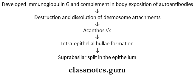
Pemphigus Histopathology:
- Formation of the vesicle or bullae within the epithelium
- Results in supra-basilar split or separation
- The basal cell layer remains attached to the lamina propria
- Loss of intercellular bridges and collection of edem fluid results in acantholysis
- Disrupts prickle cells
- Clumps of large hyperchromatic epithelial cell desquamate lie free within the vesicular fluid
- These cells are rounded and smooth- called Tzanck cells
- A small number of neutrophils and lymphocytes are present
Question 4. Lupus erythematosus
Answer:
Lupus Erythematosus
- Lupus Erythematosus is characterized by the destruction of tissue due to the deposition of autoantibody and immune complexes within it
- Lupus Erythematosus is an autoimmune disorder
- Lupus Erythematosus Forms
- Systemic – Includes
- Skin lesions
- Oral lesions
- Hepatosplenomegaly
- Pneumonia
- Cardiac problems
- Renal problems
- Neural problems
- Hematological problems
- Joint problems
- Ocular problems
- GIT disturbances
- Discoid
- Systemic – Includes
Features Systemic Form Skin Lesions
- Fixed, erythematous rashes with butterfly configuration over the malar region and across the bridge of the nose
- Produces itching or burning sensation
- Aggravates on exposure to sunlight
- Hyperpigmentation of skin
- Extensive loss of hair over the scalp
Features Systemic Form Oral Lesions
- White, hyperkeratotic plaque-like areas
- Oral and nasopharyngeal ulceration
- Presence of erythematous lesions
- Formation of hemorrhagic macules that become ulcerated
- Severe burning sensation
Discoid Forms Skin lesions
- Butterfly configuration
- Elevated, red or purple macules covered by yellow or greyscale
- Forceful removal of covering causes numerous carpet-track extensions
- Loss of hair over the scalp
Discoid From Oral lesions
- Multiple white plaques with central atrophy
- Shallow ulcers – small slit-like
- Pain and burning sensation in the mouth
- Pemphigus Histopathology
- The epithelium is atrophic, hyper ortho ortho, or para-keratinized
- Keratin plugging and acanthosis present
- Liquefactive degeneration of the basal cell layer
- Inflammatory cell infiltration occurs Deposition of antigen-antibody complexes
- Short
Diseases Of The Skin Short Question And Answers
Question 1. Nikolsky’s sign
Answer:
Nikolsky’s Sign
- Nikolsky’ssign is a characteristic feature of pemphigus
- Gentle traction or oblique pressure on an affected area around the lesion causes stripping of the normal skin or mucous membrane occurs
- This is known as Nikolsky’s sign
- It is caused due to the presence of perivascular edema that disrupts the dermal-epidermal junction
Question 2.Monro’s abscess
Answer:
Monro’s Abscess
- Monro’s abscess are histologic feature of psoriasis
- Intraepithelial microabscesses in the upper part of the Malpighian layer is known as Monro’s abscess
- They are collections of neutrophils within the parakeratin layer of epithelium
Question 3. Grinspan syndrome
Answer:
Grinspan Syndrome
- Grinspan Syndrome was described by Grinspan
- Grinspan Syndrome Refers To The Triad Of
- Diabetes mellitus
- Lichen planus
- Vascular hypertension
Question 4. Oral lichen planus
Answer:
Oral Lichen Planus
Oral Lichen Planus is a relatively common dermatological disorder occurring on skin and oral mucous membranes and refers to lace-like patterns
Oral Lichen Planus Etiology:
- Cell-mediated immune response
- Autoimmunity
- Immunodeficiency
- Genetic factor
- Psychogenic factor
- Infections
- Habits- tobacco chewing, betelnut chewing
- Vitamin deficiency
- Patients with secondary Syphilis
Oral Lichen Planus Clinical Features:
- Site Involved
- Buccal mucosa
- Tongue
- Lips
- Gingiva
- The floor of the mouth
- Palate
- initially, there is a burning sensation of oval mucosa
- It appears as radiating white and grey velvety thread-like papules in a linear, angular or region arrangement forming typical lacy, reticular pah terns, rings, and streaks
- Wickham’s striae- liny white elevated dots are present at the intersection of white lines
- It may be superimposed on candidal infections
Question 5. Erythema Multiforme
Answer:
Erythema Multiforme
Erythema multiforme is an acute self-limiting dermatitis characterized by a distinctive clinical eruption
Erythema Multiforme Clinical Features:
- Age- 2nd-4th decade of life
- Sex- males are frequently affected
- Initially, it is asymptomatic
- Later erythematous discrete macules, papules or vesicles, and bullae distributed in symmetrically
- Site Involved
- Hands and arms
- Feet and legs
- Face
- Neck
- Size- A few centimeters or less in diameter
- Appearance- a concentric ring-like appearance of lesions resulting from varying shades of erythema
- This gives rise to ‘target, ‘iris’, or ‘bull’s eye
Erythema Multiforme Oral Manifestations:
- Site Involved
- Tongue
- Palate
- Buccal mucosa
- Gingiva
- Hyporaemic macules, papules, or vesicles appear
- They become eroded or ulcerated
- Ulcers are tlliluuo, extremely painful, have Irregular borders, and are covered by slough
- They bleed profusely
- Gel and easily secondary Infected
- Font smell In the mouth
- Difficulty In eating and swallowing
- Weakness
- Dehydration
- Tracheobronchial ulceration
- Pneumonia
Question 6. Steven-Johnson syndrome
Answer:
Steven-Johnson Syndrome
Steven-Johnson Syndrome is a severe form of erythema multiforme with widespread Involvement typically Involving skin, oral cavity, eyes, and genitalia
Steven-Johnson syndrome Clinical Features:
- Fever, malaise
- Photophobia
- Eruptions on oral mucosa, genital mucosa, and skin
Steven-Johnson Syndrome Skin Lesions:
- Hemorrhagic
- Vesicles and bullae are present
- Eye Lesions:
- Photophobia
- Conjunctivitis
- Corneal ulceration
- Keratoconjunctivitis
Steven-Johnson Syndrome Genital Lesions:
- Non-specific urethritis
- Balanitis
- Vaginal ulcers
Steven-Johnson Syndrome Oral Manifestations:
- Oral mucosa may be extremely painful
- Difficulty in mastication
- Presence of mucosal vesicles or bullae
- They rupture and leave a surtax coveted with thicks white or Yellow exudate
Question 7. Lupus Erythematous Cells
Answer:
Lupus Erythematous Cells
- Lupus erythematous or LE cells ute characteristic feature of acute systemic form of lupus erythema ptosis
- Lupus Erythematous Cells are neutrophil leukocytes, which have phagocytosed other leukocytes
- Lupus Erythematous Cells are large, circular, basophilic inclusions within a neutrophil
Question 8. Tzanck cells
Answer:
Tzanck Cells
- Tzanck Cells are characteristic of pemphigus
- Tzanck Cells are acantholytic multinucleated epithelial cells
- Present freely within the vesicular space
- They have large nuclei and condensation of chromatin along the cell wall
Question 9. Paul-Bunnel test
Answer:
Paul-Bunnel Test
Paul-Bunnel Test is a diagnostic test for infectious mononucleosis
Paul-Bunnel Test Procedure:
- Collect sheep’s RBCs and human’s RBCs
- Agglunate both
Paul-Bunnel Test Result:
- Agglutination is observed
- Normal titre-1:8
- In infectious mononucleosis titer becomes-1:4096
Question 10. White sponge nevus
Answer:
White Sponge Nevus
White sponge nevus is a hereditary skin disease characterized by the occurrence of white, thickened, corrugated mucosal lesions in the oral cavity
White Sponge Nevus Clinical Features:
- Occurs in childhood
- White Sponge Nevus is congenital
- Lesions involve the cheeks, palate, gingiva, the floor of the mouth, and a portion of the tongue
- Mucosa appears thickened and folded Of corrugated with a soft and spongy texture
- Color- White
- Ragged white areas may appear which can be rt moved by gentle rubbing without any bleeding
- White Sponge Nevus are asymptomatic
Question 11. Corps, rounds, and grains
Answer:
Corps, Rounds, And Grains
- Corps, rounds, and grains are histologic font notes of keratosis follicular
- Corps And Rounds
- Slightly larger than normal squamous cells
- Present in the granular layer and superficial spinous layer
- The nucleus is round, homogenous, and basophilic
- It has a distinct cell membrane
- Grains
- Grains are small, elongated pnrakoratotic cells
- Present in the keratin layer
- Corps And Rounds
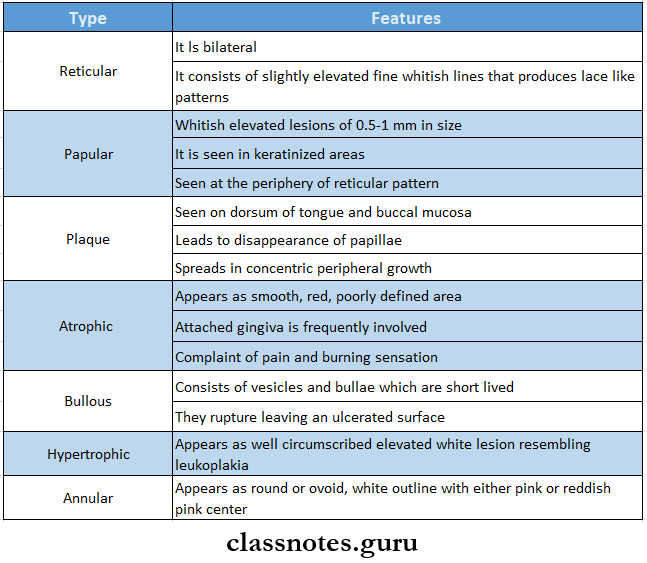
Question 12. Clinical types of lichen planus
Answer:
Clinical Types Of Lichen Planus
- Linear
- Popular
- Confluent
- Reticular
- Annular
- Pigmented
- Vesicular or bullous
- Erosive or atrophic
- Hypertrophic
Question 13. Suprabasilar split/cleft
Answer:
Suprabasilar Split Or Cleft
- Suprabasilar Split Or Cleft is a histological feature of pemphigus
- There is the formation of a vesicle or bulla entirely intraepithelial just above the basal layer
- Suprabasilar Split Or Cleft results in a suprabasal split
- Following this, the basal cell layer remains attached to the lamina propria
- This gives row-of-tomb stones appearance
Question 14. Wickham’s striae
Answer:
Wickham’s Striae
- Wickham’s striae are characteristic features of lichen planus
- The surface of the skin papules is covered by a characteristic very fine lace-like network of greyish-white lines known as Wickham’s striae
- They can occur anywhere on the skin surface but are commonly seen over
- Flexor surfaces of wrist and forearms
- The inner aspect of the knees and thighs
Question 15. Civatte bodies
Answer:
Civatte Bodies
- Civatte bodies are characteristic features of lichen planus
- Civatte Bodies are round or oval, amorphous, eosinophilic bodies present within the epithelium
- Civatte Bodies represent apoptotic keratinocytes or other necrotic epithelial components which are transported to the connective tissue for phagocytosis
- Civatte Bodies are also known as colloid, hyaline, or cytoid bodies
Question 16. Ehlers Danlos syndrome
Answer:
Ehlers Danlos Syndrome
Ehlers-Danlos syndrome is a group of hereditary disorders characterized by defective or abnormal collagen synthesis in various body organs
Ehlers-Danlos Syndrome Clinical Features:
- It affects the skin, and joints, and is characterized by hyperelasticity of skin, hyperextend- sive joints, and excessive bruising
- The patient is known as Rubber Man
- Scarred areas of skin appear as “crumpled cigarette papers”
- Gorlin’s sign- Patient can touch the tip of the nose with their tongue
- Mobility of teeth and marked periodontal weakness
- Retarded wound healing
- Enamel hypoplasia
- Large pulp stones are present
- Formation of irregular dentin
- Hypermability and subluxation of the temporo- mandibular joint
- Loss of normal scalloping of dentin enamel junction
Question 17. CREST syndrome
Answer:
CREST Syndrome
CREST syndrome is associated with scleroderma
Question 18. Target lesions
Answer:
Target Lesions
- Target lesions are characteristic features of erythema multiforme
- Target Lesions appear on extremities
- Target Lesions are concentric rings resulting from varying shades of erythema giving rise to target, iris, or Bullseye
- Target Lesions may be purpuric or paler in the center
Question 19. Tzanck test
Answer:
Tzanck Test
Tzanck smear is prepared

Tzanck test Result: Lesion shows acantholysis
Question 20. Auspit sign
Answer:
Auspit Sign
- Auspit Sign is seen in psoriasis
- If the deep scales on the surface of the lesion are removed, one or two bleeding points are often disclosed
- This phenomenon is known as the Auspilz sign
Diseases Of The Skin Viva Voce
- Butterfly-shaped cutaneous lesions of the face are seen in lupus erythema l os is
- Carpet track extensions are seen in discoid lupus erythema ptosis
- Antinuclear antibodies are seen in systemic lupus erythematosus
- Target, iris, or bull’s eye are seen in erythema multiforme
- Steven Johnson’s syndrome is a very severe bullous form of erythema multiforme
- Saw tooth retepegs are seen in lichen planus
- Monro’s abscess is seen in psoriasis
- Corps, rounds, and grains are histologic features of keratosis follicularis
- Ehler-Danlos syndrome is a group of hereditary disorders characterized by defective or abnormal collagen synthesis in various body organs
- Civatte bodies are characteristic features of lichen planus
- Kobner’s phenomenon is seen in psoriasis