
Diseases Of The Arteries Veins And Lymphatic System Long Essays
Question 1. Describe the clinical features, diagnosis, and treatment of thromboangitis obliterans.
Answer:
Thromboangitis Obliterans/Buerger’s Disease:
- Thromboangitis Obliterans is the inflammatory reaction in the arterial wall with the involvement of the neighboring vein and nerve, terminating in thrombosis of the artery.
Thromboangitis Obliterans Clinical Features:
- Age/sex – 20 – 40 years males.
- Pain while walking at the arch of the foot
- Pain increases when the muscle is exercised
- Postural color changes appear followed by trophic changes.
- Gradually ulceration and gangrene occur.
- Gangrene starts from one digit and then involves the entire foot.
- BP is normal in the normal limb but reduced in the diseased limb.
Thromboangitis Obliterans Diagnosis:
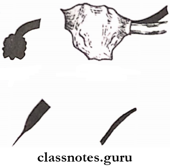
- Arteriography.
- Large arteries show abrupt areas of occlusion surrounded by extensive collateral circulation.
- It gives a tree roof or ‘spider legs’ in appearance.
- Peripheral arteries give a ‘corkscrew’ appearance.
Thromboangitis Obliterans Treatment:
- Conservative treatment.
- Quit smoking.
- Prostaglandin therapy to prevent platelet aggregation.
- Surgical treatment
- Microvascular transplantation of free grafts
- Amputation – to remove gangrenous area.
Read And Learn More: General Surgery Question and Answers
Question 2. Define gangrene. Describe the types, clinical features, and management of wet gangrene.
Answer:
Gangrene: Gangrene is the death of a portion of the body with putrefaction.
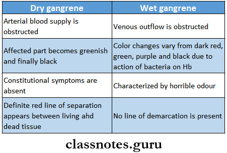
Gangrene Types:
- Dry gangrene due to slow occlusion of arteries.
- Wet gangrene – due to sudden occlusion of arteries.
Wet Gangrene: It is characterized by moist and oedematous limbs.
Wet Gangrene Clinical Features:
- The part is cold, pulseless, swollen and oedematous
- Color changes varies-dark red, green, purple, and black depending on hydrogen produced by bacteria.
- Skin becomes raised into blebs containing foul-smelling fluid.
- There is no line of demarcation present
- Crepitus may be present.
Wet Gangrene Management:
- General treatment
- Nutritious diet
- Control of diabetes
- Relief of pain.
- Local treatment.
- Conservative treatment
- The part should be kept dry
- Part may be kept elevated
- The part should be protected.
- Surgical treatment.
- Amputation – major amputation is necessary.
- Conservative treatment
Diseases of arteries, veins, and lymphatics questions and answers
Question 3. Discuss the clinical features and management of diabetic gangrene.
Answer:
Diabetic Gangrene: Diabetes makes limbs more liable to gangrene formation.
Diabetic Gangrene Clinical features:
- Pain and ulceration of the foot
- Loss of sensation.
- Absence of peripheral pulse.
- Change of color and temperature.
- There may be abscess formation.
- Dry gangrene occurs frequently in old diabetic patients while moist gangrene in young diabetics.
Diabetic Gangrene Management:
- Conservative treatment
- Diabetic control.
- Drugs used – vasodilators, dipyridamole, low-dose aspirin.
- Care of foot – keep it dean and dry.
- Antibiotics – in case of infections.
- Use of micro-cellular rubber footwear.
- Surgical treatment.
- Amputation of the part.
Question 4. Describe the signs, symptoms, and treatment of varicose veins of the leg.
Answer:
Varicose Veins: When a vein becomes dilated, elongated, and tortuous, the vein is said to be varicose
Varicose Vein Clinical features:
- Varicose Vein Symptoms:
- Visible distension of superficial veins.
- Tired and aching sensation in the affected limb.
- Sharp pain.
- Ankle swelling towards evening.
- Skin over the varicosities may itch and pigmented
- Eczema of affected skin.
- Varicose Vein Signs:
- Varicose eczema.
- Hemosiderin pigmentation.
- Atrophie blanche.
- Lipodermatosclerosis.
- Oedema.
- Ulceration.
Varicose Vein Treatment:
- Palliative Treatment:
- Avoid prolonged standing.
- Apply elastic stocking from the toes to the thigh.
- Elevation of lower extremities.
- Exercise like bicycle riding.
- Operative Treatment:
- Saphenous stripping.
- It involves the removal of all or part of the saphenous vein’s main trunk.
- Ambulatory phlebectomy.
- Vein ligation
- Cryosurgery.
- Saphenous stripping.
Vascular system disorders Q&A
Question 5. Describe the clinical features, diagnosis, and etiology, treatment of tuberculosis cervical lymphadenitis.
Answer:
Tuberculosis Cervical Lymphadenitis:
- Tuberculous cervical lymphadenitis refers to lymphadenitis of tire cervical lymph nodes associated with tuberculosis.
Tuberculosis Cervical Lymphadenitis Clinical features:
- Commonly found in children and young adults.
- Presence of chronic, painless, enlarging, or persistent mass.
- Nodes are firm and rubbery which later becomes matted.
- Skin becomes adhered to the mass and may rupture
- Systemic symptoms include.
- Fever with chills.
- Weight loss
- Malaise
Tuberculosis Cervical Lymphadenitis Diagnosis:
- Positive tuberculin test.
- Chest radiograph
- CT scan
- FNAC
- Acid-fast bacilli staining
- Mycobacterial culture.
Tuberculosis Cervical Lymphadenitis Treatment:
- Anti-tubercular drugs:
- Injection streptomycin – 0.5 – lg 1M daily.
- INH – 300 mg/ day.
- PAS – 5 – 15 g/day.
- It is continued for at least 1 and a half years.
- Supportive treatment.
- Vitamin supplements.
- High protein diet
- Surgery.
- Removal of lymph nodes
- Incision and drainage of the abscess.
Tuberculosis Cervical Lymphadenitis Etiology:
- It is caused by Mycobacterium tuberculosis.
- It has 4 pathological stages.
- Stage 1 – lymphoid hyperplasia.
- There is the formation of tubercles and granulomas without caseation necrosis.
- Stage 2 and 3 – caseation necrosis.
- Caseation necrosis in the affected lymph nodes occurs.
- There is the destruction of the capsule of lymph nodes and adherence of multiple nodes with periodontitis.
- Stage 4 – There is a rupture of caseous material into surrounding soft tissue.
- There is abscess cavity formation.
- Stage 1 – lymphoid hyperplasia.
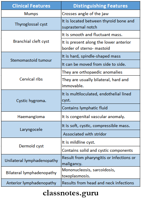
Question 6. Discuss the differential diagnosis of cervical lymphadenopathy.
Answer:
The Differential Diagnosis Of Cervical Lymphadenopathy
Arterial diseases question bank
Question 7. What are the methods of spread of carcinoma? Describe the block dissection of neck.
Answer:
Methods Of Spread Of Carcinoma:
- Through the lymphatic system.
- It is called embolization
- Through bloodstream.
- Malignant cells can break off from the tumor and travel through the bloodstream until they find a suitable place to start forming a new tumor.
- Sarcomas spread through the bloodstream.
- Through local invasion.
- Tumors invade the surrounding normal tissue.
- Through implantation or inoculation.
- It occurs rarely.
- Can happen accidentally when a biopsy is done or when cancer surgery is performed.
- Malignant cells actually drip from a needle or an instrument.
Block Dissection Of Neck:
- The main goal of the procedure is to remove the entire ipsilateral lymphatic structures.
Block Dissection Of Neck Procedure:
- Incisions are made.
- Crile’s T incision
- Martin’s double Y incision
- Ward’s Y incision
Two horizontal incisions.
↓
Skin flaps are reflected
↓
Fibro-areolar tissue of the posterior triangle is dissected away from the trapezius.
↓
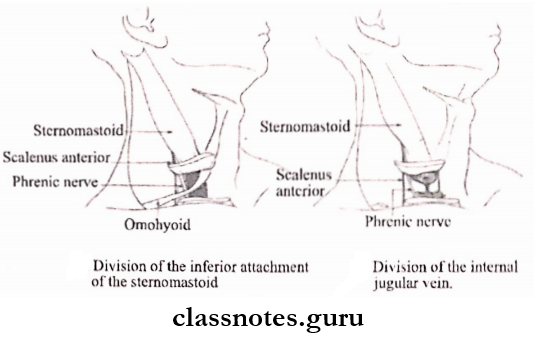
The lower end of the sternomastoid muscle is divided
↓
The internal jugular vein is separated and divided
↓
Above again sternomastoid muscle is transected.
↓
The submandibular salivary gland is dissected
↓
The spinal accessory nerve is divided in 2 places.
↓
Transaction of the jugular vein.
↓
Skin is closed with suction drainage.
Block Dissection Of Neck Structures Removed:
- Lymph nodes – submental, submandibular, upper and lower deep cervical groups, posterior cervical group, and supraclavicular group.
- Sternomastoid muscle.
- Internal jugular vein.
- Submental and submandibular salivary glands.
- Spinal accessory nerve.
- Branches of external carotied artery.
Venous diseases nursing questions
Diseases Of The Arteries Veins And Lymphatic System Important Notes
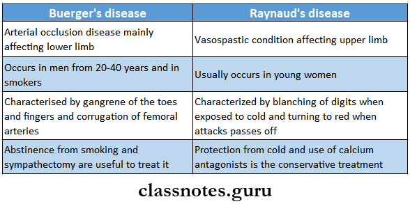
- Buerger’s Disease and Raynaud’s Disease
- Varicose Veins
- Develop in the calf when the veins above are normal
- More frequent in people who stand during their work
- Often develop during pregnancy under the influence of Estrogen and progesterone which cause the smooth muscle in the vein wall to relax
- Complications
- Superficial thrombophlebitis
- Deep vein thrombosis
- Venous ulceration
- Lymph Nodes In Different Diseases
- Soft, elastic, and rubbery – Hodgkin’s disease
- Firm, discrete – syphilis
- Stony hard – secondary carcinoma
- Matted – tuberculosis
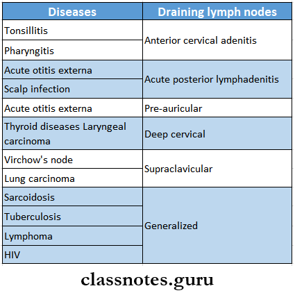
- Draining Lymph Nodes In Different Disease
- Intermittent Claudication
- Intermittent Claudication is the most common complication of the limb due to chronic arterial occlusion
- Features
- Cramp-like pain is felt in the muscles during exertion and gradually disappears within minutes upon cessation of activity
- Pain is due to accumulation of excessive P substance in the muscles
- Boyd’s classification
- Grade 1 – pain disappears if the patient continues to walk
- Grade 2 – pain continues but the patient can still walk with effort
- Grade 3 – pain compels the patient to take a rest
- Indications Of Sympathectomy
- Rest pain and minor ulceration
- Buerger’s disease
- Raynaud’s disease
- Senile gangrene
- Types Of Gangrene
- Complications Of Varicose Veins
- Thrombophlebitis
- Pigmentation
- Eczema
- Ankle flare
- Venous ulcer
- Flaemorrhage
- Periostitis
- Calcification
- Equinus deformity
- Virchow’s Triad – Considered In The Etiology Of Venous Thrombosis Which Includes
- Stasis
- Injury to the vessel wall
- Hypercoagulability of blood
Read And Learn More: General Surgery Question and Answers
Diseases Of The Arteries Veins And Lymphatic System Short Essays
Question 1. Cervical Rib.
Answer:
Cervical Rib
The cervical rib is an extra rib present in the neck.
Cervical Rib Types:
- Type 1 – The free end of the cervical rib is expanded into a hard, bony mass.
- Type 2 – complete cervical rib extending from C7 vertebra to the manubrium.
- Type 3 – Incomplete cervical rib – partly bony and partly fibrous.
- Type 4 – Complete fibrous band.
Cervical Rib Clinical Features:
- Common in females.
- Dull aching pain.
- Hand of the affected side is colder and paler
- Numbness of the fingers.
- Bruit is heard.
- Hard mass may be visible and palpable.
- Seonsory and motor disturbances in the area
Cervical Rib Treatment:
- Conservative.
- Shoulder girdle exercise.
- Correction of faulty posture.
- Surgery.
- Excision of cervical rib.
- Removal of thrombus if present
Lymphatic system disorders Q&A
Question 2. Aneurysm.
Answer:
Aneurysm Definition: Dilatation of a localized segment of the arterial system is known as aneurysm.
Aneurysm Types:
- True aneurysm – contains all three layers of the arterial wall.
- It is further classified into
- Fusiform aneurysm
- Saccular aneurysm
- Dissecting aneurysm
- It is further classified into
- False aneurysm – It has a single layer of fibrous tissue as the wall of the sac.
- Arteriovenous aneurysm.
Aneurysm Clinical Features:
- Elderly patients are commonly affected.
- Pain
- Expansile pulsatile mass
- Severe ischemia
- Bruit is heard.
Aneurysm Causes:
- Congenital
- Acquired.
- Trauma
- Infections
- Atherosclerosis
Aneurysm Treatment:
- Repair of the aneurysm with graft.
Question 3. Arteriovenous aneurysm or Arteriovenous fistula.
Answer:
Arteriovenous Aneurysm
Communication between an artery and an adjacent vein leads to an arteriovenous aneurysm.
Arteriovenous Fistula Causes:
- Congenital
- Acquired – trauma
- Iatrogenic.
Arteriovenous Fistula Clinical Features:
- Systemic effects.
- Increased cardiac output.
- Increased heart rate
- Increased systolic pressure
- Cardiac hypertrophy.
- Decreased peripheral resistance.
- Local effects.
- Aneurysmal dilatation.
- Extensive collateral circulation.
- Bruit can be heard
- Veins are enlarged.
Arteriovenous Fistula Treatment:
- Congenital lesions-excision.
- Acquired lesions.
- Reconstructive
- Ligation of involved artery
- Selective intra-arterial embolization.
Common vascular diseases questions and answers
Question 4. Venous Ulcer.
Answer:
Venous Ulcer Causes:
- Varicose veins
- Increased venous hydrostatic pressure.
Venous Ulcer Clinical Features:
- Located on the medial side of the lower 1/3rd of the leg
- It is shallow and superficial
- Painless
- Pain occurs if it is infected.
- The skin around the ulcer is pigmented
- Shows evidence of healing.
Venous Ulcer Treatment:
- Conservative treatment:
- Elevation of affected limb.
- Movement of limb
- Apply of firm elastic bandage.
- Cleaning of ulcer.
- Antibiotic administration.
- Surgical
- Sclerotherapy.
- Split skin graft.
- Ligation.
Question 5. Thrombophlebitis.
Answer:
Thrombophlebitis
- Thrombophlebitis is superficial vein thrombosis.
- Thrombophlebitis occurs more often in varicose veins or after intravenous infusion.
Thrombophlebitis Clinical Features:
- Painful cord-like inflamed area.
- Redness
- Tenderness
- Local induration.
Thrombophlebitis Treatment:
- Conservative treatment:
- Hot bath or compression
- Application of crepe bandage
- Use of anti-coagulant
- Use of aspirin.
- Intravenous infusion of antibiotics
- Surgical treatment.
- Ligation of the involved vein.
Question 6. Cystic hygroma.
Answer:
Cystic Hygroma
Cystic Hygroma is the most common form of lymphangioma.
Cystic Hygroma Clinical Features:
- Common in the neck region.
- Mostly seen in children.
- Painless swelling.
- Pain occurs when it is infected.
- Fluctuation and fluid thrill are present.
- Swellings are translucent.
- Regional lymph node enlarges in the presence of infection.
Cystic Hygroma Treatment:
- Complete excision.
Question 7. Hodgkin’s lymphoma.
Answer:
Hodgkin’s Lymphoma Definition: It is a malignant neoplasm of the lymphoreticular system.
Hodgkin’s Lymphoma Clinical Features:
- Age – 30 – 50 years
- Sex- More common in males,
- Generalised Iymphodenopathy.
- Site involved- lymph nodes in the neck, axilla, mediastinal, para-aortic, and inguinal.
- Nodes are firm without matting.
- Fever with rigors.
- Malaise, weight loss, and pallor.
- Itching of skin.
- Abdominal pain.
- Bony pain.
- Ascites.
- Superior vena cava obstruction.
Hodgkin’s Lymphoma Investigation:
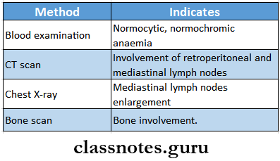
Hodgkin’s Lymphoma Treatment:
- Radiotherapy – for stages 1 and 2
- Chemotherapy – for stages 3 and 4
Short notes on vascular and lymphatic diseases
Question 8. Staging of Hodgkin’s disease.
Answer:
Hodgkin’s Disease Stage 1:
- Lymph node involvement in one anatomical region.
- Example: palpable left supraclavicular nodes.
Hodgkin’s Disease Stage 2:
- Involvement of two or more lymph nodes on the same side of the diaphragm.
- Example: Left supraclavicular and left axillary node.
Hodgkin’s Disease Stage 3:
- Involvement of lymph nodes on both sides of the diaphragm.
- Example: Left supraclavicular and left inguinal lymph nodes.
Hodgkin’s Disease Stage 4:
- Diffuse involvement of one or more extra lymphoid organs with or without lymph node involvement.
Disorders of blood vessels questions for exams
Question 9. Varicose ulcer
Answer:
Varicose Ulcer
- Varicose Ulcer is a type of venous ulcer
- Varicose Ulcer Cause
- Abnormal venous hypertension in the lower third of the leg, ankle, and dorsum of the foot
- Varicose Ulcer Features
- Shallow and superficial
- Doesn’t penetrate deep fascia
- Usually painless
- Associated with varicose veins
- The skin around the ulcer is pigmented
Diseases Of The Arteries Veins And Lymphatic System Viva Voce
- The commonest type of lymphoma is Hodgkin’s lymphoma
- Application of warmth will increase the symptoms of arterial occlusion
- Venous ulcers are commonest ulcers of the legs
- Continuous machinery murmur indicates presence of an arteriovenous fistula
- Synthetic grafts are used in aortoiliac occlusion
- Vein grafts are used in femero-popliteal occlusion
- Majority of the pulmonary emboli originates in the lower extremity