
Contents Of Orbit And Eye Question And Answers
Question 1. Write a short note on orbital fascia and bulbar fascia.
Answer:
Orbital Fascia Or Periorbital: Fascial Sheath Or Bulbar Sheath Or Tenon’s Capsule
- Tenon’s capsule forms a thin membranous sheath around the eyeball from optic nerve to the sclerocorneal junction
- Orbital Fascia Or Periorbital is loosely attached, so the eyeball can freely move within the sheath.
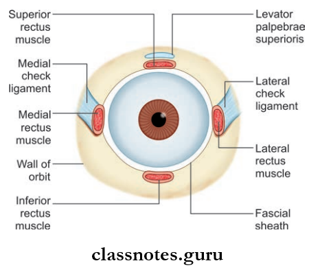
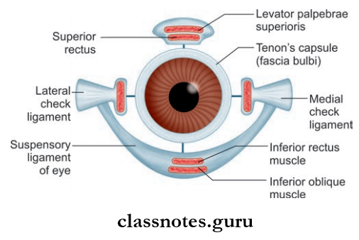
Orbital Fascia Or Periorbital Applied: Tenon’s space is a potential space around the eyeball between the sclera and Tenon’s capsule.
Question 2. Write a short note on extraocular muscles.
Answer:
Extraocular Muscles
- These are the muscles present in the orbit. Among these, there are seven voluntary muscles and three involuntary muscles
- Among the seven voluntary muscles, six muscles move the eyeball, one muscle moves the upper lid
- The Seven Voluntary Muscles Include:
- Four Rectisuperior Rectus
- Inferior Rectus
- Medial Rectus
- Lateral Rectus
- Two Oblique
- Superior oblique
- Inferior oblique
- Levator Palpebrae Superioris
- Four Rectisuperior Rectus
- The Involuntary Muscles Include:
- Superior tarsal muscle
- Inferior tarsal muscle
- Orbitalis.
Voluntary Muscles
- Voluntary Muscles Origin
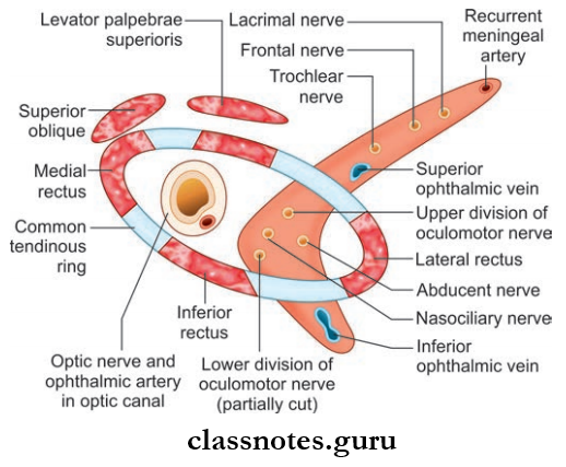
- The recti arise from a common tendinous ring
- The tendinous ring is attached to the orbital surface of the apex of orbit
- It encloses the optic canal and the middle part of the superior orbital fissure
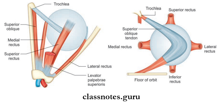
- The superior oblique arises from body of the sphenoid bone superomedial to optic canal
- Inferior oblique arises from the orbital surface of the maxilla lateral to the lacrimal groove
- Levator muscle arises from the orbital surface of the lesser wing of the sphenoid bone superior to the optic canal.
- Voluntary Muscles Insertion
- Recti muscles are inserted to the sclera posterior to the limbus
- The superior oblique is inserted into the sclera, behind the equator in the posterior superior quadrant of the eyeball between the superior and lateral rectus
- The inferior oblique is inserted into the sclera, behind the equator in the posterior superior quadrant of the eyeball below and posterior to the insertion of the superior oblique muscle
- Levator muscle’s superior lamellae is inserted into the anterior surface of the superior tarsus and into skin of the upper eyelid, whereas the inferior lamellae is inserted to upper margin of the superior tarsus, and to the superior conjunctival fornix.
- Voluntary Muscles Nerve Supply
- Superior rectus, inferior rectus, inferior oblique and levator palpebrae superioris are supplied by occulomotor nerve
- Lateral rectus is supplied by the abducent nerve
- Superior oblique is supplied by the trochlear nerve.
Mnemonics: Extraocular muscles cranial nerve innervation
LR6SO4 Rest 3:
- Lateral Rectus is 6th
- Superior Oblique is 4th rest are all 3rd cranial nerve.
Voluntary Muscles Applied
- Weakness or paralysis of a muscle causes squint or strabismus.
- Squint can be paralytic or concomitant .
- Concomitant squint is usually congenital and there is no limitation of movements and diplopia.
- In paralytic squint movements are limited, and diplopia and vertigo may be present.
Question 3. Write a short note on the actions of extraocular muscles.
Answer:
The Actions Of Extraocular Muscles
The Movements Of The Eyeball Are As Follows:
- Around Transverse Axis
- Elevation (upward rotation)
- Depression (downward rotation)
- Around Vertical Axis
- Adduction (medial rotation)
- Adduction (lateral rotation)
- Around Anterior-Posterior Axis
- Intorsion (12 o’clock position of cornea rotates medially)
- Extorsion (12 o’clock position of cornea rotates laterally)
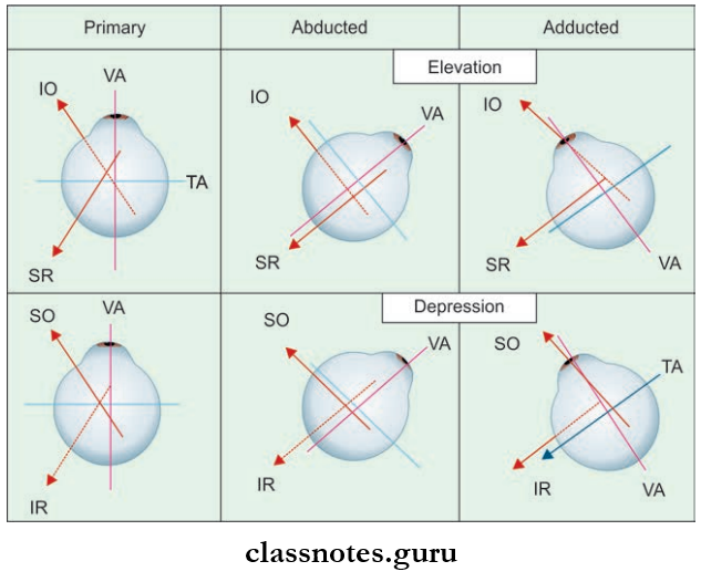
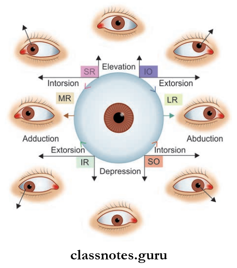
- Actions Of Individual Muscles:
- Superior Rectus: Elevation, adduction, intorsion
- Inferior Rectus: Depression, adduction, extorsion
- Medial Rectus: Adduction
- Lateral Rectus: Abduction
- Superior Oblique: Depression, abduction and intorsion
- Inferior Oblique: Elevation, abduction and extorsion.
Mnemonic: Eye rotation by oblique muscle
- I Love S and M
- Inferior Oblique: Lateral eye rotation.
- Superior Oblique: Medial eye rotation.
Contents Of Orbit And Eye Multiple Choice Questions And Answers
Question 1. All of the following muscles of the eyeball are supplied by oculomotor nerve except:
- Superior rectus
- Superior oblique
- Inferior rectus
- Inferior oblique
Answer: 1. Superior rectus
Question 2. All of the structures are derived from Tenon’s capsule except:
- Medial check ligament
- Lateral check ligament
- Ligament fascia
- Ligament of Lockwood
Answer: 3. Ligament fascia
Question 3. Which of the following branches of the ophthalmic artery is most important?
- Lacrimal artery
- The central artery of retina
- Supraorbital artery
- Supratrochlear artery
Answer: 2. Central artery of retina
Question 4. Which of the following nerves lies in the orbit outside the periorbital?
- Oculomotor
- Trochlear
- Abducent
- Zygomatic
Answer: 4. Zygomatic
Question 5. Select the incorrect statement regarding ciliary ganglion:
- It lies between optic nerve and lateral rectus in the apex of the orbit
- It is functionally connected to oculomotor nerve
- It lies between the medial rectus and optic nerve near the apex of the orbit
- Topographically it is connected to the nasociliary nerve
Answer: 3. It lies between the medial rectus and optic nerve near the apex of orbit