
Cerebrum Question And Answers
Question 1. Write a note on the lobes and external surfaces of cerebral hemispheres.
Answer:
Lobes And External Surfaces Of Cerebral Hemispheres
The External Features Of The Cerebral Hemispheres Include:
- Poles
- Surfaces
- Borders
- Sulci and Gyri.
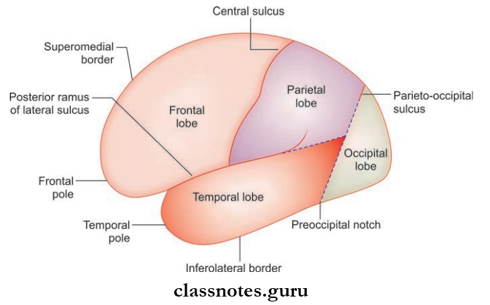
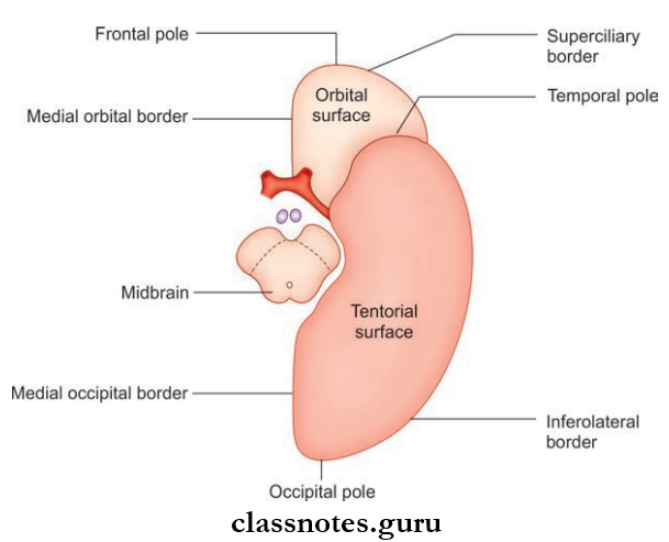
1. Poles: Each cerebral hemisphere presents the following poles
- The Frontal Pole: The anterior end of the hemisphere and is rounded
- The Occipital Pole: The posterior end of the hemisphere and is more pointed in shape
- The Temporal Pole: The anterior end of the temporal lobe.
2. Surfaces: Each cerebral hemisphere consists of the following surfaces
- The Superolateral Surface: The most extensive surface and is convex in shape
- The Medial Surface: The flat and vertical surface presents a C-shaped cut surface of the corpus callosum
- The Inferior Surface: The irregular surface corresponds to the floor of the anterior and middle cranial fossa and is divided into anterior and posterior parts by the stem of the lateral sulcus.
3. Borders: Each cerebral hemisphere presents the following borders
- The superomedial border which separates the superolateral surface from the medial surface
- The superciliary border which is present at the junction of superolateral and orbital surfaces
- The inferolateral border which separates the superolateral surface from the tentorial surface
- The medial orbital border which separates the medial surface from the orbital surface
- The inferomedial border which surrounds the cerebral peduncle
- The medial occipital border separates the medial surface from the tentorial surface.
4. Sulci And Gyri
- The cerebral cortex is thrown into a complex series of tortuous folds called gyri or convolution
- The grooves between the gyri are known as sulci
- These gyri are responsible for the increase in the surface area of the cortex.
Lobes of Cerebral Hemisphere
The superolateral surface of each hemisphere is divided into four lobes by three main sulci known as central, lateral, and parietooccipital. The lobes are as follows:
- Frontal lobe
- Parietal lobe
- Temporal lobe
- Occipital lobe.
- The central sulcus begins at the superomedial border of the hemisphere about 1 cm behind the midpoint of the frontal and occipital poles. It runs obliquely downwards and forwards and ends a little above the posterior ramus of the lateral sulcus
- The lateral sulcus of Sylvius consists of a stem and three rami. The stem begins as a deep cleft on the inferior surface of the cerebral hemisphere at the anterior perforated substance and extends laterally to reach the superolateral surface. On the superolateral surface, it divides into 3 rami:
- Anterior horizontal rami
- Anterior ascending rami
- Posterior rami.
- The parieto-occipital sulcus is present at the medial surface begins at the midpoint of the calcarine sulcus and passes upwards and slightly backward to cut the superomedial border of the hemisphere
- The calcarine sulcus is present on the medial surface and begins as a deep fissure, a little below the posterior end of the corpus callosum, the splenium, and follows an arched course with convexity directed upwards to the occipital pole
- The division is completed by drawing an imaginary line joining the parieto-occipital sulcus to the occipital notch and another line continuing backward from the posterior ramus of the lateral sulcus to meet the first line
- The frontal lobe lies anterior to the central sulcus and above the posterior ramus of the lateral sulcus
- The parietal lobe lies behind the central sulcus in front of the first imaginary line below by the posterior ramus of the lateral sulcus and a second imaginary line
- The temporal lobe lies below the posterior ramus of the lateral sulcus and the second imaginary line
- The occipital lobe lies behind the vertical line joining the parieto-occipital sulcus and the occipital notch.
Insula (Central Lobe)
- Insula is a hidden portion of the cerebral cortex in the floor of the lateral sulcus
- Insula is triangular in shape and surrounded all around by a sulcus, the circular sulcus, except anteriorly at its apex called the limen insulae which is continuous with anterior perforated substance
The insula is divided into two regions by the central sulcus:
- Anterior
- Posterior.
- The anterior region presents 3–4 short gyri called gyri breviate and the posterior region presents 1–2 long gyri called gyri longa
- The insula is overlapped by the adjacent cortical areas called opercula and they include the frontal, frontoparietal, and temporal opercula
- The superior surface of the temporal operculum presents the anterior and posterior transverse temporal gyri.
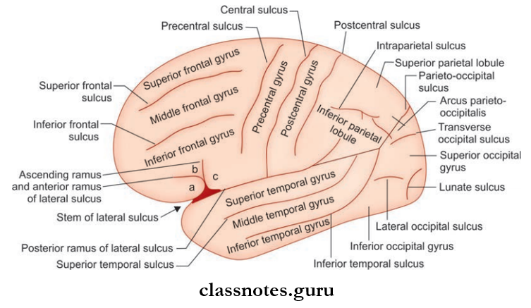
Abbreviations: a = Pars orbitalis; b = Pars triangularis; c = Pars opercularis
Question 2. Write a note on the sulci and gyri of the brain.
Answer:
The Sulci And Gyri Of The Brain Can Be Studied Under The Following Headings:
- Sulci And Gyri On The Superolateral Surface
- Sulci And Gyri On The Medial Surface
- Sulci And Gyri On The inferior surface.
1. Sulci And Gyri On The Superolateral Surface
- In The Frontal Lobe
- The prefrontal sulcus runs downwards and forward parallel and a little anterior to the central sulcus
- The area between the central and precentral sulci is called the precentral gyrus
- Anterior to the precentral sulcus there are two sulci called superior and inferior frontal sulci which run horizontally and divide the region in front of the precentral sulcus into superior, middle, and inferior frontal gyri
The Anterior And Ascending Rami Of The Lateral Sulcus Divide The Inferior Frontal Gyrus Into Three Parts:
- Pars Orbitalis: The part below the anterior ramus
- Pars Triangularis: The part between the anterior and ascending rami
- Pars Opercularis: The part posterior to the ascending ramus.
- In the Parietal Lobe
- The postcentral sulcus runs downwards and forward a little behind and parallel to the central sulcus and the area between these two sulci is known as the postcentral gyrus
- The rest of the parietal lobe is divided into superior and inferior parietal lobules by an intraparietal sulcus.
- In the Temporal Lobe
- In the temporal lobe, two sulci run parallel to the posterior ramus of the lateral sulcus and these are known as superior and inferior temporal sulci and divide the temporal lobe into superior, middle, and inferior temporal gyri.
- The superior surface of the superior temporal gyrus presents two transverse temporal gyri.
- In the Occipital Lobe: The occipital lobe presents three short sulci
- Lateral occipital sulci
- Transverse occipital sulci
- Lunate sulcus.
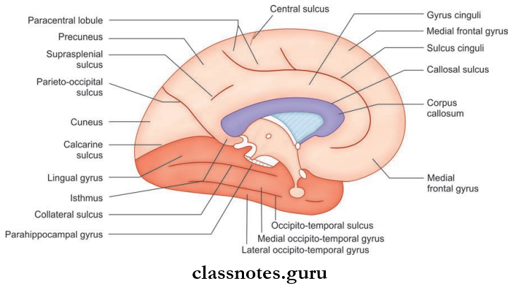
2. Sulci And Gyri On The Medial Surface: The sulci and gyri of the medial surface are as follows:
- Cingulate Sulcus: The most prominent sulcus which is parallel to the upper convex margin of the corpus callosum. Anteriorly it ends below the genu of corpus callosum and posteriorly it turns upwards to reach the superomedial border of the hemisphere just behind the upper end of the central sulcus
- Callosal Sulcus: It separates the cingulate gyrus from the corpus callosum
- The part of the medial surface between the cingulate sulcus and the superomedial border of the hemisphere is divided by a short sulcus ascending from the cingulate sulcus above the middle of the trunk of the corpus callosum into two:
- The Paracentral Lobule: A small part around the upper part of the central sulcus
- Medial Frontal Gyrus: A large medial part.
- The posterior part of the medial surface behind the paracentral lobule has two main sulci
- The Calcarine Sulcus: The small region between the splenium and calcarine sulcus is called the isthmus
- The Parieto-Occipital Sulcus: The triangular area between the posterior part of the calcarine sulcus and the parieto-occipital sulcus is called cuneus
- The quadrangular area between the parietooccipital sulcus and paracentral lobule is called the precuneus.
- A small sulcus just above and parallel to the splenium is called the supraspinal sulcus and separates the precuneus from the cingulate gyrus.
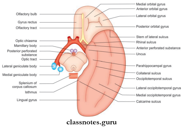
3. Sulci And Gyri On The Inferior Suface
- On The Orbital Part Of The Inferior Surface:
- Olfactory Sulcus: A straight sulcus that runs anteroposteriorly close to the medial border of the orbital surface. The area medial to this sulcus is called the gyrus rectus.
- Orbital Sulcus: It is an irregular, H–shaped sulcus which divides the rest of the orbital surface to the following gyri:
- Anterior orbital gyri
- Posterior orbital gyri
- Medial orbital gyri
- Lateral orbital gyri
- On The Tentorial Part Of The Inferior Surface:
- The surface consists of two major sulci that run anteroposteriorly. They are
- Collateral Sulcus: The medial one
- Occipitotemporal Sulcus: The lateral one
- Posteriorly the collateral sulcus is parallel to the calcarine sulcus and the area between the two is known as the lingual gyrus
- The surface consists of two major sulci that run anteroposteriorly. They are
- The lingual gyrus is continuous anteriorly with the parahippocampal gyrus. The anterior end of the parahippocampal gyrus is hook-like and is called the uncus
- Posteriorly the parahippocampal gyrus is continuous with the cingulate gyrus through the isthmus
- The area between the occipitotemporal sulcus and the collateral and rhinal sulcus is known as the medial occipitotemporal gyrus
- The area lateral to the occipitotemporal sulcus is called lateral occipitotemporal gyrus.
Question 3. Write a short note on the medial geniculate body.
Answer:
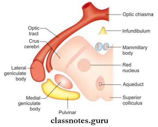
The Medial Geniculate Body
Oval elevation is present below the pulvinar of the thalamus and lateral to the superior colliculus.
Medial Geniculate Body Connections
- Afferents:
- Lateral lemniscus
- Fibers from both inferior colliculus.
- Effects: Give rise to auditory radiation joining the auditory area of the cortex through the sublentiform part of the internal capsule.
Medial Geniculate Body Function: Act as a relay station in the pathway of auditory impulses to the cerebral cortex.
Question 4. Write a short note on the lateral geniculate body.
Answer:
Lateral Geniculate Body
Small oval elevation presents anterolateral to the medial geniculate body, below the thalamus. It is connected to the superior colliculus by the superior brachium.
Lateral Geniculate Body Structure
- Six layered
- Layers 1, 4, and 6 receive contralateral optic fibers
- Layers 2, 3, and 5 receive ipsilateral optic fibers.
Lateral Geniculate Body Connection
- Afferent: Optic tract
- Efferent: Give rise to optic radiation going to the visual area of the cortex through the retro lentiform part of the internal capsule.
Lateral Geniculate Body Function: It is the last relay station in the visual pathway to the occipital cortex.
Question 5. Write a note on the hypothalamus.
Answer:
Hypothalamus
Hypothalamus is a part of the diencephalon which lies below the thalamus.
Hypothalamus Boundaries: The boundaries of the hypothalamus include
- Anteriorly: Lamina terminalis
- Posteriorly: Subthalamus
- Inferiorly: Tuber cinereum, infundibulum, mammillary body
- Superiorly: Thlamus
- Laterally: Internal capsule
- Medially: Cavity of 3rd ventricle.
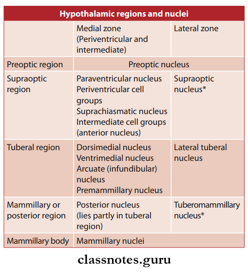
Hypothalamus Subdivisions: It is divided anteroposteriorly into the following
- Preoptic region
- Supraoptic region
- Tuberal region
- Mammillary region.
1. Preoptic Region: It lies anterior to the hypothalamus between the optic chiasma and anterior commissure.
2. Supraoptic Region: The region above the optic chiasma.
3. Tuberal Region
- It includes tuber cinereum, infundibulum, and surrounding area
- The tuber cinereum is bounded caudally by mammillary bodies and rostrally by optic chiasma
- The infundibulum connects the pituitary with the tuber cinereum
- The tuber cinereum around the base of the infundibulum is raised to form median eminence.
4. Mammillary Region: It includes the mammillary bodies and the area around it.
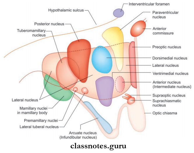
Hypothalamic Nuclei: The hypothalamus consists of numerous cell groups known as hypothalamic nuclei which are present in different regions as follows:
- Preoptic Region: Preoptic nucleus
- Supraoptic Region: Supraoptic nucleus, anterior nucleus, paraventricular nucleus
- Tuberal Region: Infundibular nucleus, ventromedial and dorsomedial nuclei
- Mammillary Region: Posterior nucleus and mammillary nuclei.
Connections Of Hypothalamus
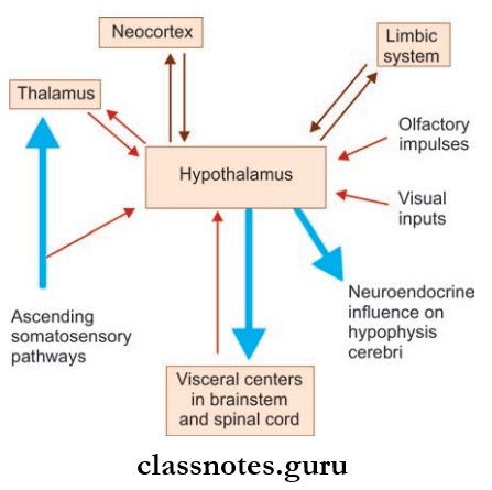
The Main Connections Of The Hypothalamus Are As Below
- Hypothalamic Hypophyseal Tract
- Formed by the axons of supraoptic and paraventricular nuclei which run in the pituitary stalk to reach the posterior pituitary
- It transports vasopressin and oxytocin produced in the supraoptic nucleus and paraventricular nucleus to the posterior pituitary
- Though Hypothalamo Hypophyseal Portal System
- The axons of tuberal nuclei reach median eminence to deliver their neurosecretory material to the hypothalamohypophyseal portal system of blood vessels
- The secretions control the secretion of hormones from the anterior pituitary.
- Mammillothalamic Tract: It connects the mammillary body to the anterior nucleus of the thalamus
- Hypothalamospinal Tract: Formed by synapse of long axons from the thalamus with the preganglionic sympathetic cells in lateral horns of thoracic and upper 2 lumbar spinal segments and with the preganglionic parasympathetic cells in lateral horns of S2, S3, and S4 spinal segments.
Hypothalamus Functions: The various functions of hypothalamus include the following
- Autonomic control
- Endocrine control
- Neurosecretion
- Regulation of water and food intake
- Emotional expression
- Sexual behavior and reproduction
- Temperature regulation
- Regulation of circadian rhythm.
Question 6. Write a short note on basal ganglia.
Answer:
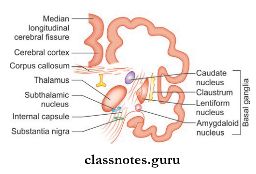
Basal Ganglia
They are large subcortical masses of grey matter present inside the white matter in the basal part of the cerebral hemisphere.
- Anatomically It Includes:
- Corpus striatum
- Claustrum
- Amygdaloid body.
- Functionally It Also Includes:
- Substantia nigra
- Red nucleus
- Subthalamus.
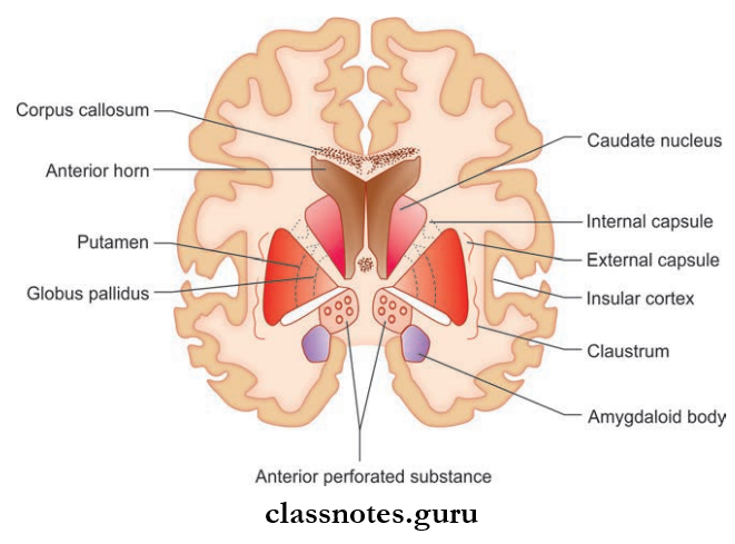
Corpus Striatum
- The Corpus Striatum is located lateral to the thalamus
- Corpus Striatum is divided into a caudate nucleus and lentiform nucleus by an internal capsule and the fibers of the anterior end are connected to the internal capsule giving a striated appearance hence the name corpus striatum.
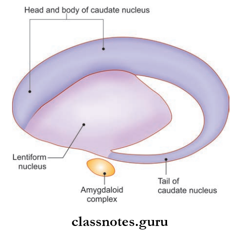
Caudate Nucleus
- The Caudate Nucleus is a large comma-shaped mass of grey matter that surrounds the thalamus.
- Caudate Nucleus convexity projects into the cavity of the lateral ventricle
Caudate Nucleus Is Divided Into:
- Head: The rounded anterior part in front of the interventricular foramen.
- Body: It is actually the tapered portion of the continuation of the head
- Tail which merges with the amygdaloid body.
Lentiform Nucleus
- The Lentiform Nucleus is a large lens-shaped mass of grey matter beneath the insula lateral to the internal capsule.
- The Lentiform Nucleus Is Divided Into:
- Putamen: The outer dark part
- Globus Pallidus: The inner lighter part
- Putamen is similar to the caudate nucleus and contains densely packed small cells.
- Globus pallidus consists of large motor cells.
- The globus pallidus is termed paleostriatum and the caudate nucleus and putamen is termed the neostriatum.
Claustrum: It is a thin saucer-shaped mass of grey matter situated in between putamen and insula.
Amygdaloid Body
- The Amygdaloid Body is an almond-shaped mass of grey matter in the temporal lobe present near the inferior horn of the lateral ventricle
- The fibers from the amygdaloid body form the stria terminalis which form the main efferent tract of the amygdaloid body
- The striae terminalis follow the inner curve of the caudate nucleus and terminate into a septal area, anterior perforated substance, and anterior hypothalamic nuclei.
Substantia Nigra
- Substantia Nigra is a curved pigmented band of grey matter located between crus cerebri and tegmentum
- Substantia Nigra consists of deeply pigmented nerve cells that contain melanin and iron.
Red Nucleus
- The Red Nucleus is a cigar-shaped mass of grey matter
- Red Nucleus is located in the tegmentum ventral to 3rd nerve nucleus and dorsomedial to substantia nigra
- The Red Nucleus is an important nucleus in the extrapyramidal system.
Subthalamus
- The subthalamus is a small nucleus in the ventral part of the thalamus
- The subthalamusis located caudal to the lateral half of the thalamus and inferior to the globus pallidus
- The subthalamus is separated from the thalamus by a small nucleus known as the zona insert.
Basal Gangila Functions
- Basal Ganglia is mainly involved in organizing and coordinating motor movements
- Basal Ganglia decreases muscle tone and inhibits unwanted muscular activity
- Basal Ganglia also controls reflex muscular activity.
Basal Ganglia Applied: Lesions of basal ganglia can result in parkinsonism, chorea, athetosis, and ballismus.
Question 7. Write a note on the corpus callosum.
Answer:
Corpus Callosum
Corpus Callosum is the largest commissure of the brain and consists of about 100 million fiers.
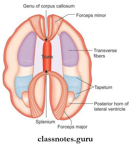
Corpus Callosum Parts: It is divided into the following four parts:
- Genu
- The thick curved anterior extremity which lies 4 cm behind the frontal pole
- The fibers of genu curve forward on either side of the frontal lobes to form a fork-like structure called forceps minor.
- Rostrum: It is the thin prolongation of the genus which joins the lamina terminalis.
- Trunk: It is the main part of the corpus callosum between genu and splenium.
- Splenium
- It is the massive posterior extremity of the corpus callosum which lies about 6 cm in front of the occipital lobe
- Its fibers connect the parietal, temporal, and occipital lobes of two cerebral hemispheres
- The fibers connecting the occipital lobe curve backward on either side above the calcarine sulcus and form a large fork-like structure known as forceps major.
Corpus Callosum External Features
- It forms a massive arched interhemispheric bridge and connects the medial surfaces of two cerebral hemispheres
- In the sagittal section of the cerebrum, it is seen as a C-shaped mass of white fibers forming the roof of the lateral ventricle
- The inferior aspect of the corpus callosum is concave and is attached to the convex superior aspect of the fornix by the septum pellucidum.
Corpus Callosum Functions: It is mainly responsible for bilateral responses and learning processes which are achieved through interhemispheric transfer of information.
Question 8. Write a note on the internal capsule.
Answer:
Internal Capsule
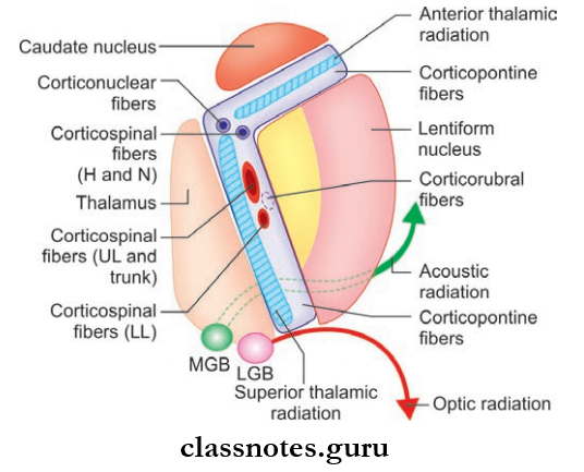
Abbreviations: LL = Lower limb; UL = Upper limb; MGB = Medial geniculate body; LGB = Lateral geniculate body
- It is a compact bundle of projection fibers between the thalamus and caudate nucleus on the medial side and lentiform nucleus on the lateral side
- It consists of ascending and descending fibers that connect the spinal cord and brain stem to the cerebral cortex
- The afferent fibers pass from the thalamus to the cerebral cortex and the efferent fibers pass from the cerebral cortex to the cerebral peduncle of the midbrain.
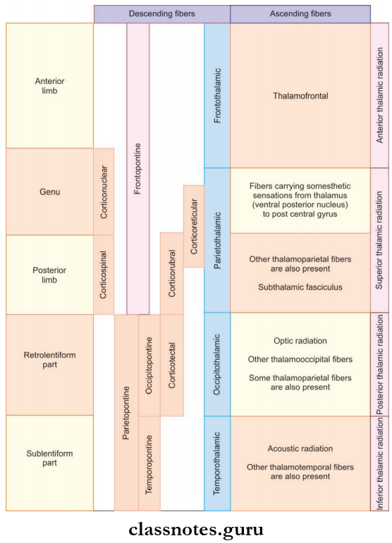
Internal Capsule Parts
- In the horizontal section of the cerebral hemisphere, the internal capsule appears as a V–V-shaped bundle of white fiers with concavity directed laterally
- It Is Divided Into The Following Parts:
- Anterior Limb
- Anterior Limb is present between the head of the caudate nucleus and the anterior part of the lentiform nucleus.
- The constituent motor fiers include corticopontine fibers and sensory includes anterior thalamic radiation.
- Genu
- Genu is the bend between anterior and posterior limbs and the concavity faces laterally.
- Genu consists of corticopontine fibers, corticonuclear and corticospinal fibers as motor fibers, and superior thalamic radiation as sensory fibers.
- Posterior Limb
- Posterior Limb is present between the thalamus and the posterior part of the lentiform nucleus.
- Posterior Limb consists of corticopontine fibers, corticospinal and corticofugal fibers as constituent motor fibers, and superior thalamic radiation as sensory fibers
- Retrolentiform Part
- The Retrolentiform Part is present behind the lentiform nucleus
- The Retrolentiform Part consists of corticopontine fibers and posterior thalamic radiation as motor and sensory fields respectively
- Sublentiform Part
- Sublentiform Part is present below the lentiform nucleus
- The Sublentiform Part consists of corticopontine fibers as motor fibers and inferior thalamic radiation as sensory fibers.
Internal Capsule Functions: The sensory and motor fibers of the internal capsule are responsible for sensory and motor innervation of the opposite side of the body.
Internal Capsule Applied: Small lesions in the internal capsule can produce widespread paralytic effects and sensory loss in the opposite half of the body due to the presence of dense motor and sensory fibers.
Cerebrum Multiple Choice Question And Answers
Question 1. Which of the following structures represents the submerged portion of the cerebral cortex?
- Frontoparietal operculum
- Insula
- Hippocampus
- Temporal operculum
Answer: 2. Insula
Question 2. The paracentral lobule is located on:
- The medial surface of the cerebral hemisphere
- The superolateral surface of the cerebral hemisphere
- The tentorial surface of the cerebral hemisphere
- The orbital surface of the cerebral hemisphere
Answer: 1. Medial surface of the cerebral hemisphere
Question 3. All of the following are examples of commissural fibers except:
- Corpus callosum
- Interthalamic adhesion
- Anterior commissure
- Habenular commissure
Answer: 2. Interthalamic adhesion
Question 4. Which of the following structures consists of both projection and commissural fibers?
- Internal capsule
- Pyramid
- Cerebral fornix
- Crus cerebri
Answer: 3. Cerebral fornix
Question 5. The area between the parieto-occipital and calcarine sulcus on the medial surfaces of the cerebral hemisphere is known as:
- Paracentral lobule
- Cuneus
- Precuneus
- Isthmus
Answer: 2. Cuneus