
Cerebellum And Fourth Ventricle Question And Answers
Question 1. Write a note on the cerebellum.
Answer:
Cerebellum
The cerebellum is the largest part of the hindbrain.
Cerebellum Dimensions, Location, And Extent
- It weighs about 150 g
- It is located in the posterior cranial fossa below tentorium cerebelli, posterior to pons and medulla
- It is separated from the pons and medulla by the cavity of the fourth ventricle
- Its surface consists of numerous slit-like sulci called fissures and parallel folds in between them called folia
- It consists of two hemispheres united by vermis
- Each hemisphere is connected to three parts of the brainstem by three pairs of large fier tracts called cerebellar peduncles.
Cerebellum External Features
- Cerebellum External Features Parts
- It consists of two large hemispheres and a narrow median worm-like portion called the vermis
- The superior and inferior aspect of vermis is termed as superior and inferior vermis, respectively.
- Cerebellum External Features Surfaces
- It consists of superior and inferior surfaces
- The superior surface is convex and the two hemispheres are continuous with each other on this surface
- The inferior surface has a deep median notch called vallecula which separates two cerebellar hemispheres.
- Cerebellum External Features Notches
- The anterior cerebellar notch is a wide shallow notch that is present on the anterior aspect and accommodates pons and medulla
- The posterior cerebellar notch is a deep and narrow notch that lodges the falx cerebelli.
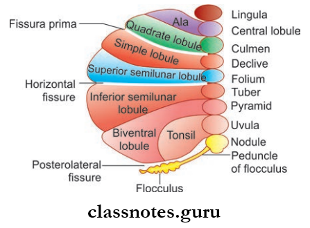
- Cerebellum External Features Fissures: Consist of three well-marked fissures
- The horizontal fissure marks the junction between the superior and inferior surfaces of the cerebellum. It runs along the lateral and posterior margins of the cerebellum
- The posterolateral fissure separates the flcculonodular lobe from the rest of the cerebellum. It lies on the inferior surface of the cerebellum
- The V-shaped fissure prima divides the cerebellum to anterior and posterior lobes and cuts the superior vermis at the junction of anterior two-thirds and posterior one-third.
Cerebellum Subdivisions
- Anatomical Subdivision: Anatomically the cerebellum is divided into
- Anterior lobe
- Posterior lobe
- Flocculonodular lobe.
- Morphological Division: Based on phylogenetic and functional criteria cerebellum is divided into
- Archicerebellum
- Paleocerebellum
- Neocerebellum.
Cerebellum Functions: Basic functions include
- Maintenance of equilibrium
- Regulation of muscle tone
- Coordination of somatic motor activities.
Cerebellum Internal Structure
- It is made up of a thin surface layer of grey matter called the cortex and the central core of the white matter.
- Within the central core masses of grey matter are embedded and they are known as intracerebellar nuclei.
- The Intracerebellar Nuclei Include The Following:
- The dentate nucleus
- The emboliform nucleus
- The globose nucleus
- The fastigial nucleus.
- The cerebellar cortex is folded so that the surface presents with a series of transverse fissures and intervening narrow leaf-like bands called folia
- The central core is arranged in the form of a branching pattern of the tree.
Cerebellum Blood Supply: Supplied by the following
- Superior cerebellar artery which supplies the superior surface
- The anterior inferior cerebellar artery which supplies the anterior part of the inferior surface
- Posterior inferior cerebellar artery which supplies the posterior part of inferior surface.
Cerebellum Applied
- The cerebellar lesions due to trauma, stroke tumors, etc. produce signs and symptoms which are collectively known as cerebellar syndrome.
- It includes generalized muscular hypotonia, intention tremors, adiadochokinesis, dysarthria, nystagmus, and generalized swaying.
Question 2. Write a note on cerebellar peduncles.
Answer:
Cerebellar Peduncles
- The efferent and afferent fibers of the cerebellum are grouped into large bundles of fibers and are known as cerebellar peduncles
- These are three in number, superior, inferior, and middle cerebellar peduncles.
Superior Cerebellar Peduncle
- The superior cerebellar peduncle connects the cerebellum to the midbrain
- The Cerebellar Peduncle emerges from the anterior cerebellar notch and forms the lateral boundary of the upper half of the fourth ventricle
- Cerebellar Peduncle consists only of efferent fibers from the dentate nucleus to the red nucleus, thalamus, and cerebral cortex of opposite sides.
Middle Cerebellar Peduncle
- Cerebellar Peduncle connects the cerebellum to the pons
- The Cerebellar Peduncle is the largest among the three
- The Cerebellar Peduncle is formed at the posterolateral margin of the pons
- The Cerebellar Peduncle consists only of afferents from pontine nuclei of the opposite side.
Inferior Cerebellar Peduncle
- Cerebellar Peduncle connects the cerebellum to the medulla
- Cerebellar Peduncle is formed at the posterolateral aspect of the medulla
- The Cerebellar Peduncle consists of both afferent and efferent fibers
- Cerebellar Peduncle consists of main afferents to the cerebellum from the spinal cord, olivary nucleus, reticular formation of the medulla and vestibular nuclei, and nerve
- The Cerebellar Peduncle also contains a few efferents from the cerebellum to the medulla, i.e. vestibular nuclei and reticular formation.
Question 3. Write a short note on the interpeduncular fossa.
Answer:
Interpeduncular Fossa
Rhomboidal space bounded by crus cerebri of cerebrum laterally, anteriorly by optic chiasma, and posteriorly by pons.
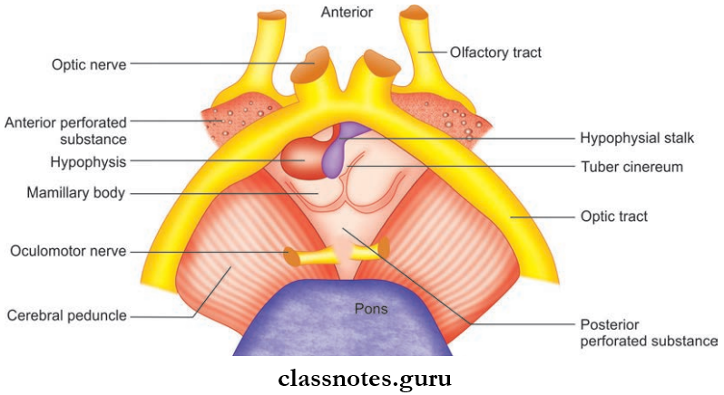
Interpeduncular Fossa Contents
- Two spherical bodies called mammillary bodies
- Raised area of grey matter anterior to mammillary bodies called tuber cinereum
- Infundibulum which connects the pituitary to tuber cinereum
- Posterior perforated substance, a layer of grey matter present in the angle between crus cerebri
- Oculomotor nerve.
Question 4. Write a note on the floor of the fourth ventricle.
Answer:
Floor Of Fourth Ventricle
- The Floor Of The Fourth Ventricle is otherwise known as the rhomboid fossa
- The floor Of the Fourth Ventricle is formed by the posterior surface of the pons and the upper part of the medulla
- The floor Of the Fourth Ventricle is divided into three parts, the upper triangular part bounded by a superior cerebellar peduncle, the lower triangular part bounded by gracile and cuneate tubercles, and the inferior cerebellar peduncles.
Floor Of Fourth Ventricle Features
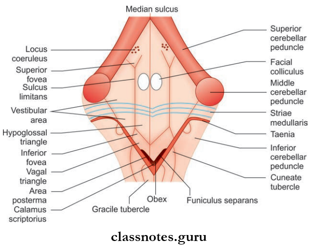
- The floor is divided into symmetrical right and left halves by the median sulcus which extends from the aqueduct of the midbrain above to the central canal below
- The widest part is transversely crossed by striae medullaris, which are white fiers from arcuate nuclei
- On the other side of the median sulcus, there is a longitudinal elevation called medial eminence which is bounded laterally by sulcus limitans
- At a lateral angle, the region lateral to sulcus limitans overlies the vestibular nuclei and is known as the vestibular area
- The upper end of the sulcus limitans widens to form a triangular depression known as the superior fovea. Above the superior fovea, it flattens and present bluish grey area called locus coeruleus
- The lower part of sulcus limitans presents a depression called the inferior fovea
- On either side of the medial eminence at the level of the superior fovea oval-shaped facial colliculus is present
- The sulcus limitans divides the medial eminence into two triangles, the hypoglossal triangle above and the vagal triangle below
- The hypoglossal triangle divided into medial and lateral parts by the nucleus of the hypoglossal nerve
- The vagal triangle overlies nuclei of the vagus, glossopharyngeal, and cranial accessory nerve
- The vagal triangle is crossed by a narrow ridge called funiculus separates and the area between the funiculus separates and the gracile tubercle is known as the area postrema
- The inferolateral margins present two narrow ridges called taenia and both taeniae meet at an inferior angle to form a fold called obex.
Cerebellum And Fourth Ventricle Multiple Choice Question And Answers
Question 1. All of the following are intracerebellar nuclei except:
- Dentate nucleus
- Fastigial nucleus
- Globose nucleus
- Red nucleus
Answer: 4. Red nucleus
Question 2. The cerebellar lesion is characterized by all of the following:
- Ataxia
- Muscular hypotonia
- Nystagmus
- Tremors at rest
Answer: 4. Tremors at rest
Question 3. Th neocerebellum is concerned with:
- Maintenance of equilibrium
- Smooth performance of fie voluntary movements
- Regulating muscle tone and posture of trunk
- Regulating muscle tone and posture of limbs
Answer: 2. Smooth performance of fie voluntary movements