Brachialis Anatomy

Arm Introduction

  • Arm extends from the shoulder joint to the elbow joint.
  • Humerus is the one and only bone present in the arm.
  • Apart from the humerus, the arm consists of neurovascular bundles located medially and muscles arranged in compartments.

Arm Question And Answers

Question 1. Briefly explain the fascial compartments of arm.
Answer:

  • The deep fascia covers the arm like a sleeve.
  • Lateral and medial intermuscular septa extend inwards from this fascial sleeve and attach to the humerus dividing the arm into anterior and posterior compartments.
  • The anterior compartment consists of muscles involved in flexion action, hence called as flexor compartment.
  • The posterior compartment consists of the triceps muscle involved in extension action, hence this compartment is called the extensor compartment.
  • Two additional septa, the transverse and anteroposterior septa divide the flexor compartment into three.

Read And Learn More: Upper Limb

Question 2. What are the contents of the anterior compartment of the arm?
Answer:

The Contents Of The Anterior Compartment Of Arm It Consists Of:

  • Three muscles
    • Biceps brachii
    • Coracobrachialis
    • Brachialis
  • Nerves
    • Musculocutaneous nerve
    • Median nerve
    • Radial nerve Nerves passing through the arm
    • Ulnar nerve
  • Brachial artery.

Anatomy of Brachialis Muscle

Question 3. Write a note on the biceps brachii.
Answer:

Biceps Brachii Origin: It has two heads of origin:

  1. Short head from the coracoid process along with coracobrachialis.
  2. Long head from the supraglenoid tubercle of the scapula and glenoid labrum.

Biceps Brachii Insertion

  • The main tendon to a posterior rough part of the radial tuberosity
  • Bicipital aponeurosis to the skin

Biceps Brachii Nerve Supply

  • Musculocutaneous nerve

Biceps Brachii Actions

  • Flexion of the elbow
  • Powerful supinator when the forearm is flexed
  • All screwing actions
  • Short head flexes the arm
  • The long head prevents the upward displacement of the head of the humerus

Biceps Brachii Clinical Anatomy

The biceps reflex is routinely tested in CNS examination to assess the integrity of the musculocutaneous nerve and C5, and C6 spinal segments.

Arm Attachments Of Biceps Brachii

Question 4. Write about the origin, insertion, nerve supply, and actions of coracobrachialis and brachialis.
Answer:

Arm Coracobrachialis And Brachialis

Arm Brachialis And Coracobrachialis

Mnemonic

  • Elbow: muscles that flex it
  • Three Bs Bend the elbow:
    • Brachialis
    • Biceps
    • Brachioradialis.

Question 5. What are the anatomical peculiarities of the insertion of the coracobrachialis muscle?
Answer:

  • The brachial artery comes to the anterior aspect from its medial position in the arm.
  • The median nerve crosses from the lateral to the medial side in front of the brachial artery.
  • Below this level, the circular shaft of the humerus becomes triangular.
  • The basilic vein pierces the deep fascia.
  • The nutrient artery enters the humerus.
  • The medial cutaneous nerve of the arm and forearm pierces the deep fascia.
  • The radial nerve pierces the deep lateral intermuscular septum and goes from the posterior compartment to the anterior compartment.
  • The ulnar nerve pierces the medial intermuscular septum and goes to the posterior compartment.

Brachialis Muscle Function

Question 6. Write a note on the cubital fossa mentioning its boundaries and contents.
Answer:

  • Cubital Fossa is a triangular hollow present in front of the elbow.
  • Cubital Fossa is homologous with the popliteal fossa in the lower limb.

Arm Cubital Fossa

Cubital Fossa Boundaries

  • The Lateral Medial border of the brachioradialis muscle
  • Medial Lateral border of pronator teres muscle
  • Base Directed upwards, represented by an imaginary line joining the front of the lateral and medial epicondyles of the humerus
  • Apex Directed downwards, meeting point of lateral and medial border
  • Roof From superficial to Deep:
    • Skin
    • Superficial fascia containing median cubital vein, medial and lateral cutaneous nerves of the forearm
    • Deep fascia with bicipital aponeurosis

Cubital Fossa Content: From the medial to the lateral side are:

  • Median nerve
  • Brachial artery
  • Tendon of biceps brachii.

Cubital Fossa Clinical Anatomy

  • A medial cubital vein in the cubital fossa is the vein of choice for intravenous injection.
  • When blood pressure is recorded, the BP cuff is wired around the arm to exert external pressure over the brachial artery.
  • Supracondylar fracture of the humerus can lead to injury to the brachial artery and median nerve. In such cases, knowledge of the cubital fossa is essential to reduce the fracture.

Question 7. What are the contents of the posterior compartment of the arm?
Answer:

The Contents Of The Posterior Compartment Of The Arm Its Contents Are:

  • 1 muscle: Triceps brachii
  • 1 nerve: Radial nerve
  • 1 artery: Profunda brachii artery.

Question 8. Write a note on the triceps brachii muscle.
Answer:

Triceps Brachii Muscle Origin It has three heads:

  • Long head from the infra glenoid tubercle of the scapula
  • The lateral head from the oblique ridge present on the upper part of the posterior surface of the humerus
  • Medial head from the large triangular area on the posterior surface of the humerus below the radial groove and the medial and lateral intermuscular septa

Arm Attachments Of Triceps Muscle

Triceps Brachii Muscle Insertion

  • The posterior part of the superior surface of the olecranon process

Triceps Brachii Muscle Nerve Supply

  • Radial nerve

Triceps Brachii Muscle Actions

  • Powerful extensor of the elbow

Triceps Brachii Muscle Clinical Anatomy

If the radial nerve is injured in the radial groove, an extension of the elbow and triceps reflex is not lost because the triceps muscle is innervated by the radial nerve in the axilla.

Brachialis Muscle Origin and Insertion

Question 9. Cross-section at the level of the middle of the arm.
Answer:

Arm Cross-section Of Arm At M idarm Level To Show The Contents Of Flexor And Extensor Compartment

Arm Multiple Choice Questions

Question 1. Which is the nerve of the anterior compartment of the arm?

  1. Axillary nerve
  2. Radial nerve
  3. Ulnar nerve
  4. Musculocutaneous nerve

Answer: 4. Musculocutaneous nerve

Question 2. Which muscle does not belong to the anterior compartment of the arm?

  1. Brachialis
  2. Brachioradialis
  3. Biceps brachii
  4. Coracobrachialis

Answer: 2. Brachioradialis

Question 3. Which among these are considered as powerful supinator?

  1. Biceps brachii
  2. Triceps brachii
  3. Coracobrachialis
  4. Deltoid muscle

Answer: 1. Biceps brachii

Brachialis Muscle Nerve Supply

Question 4. Where is the triceps brachii inserted?

  1. Medial epicondyle of humerus
  2. Lateral epicondyle of humerus
  3. Olecranon process
  4. Radial tuberosity

Answer: 3. Olecranon process

Question 5. The bicipital aponeurosis is inserted into where?

  1. Radial tuberosity
  2. Skin
  3. Ulnar tuberosity
  4. Medial lateral epicondyle of humerus

Answer: 2. Skin


Muscles of the Anterior Forearm Anatomy

Antebrachium Or Forearm Question And Answers Introduction

The forearm extends from the elbow joint to the wrist joint.

The forearm has two bones, a radius and an ulna, with their interosseous membrane.

Muscles, arteries, veins, and nerves are present in both the front and back of the forearm.

Muscles of Anterior Forearm – Full Anatomy Notes

Antebrachium Or Forearm Question And Answers

Question 1. Briefly explain the fascial compartments of the forearm.
Answer:

  • The deep fascia of the forearm covers it like a sleeve.
  • Intermuscular septa extend from this fascial sleeve, and a few of them are attached to the bones, dividing the forearm into anterior and posterior compartments.
  • Each compartment has its muscles, nerves, and blood supply.
  • The anterior compartment muscles are involved in flexion movement; hence, this compartment is called the flexor compartment.
  • Posterior compartment muscles are involved in extension movement; hence, the compartment is called the extensor compartment.
  • Near the wrist, deep fascia thickens in both the flexor and extensor compartments and is called the flexor and extensor retinaculum, respectively.

Read And Learn More: Upper Limb

Question 2. List the contents of the front forearm.
Answer:

Contents Of Front Forearm

  • 8 Muscles Arranged As:
    • 5 superficial muscles
    • 3 deep muscles.
  • 2 Arteries
    • Radial artery
    • Ulnar artery.
  • 3 Nerves
    • Medial nerve
    • Radial nerve
    • Ulnar nerve.

Flexor Muscles of the Anterior Forearm

Question 3. How are the muscles of the anterior compartment of the arm arranged? Enumerate them separately and write the origin, insertion, nerve supply, and actions of them.
Answer:

The anterior compartment of the arm has 8 muscles arranged in superficial and deep layers.

5 Superficial Muscles

  • Pronator teres
  • Flexor carpi radialis
  • Palmaris longus
  • Flexor digitorum superfiialis
  • Flexor carpi ulnaris.

All the superficial muscles take their common origin from the front of the medial epicondyle of the humerus. This is called the common flexor origin.

Muscles of the Anterior Forearm Anatomy

Mnemonic

  • Elbow: Which side has a common flexor origin?
  • FM (as in FM Radio): Flexor Medialis, so the common flexor origin is on the medial side.

Antebrachium Or Forearm Four Superfiial Muscles In The Anterior Compartment Of Forearm Arising From Common Fix Or Origin On Medial Epicondyle

Three Deep Muscles

  1. Flexor digitorum profundus
  2. Flexor pollicis longus
  3. Flexor carpi ulnaris.
  • Flexor digitorum profundus is a hybrid muscle supplied by both the ulnar nerve and the anterior interosseous nerve (a branch of the median nerve).
  • It is the most powerful and bulky muscle of the forearm contributing to the larger portion of the gripping power of hand.

Antebrachium Or Forearm Muscles Of Anterior Comaprtment Of Arm Arranged

Anterior Forearm Muscle Function

Antebrachium Or Forearm Deeper Contents Of Anterior Compartments Of Forearm After Reflection Of Superficial Muscles

Question 4. What are the contents of the back of the forearm?
Answer:

This compartment is also known as the extensor compartment as the muscles of this compartment extend the wrist joint.

Contents Of Back Of The Forearm

  • Muscles: 12 muscles arranged as 7 superficial and 5 deep muscles
  • Nerves: Posterior interosseous nerve
  • Artery: Posterior and anterior interosseous arteries.

Median and Ulnar Nerve in Anterior Forearm Muscles

Question 5. How are the muscles of the posterior compartment of the arm arranged? Enumerate them separately and write the origin, insertion, nerve supply, and actions of them.
Answer:

The posterior compartment of the arm has 12 muscles arranged in superficial and deep layers.

Seven Superficial Muscles

  • From lateral to medial.
  • They are:
    • Brachioradialis
    • Extensor carpi radialis longus
    • Extensor carpi radialis brevis
    • Extensor digitorum
    • Extensor digit minimi
    • Extensor carpi ulnaris
    • Anconeus
  • All seven muscles cross the elbow joint.
  • They have having common extensor origin from the lateral epicondyle of the humerus.
  • Dorsal digital expansion is a small triangular aponeurosis related to each tendon of the extensor digitorum.
  • It covers the metacarpophalangeal joint and dorsum of the proximal phalanx.
  • A number of muscles are inserted into dorsal digital expansion.

Antebrachium Or Forearm Contents Of Posterior Compartment Of Forearm

Antebrachium Or Forearm DFetails Of Deep Muscles Of The Externsor Compartment Of Forearm

Five Deep Muscles

  1. From above downwards, they are:
  2. Supinator
  3. Abductor pollicis longus
  4. Extensor pollicis brevis
  5. Extensor pollicis longus
  6. Extensor indicis
  • In contrast to superficial muscles, none of the deep muscles crosses the elbow joint.

Antebrachium Or Forearm Muscles Of Posterior Compartment Of Arm Arranged

Antebrachium Or Forearm Multiple Choice Questions

Question 1. Which of the following pierces the interosseous membrane?

  1. Posterior interosseous artery
  2. Anterior interosseous artery
  3. Common interosseous artery
  4. All the above

Answer: 2. Anterior interosseous artery

Question 2. Which one is not a superficial group muscle of the anterior compartment of forearm?

  1. Pronator teres
  2. Flexor carpi radialis
  3. Palmaris longus
  4. Pronator quadratus

Answer: 4. Pronator quadratus

Question 3. Which among these is a hybrid muscle?

  1. Flexor digitorum profundus
  2. Pronator teres
  3. Flexor carpi radialis
  4. Palmaris longus

Answer: 1. Flexor digitorum profundus

Flexor Muscles of the Anterior Forearm

Question 4. The anterior interosseous nerve is a branch of:

  1. Musculocutaneous nerve
  2. Median nerve
  3. Radial nerve
  4. Ulnar nerve

Answer: 2. Median nerve

Question 5. Allen’s test is performed to ascertain the proper functioning of:

  1. Median nerve
  2. Ulnar artery
  3. Radial artery
  4. Flexor digitorum profundus muscle

Answer: 3. Radial artery

Wrist And Hand Anatomy

Wrist And Hand Introduction

The hand is man’s physical asset.

A large area in the motor cortex of the brain is represented by the hand, indicating the fine and complex movements made by the hand.

Wrist And Hand Question And Answers

Question 1. What are the peculiarities of the skin and superficial fascia of the palmar aspect of the hand? What are the three modifications of the deep fascia in this region?
Answer:

Peculiarities Of Skin

  • Peculiarities Of Skin are thick and tough.
  • Peculiarities Of Skin are rich in sweat and sebaceous glands.
  • The palm creases represent the area of the skin attached to the deep fascia of the hand.
  • Skin ridges are provided for gripping, and those seen on the finger pads are called ‘fingerprints’.

Read And Learn More: Upper Limb

Peculiarities of Superfiial Fascia

  • Made of dense fibrous bands that bind skin to the deep fascia.
  • Contains subcutaneous fat and the Palmaris brevis muscle.

Peculiarities of Deep Fascia

  • The deep fascia is modified:
    • The wrist forms the flexor retinaculum.
    • In the palm to form the palmar aponeurosis.
    • The fingers form a fibrous flexor sheath.

Wrist and Hand Anatomy Notes PDF

Question 2. Write a note on the flexor retinaculum.
Answer:

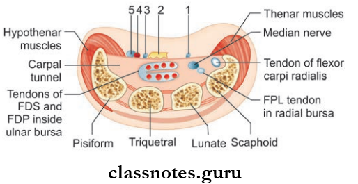
  • Flexor Retinaculum is a strong fibrous band formed by the thickening of deep fascia in front of the carpus or anatomical wrist.
  • Flexor Retinaculum is rectangular in shape and has four borders and two surfaces.

Flexor retinaculum Attachments

  • Medial to:
    • Pisiform bone
    • Hook of hamate

Wrist And Hand Attachments And Relations Of Flexor Rectinaculum

  1. Palmar cutaneous branch of the median nerve;
  2. Tendon of palmaris longus;
  3. Palmar cutaneous branch of the ulnar nerve;
  4. Ulnar artery;
  5. Ulnar nerve
  • Lateral to:
    • Tubercle of scaphoid
    • Crest of the trapezium
    • On either side, it gives off a slip.
  • Lateral Slip:
  • Attached to the medial lip of the groove of the trapezium.
  • The slip forms an osseofascial tunnel for the passage of the tendon of the flexor carpi radialis.
  • Medial Slip:
    • Attached to the pisiform bone.
    • Ulnar nerve and vessels pass deep to this slip.

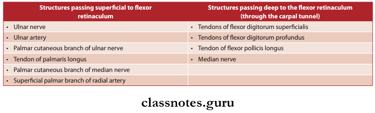
Flexor Retinaculum Relations

Wrist And Hand Flexor Retinaculum

Flxor Retinaculum Function

  • Stabilizes the flexor tendons for smooth action of muscles.

Flexor Retinaculum Clinical Anatomy

  • The median nerve can get compressed in the carpal tunnel, also known as carpal tunnel syndrome.

Wrist and Hand Joints Anatomy

Question 3. Write a short note on the palmar aponeurosis.
Answer:

It is a well-defined triangular modification of the deep fascia in the palm.

Wrist And Hand Palmar Aponeurosis And Its Attachments

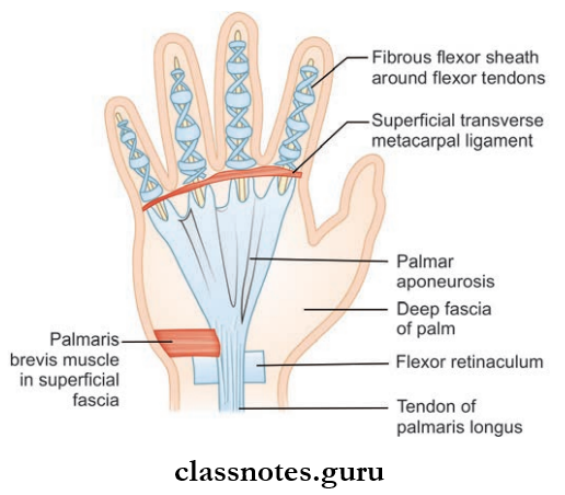
Palmar Aponeurosis Boundaries

  • Apex:
    • Directed proximally towards the wrist.
    • It blends with the flexor retinaculum.
  • Base:
    • Directed distally towards the root of the figures.
    • The base is divided into four longitudinal slips, one each for the medial 4 figures.
    • The longitudinal slip again splits into two slips, which blend with the fibrous sheath of corresponding fingers.
  • Medial Border:
    • It is continuous with the deep fascia covering the hypothenar muscles.
  • Lateral Border:
    • It is continuous with the deep fascia covering the thenar muscles.

Palmar aponeurosis Functions

  • Helps to improve the grip of the hand by fixing the skin.
  • Stabilizes and protects the underlying structures.

Question 4. Write a short note on the fibrous flexor sheath of the fingers.
Answer:

Deep fascia lying over the anterior aspect of the digits thickens to form a fibrous flexor sheath.

Firous Flexor Sheath Of Fingers Extend And Attachments

  • Proximally: Continuous with palmar aponeurosis.
  • Distally: Attached to the distal phalanx. This forms an osteofascial tunnel through which the flexor tendon passes.

Firous Flexor Sheath of the fingers Function

  • It holds the flexor tendon during the flexion of the fingers.

Hand Anatomy Viva Questions and Answers

Question 5. What are the three main synovial sheaths of flexor tendons? Describe each of them.
Answer:

There are mainly 3 synovial sheaths that enclose the tendons of the flexor muscles of the hand.

Wrist And Hand Synovial Sheaths Around Flexor Tendons In Palm

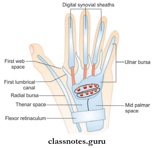
  1. Common flexor synovial sheath/ulnar bursa
    • It encloses the long tendons of both the flexor digitorum superficial and flexor digitorum profundus as they pass through the carpal tunnel.
    • It has two layers: the parietal layer lines the walls of the carpal tunnel, and the visceral layer lines the tendons.
    • Extend: Upwards up to 5–7 cm into the forearm and downwards into the palm up to the middle of the shaft of the metacarpal bones.
    • The lower medial end is continuous with the digital synovial sheath of the little finger.
  2. Radial bursa
    • It encloses the tendons of flxor pollicis longus.
    • Extend
      • Proximally, It coextends with the ulnar bursa
      • Distally: Up to the distal phalanx of the thumb
    • It joins with the digital synovial sheath of the thumb.
  3. Digital synovial sheath
    • It encloses the flexor tendons in the fingers and lines the fibrous flexor sheaths.
    • The digital synovial sheath of the little finger is continuous with the ulnar bursa, and the digital synovial sheath of the thumb is continuous with the radial bursa.
    • But the digital synovial sheaths of the index, middle, and ring fingers are independent.

Functions Of The Synovial Sheath

  • By enclosing the tendons, the synovial sheath reduces friction while the muscle acts.

Clinical Anatomy

  • Any penetrating injury caused to the digital synovial sheath can result in tenosynovitis.
  • Here, the infection causes distension of the sheath by pus and produces pain.
  • If the digital synovial sheath of the thumb or little finger is involved, the infection can spread to the radial and ulnar bursa easily due to the continuity.
  • It is more dangerous and, if the proximal end of the bursa ruptures, the infection can spread up to the space of the parona. (A fascial space) on the forearm.

Question 6. Classify and list out the intrinsic muscles of the hand. Write about their nerve supply and actions.
Answer:

  • Intrinsic Muscles are short muscles having origin and insertion within the hand.
  • Intrinsic Muscles are responsible for skilled movement and gripping of the hand.
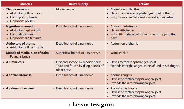
  • Intrinsic Muscles can be classified as:
  1. Thenar Muscles
    • Abductor pollicis brevis
    • Flexor pollicis brevis
    • Opponents pollicis
  2. Hypothenar Muscles
    • Abductor digiti minimi
    • Flexor digiti minimi
    • Opponents digit minimi
  3. Adductors Of Thumb
    • Adductor pollicis muscle
  4. Muscle Of The Medial Side Of The Palm
    • Palmaris brevis
  5. 4 Lubricants
    • Numbered 1–4 from lateral to medial side
  6. 4 Dorsal And 4 Palmar Interossei
    • Numbered 1–4 from lateral to medial side

Wrist And Hand Anatomical Intrinsic Muscles Of Hand

Mnemonics

Interossei Muscles: Actions of dorsal vs Palmar in hand

  • PAd And DAb:
    • The Palmar Adduct and the Dorsal Abduct
    • Use your hand to dab with a pad
  • Intrinsic Muscles Of The Hand (Palmar Surface) ‘A OF A OF A’:
    • Thenar, lateral to medial:

Anatomy of the Wrist and Hand

Question 7. Briefly explain the spaces of the hand.
Answer:

Wrist And Hand Cross-section Of Hand To Show Fascial Compartments And Fascial Spaces

  1. Abductor pollicis longus
  2. Opponents pollicis
  3. Flexor pollicis brevis
  4. Adductor pollicis
  5. Hypothenar, lateral to medial
  6. Opponents digiti minimi
  7. Flexor digiti minimi
  8. Abductor digiti minimi.
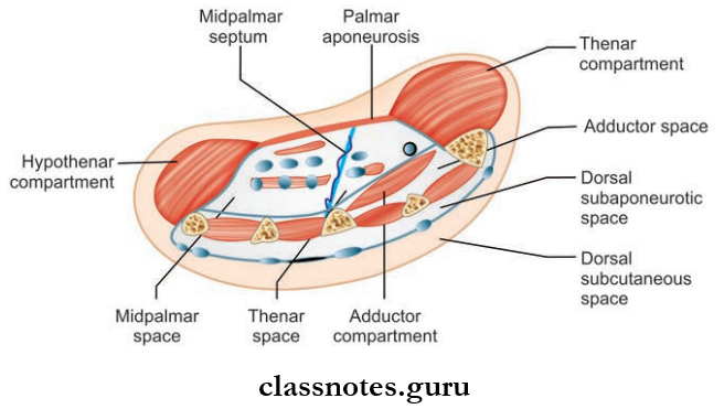
  • The fascia and fascial septa of the hand divide the hand into several fascial spaces.
  • They are potent spaces filled with loose connective tissue, but become obvious when they are infected.
  • Knowledge of these spaces is of great surgical importance as these spaces can get infected and filled with pus, causing distention and pain.
  • These spaces are:
    • Palmar Spaces
      • Midpalmar spaces
      • Thenar space
      • Pulp spaces of fingers
    • Dorsal Spaces
      • Dorsal subcutaneous space
      • Dorsal subaponeurotic space
      • The forearm space of the para

Question 8. Write a short note on the mid-palmar space of the hand.
Answer:

Mid-Palmar Space Of the Hand is a triangular-shaped space located under the medial half of the hollow of the palm.

Mid-Palmar Space Of the Hand Boundaries

  1. Anterior
    • From superficial to deep:
      • Palmar aponeurosis
      • Superfiial palmar arch
      • Digital nerve and vessels supplying medial 3-and-a-half fingers
      • Ulnar bursa with its tendons
      • 2nd, 3rd, 4th lumbricals
  2. Posterior
    • Fascia covering interossei and medial three metacarpals
  3. Lateral
    • Intermediate palmar septum
  4. Medial
    • Medial palmar septum
  5. Proximal
    • Midpalmar space is continuous with forearm space of para
  6. Distal
    • Continuous with medial three web spaces through the medial 3 lumbrical canals

Wrist Joint Classification and Structure

Mid-Palmar Space Of the Hand Clinical Anatomy

  • The major source of infection in the mid-palmar space is the ulnar bursa.
  • When infected, the pus from this space is drained by an incision in the medialmost two web spaces.

Question 9. Write a short note on the thenar space of the hand.
Answer:

Thenar Space Of The Hand is a triangular space located beneath the outer half of the hollow of the palm.

Thenar Space Of The Hand Boundaries

  1. Anterior
    • From superficial to deep, they are:
      • Palmar aponeurosis
      • Digital nerve and vessels of the lateral 1-and-a-half fingers
      • Radial bursa enclosing the tendon of flexor pollicis longus
      • Flexor tendons of index figers
      • First lumbrical
  2. Posterior
    • Fascia covering the transverse head of adductor pollicis
  3. Lateral
    • Lateral palmar septum
  4. Medial
    • Intermediate palmar septum
  5. Proximal
    • Only limited space since the anterior and posterior walls fuse in the carpal tunnel
  6. Distal
    • Communicates with the first web space through the first lumbrical canal

Thenar Space Of The Hand Clinical Anatomy

  • Infections from the radial bursa or synovial sheath of the index finger can reach the thenar space.
  • In such cases, pus is drained by an incision in the first web space.

Question 10. Write a short note on the pulp spaces of your fingers.
Answer:

  • They are subcutaneous spaces located on the palmar aspect of the tip of the fingers.
  • The pulp space is filled with subcutaneous fatty tissue.

Pulp Spaces Of Figures Boundaries

  • Superfiial: Skin and superfiial fascia.
  • Deeply: Distal 2/3rd of the distal phalanx.

Pulp Spaces Of Fingers Clinical Anatomy

  • It is the most exposed part of the digit and can get easily infected.
  • When infected, the abscess formed in the pulp space is called a whitlow.
  • The pus from the pulp space is drained by making a lateral incision.

Question 11. Briefly explain the dorsum of the hand.
Answer:

Dorsum Of Hand Skin

  • Loose and thin when the hand is relaxed.

Dorsum Of Hand Superficial Fascia

  • Contains:
    • Dorsal venous arch
    • Superficial radial nerve
    • Dorsal cutaneous branch of the ulnar nerve.

Dorsum Of Hand Deep Fascia

  • Modified to form the extensor retinaculum.

Wrist And Hand Attachments And Deep Relations Of Extensor Retinaculum

Question 12. Write a note on the extensor retinaculum.
Answer:

  • The retinaculum is a strong fibrous band formed by the thickening of deep fascia present in the back of the wrist.
  • The retinaculum is directed obliquely downwards and medially.
  • The retinaculum is 2 cm wide vertically.

Extensor Retinaculum Attachments

  • Medially To:
    • Styloid process of the ulna
    • Triquetral bone
    • Pisiform bone
  • Laterally To:
    • The lower part of the anterior border of the radius.
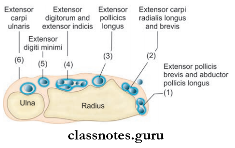
  • Extensor Retinaculum Compartments
    • The retinaculum sends down septa that are attached to the posterior surface of the lower part of the radius.
    • Thus, six osseofascial compartments are formed and each compartment is provided with synovial sheaths.
    • Structures passing through the compartments from the lateral to the medial side are:

Extensor Retinaculum Functions

  • Stabilizes the tendons of extensor muscles for their smooth action.

Question 13. Write a short note on dorsal digital expansion.
Answer:

  • They are triangular aponeuroses formed by the expansion of each tendon of the extensor digitorum muscle.
  • It covers the dorsum of the metacarpophalangeal joint.
  • It fuses anteriorly with a fibrous flexor sheath.
  • The tendons of the lumbrical and interossei are inserted into the expansion.
  • The expansion narrows as the tendons of the interossei and lumbrical converge towards it on the dorsum of the proximal phalanx.
  • From there, it splits into 3 slips.
  • The central slip is inserted into the base of the middle phalanx, and the lateral slips to the base of the terminal phalanx.

Muscles of the Wrist and Hand Anatomy

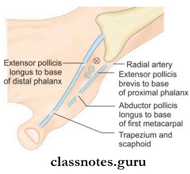
Question 14. What is an anatomical snuff box? What are its boundaries and relations?
Answer:

An anatomical Snuff Box is a triangular depression seen on the lateral side of the dorsum of the hand when the thumb is hyper-extended.

Anatomical Snuff Box Boundaries

  1. Anterolaterally
    • Tendon of the abductor pollicis longus
    • Tendon of extensor pollicis brevis
  2. Posteromedially
    • Tendon of extensor pollicis longus
  3. Floor
    • Scaphoid
    • Trapezium
  4. Roof
    • Skin
    • Superficial fascia
  5. Content
    • Radial artery

Wrist And Hand Anatomical Snuffbox And Its Contents

Structures Crossing The Roof Under The Skin:

  • Cephalic vein
  • Terminal branches of the superficial radial nerve

Anatomical Snuff Box Clinical Anatomy

  • In a scaphoid bone fracture, tenderness in the anatomical snuff box will be present.
  • The cephalic vein can be used to give intravenous fluids at this site.
  • Radial artery pulsations can be felt in the anatomical snuff box.

Wrist And Hand Anatomy

Wrist And Hand Multiple Choice Questions

Question 1. Froment’s test is done to check the integrity of the:

  1. Second palmar interosseous
  2. Second dorsal interosseous
  3. Adductor pollicis
  4. First lumbrical

Answer: 3. Adductor pollicis

Question 2. Which of the following is not a modification of the deep fascia?

  1. Extensor retinaculum
  2. Palmar aponeurosis
  3. Extensor expansion
  4. Fibrous flexor sheath

Answer: 3. Fibrous flexor sheath

Question 3. Hammer thumb deformity is due to the rupture of the tendon of

  1. Flexor pollicis longus
  2. Abductor pollicis longus
  3. Extensor pollicis brevis
  4. Extensor pollicis longus

Answer: 4. Extensor pollicis longus

Anatomy of Hand and Wrist – Medical Students Guide

Question 4. Adduction of the middle finger is brought about by:

  1. Third dorsal interosseous
  2. Third lubricants
  3. Second and third dorsal interossei
  4. Second and third lubricants

Answer: 3. Second and third dorsal interossei

Question 5. What are the four chief bony attachments of the flexor retinaculum?

  1. Hamate, pisiform, trapezium, scaphoid
  2. Hamate, capitate, trapezoid, scaphoid
  3. Lunate, hamate, capitate, scaphoid
  4. Lunate, pisiform, trapezoid, hamate
  5. Trapezium, trapezoid, capitate, hamate

Answer: 1. Hamate, pisiform, trapezium, scaphoid

Nerves Of Upper Limb

Nerves Of The Upper Limb Questions And Answers

Question 1. Outline the nerve supply of the upper limb.
Answer:

  • The brachial plexus (C5, C6, C7, C8, T1) provides nerve supply to the upper limb.
  • The major nerves originating from the brachial plexus are:
    • Axillary nerve
    • Musculocutaneous nerve
    • Radial nerve
    • Median nerve
    • Ulnar nerve

Nerves of Upper Limb Anatomy Notes PDF

Question 2. Write in detail about the brachial plexus and make notes on Erb’s and Klumpke’s paralysis.
Answer:

  • The brachial plexus is the plexus of nerves formed by the anterior (ventral) rami of the last four cervical and first thoracic spinal nerves. (C5, C6, C7, C8, T1)
  • It is divided into five subunits:
    1. Roots
    2. Trunks
    3. Division
    4. Cords
    5. Branches.

Read And Learn More: Upper Limb

Mnemonic: Brachial Plexus Subunits

  • ‘Randy Travis Drinks Cold Beer’:
    1. Roots
    2. Trunks
    3. Divisions
    4. Cords
    5. Branches
  1. Roots
    • They constitute the anterior primary rami of C5 to T1 spinal nerves.
    • They are located in the neck.
  2. Trunks
    • The upper trunk is formed by the union of C5 and C6 roots
    • The middle trunk is formed by C7 alone
    • The lower trunk is formed by the union of C8 and T1 roots
      • They are also located in the neck.
  3. Divisions
    • Each trunk is divided to form anterior and posterior divisions.
    • They are located behind the clavicle
  4. Cords
    • The lateral cord is formed by the union of the anterior division of the upper and middle trunks.
    • The medial cord is a continuation of the anterior division of the lower trunk.
    • The posterior cord is formed by the union of the posterior division of all trunks.
    • Cords are located in the axilla.
  5. Branches of the Brachial Plexus
    • From roots
      • Long thoracic nerve/nerve to serratus anterior
      • Dorsal scapular nerve/nerve to rhomboids
    • From trunks
      • Suprascapular nerve
      • Nerve to the subclavius
    • From cords
      • From the lateral cord
        • Lateral pectoral nerve
        • Lateral root of the median nerve
        • Musculocutaneous nerve
      • From the medial cord
        • Medial pectoral nerve
        • Medial cutaneous nerve of the arm
        • Medial cutaneous nerve of the forearm
        • Medial root of the median nerve
        • Ulnar nerve
      • From the posterior cord
        • Radial nerve
        • Axillary nerve
        • Thracodorsal nerve/nerve to latissimus dorsi
        • Upper subscapular nerve
        • Lower subscapular nerve

Nerves Of Upper Limb Brachial Plexus

Mnemonic: Brachial plexus: Branches of the posterior cord

STAR

  • Subscapular [upper and lower]
  • Thoracodorsal
  • Axillary
  • Radial

Clinical Anatomy: Two types of lesions occurring in the brachial plexus are important.

  1. Erb’s paralysis:
    • There is a point in the brachial plexus where six nerves meet called Erb’s point. These meeting nerves are:
      • 5th and 6th cervical roots
      • The upper trunk is formed by the union of these nerve roots
      • Suprascapular nerve and nerve to subclavius, branching from the upper trunk
    • Any accident which causes an increase in angle between the head and shoulder can cause injury to the upper brachial plexus most commonly at Erb’s point.
    • It results in a specific type of paralysis of the upper limb known as Erb’s paralysis
    • For example, A fall on the shoulder, birth injury, following anesthesia, etc.
    • The clinical features are given in the table.
  2. Klumpke’s paralysis.
    • It is another type of paralysis of the upper limb caused by accidents that increases angle between the trunk and shoulder making injury to lower brachial plexus.
    • For example, Sudden upward pulling of the arm, birth injury, etc.

Nerves Of Upper Limb Erb's Point

Nerves Of Upper Limb Erb's And Klumpke's Paralysis

Upper Limb Nerve Injuries – Essay and MCQs

Nerves Of Upper Limb Claw-hand Deformity Due To Lesion Of Lower Trunk Of Brachial Plexus

Question 3. Explain in detail about the axillary nerve under headings—origin, root value, course, branches, and innervation. Make a note on the injury to the nerve.
Answer:

  • It is called axillary because it runs through the upper part of the axilla.
  • It is called circumflex because it courses around the surgical neck of the humerus.

Axillary Nerve Origin

  • It is a smaller terminal branch of the posterior cord of the brachial plexus.

Axillary Root Value

  • Ventral rami of C5, C6 segments.

Axillary Nerve Course

  • From the posterior cord, it passes backward through the quadrangular intermuscular space.
    • After reaching back, it divides into anterior and posterior divisions below the capsule of the shoulder joint.
    • The posterior division again divides and one part continues as upper lateral cutaneous nerve and the other part goes to supply deltoid and teres minor with a pseudoganglion in it.
    • The anterior division supplies the deltoid muscle and skin over its anteroinferior part (regiment badge).

Brachial Plexus and Nerves – Medical Students Guide

Axillary Nerve Branches and Innervation

  • Trunk Of Axillary Nerve
    • Articular branch to the shoulder joint
  • Anterior Division
    • Muscular branch to the deltoid- cutaneous branch to the skin over deltoid’s anteroinferior part (regimen badge)
  • Posterior Division
    • The cutaneous branch continues as the upper cutaneous nerve of the arm.
    • Muscular branch to the teres minor and posterior part of the deltoid.

Nerves Of Upper Limb Axillary Nerve

Axillary Nerve Clinical Anatomy

  • The axillary nerve can easily get injured in inferior dislocation of the humerus or in injury to the surgical neck of the humerus.
  • The presentation will be:
    • Impaired abduction.
    • Loss of contour of the shoulder due to deltoid muscle wasting.
    • Loss of sensation over the lower half of the deltoid (regimen badge)

Nerves of Arm and Hand – Short Notes

Question 8. Write a note on the cutaneous supply of the upper limb.
Answer:

Nerves Of Upper Limb

  • The upper limb is supplied by C3–T2 spinal segments
  • This is via:
    • Supraclavicular nerves (C3 and C4)
    • Nerves from brachial plexus (C5–T1)
    • Intercostobrachial nerve (T2)
  1. Pectoral Region
    • Above the 2nd rib by supraclavicular nerves (C3, C4)
    • Below the 2nd rib by intercostal nerves (T2–T6).
  2. Axilla
    • Intercostobrachial nerve (T2)
    • Small branches from (T3).
  3. Shoulder
    • Upper half of deltoid by supraclavicular nerves (C3, C4)
    • The lower half of the deltoid by the upper lateral cutaneous nerve of the arm.
  4. Arm
    • Upper medial part by the intercostobrachial nerve (T2)
    • Lower medial part by the medial cutaneous nerve of the arm (T1, T2)
    • Upper lateral half by upper lateral cutaneous nerve of arm
    • Lower lateral part by lower lateral cutaneous nerve of arm (C5, C6)
    • Posterior aspect of the arm by the posterior cutaneous nerve of the arm (C5).
  5. Forearm
    • The medial side of the forearm by the medial cutaneous nerve of the forearm (C8, T1)
    • The lateral side of the forearm by lateral cutaneous nerve of the forearm (C5, C6)
    • Posterior aspect of the forearm by the posterior cutaneous nerve of the forearm (C6, C7, C8).
  6. Hand
    • Palmar Surface
      • Lateral 2/3rd of the palm by a palmar cutaneous branch of the median nerve
      • Medial 1/3rd of the palm is supplied by the palmar cutaneous branch of the ulnar nerve.
    • Dorsal surface
      • Lateral 2/3 rd of the dorsum by superficial terminal branch of radial nerve
      • Medial 1/3 rd by dorsal branch/posterior cutaneous branch of the ulnar nerve.
  7. Digits
    • Palmar Surface
      • Lateral 3½ digits up to distal half of the middle phalanges by digital branches of median nerve
      • Medial 1½ digits up to distal half of the middle phalanges by palmar digital branch of ulnar nerve
    • Dorsal Surface
      • Lateral 3½ digits up to the proximal half of their middle phalanges by digital branches of radial nerve
      • Lateral 3½ digits up to distal half of the middle phalanges by digital branches of median nerve
      • Medial 1½ digits up to their middle phalanges by digital branches of the ulnar nerve
      • Medial 1½ digits up to the distal half of the middle phalanges by a palmar digital branch of the ulnar nerve.

Nerves Of Upper Limb Cutaneous Nerve Supply Of The Front Of The Upper Extremity

Nerves Of Upper Limb Cutaneous Nerve Supply Of The Back Of The Upper Extremity

Question 9. Draw the dermatomes of the upper limb.
Answer:

  • The area of the skin supplied by one spinal segment is called a dermatome.
  • Dermatomes of the upper limb are given in the picture.

Nerves Of Upper Limb Cutaneous Nerve Supply Of The Hand

Nerves Of Upper Limb Dermatomes Of upper Limb From Ventral And Dorsal Aspects

Nerves Of Upper Limb Multiple Choice Questions

Question 1. What is the continuation of the ventral rami of the 7th spinal cord called?

  1. Medial cord
  2. Upper trunk
  3. Middle trunk
  4. Lateral cord

Answer: 3. Middle trunk

Question 2. A patient presents with loss of abduction and weakness of lateral rotation of the arm. This is due to injury to a nerve caused by a fracture of the humerus at:

  1. Anatomical neck
  2. Midshaft
  3. Surgical neck
  4. Medial epicondyle

Answer: 3. Surgical neck

Question 3. Which nerve is injured if on trying to make a circle by touching the tip of index finger and thumb, the approximation of palmar spaces of distal phalanx occurs (as in pinching)?

  1. Median nerve at wrist
  2. Anterior interosseous nerve
  3. Recurrent branch of the median nerve
  4. Deep branch of ulnar nerve

Answer: 2. Anterior interosseous nerve

Question 4. A sportsman with a severe injury to their right leg had to use crutches for several months. Subsequently, his doctor found that he had restricted abduction of shoulder and extension of the elbow. What is the site of injury to the brachial plexus?

  1. Middle trunk
  2. Posterior cord
  3. Lateral cord
  4. Medial cord

Answer: 2. Lateral cord

Upper Limb Nerve Supply Viva Questions

Question 5. Which dermatome overlies the thumb?

  1. T1
  2. C8
  3. C7
  4. C6

Answer: 4. C6

Question 6. The skin overlying the thenar eminence is supplied by:

  1. Recurrent branch of the median nerve
  2. Palmar cutaneous branch of ulnar nerve
  3. Palmar cutaneous branch of the median nerve
  4. Lateral proper digital branch of the median nerve

Answer: 3. Palmar cutaneous branch of the median nerve