Nasal Cavity Anatomy Question And Answers

Nose And Paranasal Sinuses Question And Answers

Question 1. Write a note on the nose and nasal septum.
Answer:

Nose

  • Act as a respiratory passage as well as the organ of smell
  • Consist Of Two Parts:
    1. External nose formed by root, dorsum, tip, anterior nares, nasal septum and columella
    2. The nasal cavity extends from external nares to posterior nasal apertures divided into right and left, halves by nasal septum.
  • Each half has a roof, floor, medial and lateral walls
  • The roof is formed by parts of ethmoid, frontal, and nasal bones and is involved in olfactory function
  • The floor is formed by the palatine process of the maxilla and palatine bone.

Nasal Septum

  • Nasal Septum is a osteocartilaginous partition between two halves of the nasal cavity
  • Nasal Septum is covered by mucous membrane and forms the medial wall of the nasal cavity

Nose And Paranasal Sinuses Skeletal Basis Of the Nasal Septum

The nasal septum consists of three parts:

  • Columellar septum: Formed by the alar cartilage covered with fatty tissue and skin
  • Membranous septum: Formed by a dual layer of skin and lies between the columella and septal cartilage
  • Septum proper: Formed by cartilage and bone

The cartilaginous part is formed by septal cartilage and the septal process of inferior nasal cartilage

The bony part is formed by:

  • Perpendicular plate of the ethmoid
  • Vomer
  • Also parts of the frontal bone, sphenoid, palatine, and maxillary bones.

Nose And Nasal Septum Blood Supply

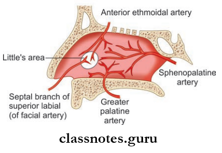
  • Arterial Supply: Mainly by the following arteries:
    1. Anterosuperior by anterior ethmoidal artery
    2. Anteroinferior part by superior labial artery (branch of a facial artery)
    3. Posterosuperior part by greater palatine artery
    4. Posteroinferior part by the sphenopalatine artery.
  • Venous Drainage: Drains into facial vein anteriorly and sphenopalatine vein posteriorly and ultimately to pterygoid venous plexus.

Nose And Nasal Septum Nerve Supply

  • General
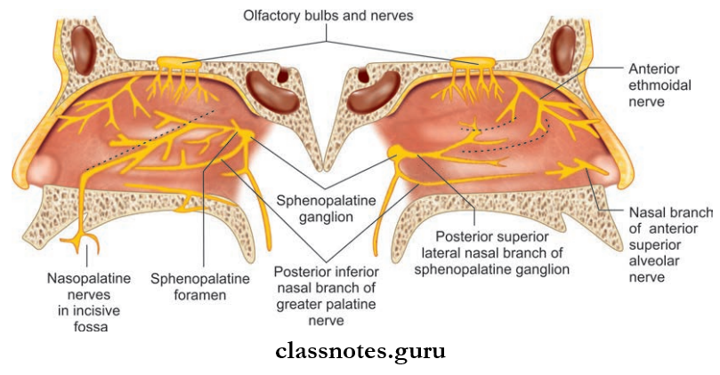
    • Anterosuperior part by nasal branch of anterior ethmoidal nerve
    • Anteroinferior part by anterior superior alveolar nerve
    • Posterosuperior part by a medial posterosuperior branch from pterygopalatine ganglion
    • Posteroinferior part by nasopalatine branch of the pterygopalatine ganglion.
  • Special Sensory: In the form of olfactory rootlets in the roof and adjoining area.

Nose And Nasal Septum Lymphatics

  • Anterior part to submandibular nodes
  • Posterior part to deep cervical nodes and retropharyngeal nodes.

Nose And Nasal Septum Applied

  • Rhinitis refers to the inflammation of the nasal mucosa as a result of infection or an allergic reaction.
  • Rhinitis can be caused by viruses as in the common cold or by allergies as in allergic rhinitis.
  • The nasal septum commonly deviates to the sides from the midline which can be anterior deviation, C­shaped deviation, S­shaped, etc.
  • Nasal septum deviation is corrected by procedures like septoplasty or submucous resection.
  • Septal hematoma refers to the collection of blood under the periosteum of nasal septum.
  • Secondary infection of septal hematoma can lead to septal abscess.
  • Bleeding from the nose is known as epistaxis and is most commonly caused due to rupture of blood vessels in the Little’s area (anterior inferior part of nasal septum just above vestibule).

Question 2. Write a short note on the lateral wall of the nose.
Answer:

Lateral Wall Of Nose

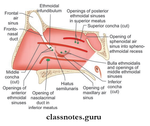
Nose And Paranasal Sinuses Lateral Wall Of The Nasal cavity After Removing The Conchae To Reveal Structures Deep To Them

  • The lateral wall is highly irregular due to the presence of bony shelf-like elevation called turbinates/conchae and space between them called meatuses.
  • These are mainly present in order to increase surface area for air conditioning of inspired air.

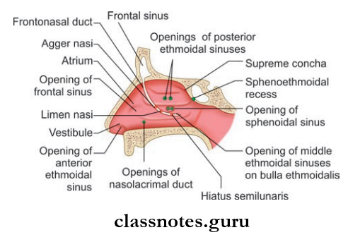
Nose And Paranasal Sinuses Openings In Lateral Wall Of Nasal Cavity

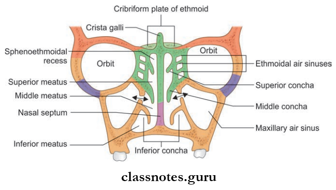
Nasal Conchae/Turbinate: These are curved bony projections directed downwards and medially and are three in number.

  1. Superior Turbinate: Smallest of the three and is present at the posterior part of nasal cavity and is the projection from the ethmoid bone
  2. Middle Turbinate:  Middle Turbinate is a projection from medial surface of the ethmoid bone
  3. Inferior Turbinate:  Inferior Turbinate is an independent bone.

Lateral Wall Of Nose Meatuses: These are spaces between or enclosed by conchae and include the following:

  • Inferior Meatus: Present below the inferior turbinate and is the largest one. The nasolacrimal duct opens here and is guarded by valve of Hasner.
  • Middle Meatus: Middle Meatus is present between the middle turbinate and inferior turbinate.
  • It consist Of The Following:
    • Bulla Ethmoidal: A rounded elevation produced by underlying ethmoid sinuses
    • Hiatus Semilunaris: Hiatus Semilunaris is a deep curved cleft/sulcus below bulla ethmoidalis
    • Infundibulum: Infundibulum is a short passage present at the anterior end of hiatus semilunaris and the anterior ethmoidal sinus open here
    • Opening Of Frontal Sinus: Seen in the anterior part of hiatus semilunaris
    • Two opening of the maxillary sinus is seen in the posterior part of hiatus semilunaris
    • Opening of the middle ethmoidal sinus is present at the upper margin of bulla ethmoidal.
  • Superior meatus lies between the superior turbinate and the middle turbinate and has openings of posterior ethmoidal sinus.
  • Sphenoethmoidal recess is present just above superior turbinate and has opening of sphenoid sinus.

Lateral Wall Of Nose Blood Supply

Nose And Paranasal Sinuses Arterial Supply Of nasal Septum

  • Arterial Supply
    • Anterosuperior quadrant by anterior ethmoidal artery and assisted by posterior ethmoidal and facial artery
    • The anteroinferior quadrant is supplied by the branch of the facial artery and the greater palatine artery
    • Posterosuperior quadrant is supplied by the sphenopalatine artery
    • The posterior inferior quadrant is supplied by branches from greater palatine artery.
  • Venous Drainage
    • Anteriorly it drains to the facial vein
    • Posteriorly it drains to the pharyngeal plexus
    • The middle part drains to the pterygoid plexus of veins.

Lateral Wall Of Nose Nerve Supply

Nose And Paranasal Sinuses Nerve Supply Of Lateral Wall And Medial Wall Of Nasal Cavity

  • General Sensory Supply
    • The anterosuperior quadrant is supplied by the anterior ethmoidal nerve a branch of ophthalmic nerve
    • The anteroinferior quadrant is supplied by the anterior superior alveolar nerve branch of the infraorbital nerve
    • The posterosuperior quadrant is supplied by posterior superior lateral nasal branches from the pterygopalatine ganglion
    • Posteroinferior quadrant is supplied by greater palatine branch of the pterygopalatine ganglion.
  • Special Sensory Supply: Olfactory nerves are present in the upper part of lateral wall ust below the cribriform plate of ethmoid to superior turbinate.

Lateral Wall Of Nose Lymphatic Drainage

  • Anterior half drains into submandibular lymph nodes
  • Posterior half to retropharyngeal and upper deep cervical nodes.

Lateral Wall Of Nose Applied

  • Rhinosporidiosis is a common fungal infection of the nose where a pink granulomatous mass protrudes from the nose which usually arises from the lateral wall. It usually bleeds on touch.
  • The adhesions formed between the nasal septum and turbinates by scar tissue is known as nasal synechia which is most commonly a result of injuries to the nose.
  • Nasal polyps refer to the non­neoplastic masses of edematous nasal or sinus mucosa.

Question 3. Write a short note on the maxillary sinus.
Answer:

Maxillary Sinus

Nose And Paranasal Sinuses Paranasal Air Sinuses Around The Nasal Cavity

  • One of the air filed spaces present within the body of the maxilla
  • The largest paranasal sinus of the body.

Maxillary Sinus Dimensions

  • Variable in size
  • Measures about 3.5 cm in height, 2.5 cm in width, and has an anteroposterior depth of 3.5 cm.

Maxillary Sinus Features

  • Maxillary Sinus is pyramidal in shape and has a base and apex
  • The apex is directed laterally in the zygomatic process of the maxilla and base is directed toward the lateral wall of nose
  • Maxillary Sinus also has a roof and a floor
  • The roof is formed by the floor of the orbit and flor by the alveolar process of the maxilla
  • Maxillary Sinus opens into the middle meatus of nose in the lower part of the hiatus semilunaris and a second opening is present at the posterior end of the hiatus.

Maxillary Sinus Blood Supply

  • Arterial: By facial artery, infraorbital artery, and greater palatine artery
  • Venous Drainage: Drains into facial vein and pterygoid plexus of veins.

Maxillary Sinus Lymphatics: Drain into submandibular lymph nodes.

Maxillary Sinus Nerve Supply: By infraorbital nerve, anterior, middle, and posterior superior alveolar nerves.

Maxillary Sinus Applied

  • Inflammation of the sinus mucosa is called sinusitis.
  • Maxillary sinusitis occurs commonly as a result of dental infections.
  • Pain of maxillary sinusitis may be referred to upper teeth and infraorbital skin.
  • Oroantral fistula refers to abnormal connections between the oral cavity and the maxillary antrum. It occurs usually as a result of dental extraction

Nose And Paranasal Sinuses Multiple Choice Question And Answers

Question 1. All of the following sinuses open into hiatus semilunar is except:

  1. Frontal
  2. Anterior ethmoidal
  3. Posterior ethmoidal
  4. Maxillary

Answer: 3. Posterior ethmoidal

Question 2. Toothache in maxillary sinusitis occurs due to stimulation of:

  1. Inferior alveolar nerve
  2. Superior alveolar nerves
  3. Greater palatine nerves
  4. Nasopalatine nerves

Answer: 2. Superior alveolar nerves

Question 3. The nasolacrimal duct opens into:

  1. Middle meatus
  2. Inferior meatus
  3. Superior meatus
  4. Vestibule

Answer: 2. Inferior meatus

Question 4. The chief artery supplying nasal mucosa is:

  1. Greater palatine
  2. Sphenopalatine
  3. Anterior ethmoidal
  4. Posterior ethmoidal

Answer: 2. Sphenopalatine

Question 5. The chief nerve innervating the nasal mucosa is:

  1. Anterior ethmoidal
  2. Greater palatine
  3. Nasopalatine
  4. Posterior ethmoidal

Answer: 3. Nasopalatine

Submandibular Gland Question And Answers

Submandibular Region Question And Answers

Question 1. Write a short note on suprahyoid muscles.
Answer:

Suprahyoid Muscles

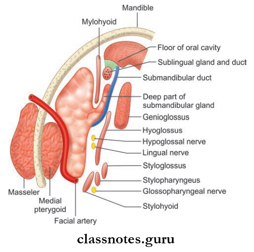
Submandibular Region Boundaries Of Digastric Triangle

Submandibular Region Attachments Of Mylohyoid Muscles And Formation Of Oral Diaphragm As Seen From Superior Aspect

Submandibular Region Superficial And Deep Relations Of The Mylohyoid Muscle

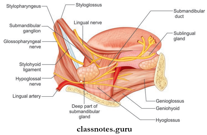
Submandibular Region Relations Of Hyoglossus Muscle

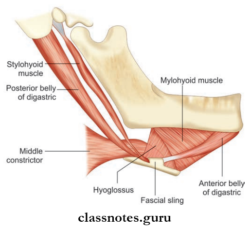
These include the digastric, the stylohyoid, the mylohyoid, and the geniohyoid.

Digastric Muscle: It has 2 bellies united by a tendon.

  • Digastric Muscle Origin
    • Anterior Belly: From digastric fossa of mandible
    • Posterior Belly: Mastoid notch of the temporal bone.
  • Digastric Muscle Insertion: Both heads are united by the intermediate tendon which pierces stylohyoid muscle and is held by a fibrous pulley to hyoid bone.
  • Digastric Muscle Nerve supply
    • Anterior Belly: Nerve to mylohyoid
    • Posterior Belly: Facial nerve.
  • Digastric Muscle Action
    • Depresses mandible
    • Elevates hyoid bone.

Styloid Muscle

  • Styloid Muscle Origin: Posterior surface of the styloid process
  • Styloid Muscle Insertion: Junction of body and greater cornua of the hyoid bone
  • Styloid Muscle Nerve supply: Facial nerve
  • Styloid Muscle Action
    • Fixing of hyoid bone
    • Pulls hyoid bone upwards and backward.

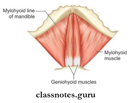
Mylohyoid Muscle

  • Mylohyoid Muscle Origin: Mylohyoid line of mandible
  • Mylohyoid Muscle Insertion
    • Posterior fibers to the body of hyoid bone
    • Anterior and middle fingers to the median raphe between the mandible and hyoid bone
  • Mylohyoid Muscle Nerve supply: Nerve to mylohyoid
  • Mylohyoid Muscle Action
    • Elevates the floor of the mouth during deglutition
    • Helps in the depression of the mandible and elevation of the hyoid bone.

Geniohyoid Muscle

  • Geniohyoid Muscle Origin: Inferior mental spine/genial tubercle
  • Geniohyoid Muscle Insertion: Anterior surface of body of hyoid bone
  • Geniohyoid Muscle Nerve supply: C1 through hypoglossal nerve
  • Geniohyoid Muscle Action: Elevates hyoid bone.

Hyoglossus Muscle

  • Hyoglossus Muscle Origin: Whole length of greater cornua and lateral part of body of hyoid bone
  • Hyoglossus Muscle Insertion: Side of the tongue between Styloglossus and inferior longitudinal muscle of tongue
  • Hyoglossus Muscle Nerve supply: Hypoglossal nerve
  • Hyoglossus Muscle Action
    • Depresses tongue
    • Retracts protruded tongue and makes dorsum convex.

Question 2. Write a note on the submandibular salivary gland.
Answer:

Submandibular Salivary Gland

  • It is a J-shaped gland present in the anterior part of the digastric triangle
  • It is indented by the posterior border of the mylohyoid which divides the gland to superficial and deep parts.

Superficial Part (Larger Part)

  • It extends upwards deep to the mandible up to the mylohyoid line
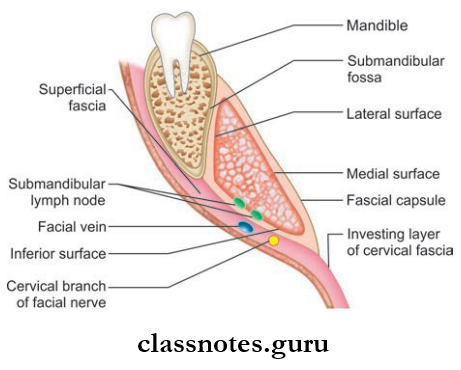
  • It consists of 3 surfaces: Inferior, lateral, and medial
  • It is partially enclosed by the layers of deep cervical fascia
  • The inferior surface is covered by the superficial layer and the medial surface is covered by the deep fascial layer.

Superficial Part Relations

  • Inferior Surface:
    • Skin
    • Platysma
    • Cervical branch of the facial nerve
    • Deep fascia
    • Facial vein
    • Submandibular lymph nodes
  • Lateral Surface:
    • Submandibular fossa on mandible
    • Insertion of the medial pterygoid
    • Facial artery
  • MedialSurface: Mylohyoid, hyoglossus, styloglossus muscles.

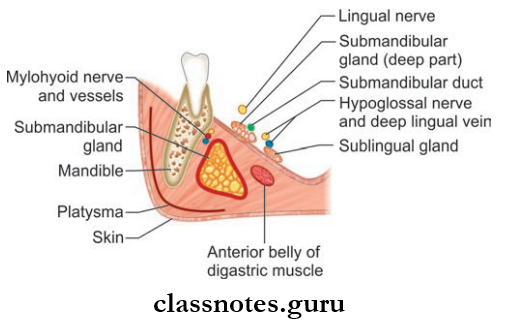
Submandibular Region Fascial Capsule And Surfaces Of Superficial Part Of Submandibular Salivary Gland

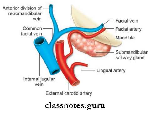
Submandibular Region Relation Of Facial Vessels To Submandibular Salivary Gland

Submandibular Region Origin And Termination Of Submandibular Duct

Deep Part (Smaller Part)

  • Lies deep to mylohyoid and superficial to hyoglossus and styloglossus
  • Posteriorly, it is continuous with superficial part
  • Anteriorly, it extends up to the posterior end of the sublingual gland.

Submandibular Duct/Wharton’s Duct

  • It is a 5 cm-long thin-walled structure
  • It emerges at the anterior end of the deep part of gland
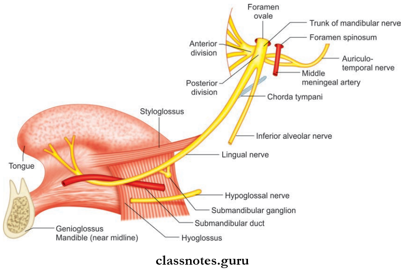
  • It then runs forwards on the hyoglossus muscle between the hypoglossal and lingual nerves
  • The duct is crossed by a lingual nerve at the anterior border of the hyoglossus muscle
  • It opens on the floor of mouth on the summit of sublingual papillae at the sides of the frenulum of the tongue.

Salivary Gland Blood Supply

  • Arterial: By facial artery
  • Venous drainage: Drain into common facial/lingual vein.

Submandibular Salivary Gland Lymphatic Drainage: Drains into submandibular lymph nodes.

Submandibular Salivary Gland Nerve Supply

  • Sensory fibers from the lingual nerve
  • Vasomotor sympathetic fibers from the plexus on the facial artery
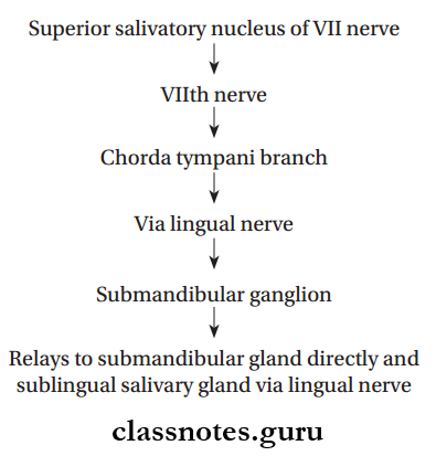
  • Secretomotor fiers from the superior salivatory nucleus and through chorda tympani and lingual nerve.

Submandibular Salivary Gland Applied

  • The salivary calculi in the submandibular duct occur due to stasis of secretion. The gland swells during eating when a stone blocks the duct.
  • Excision of the submandibular gland for calculus or tumor is done by an incision below the angle of jaw.
  • Since the marginal mandibular branch of the facial nerve passes posteroinferior to angle of the jaw, the incision is placed 4 cm below the angle to preserve the nerve.

Question 3. Write a short note on the sublingual gland.
Answer:

The Sublingual Gland

  • Smallest of the three salivary glands
  • Almond-shaped and weighs 3–4 g

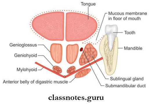
Sublingual Gland Location: Lies above the mylohyoid muscle below the mucosa of the floor of the mouth, medial to the sublingual fossa of the mandible, and lateral to the genioglossus.

Sublingual Gland Features

  • About 15 ducts arise from the gland
  • The majority of the ducts open into the floor of mouth on the summit of the sublingual fold
  • Few of the ducts join the submandibular duct.

Submandibular Region Schematic Coronal Section Through Anterior Part Of Tongue And Mouth To Show Relationships Of The Sublingual Salivary Gland, And The Oral Diaphragam

Sublingual Gland Blood Supply

  • Arterial supply from lingual and submental arteries
  • Venous drainage by lingual vein.

Sublingual Gland Nerve Supply: By the branches from the submandibular ganglion.

Sublingual Gland Lymphatic Drainage: Drains into submandibular lymph nodes.

Sublingual Gland Applied: Ranula is the cystic swelling of the sublingual gland. It is soft and bluish in color and projects in the floor of the mouth.

Submandibular Region Multiple Choice Question And Answers

Question 1. The superficial relation of hyoglossus muscle includes all except:

  1. Hypoglossal nerve
  2. Lingual artery
  3. Lingual nerve
  4. Deep part of sublingual gland

Answer: 2. Lingual artery

Question 2. Select the incorrect statement about the submandibular gland:

  1. It develops from endoderm
  2. It consists of superficial and deep parts
  3. Its ducts open into the vestibule of the oral cavity
  4. It is grooved by the facial artery

Answer: 3. Its ducts open into the vestibule of the oral cavity

Question 3. Which of the following nerves crosses the submandibular gland?

  1. Glossopharyngeal nerve
  2. Hypoglossal nerve
  3. Lingual nerve
  4. Chorda tympani nerve

Answer: 3. Lingual nerve

Question 4. All of the following pass deeper to hyoglossus except:

  1. Glossopharyngeal nerve
  2. Stylohyoid muscle
  3. Stylohyoid ligament
  4. Lingual artery

Answer: 2. Stylohyoid muscle

Question 5. A correct statement about the submandibular ganglion is:

  1. It is functionally related to facial nerve
  2. It is situated in the inner surface of hyoglossus muscle
  3. It lies above lingual nerve
  4. It supplies preganglionic parasympathetic fibers to submandibular and sublingual glands.

Answer: 1. It is functionally related to the facial nerve

Infratemporal Fossa Boundaries Question And Answers

Temporal And Infratemporal Regions Question And Answers

Question 1. Write a short note on temporal fossa, infratemporal fossa, pterygopalatine fossa.
Answer:

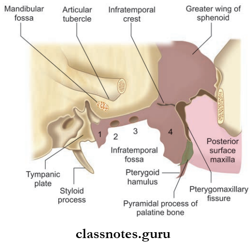
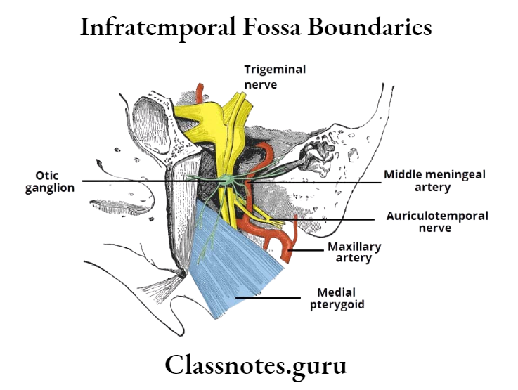
Temporal And Infratemporal Regions Bony Boundaries Of Infratemporal Fossa

  1. Spine of the sphenoid,
  2. Foramen spinosum,
  3. Foramen ovale,
  4. Lateral pterygoid plate

Temporal Fossa: Lies on the side of the skull, bounded by superior temporal line and zygomatic arch.

Temporal Fossa Boundaries

  • Anterior: Zygomatic and frontal bone
  • Posterior: Inferior temporal line and supramastoid crest
  • Superior: Superior nuchal line
  • Inferior: Zygomatic arch
  • Floor Parts of the frontal, parietal, and temporal bone, temporalis muscle, and greater wing of the sphenoid

Temporal Fossa Contents

  • Temporalis muscle
  • Zygomatic temporal nerve and artery
  • Middle temporal artery
  • Deep temporal nerve and artery.

Infratemporal Fossa: Irregular space below the zygomatic arch.

Infratemporal Fossa Boundaries

  • Anterior: Posterior surface of a body of the maxilla
  • Roof: Infratemporal surface of greater wing of the sphenoid
  • Medial: Lateral pterygoid plate
  • Lateral: Ramus of mandible

Infratemporal Fossa Contents

  • Lateral pterygoid muscle
  • Medial pterygoid muscle
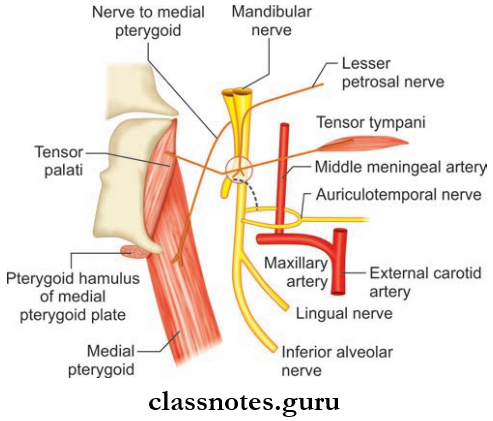
  • Mandibular nerve and maxillary nerve
  • Chorda tympani nerve
  • 1st and 2nd parts of maxillary artery.

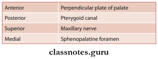
Pterygopalatine Fossa: Lies in the depth of pterygomaxillary fissures.

Pterygopalatine Fossa Boundaries

  • Anterior: Posterior surface of maxilla
  • Posterior: Pterygoid process and greater wing of the sphenoid
  • Medial: Perpendicular plate of palatine bone
  • Floor: Union of anterior and posterior walls

Pterygopalatine Fossa Contents

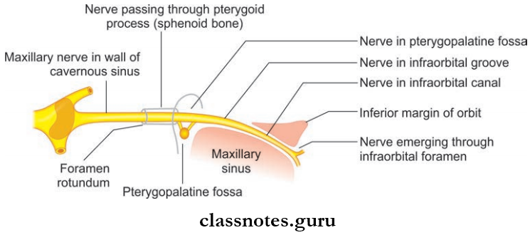
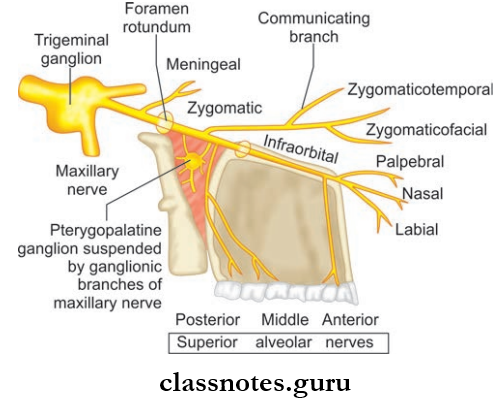
  • Maxillary nerve and its branches
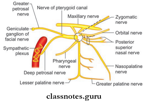
  • Pterygopalatine ganglion and its branches
  • 3rd part of the maxillary artery and its branches.

Question 2. Write a short note on muscles of mastication.
Answer:

Muscles Of Mastication

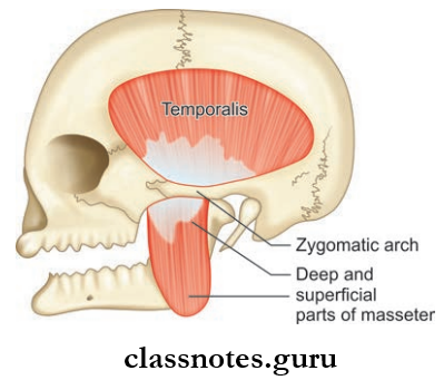
Temporal And Infratemporal Regions Attachements Of masseter And Temporalis Muscles

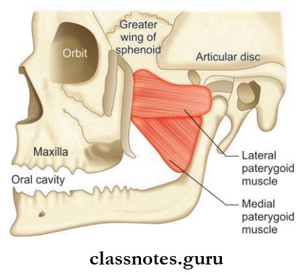
Temporal And Infratemporal Regions Attachements Of Lateral And Medical Pterygoid Muscles

  • These are the muscle that moves the mandible during mastication and speech
  • The muscles include the masseter, the temporalis, the lateral pterygoid, and the medial pterygoid.

Masseter

  • Masseter is quadrilateral in shape and has 2 layers
  • Covers the lateral surface of ramus of the mandible.
  • Masseter Origin
    • Superficial layer: Anterior 2/3rd of the lower border of the zygomatic arch and zygomatic process of maxilla
    • Deep layer: Deep surface of the zygomatic arch.
  • Masseter Insertion
    • Superficial layer: Lower part of lateral surface of ramus of the mandible
    • Deep layer: Rest of the ramus of the mandible.
  • Masseter Nerve supply: Masseteric nerve branch of anterior division of mandibular nerve.
  • Masseter Action: Elevation of mandible to close mouth to bite.

Temporalis

  • Fan-shaped muscle
  • Present in the temporal fossa
  • Temporalis Origin: Temporal fossa and temporal fascia
  • Temporalis Insertion
    • Margins and deep surface of coronoid process of mandible
    • Anterior border of ramus of mandible.
  • Temporalis Nerve supply: Deep temporal branches from the anterior division of mandibular nerve
  • Temporalis Action: Elevates mandible, helps in side to side grinding movements, retraction of protruded mandible.

Lateral Pterygoid

  • Lateral Pterygoid is a short conical-shaped muscle having upper and lower heads.
  • Temporalis Origin:
    • Upper Head: Arise from the infratemporal surface and crest of greater wing of sphenoid bone
    • Lower Head: Lateral surface of lateral pterygoid plate
  • Temporalis Insertion:
    • Pterygoid fovea on the anterior surface of neck of mandible
    • Anterior margin of articular disc and capsule of temporomandibular joint
  • Temporalis Nerve supply: Branch from anterior division of mandibular nerve
  • Temporalis Actions:
    • Depresses mandible, protrusion of mandible
    • Grinding movements.

Infratemporal Fossa Boundaries

Medial Pterygoid

  • Medial Pterygoid is a quadrilateral muscle
  • Medial Pterygoid has a small superficial and large deep head.
  • Medial Pterygoid Origin:
    • Superficial head: From tuberosity of maxilla and adjoining bone
    • Deep head: From the medial surface of the lateral pterygoid plate and adjoining process of palatine bone.
  • Medial Pterygoid Insertion: Roughened area on the medial surface of angle and ramus of mandible, behind mandibular foramen and mylohyoid groove.
  • Medial Pterygoid Nerve supply: Nerve to medial pterygoid, a branch of main trunk of mandibular nerve
  • Medial Pterygoid Actions:
    • Elevates mandible
    • Protrusion of mandible, grinding movements.

Question 3. Write a short note on the relations of medial and lateral pterygoid.
Answer:

Relations Of Medial And Lateral Pterygoid

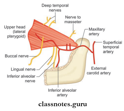
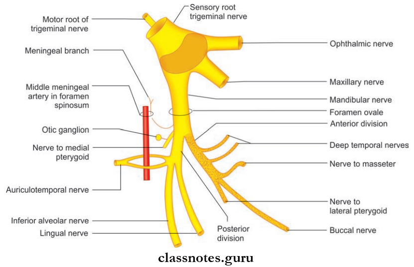
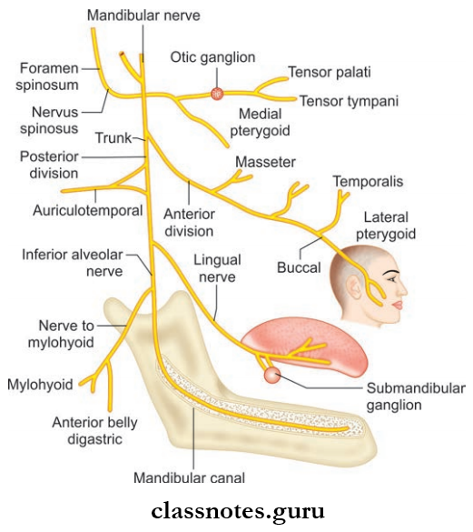
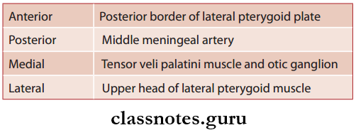
Temporal And Infratemporal Regions Neural And Vascular Relations Of lateral Pterygoid Muscle In Infratemporal Fossa

Relations Of Lateral Pterygoid: This key muscle of the region as it provides ideas about the layout of structures of the infratemporal fossa.

  • Superficial
    • Masseter
    • Ramus of mandible
    • Maxillary artery
    • Tendon of temporalis.
  • Deep
    • Mandibular nerve
    • Middle meningeal artery
    • Deep head of the medial pterygoid
    • Sphenomandibular ligament.
  • Structures Emerging At The Upper Border
    • Deep temporal nerves
    • Masseteric nerve.
  • Structures Emerging At Lower Border
    • Lingual nerve
    • Inferior alveolar nerve
    • Middle meningeal artery.
  • Structures Passing In Between Two Heads
    • Maxillary artery
    • Buccal branch of mandibular nerve.

Relations Of Medial Pterygoid

  • Superficial
    • The lateral pterygoid plate
    • The lingual nerve
    • Inferior alveolar nerve
    • Maxillary artery
    • Sphenomandibular ligament.
  • Deep Relations
    • Tensor veli palatini
    • Superior constrictor of pharynx
    • Styloglossus
    • Stylopharyngeus.

Mnemonic: Pterygoid muscles (function)

Look at how your jaw ends up when saying first syllable of ‘Lateral’ or ‘Medial’

‘La’: Your jaw is now open, so lateral opens your mouth.

‘Me’: Your jaw is still closed, so the medial closes the mandible.

Question 4. Write a note on the temporomandibular joint.
Answer:

Temporomandibular Joint

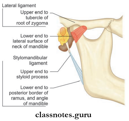
Temporal And Infratemporal Regions Ligaments Of The Temporomandibular Joint As Seen From The Lateral Side

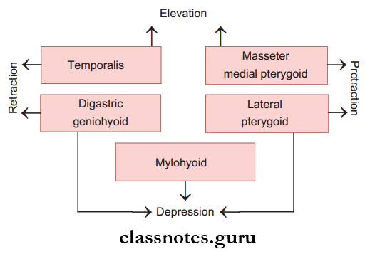
Temporal And Infratemporal Regions Muscles Responsible For Movements At The Temporomandibular Joint

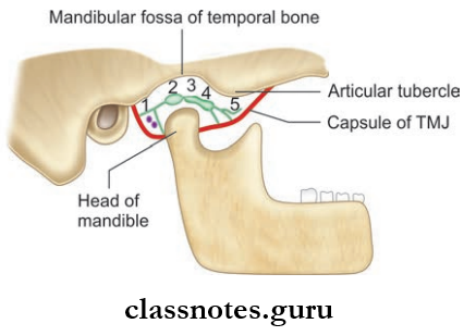
Temporal And Infratemporal Regions Interior Of Temporomandibular Joint Showing Parts Of Articular Disc From Posterior To Anterior Side

  1. Bilaminar part with venous plexus,
  2. Posterior thick band,
  3. Intermediate zone,
  4. Anterior thick band,
  5. Anterior extension

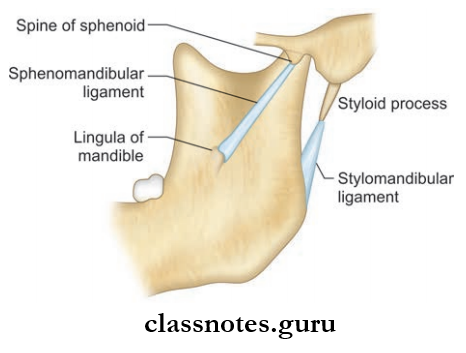
Temporal And Infratemporal Regions Accessory Ligaments Of TMJ Sphenomandibular Ligament And Stylomandibular Ligament

  • Temporomandibular Joint is a joint on each side of head between the temporal bone and mandible
  • Temporomandibular Joint accounts for the movement of the mandible for speech and mastication
  • Temporomandibular Joint is a synovial joint of condylar variety.

Temporomandibular Joint Articular Surfaces

  • Upper articular surface is formed by the mandibular fossa and articular eminence of the temporal bone
  • The lower articular surface is formed by the head of the mandible
  • Both articular surfaces is covered by fibrocartilage so the joint is an atypical joint.

Temporomandibular Joint Cavity: The joint is divided into:

  • Upper meniscotemporal compartment for gliding movements only
  • Lower meniscomandibular compartment for gliding as well as rotational movements.

Temporomandibular Joint  Articular Disc

  • Temporomandibular Joint  Articular Disc is an oval plate of fibrocartilage and consists of mainly collagen fibers and few cartilage cells
  • Its superior surface is concavo-convex (before backward) and its inferior surface is concave
  • The periphery is attached to firous capsule
  • The disc consists of a thin intermediate zone and anterior and posterior branch when cut sagittaly.
  • The anterior band is continuous with the tendon of the lateral pterygoid
  • The posterior band is bilaminar and upper lamina is attached to the squamotympanic fissure and lower lamina is attached to back of condyle.

Temporomandibular Joint Ligaments:

  • The Ligaments Include:
    1. Fibrous capsule
    2. Temperomandibular ligament
    3. Sphenomandibular ligament
    4. Stylomandibular.
  1. Fibrous Capsule
    • It is a fibrous sac which encloses the joint cavity
    • It is attached to the articular tubercle, the circumference of the articular fossa, and squamotympanic figure above and neck of the mandible below
    • It is loose above the intra-articular disc and tight below the disc.
  2. Temporomandibular Ligament
    • It is attached to the articular tubercle in the root of zygoma above and neck of the mandible below.
  3. Sphenomandibular Ligament
    • It is attached to the spine of the sphenoid above and the lingula and lower margin of the mandibular foramen below
    • It represents the unossified intermediate part of the sheath of Meckel’s cartilage.
  4. Stylomandibular Ligament
    • It is an accessory ligament of temporomandibular joint
    • It is attached to the lateral surface of the styloid process above and the angle and adjoining border of the ramus of the mandible below
    • It is a thickening of investing layer of deep cervical fascia.

Relations Of Temporomandibular Joint

  • Anterior
    • Lateral pterygoid
    • Masseteric nerve and artery
  • Posterior
    • Parotid gland
    • Superficial temporal vessels
    • Auriculotemporal nerve
  • Lateral
    • Skin and fascia
    • Parotid gland
    • Temporal branches of the facial nerve
  • Medial
    • The tympanic plate
    • Spine of sphenoid
    • Auriculotemporal and chorda tympani nerves
    • Middle meningeal artery
  • Superior
    • Middle cranial fossa
    • Middle meningeal vessels
  • Inferior: Maxillary artery and vein

Temporomandibular Joint Nerve Supply: The auriculotemporal nerve which enters through posterior aspect of joint Masseteric nerve enters through anterior aspect of joint.

Temporomandibular Joint Blood Supply

  • Arterial supply: Maxillary artery and superficial temporal artery enters the joint through posterior aspect of the capsule.
  • Lymphatic Drainage: Drain into preauricular nodes, parotid nodes, and upper deep cervical nodes.

Temporomandibular Joint Stability

  • More stable when mouth closed
  • Forward movement of the condyle is discouraged by articular eminence and contraction of posterior fibers of the temporalis muscle
  • Backward movement is prevented by lateral ligament and contraction of the lateral pterygoid muscle.

Temporomandibular Joint Movements: The movements occurring at the joint involve two basic movements.

  1. Gliding Movements
  2. Rotational Movements.

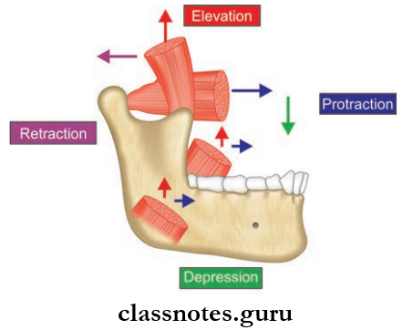
Temporal And Infratemporal Regions Movements Of Mandible Produced By Muscles Of Mastication

1. Gliding Movements: occurs between disc and articular eminence with disc and condyle moving forward and backward, down or up

2. The Rotational Movements occur between the disc and condyle

The Movements Include:

  • Protraction: Done by lateral and medial pterygoid and masseter
  • Retraction: Done by posterior fibers of the temporalis
    Elevation: By medial pterygoid, masseter, and vertical fibers of temporalis
  • Depression: Caused by the lateral pterygoid
  • Lateral movements: To the left side by the right lateral pterygoid and vice versa.

Temporomandibular Joint Applied

  • When the articular disc is detached from the fibrous capsule, it is known as the derangement of the joint.
  • Temporomandibular causes painful movements and clicking sounds while opening and closing the mouth.
  • In case of surgeries of the temporomandibular joint, the facial nerve, auriculotemporal nerve, and mandibular division of the trigeminal nerve are preserved with care.

Temporal And Infratemporal Regions Multiple Choice Questions And Answers

Question 1. Which of the following muscles opens the mouth?

  1. Temporalis
  2. Lateral pterygoid
  3. Medial pterygoid
  4. Masseter

Answer: 2. Lateral pterygoid

Question 2. The temporomandibular joint is commonly dislocated:

  1. Medially
  2. Laterally
  3. Anteriorly
  4. Posteriorly

Answer: 3. Anteriorly

Question 3. The nerve to the medial pterygoid supplies all of the muscles except:

  1. Tensor palate
  2. Medial pterygoid
  3. Lateral pterygoid
  4. Tensor tympani

Answer: 3. Lateral pterygoid

Question 4. All of the structures occupy infratemporal fossa except:

  1. Mandibular nerve
  2. Chorda tympani nerve
  3. Pterygoid venous plexus
  4. Masseter muscle

Answer: 4. Masseter muscle

Question 5. Select the incorrect statement regarding otic ganglion:

  1. It is functionally related to the mandibular nerve
  2. It is topographically related to the mandibular nerve
  3. It is 2–3 mm in size
  4. It is located lateral to the tensor palate muscle

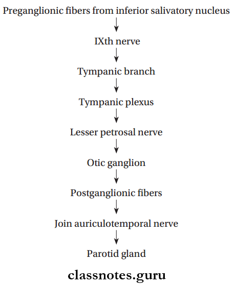
Answer: 1. It is functionally related to the mandibular nerve

Contents Of Orbit And Eye Question And Answers

Contents Of Orbit And Eye Question And Answers

Question 1. Write a short note on orbital fascia and bulbar fascia.
Answer:

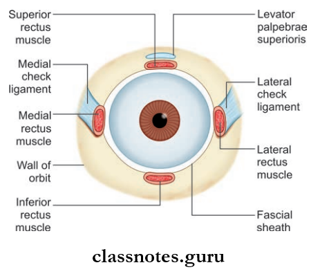
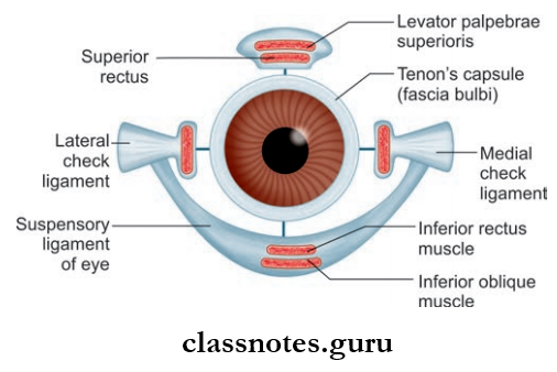
Orbital Fascia Or Periorbital: Fascial Sheath Or Bulbar Sheath Or Tenon’s Capsule

  • Tenon’s capsule forms a thin membranous sheath around the eyeball from optic nerve to the sclerocorneal junction
  • Orbital Fascia Or Periorbital is loosely attached, so the eyeball can freely move within the sheath.

Contents Of Orbit And Eye Parts Of The Fascial Sheath Of The Eyeball

Contents Of Orbit And Eye Fascial Covering Of Eyeball

Orbital Fascia Or Periorbital Applied: Tenon’s space is a potential space around the eyeball between the sclera and Tenon’s capsule.

Question 2. Write a short note on extraocular muscles.
Answer:

Extraocular Muscles

  • These are the muscles present in the orbit. Among these, there are seven voluntary muscles and three involuntary muscles
  • Among the seven voluntary muscles, six muscles move the eyeball, one muscle moves the upper lid
  • The Seven Voluntary Muscles Include:
    • Four Rectisuperior Rectus
      • Inferior Rectus
      • Medial Rectus
      • Lateral Rectus
    • Two Oblique
      • Superior oblique
      • Inferior oblique
    • Levator Palpebrae Superioris
  • The Involuntary Muscles Include:
    • Superior tarsal muscle
    • Inferior tarsal muscle
    • Orbitalis.

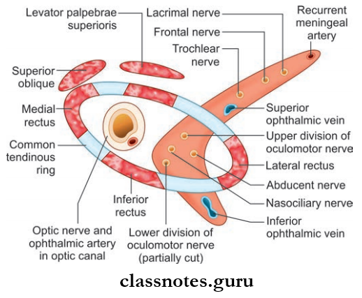
Contents Of Orbit And Eye Structures Passing Through Optic Canal And Superior Orbital Fissure And Muscles Taking Origin From Common Tendinous Ring At The Apex Of Orbit

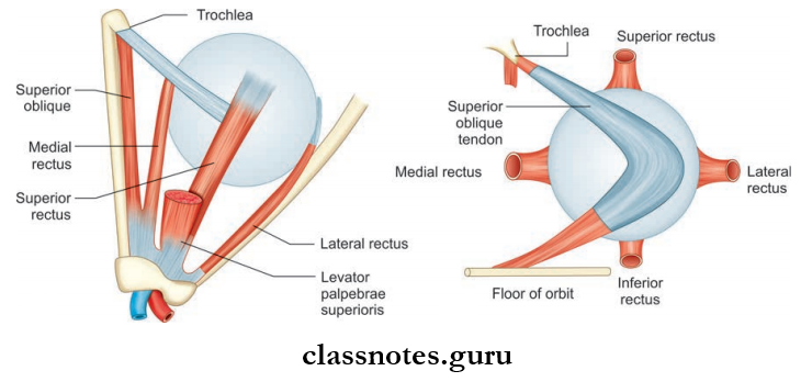
Contents Of Orbit And Eye Insertion Of Extraocular Muscles In The Sclera

Voluntary Muscles

  • Voluntary Muscles Origin
    • The recti arise from a common tendinous ring
    • The tendinous ring is attached to the orbital surface of the apex of orbit
    • It encloses the optic canal and the middle part of the superior orbital fissure
    • The superior oblique arises from body of the sphenoid bone superomedial to optic canal
    • Inferior oblique arises from the orbital surface of the maxilla lateral to the lacrimal groove
    • Levator muscle arises from the orbital surface of the lesser wing of the sphenoid bone superior to the optic canal.
  • Voluntary Muscles Insertion
    • Recti muscles are inserted to the sclera posterior to the limbus
    • The superior oblique is inserted into the sclera, behind the equator in the posterior superior quadrant of the eyeball between the superior and lateral rectus
    • The inferior oblique is inserted into the sclera, behind the equator in the posterior superior quadrant of the eyeball below and posterior to the insertion of the superior oblique muscle
    • Levator muscle’s superior lamellae is inserted into the anterior surface of the superior tarsus and into skin of the upper eyelid, whereas the inferior lamellae is inserted to upper margin of the superior tarsus, and to the superior conjunctival fornix.
  • Voluntary Muscles Nerve Supply
    • Superior rectus, inferior rectus, inferior oblique and levator palpebrae superioris are supplied by occulomotor nerve
    • Lateral rectus is supplied by the abducent nerve
    • Superior oblique is supplied by the trochlear nerve.

Mnemonics: Extraocular muscles cranial nerve innervation

LR6SO4 Rest 3:

  • Lateral Rectus is 6th
  • Superior Oblique is 4th rest are all 3rd cranial nerve.

Voluntary Muscles Applied

  • Weakness or paralysis of a muscle causes squint or strabismus.
  • Squint can be paralytic or concomitant .
  • Concomitant squint is usually congenital and there is no limitation of movements and diplopia.
  • In paralytic squint movements are limited, and diplopia and vertigo may be present.

Question 3. Write a short note on the actions of extraocular muscles.
Answer:

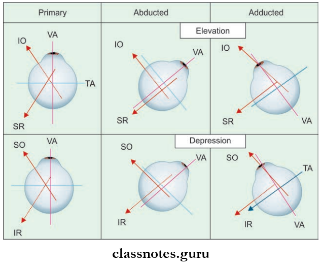
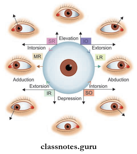
The Actions Of Extraocular Muscles

Contents Of Orbit And Eye Actions Of Extraocular Muscles

Contents Of Orbit And Eye Pictorical Depiction Of Actions Of Extraocular Muscles

The Movements Of The Eyeball Are As Follows:

  • Around Transverse Axis
    • Elevation (upward rotation)
    • Depression (downward rotation)
  • Around Vertical Axis
    • Adduction (medial rotation)
    • Adduction (lateral rotation)
  • Around Anterior-Posterior Axis
    • Intorsion (12 o’clock position of cornea rotates medially)
    • Extorsion (12 o’clock position of cornea rotates laterally)
  • Actions Of Individual Muscles:
    • Superior Rectus: Elevation, adduction, intorsion
    • Inferior Rectus: Depression, adduction, extorsion
    • Medial Rectus: Adduction
    • Lateral Rectus: Abduction
    • Superior Oblique: Depression, abduction and intorsion
    • Inferior Oblique: Elevation, abduction and extorsion.

Mnemonic: Eye rotation by oblique muscle

  • I Love S and M
  • Inferior Oblique: Lateral eye rotation.
  • Superior Oblique: Medial eye rotation.

Contents Of Orbit And Eye Multiple Choice Questions And Answers

Question 1. All of the following muscles of the eyeball are supplied by oculomotor nerve except:

  1. Superior rectus
  2. Superior oblique
  3. Inferior rectus
  4. Inferior oblique

Answer: 1. Superior rectus

Question 2. All of the structures are derived from Tenon’s capsule except:

  1. Medial check ligament
  2. Lateral check ligament
  3. Ligament fascia
  4. Ligament of Lockwood

Answer: 3. Ligament fascia

Question 3. Which of the following branches of the ophthalmic artery is most important?

  1. Lacrimal artery
  2. The central artery of retina
  3. Supraorbital artery
  4. Supratrochlear artery

Answer: 2. Central artery of retina

Question 4. Which of the following nerves lies in the orbit outside the periorbital?

  1. Oculomotor
  2. Trochlear
  3. Abducent
  4. Zygomatic

Answer: 4. Zygomatic

Question 5. Select the incorrect statement regarding ciliary ganglion:

  1. It lies between optic nerve and lateral rectus in the apex of the orbit
  2. It is functionally connected to oculomotor nerve
  3. It lies between the medial rectus and optic nerve near the apex of the orbit
  4. Topographically it is connected to the nasociliary nerve

Answer: 3. It lies between the medial rectus and optic nerve near the apex of orbit

Cranial Cavity Question And Answers

Cranial Cavity Question And Answers

Question 1. Write a short note on cerebral dura mater.
Answer:

Cerebral Dura Mater

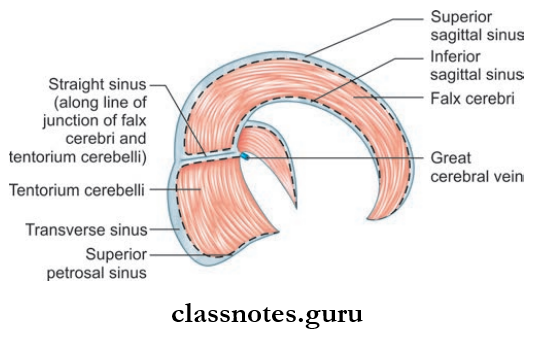
Cranial Cavity Falx Cerebri And Tentorium Cerebelli And The Dural Venous Sinuses Contained In Their Attached And Free Margins

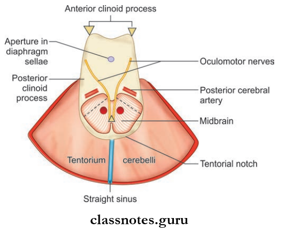
Cranial Cavity Tentorium Cerebelli And Structures Inside The U-shaped Tentorium Notch

It Is A Strong Fibrous Membrane Consisting Of Two Layers:

  1. Inner meningeal layer
  2. Outer endosteal layers

The two layers are fused with each other except where venous sinuses are present.

Cerebral Dura Mater Outer Layer

  • The outer layer is the periosteum lining the inner surface of skull bones and is continuous with periosteum.
  • Cerebral Dura Mater Outer Layer is loosely attached to the inner surface of the cranial vault but is fimly attached to base of skull and around the foramen magnum.

Cerebral Dura Mater Inner Layer

  • Cerebral Dura Mater Inner Layer encloses brain and at the foramen magnum, it is continuous with dura of spinal cord.
  • The meningeal layer is folded on itself at some places to form dural folds.

Folds Of Dura Mater: The folds are as follows

  • Falx cerebri
  • Tentorium cerebelli
  • Falx cerebelli
  • Diaphragma sellae

Falx Cerebri

  • The large sickle-shaped fold of dura is situated between the two cerebral hemispheres and occupy the median longitudinal fissure between them.
  • The narrow anterior end is attached to the crista galli and the broad posterior end to the upper surface of the tentorium cerebelli.
  • Falx Cerebri encloses superior and inferior sagittal sinuses in its upper and lower margins respectively.
  • Falx Cerebri also encloses straight sinus along the attachment with tentorium cerebelli.

Tentorium Cerebelli

  • Tentorium Cerebelli is a tent-shaped fold of dura forming roof of posterior cranial fossa
  • Tentorium Cerebelli separates the cerebellum from the occipital lobe of the cerebrum
  • Tentorium Cerebelli consists of inner margin and outer margin, which are attached to anterior and posterior clinoid processes respectively.
  • Consists of upper surface and inferior surface to which falx cerebri and falx cerebelli are attached respectively.
  • Tentorium Cerebelli encloses transverse sinus, superior petrosal sinus, and straight sinus.

Falx Cerebelli

  • Small sickle-shaped dural fold is a sagittal plane extending from internal occipital protuberance to the posterior margin of foramen magna
  • Falx Cerebelli has a free anterior margin and attached posterior margin
  • Falx Cerebelli encloses occipital sinus.

Question 2. Write a short note on intracranial dural venous sinuses.
Answer:

Intracranial Dural Venous Sinuses

  • These are venous channels present in the cranial dura.
  • These are formed by either separation of two layers of dura or by the reduplication of the meningeal layer.
  • The sinuses have thin walls lined by endothelium and drain blood from brain.
  • The blood is ultimately drained to internal jugular vein.

The Venous Sinuses Are Divided Into:

  • Paired Sinuses Consisting Of:
    • Cavernous sinus
    • Superior petrosal sinus
    • Inferior petrosal sinus
    • Transverse sinus
    • Sigmoid sinus
    • Sphenoparietal sinus
    • Petrosquamous sinus
    • Middle meningeal sinus
  • Unpaired Sinuses Consisting Of:
    • Superior sagittal sinus
    • Inferior sagittal sinus
    • Straight sinus
    • Occipital sinus
    • Anterior intercavernous sinus
    • Posterior intercavernous sinus
    • Basilar venous plexus.

Cranial Cavity Position Of dural Venous Sinuses Inside The Cranium

Question 3. Write a short note on cavernous sinus.
Answer:

Cavernous Sinus

Cranial Cavity RFelations Of Cavernous Sinus

Cranial Cavity Directt Tributraries, Draining Channels And Communications Of Cavernous Sinus

Cavernous Sinus Situation And Extent

  • Situated in the middle cranial fossa along the body of the sphenoid and sella turcica
  • Anteriorly, it extends up to the medial end of the superior orbital fissure and posteriorly up to apex of the petrous temporal bone
  • Each sinus is divided into number of small spaces/ caverns by trabeculae
  • The floor is formed by the endosteal layer, the lateral and medial wall, and roof is formed by the meningeal dura mater.

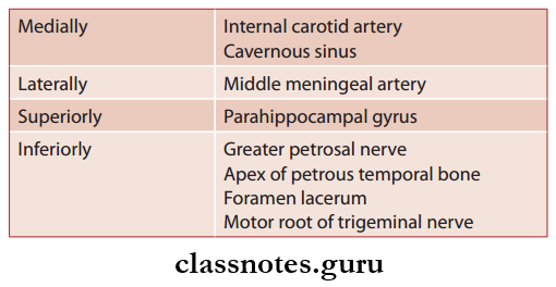
Cavernous Sinus Relations

Cranial Cavity Cavernous Sinus Relations

Structures Present In The Lateral Wall Of Sinus

  • Oculomotor nerve
  • Trochlear nerve
  • Ophthalmic nerve
  • Maxillary nerve
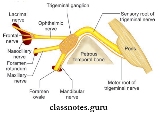
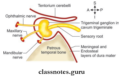
  • Trigeminal ganglion

Structures Passing Through Sinus

  • Internal carotid artery and sympathetic plexus of nerves
  • Abducent nerve

Tributaries Of Cavernous Sinus: Receives blood from three sources—orbit, meninges and brain

  • From Orbit
    • Superior ophthalmic vein
    • Inferior ophthalmic vein
    • Central vein of retina
  • From Meninges
    • Sphenoparietal sinus
    • Anterior trunk of middle meningeal vein
  • From Brain
    • Superficial middle cerebral vein
    • Inferior cerebral vein

Communications Of Cavernous Sinus

  • Transverse sinus through superior petrosal sinus
  • Internal jugular vein through inferior petrosal sinus
  • Pterygoid venous plexus via emissary’s veins
  • Facial vein through a superior ophthalmic vein
  • Opposite cavernous sinus through anterior and posterior intercavernous sinuses
  • Superior sagittal sinus through superfiial middle cerebral vein
  • Internal vertebral venous plexus through basilar venous plexus.

Applied For Cavernous Sinus

  • Cavernous Sinus Thrombosis – the sinus gets infected from different septic foci like:
    • Meningitis, brain abscess through a superficial middle cerebral vein, inferior cerebral vein, shenoparietal sinus
    • Infections of face, nose, paranasal sinus, and orbit it reaches the sinus through a superior and inferior ophthalmic vein
    • Otitis media (labyrinthine veins) mastoid abscess (mastoid emissary veins) through superior and inferior petrosal sinus reach the cavernous sinus.
  • Infections from the face spread in a retrograde direction via the facial vein and pterygoid plexus and cause thrombosis of cavernous sinus
  • Cavernous sinus syndrome caused by various parasellar pathological conditions that involves the cavernous sinus along with cranial nerves (3,4,5,6), internal carotid artery, and sympathetic plexus
  • Carotid cavernous fitula

Question 4. Write a short note on superior sagittal sinus.
Answer:

Superior Sagittal Sinus

  • Located between two layers of falx cerebri
  • Begins at crista galli and passes backward and deviates to the right at internal occipital protuberance to become continuous with right transverse sinus.
  • The right transverse sinus becomes continuous with right sigmoid sinus.

Superior Sagittal Sinus Features

  • Triangular in shape
  • Drains the diploic and meningeal veins
  • Arachnoid granulations project to the lumen

Superior Sagittal Sinus Tributaries

  • Superior cerebral vein
  • Parietal emissary vein
  • Veins of frontal air sinus
  • Small vein from nasal cavity

Question 5. Write a short note on sigmoid sinus.
Answer:

Sigmoid Sinus

  • S-shaped sinus
  • Each sinus in the direct continuation of transverse sinuses
  • It grooves the inner surface of mastoid part of the petrous bone
  • The terminal part curves downwards and forwards to the posterior margin of the jugular foramen through which it passes and continues as the internal jugular vein.

Sigmoid Sinus Tributaries

  • Mastoid and condylar emissary veins
  • Cerebellar veins
  • Internal auditory veins

Question 6. Write a note on hypophysis cerebri.
Answer:

Hypophysis Cerebri

Cranial Cavity Subdivisions Of Pituitary Gland

Cranial Cavity Arterial Supply Of Pituary Gland

Hypophysis Cerebri Location And Extent

  • It is a small oval-shaped endocrine gland situated in the hypophyseal fossa of the sphenoid bone.
  • It is suspended from the floor of the third ventricle by a narrow stalk called an infundibulum.
  • It measures about 8 mm anteroposteriorly and 12 mm transversely and weighs about 500 mg.

Hypophysis Cerebri Relations

  • Superiorly: Optic chiasma, diaphragm a sellae
  • Inferiorly: Sphenoid air sinuses, hypophyseal fossa
  • Laterally: Cavernous sinus on each side

Hypophysis Cerebri Subdivisions: It is divided into adenohypophysis and neurohypophysis

  • Adenohypophysis consists of
    • Pars anterior
    • Pars intermedius
    • Pars tuberalis
  • Neurohypophysis consists of
    • Pars posterior
    • Infundibulum

It Is Associated With The Hypothalamus Through The Following:

  • Hypothalamohypophyseal tract
  • Hypothalamohypophyseal portal system

Hypophysis Cerebri Blood Supply

  • Arterial supply is mainly through superior and inferior hypophyseal arteries (branches of internal carotid artery)
  • Venous drainage is through short veins which drain into cavernous and intercavernous sinuses.

Hypophysis Cerebri Applied

  • A pituitary adenoma is the most common tumor of the pituitary gland arising from the chromophobe cells.
  • Hypophysis Cerebri produces symptoms due to pressure on adjacent structures and due to endocrine disturbances.

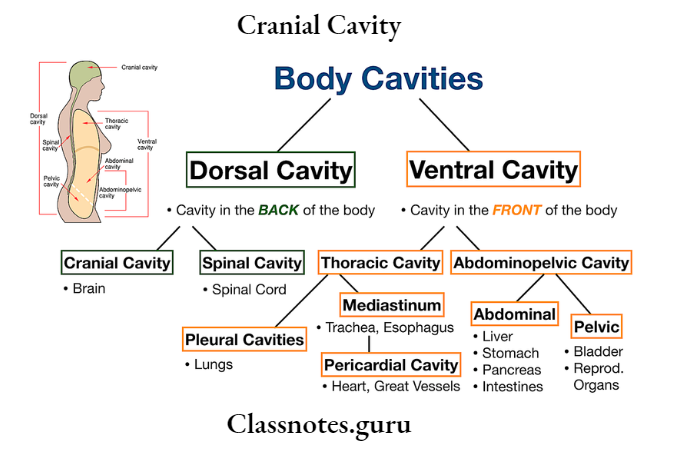
Cranial Cavity Body Cavities

Cranial Cavity Multiple Choice Question And Answers

Question 1. The contents of the vertebral canal include all of the following structures except:

  1. Spinal nerve roots
  2. Spinal cord
  3. Spinal ganglia
  4. Spinal meninges

Answer: 3. Spinal ganglia

Question 2. Select the incorrect statement regarding vertebral venous plexus:

  1. It is a network of veins in subdural space
  2. It receives basivertebral veins
  3. It drains into segmental veins
  4. It is continuous with intracranial dural venous sinuses

Answer: 1. It is a network of veins in subdural space

Question 3. Select the incorrect statement regarding the dural venous sinuses:

  1. They are lined by endothelium
  2. They have no valves
  3. They communicate with extracranial veins
  4. They possess thin muscular coats in their wall

Answer: 4. They possess thin muscular coat in their wall

Question 4. All of the following dural venous sinuses are paired except:

  1. Superior petrosal
  2. Inferior petrosal
  3. Inferior sagittal
  4. Cavernous

Answer: 3. Inferior sagittal

Question 5. Lateral wall of cavernous sinus contains all of the following nerves except:

  1. Oculomotor
  2. Trochlear
  3. Ophthalmic
  4. Abducent

Answer: 4. Abducent

Major Arteries of the Head and Neck Question And Answers

Blood Vessels Of Head And Neck Question And Answers

Question 1. Write a short note on common carotid artery.
Answer:

Common carotid artery
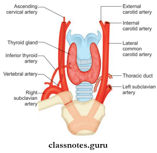
Blood Vessels Of Head And Neck Realtionship Of Common Cartooid Artery To The Larynx, Trachea And Thyroid

There are two common carotid arteries right and left which form chief arteries of head and neck.

Common Carotid Artery Origin

  1. Right common carotid artery arises in neck from the brachiocephalic trunk behind sternoclavicular joint
  2. Left common carotid artery arises directly from arch of aorta and ascends back to sternoclavicular joint.

Common Carotid Artery Course

  • Both right and left arteries in neck have a similar course
  • Each artery runs upwards from the sternoclavicular joint of respective sides to the upper border of lamina of thyroid which lies to body of C4 vertebrae
  • At the level of lamina thyroid cartilage, it divides into internal and external carotid arteries
  • They are named so because external carotid artery supplies the structures outside the skull and internal carotid artery supplies inside the skull
  • Each common carotid artery lies in the front of transverse process of the lower cervical vertebrae and under cover of the anterior border of sternocleidomastoid
  • Internal carotid artery is considered as continuation of common carotid artery.
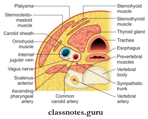
Blood Vessels Of Head And Neck Right Lateral View Showing Structures Crossing Superficial To The Common Cartoid Artery

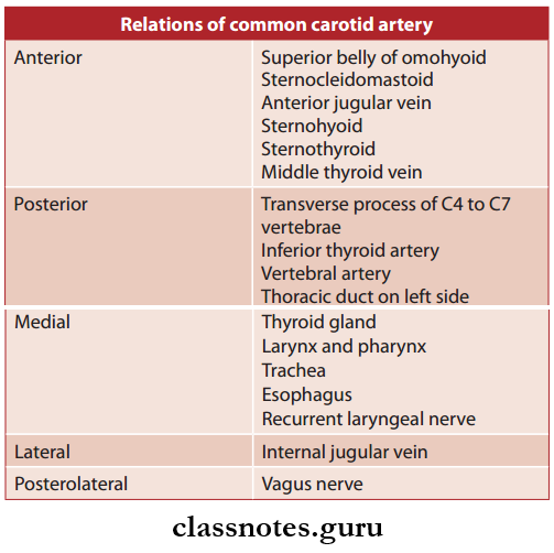
Relations of common carotid artery

Blood Vessels Of Head And Neck Realtionship Of Common Cartooid Artery

Carotid Sinus

  • It is a fusiform dilation at the termination of common carotid artery or the beginning of the internal carotid artery
  • It receives rich innervation from the glossopharyngeal and sympathetic nerves
  • It is a baroreceptor and regulates blood pressure.

Carotid Body

  • Small oval reddish-brown structures situated behind the bifurcation of common carotid artery
  • It receives rich innervation from glossopharyngeal, vagus, and sympathetic nerves similar to carotid sinus
  • But carotid body act as a chemoreceptor and respond to the changes in O2 and CO2, the pH content of blood.
  • Common Carotid Artery Applied
  • The th carotid pulse is the most constant pulse of the body and is felt at 4cm above the sternoclavicular joint the level of cricoid cartilage.

Question 2. Write a short note on external carotid artery.
Answer:

External carotid artery
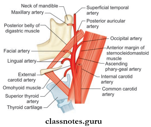
Blood Vessels Of Head And Neck landmarks To Which The External Cartoid Artery, And Its Bracnhes, Are Related

  • One of the terminal branches of common carotid artery
  • Supplies the structures in front of the neck and external to head.

External Carotid Artery Course

  • It extends upwards from the upper border of lamina of thyroid cartilage to a point behind the neck of mandible
  • It is a slightly curved course where it is anteromedial to internal carotid artery in the lower part and anterolateral in the upper part
  • At the point behind the neck of mandible, it terminates in the substance of parotid gland and divides into superficial temporal and maxillary arteries
  • In the carotid triangle, its comparatively superficial and lies in under cover of the sternocleidomastoid, and above the carotid artery lie deep in substance of the parotid gland.

External Carotid Artery Branches

  • Superior Thyroid Artery: Arises from the front of external carotid artery just below the level of greater cornu of hyoid bone and reaches upper pole of thyroid gland to supply it.
  • Lingual Artery
    • Arises from the front of the external carotid artery opposite to tip of greater cornu of hyoid bone and is divided into 3 parts by hyoglossus muscle
    • It is the main artery to the tongue.
  • Facial Artery
    • Arises from the front of external carotid artery just above the tip of greater cornu of hyoid bone
    • It has 2 parts: The cervical part and facial part.
  • Occipital Artery
    • Arises from the posterior aspect of external carotid artery at the same level of facial artery
    • It supplies most of the back of scalp.
  • Posterior Auricular Artery
    • Also, arise from posterior aspect a little above the occipital artery
    • It mainly supplies the ear.
  • Ascending Pharyngeal Artery
    • Slender artery arising from the medial aspect of the external carotid artery near its lower end.
  • Superficial Temporal Artery
    • Smaller terminal branches of the external carotid artery
    • Divides into anterior and posterior branches to supply temple and scalp.
  • Maxillary Artery
    • Larger terminal branches of the external carotid artery
    • Has three parts mandibular, pterygoid part and pterygopalatine part
    • Supplies both jaws, temporal and infratemporal fossa, nose and paranasal sinuses, palate, external and middle ear, and dura mater.
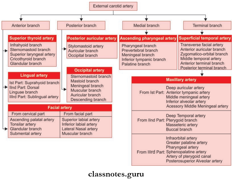
Blood Vessels Of Head And Neck External Cartoid Artery

External Carotid Artery Applied: The descending branch of the occipital artery provides the chief collateral circulation after ligation of external carotid artery.

Question 3. Write a note on the facial artery.
Answer:

Facial artery

It is a branch of external carotid artery and is the chief artery of the face.

Facial Artery Origin; It arises from front of the external carotid artery above the tip of greater cornu of hyoid bone.
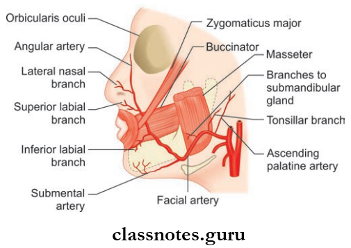
Blood Vessels Of Head And Neck Braches Of the Facial Artery

Facial Artery Course: It is divided into 2 parts

  1. Cervical and
  2. Facial part.

1. Cervical Part

  • After a looped course in the submandibular region, it enters into the face by winding around lower border of body mandible by piercing deep cervical fascia
  • Looped course—ascends upwards on superior constrictor deep to digastric and stylohyoid muscle and passes deep to ramus of mandible and grooves the posterior border of the submandibular gland and makes ‘s-shaped bend (fist bending over the submandibular gland, then up over the base of mandible).

2. Facial Part

  • Begins at the lower border of body of mandible at anteroinferior angle of the masseter by piercing fascia colli and pass fist upwards and forward to a point 1.25 cm lateral to angle of mouth
  • It ascends along the side of nose to medial canthus/medial angle of eye
  • Near the medial angle it ends by anastomosing with nasal branch of ophthalmic artery (anastomosis between internal carotid artery and external carotid artery)
  • The terminal part is known as angular artery.

Facial Artery Branches

  • Branches of Cervical Part
    • Ascending palatine artery which mainly supply the palate
    • Tonsillar artery which supplies tonsil
    • The glandular branch which supply the submandibular gland
    • Submental artery which supplies mylohyoid, submandibular and sublingual glands.
  • Branches of Facial Part
    • Inferior labial artery which supply the lower lip
    • Superior labial artery which supply lower lip
    • Lateral nasal artery which supply alae and dorsum of nose
    • Muscular branches.

Facial Artery Applied: Tortuosity of facial artery prevents its wall from being stretched during movements of face.

Question 4. Write a short note on occipital artery.
Answer:

Occipital Artery Origin: Arises from external carotid artery opposite to the origin of facial artery.

Occipital Artery Course

  • Runs backwards and upwards deep to the posterior belly of digastric crossing carotid sheath; 11th and 12th cranial nerves
  • Then it runs deep to the mastoid process and the muscles attached to it
  • It then crosses muscles at the apex of the posterior triangle and it pierces trapezius 2.5 cm from the midline
  • It has a tortuous course in the superficial fascia of scalp.

Occipital Artery Branches

  • Mastoid branches
  • Meningeal branches
  • Muscular branches
    • Descending branch
      • The superficial branch which anastomoses with superficial branch of transverse cervical artery
      • The deep branch which anastomoses with vertebral and deep cervical arteries.
    • Sternocleidomastoid branch.

Question 5. Write a short note on the maxillary artery.
Answer:

Large terminal branch of external carotid artery.

Maxillary Artery Course

  • Arises behind the neck of mandible
  • It then runs horizontally and forwards to the lower border of lower head of lateral pterygoid
  • Then it runs upwards and forwards and cross the lower head of lateral pterygoid
  • It then emerges between 2 heads of lateral pterygoid and enter the pterygopalatine fossa and terminates by dividing into branches.

Maxillary Artery Areas Supplied

  • Upper and lower jaw
  • Muscles of temporal and infratemporal fossa
  • Nose and paranasal sinuses
  • Palate and root of the pharynx
  • External and middle ear
  • Eustachian tube and dura mater.

Maxillary Artery Parts: It is divided into 3 parts by lower head of the lateral pterygoid.

  1. The first part (mandibular part)
    1. Extends from origin to lower border of lateral pterygoid
    2. Lies between the neck of mandible and the sphenomandibular ligament.
  2. Second part (pterygoid part): From lower border to upper border of lower head of lateral pterygoid muscle.
  3. Third part (pterygopalatine part): It extends from upper border of lower head of lateral pterygoid to the pterygopalatine fossa.
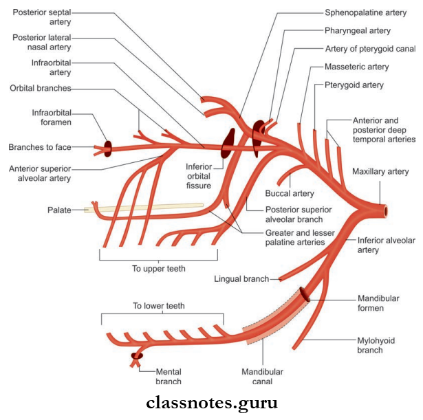
Blood Vessels Of Head And Neck Braches Of The Maxillary Artery

Maxillary Artery Branches

  • Branches from 1st Part: Consist of five branches
    • Deep auricular artery which supplies skin of external acoustic meatus and tympanic membrane
    • Anterior tympanic artery which supplies inner surface of tympanic membrane
    • The middle meningeal artery which supplies the meninges
    • The accessory middle meningeal artery which supplies meninges and structures in the infratemporal fossa
    • The inferior alveolar artery supplies the teeth, gums, and skin of chin.
  • Branches from 2nd Part: Consist of four branches
    • Deep temporal artery which supplies temporalis muscle
    • Pterygoid branches which supplies the medial and lateral pterygoid
    • Masseteric artery which supplies masseter muscle
    • Buccal branches which supply buccinators’ muscle.
  • Branches from 3rd Part: Consist of six branches
    • Posterior superior alveolar artery which supplies molar and premolar teeth and mucous membrane of maxillary air sinus
    • Infraorbital artery which supplies lower orbital muscles, maxillay sinus, canine and incisor teeth of upper jaw, lacrimal sac, medial angle of eye, upper lip.
    • Greater palatine artery which supplies roof of mouth and adjoining gum, soft palate, and tonsil
    • The pharyngeal artery which supplies submucosal membrane of nasopharynx, auditory tube, and sphenoid sinus
    • Artery of pterygoid canal which supplies the pharynx, auditory tube and tympanic cavity
    • Sphenopalatine artery supplies lateral wall of nose, sphenoid and ethmoid sinus.

Question 6. Write a short note on internal carotid artery.
Answer:

Internal carotid artery

  • One of the terminal branches of common carotid artery
  • Considered as an upward continuation of common carotid artery and supplies structures inside the skull and orbit and forms the principal artery to brain and eye.

Internal Carotid Artery Origin: Begins at the upper border of lamina of thyroid cartilage at the level of C4 vertebrae as a continuation of common carotid artery.
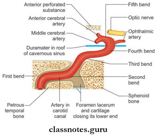
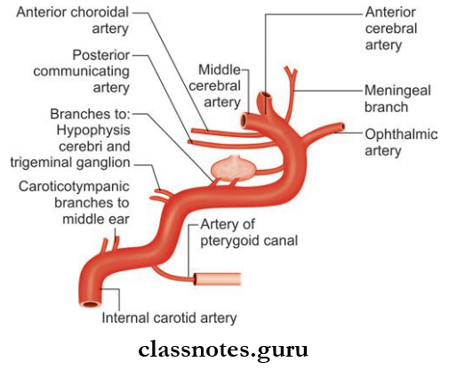
Blood Vessels Of Head And Neck Intracranial Course Of the Internal Cartoid Artery

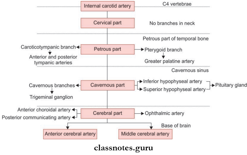
Blood Vessels Of Head And Neck Braches Given By The Internal Cartoid Artery

Internal Carotid Artery Course: It is divided into four parts for convenience.

  1. Cervical Part
    • It ascends upward from origin to the base of skull and reach the lower end of the carotid canal and lies in front of transverse process of upper cervical vertebrae
    • In the neck, it is enclosed within carotid sheath along with internal jugular vein and vagus nerve
    • The lower part of the artery lies superficial in the carotid triangle
    • Upper part lies deep to posterior belly of the digastric, styloid process and parotid gland
    • In the upper end, the jugular vein lies posterior and last four cranial nerves lie in between internal jugular vein and internal carotid artery.
  2. Petrous Part
    • Enters the petrous part of temporal bone through the carotid canal and runs upwards, forwards and medially
    • It emerges at the apex of petrous temporal bone in the posterior wall of foramen lacerum and passes upwardsm to cranial cavity.
  3. Cavernous Part
    • From foramen lacerum it ascends and enters cavernous sinus. Here, it passes forwards alongside of sella turcica and medial wall of the sinus and abducens nerve lies inferolaterally
    • In the anterior part of sinus artery pierce the dura in between anterior and middle clinoid process to reach beneath the cerebrum.
  4. Cerebral Part
    • After emerging through the dura, it turns backward in subarachnoid space along the roof of sinus and lies inferior to optic nerve
    • Later, it passes upwards by the side of optic chiasma and reaches anterior perforated substance of brain and divides into anterior and middle cerebral arteries.

Internal Carotid Artery Branches

  • No branches are given in the neck
  • Branches from petrous part
    • Caroticotympanic branches anastomose with posterior and anterior tympanic arteries
    • The pterygoid branch which anastomoses with greater palatine artery.
  • Branches from cavernous part
    • Cavernous branches to trigeminal ganglion
    • Superior and inferior hypophyseal arteries to pituitary.
  • Branches from cerebral part
    • Ophthalmic artery
    • Anterior choroid artery
    • Posterior communicating artery.
  • Terminal branches
    • Anterior cerebral artery
    • Middle cerebral artery.

Blood Vessels Of Head And Neck Braches Of Internal Cartoid Artery

Internal Carotid Artery Applied

  • The complete or partial obstruction of internal carotid arteries due to arteriosclerosis is the common cause of stroke or cerebral insufficiency.
  • The curvatures of the petrous, cavernous, and cerebral parts of the internal carotid artery together form a ‘s-shaped figure which can be seen in the angiograms as a ‘carotid siphon’.

Question 7. Write a short note on ophthalmic artery.
Answer:

Ophthalmic artery

Branch of internal carotid artery.
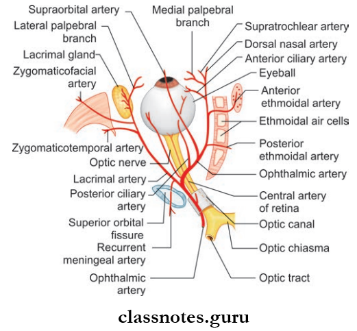
Blood Vessels Of Head And Neck Braches Of The Opthalmic Artery

Ophthalmic Artery Course

  • Arises from internal carotid artery and emerges from roof of cavernous sinus close to the optic canal
  • It then enters the orbit through the optic canal inferolateral to optic nerve
  • It then pierces duramater, ascends over the lateral side of optic nerve, and cross it from lateral to medial
  • It then runs forwards along medial wall of the orbit and terminates by dividing into supratrochlear and dorsal nasal arteries at the medial angle of eye.
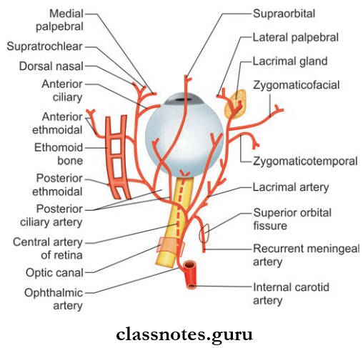
Blood Vessels Of Head And Neck Origin, Course And Branches Of Ophthalmic Artery On Right Side

Ophthalmic Artery Branches

  • Central artery of retina: Arises below optic nerve and run forward in dural sheath and pierces optic nerve and reaches optic disc and supply optic nerve and retina
  • Lacrimal artery: Arises just before the crossing of optic nerve. It passes along the upper part of lateral rectus and supply lacrimal gland, eyelids and conjunctiva
  • Posterior ciliary arteries: Long and short types which supply sclera and choroid
  • Supraorbital arteries: Accompany supraorbital nerve
  • Posterior ethmoidal artery which supply the ethmoid sinus, nasal cavity, and dura
  • Anterior ethmoidal artery which supply ethmoid sinus, medial and lateral wall of nose, and dura
  • Dorsal nasal artery: Supplying the dorsum of nose
  • Supratrochlear artery: Accompanying supratrochlear nerve and supply the forehead
  • Medial palpebral branches: Supplying eyelids.

Question 8. Write a short note on subclavian artery.
Answer:

Subclavian artery

Main source of arterial supply to upper limb.
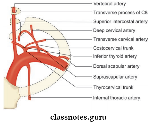
Blood Vessels Of Head And Neck Braches Of The Subclavian Artery

Subclavian Artery Origin

  • Right subclavian artery arises from the brachiocephalic trunk behind the right sternoclavicular joint
  • Left subclavian artery arises from arch of aorta and runs upward and makes groove on left lung and enters neck by passing behind left sternoclavicular joint.

Subclavian Artery Course

  • On each side, it arches laterally across the cervical pleura into the fist rib posterior to scalenus anterior muscle
  • For convenience, the subclavian artery is divided into 3 parts by scalenus anterior muscle:
    1. 1st part: From origin to medial border of scalenus anterior muscle
    2. 2nd part: Lies behind scalenus anterior muscle
    3. 3rd part: Lateral border of scalenus muscle to outer border of 1st rib.
  • It continues as axillary artery at the outer border of 1st rib.

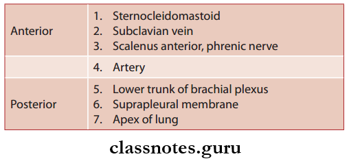
Subclavian Artery Relations

First Part (Anterior To Posterior)
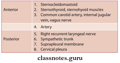
Blood Vessels Of Head And Neck Ophthalmic Artery Relation First Part

Second Part (Anterior To Posterior)

Blood Vessels Of Head And Neck Ophthalmic Artery Relation Second Part

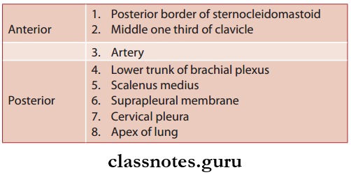
Third Part (Anterior to Posterior)

Blood Vessels Of Head And Neck Ophthalmic Artery Relation Third Part

Subclavian Artery Branches: Gives four branches

  1. Vertebral artery
  2. Internal thoracic arteries
  3. Throcervical trunk
  4. Costocervical trunk
  • Among these branches fist 3 arises from 1st part of the artery whereas costocervical trunk arises from 2nd part of artery
  • Occasionally, a large branch arises from third part of artery called the dorsal scapular artery.
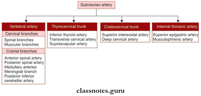
Blood Vessels Of Head And Neck Ophthalmic Artery Branches

Subclavian Artery Applied

  • In case of obstruction of subclavian artery proximal to the origin of vertebral artery, some amount of blood from the opposite vertebral artery will pass in a retrograde manner to the subclavian artery of affcted side through vertebral artery of that side to provide collaterals to upper limb on side of lesion.
  • Hence, some amount of blood from brain is stolen by subclavian artery which is called as subclavian steal syndrome.

Question 9. Write a short note on the thyrocervical trunk.
Answer:

Thyrocervical Trunk

Branch from 1st part of subclavian artery.

Thyrocervical Trunk Origin: Arises from 1st part of the subclavian artery near medial border of the scalenus anterior muscle between phrenic and vagus nerves.

Thyrocervical Trunk Branches: Inferior thyroid artery which supply the thyroid gland and gives the following branches

  1. Ascending cervical artery
  2. Inferior laryngeal artery
  3. Branches to the pharynx, trachea, and esophagus.
  • The suprascapular artery which takes part in anastomosis around scapula and also supplies muscle around the scapula, clavicle, and the acromioclavicular joint
  • Transverse cervical artery which divides into superficial and deep branch of which the deep branch takes part in anastomosis around scapula and superfiial branch accompanies the spinal root of the accessory nerve.

Question 10. Write a short note on vertebral artery.
Answer:

Vertebral Artery
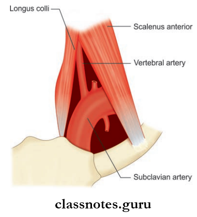
Blood Vessels Of Head And Neck Positions Of The First Part Of The Vertebral Artery

Blood Vessels Of Head And Neck Course Of The Upper Part Of The Vertebral Artery

One of the two principal arteries that supply brain and also supplies the spinal cord, meninges, the surrounding muscles, and bones.

Vertebral Artery Origin: It arises from the posterior superior aspect of fist part of subclavian artery.
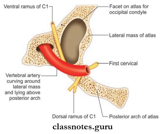
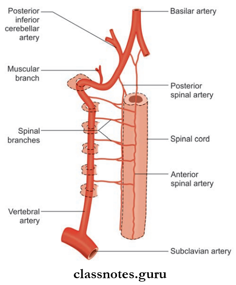
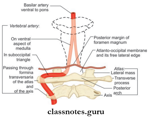
Blood Vessels Of Head And Neck Relationship Of The Vertebral Artey To The Atlas Vertebra

Blood Vessels Of Head And Neck Braches Of The Vertebral Artery

Vertebral Artery Course: It has a very long course and it is divided into four parts.

  • Vertebral Artery Course First Part: Extends from the origin to foramen transversarium of sixth cervical vertebra.
    • Vertebral Artery Course First Part Relations: It lies in the triangular space between scalenus anterior and longus colli muscle called scaleno­vertebral triangle.
      • Anteriorly­ Vertebral Vein
        • Carotid sheath
        • Inferior thyroid artery
        • Thracic duct on left side.
      • Posteriorly–Transverse Process Of C7 Vertebra
        • Ventral rami of C7, C8 nerves
        • Stellate ganglion.
  • Vertebral Artery Course Second Part: Located within foramina transversaria of upper 6 cervical vertebrae.
    • Vertebral Artery Course Second Part Relations
        • Ventral rami of C2–C6 nerves posteriorly
        • Accompanies vertebral venous plexus and large branch from the stellate ganglion.
  • Vertebral Artery Course Third Part: 
    • Extends from foramen transversarium of atlas to foramen magnum
    • Lies in the suboccipital triangle
    • The artery winds medially around the lateral process of atlas.
      • Vertebral Artery Course Third Part Relations
        • Anterior: Lateral mass of atlas
        • Posterior: Semispinalis capitis
        • Lateral: Rectus capitis lateralis
        • Medial: Ventral ramus of C1 nerve
        • Inferiorly: Dorsal ramus of C1 nerve and posterior arch of atlas.
  • Vertebral Artery Course Fourth Part
    • Intracranial part of artery
    • Extends from posterior atlantooccipital membrane to lower border of Pontomedullary junction to form basilar artery
    • In vertebral canal, it pierces dura and arachnoid and ascends in front of roots of hypoglossal nerve.

Vertebral Artery Branches

  • Extracranial Branches
    • Spinal branches arise from 2nd part and supply spinal cord, meninges, and vertebra
    • Muscular branches arise from 3rd part and supply suboccipital muscles.
  • Intracranial Branches
    • Meningeal Branches: Supply bone and meninges of posterior cranial fossa
    • Anterior Spinal Artery: Arises at termination and, supplies anterior 2/3rd of spinal cord and part of medulla
    • Posterior Spinal Artery: Arises at the side of medulla and supplies posterior 1/3rd of the spinal cord
    • Posterior Inferior Cerebellar Artery: Largest branch arises at the lower end of olive and supplies posterior lateral aspect of medulla, lower part of pons, and cerebellum
    • Medullary artery: Supply medulla.

Question 11. Write a short note on subclavian vein.
Answer:

Subclavian vein

Subclavian vein is the continuation of the axillary vein.

Subclavian Vein Extent: Begin at the outer border of first rib and extends up to medial border of scalenus anterior.

Subclavian Vein Course

  • Begin at the outer border of first rib and forms an arch across pleura below the subclavian artery
  • It ends at the medial border of the scalenus anterior by joining the internal jugular vein to form the brachiocephalic vein.

Subclavian Vein Tributaries

  • External jugular vein
  • Dorsal scapular vein
  • Thracic duct on left side and right lymphatic duct on right side.

Subclavian Vein Relations

  • Anteriorly: Clavicle and subclavius muscle
  • Posteriorly: Subclavian artery, scalenus anterior muscle, and phrenic nerve
  • Inferiorly: First rib and pleura.

Subclavian Vein Applied: The subclavian vein is commonly used to measure intracardiac pressure.

Question 12. Write a short note on external jugular vein.
Answer:

External Jugular Vein Formation: Formed by the union of posterior division of retromandibular vein and posterior auricular vein just below the angle of mandible.

External Jugular Vein Course:

  • External Jugular Vein Course then runs downwards under the cover of platysma,vertically across the sternocleidomastoid to pierce deep cervical fascia in posterior triangle near its anteroinferior angle to enter the supraclavicular space
  • Then it terminates in subclavian vein.
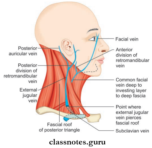
Blood Vessels Of Head And Neck Formation And Termination Of External Jugular Vein

External Jugular Vein Tributaries

  • Formative Tributaries:
    • Posterior auricular vein
    • Retromandibular vein
  • TerminalTributaries:
    • Transverse cervical vein
    • Suprascapular vein
    • Anterior jugular vein
  • Other Tributaries:
    • Posterior external jugular vein
    • Oblique jugular vein.

Mnemonic

  • External Jugular Vein: Tributaries
  • PAST PR
  • Posterior external jugular vein
  • Anterior jugular vein
  • Suprascapular vein
  • Transverse cervical vein
  • Posterior auricular vein
  • Retromandibular vein.

Question 13. Write a short note on the internal jugular vein.
Answer:

Internal Jugular Vein
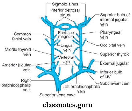
Blood Vessels Of Head And Neck Formation, Tributaries And Termionation Of Internal Jugular Vein

Internal Jugular Vein Formation: Begin as the direct continuation of sigmoid sinus at the base of skull below the jugular foramen.

Internal Jugular Vein Course: Then it descends vertically downwards and ends posterior to clavicle near its sternal end by joining the subclavian vein to form brachiocephalic vein.

Internal Jugular Vein Features

  • The Origin And Termination Presents With 2 Dilatations:
  1. The dilatation present at the origin is called superior bulb which lies in jugular fossa of the temporal bone, beneath the flor of middle ear
  2. The dilatation present at the termination is called inferior bulb which lies in the lesser supraclavicular fossa.

Internal Jugular Vein Tributaries

  • Inferior petrosal sinus
  • Pharyngeal vein
  • Common facial vein
  • Lingual vein
  • Superior thyroid vein
  • Middle thyroid vein
  • Thracic duct opens into the junction between the left internal jugular and left subclavian and right lymphaticm duct to right side simultaneously.

Internal Jugular Vein Relation

  • Superfiial (anterolateral)
    • Sternocleidomastoid
    • Posterior belly of digastric
    • Superior belly of omohyoid
    • Parotid gland
    • Styloid process
    • Internal carotid artery, glossopharyngeal nerve, vagus, spinal accessory.
  • Deep (posterior)
    • Transverse process of atlas
    • Cervical plexus
    • Scalenus anterior
    • The first part of subclavian artery.
  • Medial
    • Internal carotid artery
    • Common carotid artery
    • Vagus nerve.

Internal Jugular Vein Applied

  • The internal jugular vein is commonly used for central venous access for measuring pulmonary artery pressure, prolonged intravenous feeding, and for the introduction of cardiac pacemaker
  • The internal jugular vein is commonly injured during removal of tuberculous or neoplastic lymph nodes.

Mnemonics: Tributaries of internal jugular vein:

‘Medical Schools Let Confident People In’

From inferior to superior:

  • Middle thyroid
  • Superior thyroid
  • Lingual
  • Common facial
  • Pharyngeal
  • Inferior petrosal sinus.

Question 14. Write a short note on the brachiocephalic vein.
Answer:

Brachiocephalic vein

  • Formed by the union of internal jugular vein and subclavian vein
  • The right vein (2.5 cm) is shorter than left vein (6 cm).

Brachiocephalic Vein Course

  • Right vein runs downwards vertically and the left vein runs downwards obliquely towards the right fist coastal cartilage behind upper half of manubrium sterni
  • They unite at the lower border of 1st costal cartilage on right side to form superior vena cava.

Brachiocephalic Vein Tributaries

  • Right Brachiocephalic
    • Vertebral vein
    • Internal thoracic vein
    • Inferior thyroid vein
    • First posterior intercostal vein.
  • Left Brachiocephalic
    • Vertebral vein
    • Internal thoracic vein
    • Inferior thyroid vein
    • First posterior intercostal vein
    • Left superior intercostal vein
    • Thymic vein
    • Pericardial vein.

Question 15. Write a short note on ophthalmic veins.
Answer:

Ophthalmic Veins
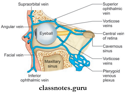
Blood Vessels Of Head And Neck Formation, Termination And Communications Of Superior And Inferior Ophthalmic Veins

  • They drain the orbit and receive tributaries which correspond to the branches of the ophthalmic artery
  • The ophthalmic Veins Include:
    • Superior Ophthalmic Vein: It accompanies ophthalmic artery and begins at medial palpebral ligament and run along with the ophthalmic artery and drains into the cavernous sinus after passing through superior orbital fissure.
    • Inferior Ophthalmic Vein
      • Runs below the optic nerve
      • It drain to either the cavernous sinus directly or through superior ophthalmic vein.

Blood Vessels Of Head And Neck Multiple Choice Question And Answers

Question 1. The vertebral artery does not pass through the foramen transversarium of:

  1. First cervical vertebra
  2. Second cervical vertebra
  3. Sixth cervical vertebra
  4. Seventh cervical vertebra

Answer: 4. Seventh cervical vertebra

Question 2. All of the following arteries arise from the thyrocervical trunk except:

  1. Inferior thyroid artery
  2. Suprascapular artery
  3. Dorsal scapular artery
  4. Superficial cervical artery

Answer: 3. Dorsal scapular artery

Question 3. The subclavian artery is divided into 3 parts by

  1. Scalenus posterior muscle
  2. Scalenus medius muscle
  3. Scalenus anterior muscle
  4. Scalenus minimus muscle

Answer: 3. Scalenus anterior muscle

Question 4. The cervical part of the facial artery gives of all of the following branches except

  1. Tonsillar artery
  2. Inferior alveolar artery
  3. Branches to submandibular gland
  4. Submental artery

Answer: 1. Tonsillar artery

Question 5. Select the incorrect statement regarding the internal jugular vein:

  1. It begins as the direct continuation of the sigmoid sinus
  2. It presents 2 dilations
  3. It is crossed by two muscles on its superficial aspect
  4. Its lower part lies in the greater supraclavicular fossa

Answer: 4. Its lower part lies in the greater supraclavicular fossa
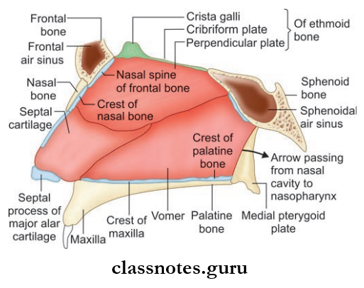
Anatomy Of Head And Neck Question and Answers

Nerves Of Head And Neck Question And Answers

Question 1. Write a short note on ansa cervicalis or ansa hypoglossi.
Answer:

Ansa Cervicalis

Nerves Of Head And Neck Mode Of Innervation Of The Infrahyoid Muscles From The Cervical Plexus

  • U-shaped nerve loop present in carotid triangle embedded to the anterior wall of the carotid sheath
  • Derived from vertebral rami of C1, C2, and C3 spinal nerves.

Ansa Cervicalis Or Ansa Hypoglossi Formation

  • Superior Root (Descendants Hypoglossi)
    • Continuation of descending branch of hypoglossal nerve whose fibers are derived from C1 spinal nerve
    • It descends over the internal and common carotid artery.
  • Inferior Root (Descendens Cervicalis)
    • Derived from C2 and C3 spinal nerves
    • It winds around the internal jugular vein and continues anteroinferior (takes a U-turn) to join the superior root in front of the common carotid artery.

Ansa Cervicalis Or Ansa Hypoglossi Distribution

  • Supplies all infrahyoid muscle except thyrohyoid
  • Throhyoid and geniohyoid are supplied by branches from C1 spinal nerve through the hypoglossal nerve
  • Superior root supplies superior belly of omohyoid.

Ansa Cervicalis Or Ansa Hypoglossi Applied: In case of paralysis of recurrent laryngeal nerve as in complicated esophageal carcinoma, nerve transplantation is being done by using ansa cervicalis after resection of recurrent laryngeal nerve.

Mnemonics: Ansa cervicalis nerve supply:

Anatomy Of Head And Neck Mneomonics

Question 2. Write a short note on the cervical part of the sympathetic trunk.
Answer:

Cervical Part Of The Sympathetic Trunk

The cervical part of the sympathetic trunk is situated on each side of the vertebral column.

Sympathetic Trunk Formation: Formed by nerves that emerge from T1 to T4 segments of the spinal cord which ascends to the neck.

Sympathetic Trunk Relations

Nerves Of Head And Neck Cervical Sympathetic Chain And Its Branches

  • Anterior
    • Carotid sheath and contents
    • Inferior thyroid artery.
  • Posterior
    • Prevertebral fascia
    • Longus capitis and cervicis muscles
    • Transverse process of C2–C7 vertebrae.

Ganglia: Due to the fusion of cervical nerves three ganglions are formed.

  • Superior Cervical Ganglion
    • Lies at the level of C2, and C3 vertebrae formed by fusion of upper four cervical connections
    • It communicates with 9, 10, and 12 cranial nerves and with external and recurrent laryngeal nerve
    • It gives internal carotid and external carotid branches to form plexus around them and also pharyngeal branches and superior cardiac branches to form pharyngeal and cardiac plexus respectively.
  • Middle Cervical Ganglion
    • Lies at the level of C6 vertebrae formed by fusion of 5th and 6th cervical ganglia connections
    • It gives grey rami communicans to C5 and C6 ventral rami and branches to thyroid, parathyroid gland, trachea, and esophagus, and also gives branches to the cardiac plexus.
  • Inferior Cervical Ganglion (Stellate Ganglion)
    • Lies at the level of C7 vertebrae
    • Formed by the fusion of 7th and 8th cervical ganglia connections
    • It gives grey rami communicans to C7 and C8 ventral rami, branches to the vertebral artery and subclavian artery to form plexus around them
    • It also give a cardiac branch to the deep cardiac plexus.

Sympathetic Trunk Applied

  • The head and neck region is supplied by sympathetic fiers arising from upper four thoracic spinal segments.
  • The preganglionic fibers pass though the stellate ganglion to relay in the superior cervical ganglion and the postganglionic fibers arise from the stellate ganglion and supply structures in the head and neck.
  • An injury to the cervical part of the sympathetic trunk produces a clinical condition known as Horner’s syndrome.
  • Horner’s Syndrome Is Characterized By The Following:
    • Partial ptosis (drooping of upper eyelid) due to paralysis of smooth part levator palpebrae superioris.
    • Miosis (constriction of pupil) due to paralysis of dilator pupillae
    • Anhydrosis (loss of sweating on that side of face) due to vasomotor denervation
    • Loss of ciliospinal reflux (pinching of the skin of neck produces dilation of pupil)
    • Enophthalmos (sunken eyeball).
  • Horner’s syndrome also can be caused due to lesion within the central nervous system anywhere at or above the 1st thoracic spinal segment.

Question 3. Write a short note on the cervical plexus.
Answer:

Cervical Plexus

Nerves Of Head And Neck Formation And Branches Of Cervical Plexus

Cervical Plexus Formation

  • Formed by ventral rami of upper four cervical nerves(C1–C4)
  • The ventral rami of C2–C4 divide to form upper and lower branches
  • The ventral ramus of C1 and branches of C2–C4 are connected with one another to form three loops
  • The first loop is directed forwards in front of the transverse process of atlas and others two loops are directed backward.

Cervical Plexus Position And Relations

  • Anteriorly
    • Prevertebral fascia
    • Internal jugular vein
    • Sternocleidomastoid.
  • Posteriorly
    • Levator scapulae
    • Scalenus medius.

Cervical Plexus Branches

  • Superficial Branches
    • Lesser occipital
    • Greater auricular
    • The transverse cutaneous nerve of neck
    • Supraclavicular.
  • Deep Branches
    • Communicating Branches
      • Grey rami from superior cervical ganglion to C1–C4 nerve roots
      • Branch from C1 join the hypoglossal nerve and take part in ansa cervicalis
      • Branch from C2 to sternocleidomastoid and branches from C3 and C4 to trapezius.
    • Muscular Branches
      • Muscles supplied only by the cervical plexus
        • Rectus capitis anterior from C1
        • Rectus capitis lateralis from C1 and C2
        • Longus capitis from C1–C3
        • Lower root of ansa cervicalis from C2, C3 to sternohyoid, sternothyroid, inferior belly of omohyoid.
      • Muscles are supplied by cervical plexus along with the brachial plexus or spinal accessory.
        • Sternocleidomastoid from C2 and spinal accessory
        • Trapezius from C3, C4 and spinal accessory
        • Levator scapulae from C3, C4 and C5
        • Phrenic nerve from C3, C4, C5
        • Longus colli C3–C8
        • Scalenus medius C3–C8
        • Scalenus anterior C4–C6
        • Scalenus posterior C6–C8.

Mnemonics: Branches of cervical plexus

‘GLAST’: Four compass points—clockwise from north on the right side of neck

Nerves Of Head And Neck Mnemonics Glast

  • Great auricular
  • Lesser occipital
  • Accessory nerve pops out between
  • L and S
  • Supraclavicular
  • Transverse cervical.

Question 4. Write a short note on phrenic nerve.
Answer:

Phrenic Nerve

Mixed nerve-carrying motor fibers to the diaphragm and sensory fibers from pleura and pericardium, diaphragm, and peritoneum (some part).

Phrenic Nerve Origin: Arises chiefly from ventral rami of the C4 nerve and receives a contribution from C3 and C5 nerves.

Phrenic Nerve Course:

  • Forms at the lateral border of scalenus anterior at the level of upper border of thyroid cartilage opposite to middle of the sternocleidomastoid
  • Then it runs vertically downwards on the anterior surface of scalenus anterior (oblique, so the nerve appears crossing obliquely from lateral border to medial border)
  • Then it runs downwards on the cervical pleura behind the brachiocephalic vein to enter thorax behind the fist costal cartilage.

Phrenic Nerve Distribution

  • Phrenic Nerve provides sensory innervation to the diaphragm, pleura, pericardium, and subdiaphragmatic pleura.
  • Phrenic Nerve provides motor supply to the diaphragm.

Phrenic Nerve Applied

  • Accessory phrenic nerve is a branch from nerve to the subclavius having C5 fiers and lies lateral to the phrenic nerve and usually join the main nerve near 1st rib
  • Pain arising from structures supplied by the phrenic nerve is referred regions supplied by spinal nerves C3–C5
  • A subphrenic abscess beneath the right diaphragm can cause pain in the right shoulder
  • Irritation of the phrenic nerve (or the tissues supplied by it) leads to the hiccup reflex. A hiccup is a spasmodic contraction of the diaphragm, which pulls air against the closed folds of the larynx.

Question 5. Write a short note on the greater occipital nerve.
Answer:

Greater Occipital Nerve

  • Large medial branch of dorsal ramus of C2 nerve
  • Thickest cutaneous nerve in the body.

Greater Occipital Nerve Course

  • Winds around the middle of the lower border of inferior oblique muscle and passes upwards and medially
  • Greater Occipital Nerve Course then crosses suboccipital triangle and pierces semispinalis capitis and trapezius.

Greater Occipital Nerve Areas Supplied: Semispinalis capitis and scalp.

Question 6. Write a short note on submandibular ganglion.
Answer:

Submandibular Ganglion

Nerves Of Head And Neck Triple Relation Of Lingual Nerve To Submandibular Duct

  • Peripheral parasympathetic ganglion
  • Relay station for secretomotor fiers to submandibular and sublingual glands
  • Topographically it is related to lingual nerve but functionally connected to chorda tympani nerve.

Submandibular Ganglion Situation: Lies on the hyoglossus muscle just above deep part of submandibular gland.

Submandibular Ganglion Relation:

Nerves Of Head And Neck Submandibular Ganglion Relation

Submandibular Ganglion Connections

  • Sensory fibers are through lingual nerve
  • Sympathetic Fibers: Derived from the sympathetic plexus around facial artery and contain postganglionic fibers from the superior cervical ganglion
  • Secretomotor Fibers: Though posterior root from lingual nerve and contain preganglionic fibers from the superior salivatory nucleus through nervus intermedius via chorda tympani and lingual nerve and relay in the ganglion.

Submandibular Ganglion Branches

  • Six branches which supply submandibular gland
  • Other fires join lingual nerve to supply the sublingual and anterior lingual nerve.

Submandibular Ganglion Secretomotor Fibers

Nerves Of Head And Neck Submandibular Ganglion Secretomotor Fibers

Question 7. Write a short note on mandibular nerve.
Answer:

Mandibular Nerve

Nerves Of Head And Neck Branches Of The Mandibular Nerve

Nerves Of Head And Neck Motor And Sensory Braches Of Mandibular Nerve In Infratemporal Fossa

  • Largest division of trigeminal nerve
  • Mixed nerve having both sensory and motor fibers
  • Nerve of fist pharyngeal arch and supply all structures derived from this arch.

Mandibular Nerve Course

  • Mandibular Nerve Course begins in the middle cranial fossa from a large sensory root and a small motor root
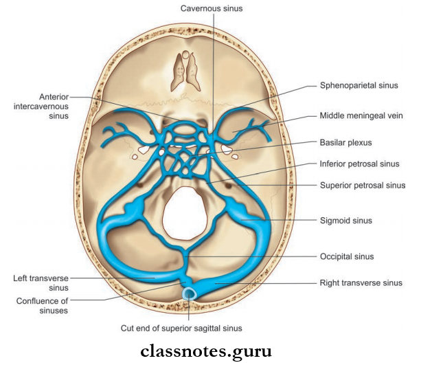
  • Sensory root arises from the lateral convex part of trigeminal ganglion whereas small motor root arises from the pons
  • Both roots pass through the foramen ovale and join in the infratemporal fossa and form main trunk
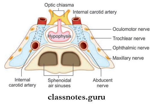
  • The trunk is short and soon divides to anterior (small) and posterior (large) division.

Relations In Infratemporal Fossa
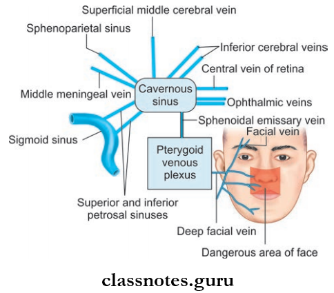
Nerves Of Head And Neck Mandibular Nerve Relations In Infratemporal Fossa
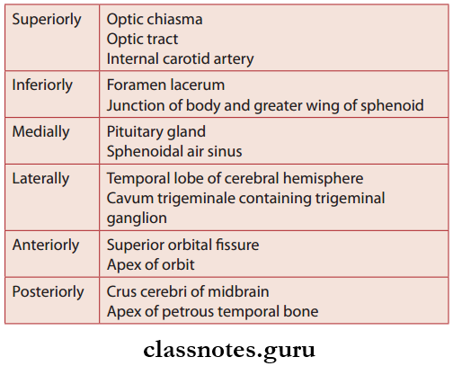
Mandibular Nerve Branches

  • From Main Trunk
    • Nervus spinosus which supplies the dura of middle cranial fossa
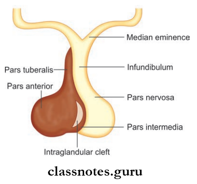
    • Nerve to medial pterygoid which supplies medial pterygoid, tensor veli palatine and tensor tympani muscles.
  • From Anterior Division
    • Masseteric nerve which supplies masseter and temporomandibular nerve
    • Deep temporal nerve which supplies the temporalis muscle
    • Nerve to lateral pterygoid which supplies lateral pterygoid muscle
    • Buccal nerve which supplies the mucous membrane of the cheek and gum of lower jaw opposite to the molars and 2nd premolar.
  • From The Posterior Division
    • The auriculotemporal nerve which supplies skin of the tragus, upper part of the pinna external auditory meatus, temporomandibular joint, the skin of the temple, secretomotor fibers to the parotid gland
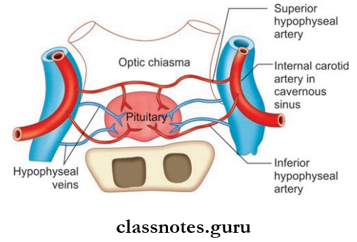
    • The lingual nerve which supplies sensory supply to floor of mouth, lingual surface of the gum, anterior 2/3rd of tongue, carries secretomotor fibers to submandibular and sublingual glands
    • Inferior alveolar nerve which supplies mylohyoid, nanterior belly of digastric, molar and premolar teeth, skin of chin and lower lip, canine and incisor teeth.

Question 8. Write a short note on the maxillary nerve.
Answer:

Maxillary Nerve

Arises from trigeminal ganglion.

Maxillary Nerve Course

  • It then runs forwards in the lateral wall of cavernous sinus just below the ophthalmic nerve
  • It then leaves the middle cranial fossa by passing through foramen rotundum and crosses upper part of the pterygopalatine fossa
  • Then it passes straight in the upper part of the fossa and enter the orbit through the inferior orbital fissure and continues as the infraorbital nerve
  • In the pterygopalatine fossa, it is related to pterygopalatine ganglion and gives ganglionic fibers, posterior superior alveolar nerves, and zygomatic nerves.

Nerves Of Head And Neck Course Of The Maxillary Nerve

Nerves Of Head And Neck Course And Distribution Of Maxillary Nerve Through Its various Sensory Branches

Zygomatic Nerve

  • Enters orbit through inferior orbital fissure and enters zygomatic bone and divides to
    • Zygomaticotemporal nerve
    • Zygomaticofacial nerve.
  • These supply skin of face and anterior part of the temple.

Posterior Superior Alveolar Nerve

  • Posterior Superior Alveolar Nerve supplies the upper molar of the same side.

Infraorbital Nerve

  • Continuation of the maxillary nerve
  • Enters orbit through the inferior orbital foramen
  • It has a common course in the orbit and then emerges on the face through the infraorbital foramen and divides into palpebral, nasal, and labial branches.
  • Infraorbital Nerve Branches
    • Middle superior alveolar nerve
    • Anterior superior alveolar nerve
    • Terminal branches
      • Palpebral
      • Nasal
      • Labial.

Maxillary Nerve Branches

  • In Middle Cranial Fossa
    • The meningeal branch which supplies dura mater of the middle cranial fossa.
  • In Pterygopalatine Fossa
    • Ganglionic branches to pterygopalatine ganglion.
    • Zygomatic nerve
      • The zygomaticotemporal branch which supplies the skin of temple
      • The zygomaticofacial branch which supplies skin of face.
    • Posterior superior alveolar nerve.
    • Supplies mucous membrane of the maxillary sinus, molar teeth, and adjoining part of gum.
  • In The Orbit
    • Middle superior alveolar nerve which supplies premolar teeth
    • Anterior superior alveolar nerve supplies canine and incisor teeth, the mucous membrane of the lateral wall, and floor of nasal cavity.
  • On Face
    • Palpebral branch which supplies the skin of lower eyelid
    • Nasal branch which supplies the skin of side of nose and mobile part of the nasal septum
    • Superior labial branch which supplies the skin and mucous membrane of upper lip.

Question 9. Write a short note on optic ganglion.
Answer:

Optic Ganglion

Peripheral parasympathetic ganglion which relays secretomotor fibers to the parotid gland.

Nerves Of Head And Neck Roots And Branches Of Optic Ganglion In Infratemporal Fossa

Optic Ganglion Size And Situation

  • 2–3 mm in size
  • Situated in infratemporal fossa below foramen ovale
  • Lies medial to mandibular nerve lateral to tensor veli palatine.

Optic Ganglion Relations

Nerves Of Head And Neck Optic Ganglion Relations

Optic Ganglion Connections And Branches

  • Motor root is formed by lesser petrosal nerve
  • The sympathetic root is derived from the plexus in the middle meningeal artery and contain postganglionic fibers arising in superior cervical ganglion and fiers pass without relay in the ganglion and reach parotid gland through the auriculotemporal nerve
  • Sensory root is from the auriculotemporal nerve
  • Other Fibers
    • Nerve to medial pterygoid: Gives a motor root to ganglion and do not relay and supplies tensor veli palatini and tensor tympani muscles
    • Chorda tympani nerve: Connected to otic ganglion and nerve of pterygoid canal and forms alternate taste pathway from anterior 2/3rd of tongue.

Secretomotor Fibers To Parotid Gland

Nerves Of Head And Neck Optic Ganglion Secretomotor Fibers To Parotid Gland

Question 10. Write a short note on pterygopalatine ganglion.
Answer:

Pterygopalatine Ganglion

  • Largest parasympathetic peripheral ganglion
  • Act as a relay station for secretomotor fibers to the lacrimal gland and to the mucous gland of nose, paranasal sinuses, palate, and pharynx
  • Topographically related to the maxillary nerve and functionally connected to the facial nerve through the greater petrosal branch.

Pterygopalatine Ganglion Location: Lies in the pterygopalatine fossa.

Nerves Of Head And Neck Pterygopalatine Ganglion Showing Its Roots And Branches

Pterygopalatine Ganglion Relations

Nerves Of Head And Neck Pterygopalatine Ganglion Relations

Pterygopalatine Ganglion Connections

  • The sensory root arises from the maxillary nerves and fibers do not relay in the ganglion
  • The fibers emerge as the branches of ganglion
  • The sympathetic root is derived from sympathetic plexus around internal carotid artery through nerve of pterygoid canal and contains postganglionic fibers from the superior cervical sympathetic ganglion
  • These fibers also do not rely in the ganglion and supply mucous membranes of nose, paranasal sinuses, nasopharynx, and palate
  • The motor root is formed by nerve of the pterygoid canal and contains preganglionic fibers from neurons present near the superior salivatory nucleus and lacrimatory nucleus
  • The fibers relay in the ganglion and postganglionic fibers supply secretomotor nerves to the lacrimal gland, mucous glands of nose, nasopharynx, paranasal sinuses, and palate.

Pterygopalatine Ganglion Branches

  • These are actually branches of the maxillary nerve which forms the sensory root
  • The Branches Include:
    • Orbital Branches: These supplies the periosteum of orbit, orbitalis muscle, ethmoid sinus, and secretomotor fibers to lacrimal gland
    • Palatine Branches: Th greater and lesser palatine nerve.
      • The greater palatine nerve supplies hard palate and lateral wall of nose while lesser palatine nerve supplies the soft palate and tonsil.
    • Nasal Branches: Supply posterior part of superior and middle conchae, roof of nose, nasal septum
    • Pharyngeal Branches: Supply part of nasopharynx behind auditory tube
    • Lacrimal Branches: Supply secretomotor fiers to lacrimal gland.

Secretomotor Fibers

Nerves Of Head And Neck Pterygopalatine Ganglion Secremotor Fibers

Question 11. Write a short note on ciliary ganglion.
Answer:

Ciliary Ganglion

  • Ciliary Ganglion is a peripheral parasympathetic ganglion
  • Topographically, it is connected to the nasociliary nerve which is branch of ophthalmic division of the trigeminal nerve but functionally it is connected to the oculomotor nerve.

Ciliary Ganglion Situation: It is a small structure measuring about 2 mm in diameter near the apex of orbit between the optic nerve and lateral rectus muscle.

Ciliary Ganglion Connections: Three roots enter the ganglion through the posterior end.

  1. Sensory root: Derived from nasociliary nerve and consist of sensory fibers from eyeball and do not relay here.
  2. Sympathetic root: Derived from the sympathetic plexus around the internal carotid artery and consist of postganglionic fibers from superior cervical ganglion and do not relay and pass through short ciliary nerve to supply dilator pupillae and blood vessels.
  3. Motor root: Derived from nerve to inferior oblique and contain preganglionic fibers from Edinger-Westphal nucleus and fiers relay here and postganglionic fibers pass through short ciliary nerve to supply ciliary muscle and sphincter pupillae.

Ciliary Ganglion Branches: Branches are short ciliary nerves about 8–10 in number.

Secretomotor Fibers

Nerves Of Head And Neck Ciliary Ganglion Secretomotor Fibers

Question 12. Write a short note on the nasociliary nerve.
Answer:

Nasociliary Nerve

One of the terminal branches of the ophthalmic division of trigeminal nerve.

Nasociliary Nerve Course

  • Begins in the lateral wall of anterior part of the cavernous sinus and enters orbit through middle part of superior orbital fissure
  • Nasociliary Nerve Course cross the optic nerve lateral to medially and runs along medial wall of orbit between the medial rectus and superior oblique
  • Nasociliary Nerve Course ends by dividing into infratrochlear and anterior ethmoidal nerves at the anterior ethmoidal foramen.

Nasociliary Nerve Branches

  • Anterior ethmoidal nerve: Large terminal branch and supplies the nose and nasal septum
  • Posterior ethmoidal nerve: Supply ethmoidal and sphenoidal air sinuses
  • Infratrochlear nerve: Small terminal branch and supply conjunctiva, lacrimal sac and caruncle, medial end of eyelid, and upper half of external nose
  • Long ciliary nerves: Supply cornea, iris, and ciliary body
  • A communicating branch to the ciliary ganglion.

Question 13. Write a note on trigeminal ganglion.
Answer:

Trigeminal Ganglion

  • Trigeminal Ganglion is the sensory ganglion of 5th cranial nerve
  • Trigeminal Ganglion is semilunar in shape with convexity directed anterolaterally.

Trigeminal Ganglion Situation And Extent

  • Trigeminal Ganglion lies in the impression on the anterior surface of petrous temporal bone near its apex
  • Trigeminal Ganglion occupies a special space of Dura called Meckel’s cave.

Nerves Of Head And Neck Trigeminal Ganglion

Nerves Of Head And Neck Dual Relations Of Trigeminal Ganglion

Trigeminal Ganglion Relations

Nerves Of Head And Neck Trigeminal Ganglion Relations

Trigeminal Ganglion Roots And Branches

  • The central process of the ganglion forms the large sensory root which is attached to the pons
  • The peripheral processes of ganglion cells form the three divisions of the trigeminal nerve—ophthalmic, maxillary, and mandibular.
  • The three divisions of trigeminal nerve originate from the convex distal surface of the ganglion.
  • The small motor root is attached to pons superomedial to the sensory root.

Trigeminal Ganglion Blood Supply: Supplied by branches of internal carotid, middle meningeal, accessory meningeal and meningeal branch of ascending pharyngeal artery.

Trigeminal Ganglion Applied

  • Trigeminal neuralgia is a paroxysmal severe pain of sudden onset and short duration in area of distribution of three divisions of trigeminal nerve
  • The sensory root of the trigeminal nerve is cut in the
  • Meckel’s cave in order to relieve the pain of trigeminal neuralgia.

Nerves Of Head And Neck Mulltiple Choice Question And Answers

Question 1. Select the incorrect statement about the inferior cervical ganglion:

  1. It is formed by the fusion of 7th and 8th cervical sympathetic ganglia
  2. It lies in front of transverse process of the C6 vertebra
  3. It is also called stellate ganglion
  4. It is connected to middle cervical sympathetic ganglia by ansa cervicalis

Answer: 2. It lies in front of the transverse process of the C6 vertebra

Question 2. Select the incorrect statement about the cervical plexus:

  1. It is formed of the ventral rami of C1 to C4 cervical spinal nerves
  2. It is called the plexus of loops
  3. It lies superfiial to prevertebral fascia
  4. Its superficial branches are cutaneous in nature

Answer: 3. It lies superficial to the prevertebral fascia

Question 3. A correct statement about the submandibular ganglion is:

  1. It is functionally related to the facial nerve
  2. It is situated on the inner surface of the hyoglossus muscle
  3. It lies above the lingual nerve
  4. It supplies preganglionic parasympathetic fibers to the submandibular gland

Answer: 1. It is functionally related to the facial nerve

Question 4. All of the following statements are true about the ansa cervicalis except:

  1. It is present in the region of the carotid triangle
  2. It is formed by the ventral rami of C1, C2, and C3 spinal nerves
  3. It supplies all the infrahyoid muscles
  4. It is embedded in the anterior wall of the carotid sheath

Answer: 3. It supplies all the infrahyoid muscles

Question 5. All of the following nerves derived from anterior division of the mandibular nerve are motor except:

  1. Masseteric nerve
  2. Buccal nerve
  3. Nerve to lateral pterygoid
  4. Deep temporal nerve

Answer: 2. Buccal nerve

Vertebral Canal Anatomy Question And Answers

Vertebral Canal And Contents Question And Answers

Question 1. Write a short note on the vertebral canal and its contents.
Answer:

Vertebral Canal

  • The continuous canal formed by vertebral foramina of vertebrae lying one below the other
  • It contains the three meninges and their spaces and the spinal cord
  • It also contains the vertebral venous plexus

The Contents Outside To Inwards Are The Following:

  • Epidural space
  • Dura mater
  • Subdural space
  • Delicate arachnoid mater
  • Subarachnoid space and CSF
  • Pia mater
  • Spinal cord and cauda equina.

Question 2. Write a short note on the vertebral venous plexus.
Answer:

Vertebral Venous Plexus

Vertebral Canal And Contents Vertebral Venous Plexus And Basivertebal Vein, Which Drains The Blood Cells From The Body Of Vertebra Into Internal Vertebral Venous Plexus

  • It is a venous system made up of a valveless complicated network of veins in a longitudinal pattern.
  • It runs parallel to anastomoses with superior and inferior vena cava.

It Has Three Subdivisions Which Interconnect With Each Other:

  1. Epidural Plexus:
    • It lies outside the dura mater in the vertebral canal
    • It drains the structures in vertebral canal and itself is drained by segmental veins (vertebral, lumbar, lateral sacral, etc.)
  2. Plexus Within Vertebral Bodies: Drains backward to epidural plexus and anterolaterally to external vertebral plexus
  3. External Vertebral Venous Plexus
    • Consists of anterior vessels in front of vertebral bodies and posterior vessels posterior to vertebral arches and on adjacent muscles
    • Drained by segmental vein
    • It also includes the suboccipital plexus.

Vertebral Venous Plexus Communication: Communicates with:

  • Intracranial venous sinuses above
  • Pelvic vein, portal vein, and caval system of vein below.

Vertebral Venous Plexus Applied

Responsible for tumors or infection from the pelvic, abdominal, or thoracic area to the spinal cord, skull, or brain.

Vertebral Canal And Contents Multiple Choice Question And Answers

Question 1. The contents of the vertebral canal include all of the following structures except:

  1. Spinal nerve roots
  2. Spinal cord
  3. Spinal ganglia
  4. Spinal meninges

Answer: 3. Spinal ganglia

Question 2. Select the incorrect statement about the internal vertebral venous plexus:

  1. It is a network of veins in the subdural space
  2. It receives basivertebral veins
  3. It drains into segmental veins
  4. It is continuous with the intracranial dural venous sinuses

Answer: 1. It is a network of veins in the subdural space