Diseases Of Thyroid And Parathyroid Glands Question And Answers

Diseases Of Thyroid And Parathyroid Glands Important Notes

  1. Hyperthyroidism and hypothyroidismDiseases Of Thrroid And Parathyroid Glands 
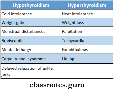
Hyperthyroidism And Hypothyroidism
  2. Goitre
    • It is the enlargement of the thyroid gland irrespective of its cause
    • Goitre Classification
      • Simple goitre
        • Diffuse hyperplastic goitre
        • Nodular goitre
        • Colloid goitre
      • Toxic goitre
        • Diffuse toxic goitre
        • Toxic nodular goitre
        • Toxic nodule
      • Neoplastic goitre
        • Benign tumours
        • Malignant tumours
      • Thyroiditis
        • Autoimmune
        • Subacute
        • Reidel’s thyroiditis
      • Other rare causes
    • Goitre Treatment
      • Addition of iodide
      • Discontinuation of offending drug
      • Administration of thyroxin
      • Surgical treatment for cosmetic purposes
  3. Retrosternal Goitre
    • Mainly derived from the lower pole of the multinodular goitre
    • Retrosternal Goitre Classification
      • Substernal
      • Plunging
      • Intra-thoracic
    • Retrosternal Goitre Treatment – resection
  4. Thyrotoxicosis Or Toxic Goitre
    • Thyrotoxicosis Or Toxic Goitre Causes
      • Grave’s disease
      • Toxic multinodular goitre
      • Toxic adenoma
      • Other rare causes
    • Thyrotoxicosis Or Toxic Goitre Types
      • Primary toxic goitre / Grave’s diseases
      • Secondary toxic goiter
  5. Thyroid carcinomaDiseases Of Thrroid And Parathyroid Glands Thyroid Carcinoma

Diseases Of Thyroid And Parathyroid Glands Long Essays

Question 1. Describe clinical features, diagnosis and management of carcinoma of the thyroid.
Answer:

Thyroid carcinoma is classified into four types:

  1. Follicular Carcinoma:
    • Follicular Carcinoma Clinical Features:
      • Occurs in old age
      • Common in females
      • An asymptomatic slow-enlarging thyroid nodule is present
      • Regional lymph nodes are involved
      • Pain is present
      • It invades adjacent structures
      • Distant metastasis occurs
    • Follicular Carcinoma Treatment:
      • Low-grade carcinoma- treated by thyroid lobectomy
      • If regional lymph nodes are involved- Dissection of lymph nodes
      • Suppressive thyroid hormone therapy
      • Aggressive follicular carcinoma- Treated by near-total thyroidectomy and postoperative radiotherapy
  2. Papillary Carcinoma
    • Papillary Carcinoma Clinical Features:
      • Occurs at any age
      • Common in females
      • Slow growing tumour
      • Tends to become malignant
      • Asymptomatic nodules appear within the thyroid gland
      • Regional lymph nodes are enlarged
    • Papillary Carcinoma Treatment:
      • Surgical excision
      • Near Total thyroidectomy
      • Suppressive dose of thyroid hormone post-operatively
  3. Medullary Carcinoma:
    • Arises from parafollicular C cells
    • Medullary Carcinoma Types:
      • Sporadic
      • Familial variety
    • Medullary Carcinoma Clinical Features:
      • Associated with mucocutaneous neuroma
      • It is present as a solid, hard, nodular tumour
    • Medullary Carcinoma Treatment:
      • Near total thyroidectomy
      • Radical block dissection of lymph nodes
  4. Anaplastic carcinoma:
    • Age- Over 50 years of age o Enlarged thyroid occurs
    • Gland is fixed
    • Consistency is hard to firm
      • Anaplastic Carcinoma Treatment:
        • Total thyroidectomy with modified neck dissection
        • Tracheostomy
        • External irradiation

Read And Learn More: General Surgery Question and Answers

Question 2. Describe clinical features, diagnosis and management of primary thyrotoxicosis.
Answer:

Primary Thyrotoxicosis Clinical Features:

  • Common in females a Loss of weight
  • Increased smooth muscle activity
  • Intolerance to heat
  • Fine tremors
  • Excitability
  • Hyperkinetic movements
  • Excessive sweating
  • Raised pulse rate
  • Diarrhoea
  • Polyuria
  • Hot moist palms
  • Systolic hypertension
  • Exophthalmos
  • Moebius sign- loss of convergence of eyeball
  • Stell Wag’s sign- infrequent blinking
  • Von Giraffe’s sign-lid lag sign

Primary Thyrotoxicosis Diagnosis:

  • Thyroid function tests- elevation of T3, T4 and decrease in TSH levels
  • Thyroid scan
  • Ultrasound- for the search of cysts

Primary Thyrotoxicosis Management:

  1. To restore the patient to the euthyroid state
    • Antithyroid drugs
      • Carbimazole-10 mg 6th hourly for 2-3 weeks
      • Propranolol-10-20 mg, two-three times a day
      • Lugol’s iodine-10-12 drops three times a day
    • Potassium perchlorate- 20 mg three times a day
  2. To reduce the functioning of thyroid mass
    • Subtotal thyroidectomy
    • Radio-iodine therapy
  3. To minimize complications
    • Proper anaesthetic measures
    • Good post-operative care

Question 3. Discuss Grave’s disease in detail.
Answer:

Grave’s Disease: It is a diffuse goitre affecting the whole functioning thyroid tissue causing hypertrophy and hyperplasia due to abnormal thyroid stimulants

Grave’s Disease Etiology

  • It is a systemic autoimmune disease
  • It is hereditary
  • Another causative factor is emotional disturbances

Grave’s Disease Clinical Features:

It consists of a classical triad

  1. Grave’s Disease Goitre
    • Characterised by an enlarged thyroid gland
    • On palpation, the gland is smooth and irregular
    • Bruit is heard
    • Extreme vascularity is present.
  2. Grave’s Disease Thyrotoxicosis
    • Common in females
    • Loss of weight
    • Increased smooth muscle activity
    • Intolerance to heat
    • Fine tremors
    • Excitability
    • Hyperkinetic movements
    • Excessive sweating
    • Raised pulse rate
    • Diarrhoea
    • Polyuria
    • Hot moist calms
    • Systolic hypertension
  3. Grave’s Disease Exophthalmos
    • It means oro tarsi on of eyeball
    • Spasm of the upper eyelid
    • Proptosis with the widening of the palpebral fissure
    • Supraorbital and infraorbital fissure
    • Congestion and edema of the conjunctive
    • Corneal ulceration

Diseases Of Thyroid And Parathyroid Glands Thyroid Simulating harmones

Grave’s Disease Investigations:

  1. Thyroid function tests
    • T3 T4 levels are high
    • TSH levels are low
  2. Presence of thyroid autoantibodies

Grave’s Disease Management

  1. To restore the patient to the euthyroid state
    • Antithyroid drugs
      • Carbimazole- 10 mg 6th hourly for 2-3 weeks
      • Propranolol-10-20 mg two-three times a day
      • Lugol s iodine- 10-12 drops three times a day
    • Potassium perchlorate- 20 mg three times a day
  2. To reduce the functioning of thyroid mass
    • Subtotal thyroidectomy
    • Radio-iodine therapy
  3. To minimize complications
    • Proper anaesthetic measures
    • Good post-operative care

Question 4. Classify thyroid tumours. Discuss etiopathology.

Answer:

Thyroid Tumours Classification

  1. Benign tumours
    • Adenoma
      • Follicular adenoma
      • Papillary adenoma
  2. Malignant tumours
    • Follicular cell origin
      • Differentiated tumours
        • Papillary carcinoma
        • Follicular carcinoma
        • Mixed papillary and follicular carcinoma
      • Undifferentiated tumours
        • Anaplastic carcinoma
    • Parafollicular cell origin
      • Medullary carcinoma
    • Non-thyroid cell origin
      • Malignant lymphoma
      • Sarcoma
      • Metastatic carcinoma

Differentiated Thyroid Tumours:

  • Differentiated thyroid tumours are:
    • Papillary carcinoma
    • Follicular carcinoma

Thyroid Tumours  Etiology:

  • Iodide deficiency
  • External irradiation
  • Ionizing radiation
  • Autoimmune thyroiditis

Question 5. Describe the etiopathology, clinical features and treatment of solitary nodules in the thyroid gland.
Answer:

Solitary Nodule In Thyroid Gland: It can be classified into benign and malignant nodules

Solitary Nodule In Thyroid Gland Etiopathogenesis:

  • Thyroiditis
  • Follicular adenoma
  • Carcinoma
  • Thyroid cyst
  • Colloid nodules

Solitary Nodule In Thyroid Gland Clinical Features:

  • Age-20-40 Years of age
  • Sex- common in females
  • Stridor
  • Tracheal deviation
  • Neck vein engorgement
  • Dyspnoea
  • Dysphagia
  • The solitary nodule is present

Solitary Nodule In Thyroid Gland Treatment:

  • Excision of solitary nodule along with a margin of normal thyroid tissue
  • If the nodule is present at the junction of the isthmus and lobe- hemithyroidectomy is done
  • In the presence of malignancy- immediate total thyroidectomy is done

Question 6. Discuss clinical features and treatment of multinodular goitre. Mention four complications of it.
Answer:

Multinodular Goitre: Multinodular goitre is the end-stage result of diffuse hyperplastic goitre

Multinodular Goitre Clinical Features:

  • Age- 40-60 years of age
  • Sex- common in females
  • Exophthalmos rarely occurs
  • The presence of long-standing swelling in front of the neck
  • Dyspnoea
  • Dysphagia
  • The gland is firm and irregular

Multinodular Goitre Treatment:

  1. Total or near-total thyroidectomy
    • It is widely used to
      • Avoid nerve injury
      • Avoid recurrence
    • Subtotal thyroidectomy
      • In it, parts of the right and left lobes are removed along with the isthmus
      • It avoids right laryngeal nerve paralysis and hypothyroidism

Multinodular Goitre Complications:

  • Calcification
  • Sudden haemorrhage
  • Dyspnoea
  • Secondary thyrotoxicosis with CVS involvement- called Plummer’s disease
  • Development of follicular carcinoma

Question 8. Discuss clinical features and management of toxic goitre.

Answer:

Toxic Goitre Or Thyrotoxicosis: It refers to an accumulation of clinical manifestations which are due to excess secretion of acute thyroid hormones.

Toxic Goitre Or Thyrotoxicosis Causes:

  • Grave’s disease
  • Toxic multinodular goitre
  • Toxic adenoma
  • The early stage of thyroiditis
  • Neonatal thyrotoxicosis
  • Iatrogenic hyperthyroidism

Toxic Goitre Or Thyrotoxicosis Types:

  • Primary toxic goitre- Grave’s disease
  • Secondary toxic goitre

Toxic Goitre Or Thyrotoxicosis Clinical Features:

  1. Primary toxic goitre
    • It consists of a classical triad
      • Goitre
        • Characterised by an enlarged thyroid gland
        • On palpation, the gland is smooth and irregular
        • Bruit is heard
        • Extreme vascularity is present
      • Thyrotoxicosis
        • Common in females
        • Loss of weight
        • Increased smooth muscle activity
        • Intolerance to heat
        • Fine tremors
        • Excitability
        • Hyperkinetic movements
        • Excessive sweating
        • Raised pulse rate
        • Diarrhoea
        • Polyuria
        • Hot moist palms
        • Systolic hypertension
    • Exophthalmos
      • It means protrusion of the eyeball
      • Spasm of the upper eyelid
      • Proptosis with the widening of the palpebral fissure
      • Supraorbital and infraorbital swelling
      • Congestion and edema of the conjunctiva
      • Corneal ulceration
  2. Secondary toxic goitre
    • Toxic multinodular goitre
      • Age- 40-60 years of age
      • Sex- common in females
      • Exophthalmos rarely occurs
      • The presence of long-standing swelling in front of the neck
      • Dyspnoea
      • Dysphagia
      • The gland is firm and irregular
    • Toxic nodule
      • Occurs at a young age
      • Females are commonly affected
      • Thyroid swelling occurs
      • Presence of hyperthyroidism
      • There is a sudden increase in the size of swelling due to necrosis and intraglandular haemorrhage

Toxic Goitre Or Thyrotoxicosis Treatment:

  1. Toxic multinodular goitre
    • To restore the patient to the euthyroid state
      • Antithyroid drugs
        • Carbimazole-10 mg 6th hourly for 2-3 weeks
        • Propranolol-10-20 mg, two-three times a day
        • Lugol’s iodine-10-12 drops three times a day
      • Potassium perchlorate- 20 mg three times a day
    • To reduce the functioning of thyroid mass
      • Subtotal thyroidectomy
      • Radio-iodine therapy
    • To minimize complications
      • Proper anaesthetic measures
      • Good post-operative care
  2. Toxic nodule
    • Anti-thyroid drugs
    • Radiotherapy
    • Subtotal thyroidectomy

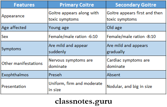
Question 9. What is toxic goitre? What are the differences between primary and secondary goitre? Outline the treatment of primary toxic goitre of the thyroid gland.
Answer:

Toxic Goitre Or Thyrotoxicosis: It refers to an accumulation of clinical manifestations which are due to an excess section of acute thyroid hormones.

Difference Between Primary and Secondary Goitre:

Diseases Of Thrroid And Parathyroid Glands Difference Between Primary And Secondary Goitre

Question 10. Define goitre. Classify various types of goitre.
Answer:

Goitre Definition: Goitre refers to as enlargement of the thyroid gland irrespective of the cause

Goitre Classification

  • Simple goitre
    • Diffuse hyperplastic goitre
    • Nodular goitre
    • Colloid goitre
  • Toxic goitre
    • Diffuse toxic goitre
    • Toxic nodular goitre
    • Toxic nodule
  • Neoplastic goitre
    • Benign tumours
    • Malignant tumours
  • Thyroiditis
    • Autoimmune thyroiditis
    • Subacute thyroiditis
    • Reidel’s thyroiditis
  • Other rare causes

Diseases Of Thyroid And Parathyroid Glands Short Essays

Question 1. Hypocalcaemia Or Tetany
Answer:

Hypocalcaemia Or Tetany

Hypocalcaemia is defined as the presence of low calcium levels in the blood

Hypocalcaemia Or Tetany Causes:

  • Acute pancreatitis
  • Acute and chronic renal failure
  • Pancreatic and small intestinal fistulae
  • Hypoparathyroidism
  • Transient hypocalcaemia

Hypocalcaemia Or Tetany Clinical Features:

  • Numbness and tingling sensation in the circumoral region and the tips of the fingers and toes
  • Hyperactive tendon jerks
  • Muscle cramps
  • Carpopedal spasms
  • Positive Chvostek’s sign

Hypocalcaemia Or Tetany Treatment:

  • Intravenous administration of calcium gluconate or chloride
  • Oral administration of calcium lactate along with vitamin D

Question 2. Thyroid crisis Or Thyroid storm

Answer:

Thyroid crisis Or Thyroid storm

  • It is a severe thyrotoxic reaction
  • It appears suddenly within 3-4 days after the operation
  • It is an acute exacerbation of hyperthyroidism

Thyroid crisis Or Thyroid storm Clinical Features:

  • Tachycardia
  • Fever
  • Restlessness
  • Delirium

Thyroid crisis Or Thyroid storm Treatment:

  • Sedatives- morphine or pethidine
  • Control of hyperpyrexia- by use of ice bag, tepid sponging, hypothermic blanket
  • Oxygen administration
  • Potassium administration
  • Use of 100 mg cortisone
  • Lugol’s iodine is given intravenously
  • 20-40 mg propranolol
  • Digitalis to treat atrial fibrillation

Question 3. Adenoma Thyroid
Answer:

Adenoma Thyroid

All benign tumours of the thyroid gland arise from glandular tissues and are called adenomas

Adenoma Thyroid Types:

  • Follicular adenoma
  • Papillary adenoma

Adenoma Thyroid Clinical Features:

  • Gradual swelling of the thyroid gland
  • Swelling is palpable
  • Bleeding in the tumour causes pain and a rapid increase in the size of the swelling

Adenoma Thyroid Investigations:

X-ray of the neck- for swelling

Adenoma Thyroid Treatment:

  • Excisional biopsy
  • Lobectomy and removal of adenoma with surrounding healthy tissue
  • Replacement doses of L-thyroxine

Question 4. Hashimoto’s thyroiditis
(or)
Autoimmune thyroiditis
Answer:

Autoimmune thyroiditis

It is also called Hashimoto’s disease or lymphedema- noid goitre

Hashimoto’s Thyroiditis Etiology:

  • It is an autoimmune disease
  • Four antigens are detected
    • Thyroid cell microsomes
    • Thyroid cell nuclear component
    • Thyroglobulin
    • Nonthyroglobulin colloid

Hashimoto’s Thyroiditis Clinical Features:

  • Females are commonly affected n Onset is insidious
  • Thyroid enlargement with slight pain
  • Tenderness in the region of the thyroid
  • Mild hyperthyroidism
  • Shortness of breath
  • Fatigue
  • Increase in weight
  • Thyroid gland examination
    • Diffuse swelling
      • Nodular
      • Firm or rubbery in consistency
  • Increased incidence of rheumatoid arthritis, hemo- Iyitc anaemia, myasthenia gravis and pernicious anaemia

Hashimoto’s Thyroiditis  Treatment: Surgical excision

Question 5. Solitary nodule of thyroid
(or)
Thyroid nodule
Answer:

Solitary nodule of thyroid

It can be classified into benign and malignant nodules

Thyroid Nodule Etiopathogenesis:

  • Thyroiditis
  • Follicular adenoma
  • Carcinoma
  • Thyroid cyst
  • Colloid nodules

Thyroid Nodule Clinical Features:

  • Age- 20-40 Years of age
  • Sex- common in females
  • Stridor
  • Tracheal deviation
  • Neck vein engorgement
  • Dyspnoea
  • Dysphagia
  • The solitary nodule is present

Thyroid Nodule Treatment:

  • Excision of solitary nodule along with a margin of normal thyroid tissue
  • If the nodule is present at the junction of the isthmus and lobe- hemithyroidectomy is done
  • In the presence of malignancy- immediate total thyroidectomy is done

Question 6. Retrosternal Goitre
Answer:

Retrosternal Goitre

Retrosternal goitre is derived from the lower pole of the multinodular goitre

Retrosternal Goitre Clinical Features:

  • Usually asymptomatic
  • Symptoms occur as obstruction
    • Obstruction of trachea- Dyspnoea
    • Obstruction of the oesophagus- Dysphagia
    • Obstruction to the major veins in the thorax- Engorgement of neck veins

Retrosternal Goitre Investigations:

  • Thyroid scan
  • Thyroid function tests
  • Barium swallow
  • Chest X-ray

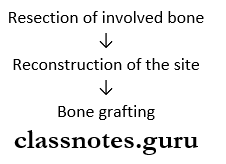
Retrosternal Goitre Treatment: Resection of retrosternal goitre

Question 7. Gasification of goitre.
Answer:

Gasification of goitre

Goitre refers to the enlargement of the thyroid gland irrespective of the cause

Gasification Of Goitre Classification:

  1. Simple goitre
    • Diffuse hyperplastic goitre
    • Nodular goitre
    • Colloid goitre
  2. Toxic goitre
    • Diffuse toxic goitre
    • Toxic nodular goitre
    • Toxic nodule
  3. Neoplastic goitre
    • Benign tumours
    • Malignant tumours
  4. Thyroiditis
    • Autoimmune thyroiditis
    • Subacute thyroiditis
    • Reidel’s thyroiditis
  5. Other rare causes

Question 8. Thyroglossal Fistula
Answer:

Thyroglossal Fistula

  • A Thyroglossal cyst may rupture unexpectedly resulting in a draining sinus known as a Thyroglossal fistula
  • It can develop when the removal of the cyst has not been fully completed
  • It is usually noticed when bleeding in the neck occurs causing swelling and fluid ejection around the original wound of removal
  • It is lined by columnar epithelium

Thyroglossal Fistula Types

  • Congenital
    • Thyroglossal fistula is a disorder of the Thyroglossal duct which develops from the thyroid primordium in the floor of the primitive pharynx at the site of foramen caecum
    • If involution of the Thyroglossal duct is incomplete, the Thyroglossal fistula may develop
  • Acquired
    • Incomplete removal of Thyroglossal cyst
    • Rupture of Thyroglossal cyst

Thyroglossal Fistula Treatment: Sistrunk operation

Diseases Of Thyroid And Parathyroid Glands Short Answers
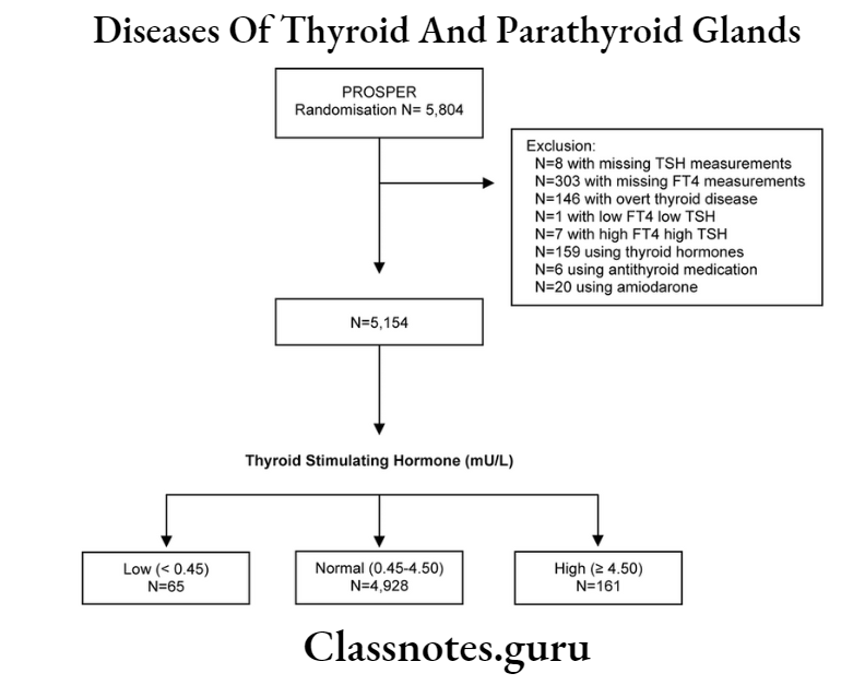
Question 1. TSH
Answer:

TSH

  • Thyroid stimulating hormone is a peptide hormone synthesized and secreted by thyrotrope cells in the anterior pituitary gland
  • It regulates the endocrine function of the thyroid gland
  • It controls the rate of secretion of thyroxine and triiodothyronine by the thyroid gland
  • It controls the rates of most intracellular chemical reactions in the body
  • TSH secretion is regulated via a negative feedback loop by T4 and T3, when their levels are low, the production of TSH is increased
  • When their levels are high, TSH production is decreased

Question 2. Goitre
Answer:

Goitre

Goitre refers to the enlargement of the thyroid gland

Goitre Clinical Features:

  • Hoarseness of voice
  • Coughing
  • Dysphagia
  • Dyspnoea

GoitreTreatment:

  • Use of antithyroid drugs
  • Use of Levothyroxine in euthyroid patients
  • Thyroidectomy

Question 3. Thyroid scan I131
Answer:

Thyroid scan I131

I131 is obtained at 24 hours

Thyroid Scan I131 Indications:

  • When a solitary nodule is palpated
  • In retrosternal goitre
  • In ectopic thyroid tissue
  • In toxic nodular goitre

Thyroid Scan I131 Uses:

  • To distinguish functioning and non-functioning thyroid nodules
  • To diagnose pathology of the thyroid gland
  • To assess the nature of the nodule
  • Detect areas of abnormality
  • Determine the spread of thyroid cancer
  • Evaluate changes in the gland

Question 4. Exophthalmos
Answer:

Exophthalmos

  • It means protrusion of the eyeball
  • Spasm of the upper eyelid
  • Proptosis with the widening of the palpebral fissure
  • Supraorbital and infraorbital swelling
  • Congestion and edema of the conjunctiva
  • Corneal ulceration

Question 5. Hoarseness of voice
Answer:

Hoarseness of voice

It is an abnormal change in voice

Hoarseness Of Voice Causes:

  • Acid reflux
  • Smoking
  • Alcohol
  • Screaming
  • Allergies
  • Inhaling toxic substances
  • Coughing
  • Polyps on the vocal cords
  • Throat, thyroid or lung cancer
  • Damage to the throat
  • Aortic aneurysm

Hoarseness Of Voice Treatment:

  • Rest your voice for a few days
  • Avoid talking and shouting B Drink plenty of fluids
  • Take hot showers n Use lozenges Avoid smoking
  • Eliminate allergens

Question 6. Causes of dyspnoea in goitre
Answer:

Causes of dyspnoea in goitre

  • Pressure due to enlarged thyroid gland
  • Formation of haematoma
  • Tracheomalacia
  • Bilateral recurrent laryngeal nerve injury
  • Hypocalcaemia

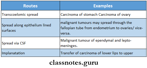
Question 7. Metastasis in papillary carcinoma of the thyroid.
Answer:

Metastasis in papillary carcinoma of the thyroid

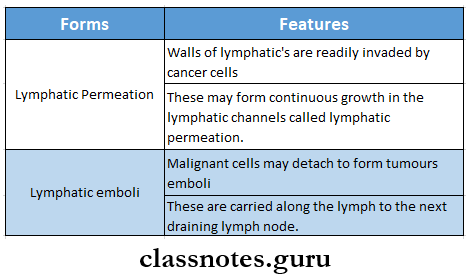
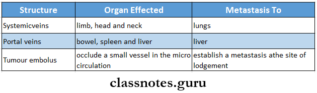
  • Papillary carcinoma of the thyroid invades the lymphatic’s and spreads to other sites
  • Vascular invasion is rare
  • If occurs, it occurs through the lungs and bones
  • Direct extension into soft tissues of the neck occurs in approx. 25% of cases

Question 8. Post-operative complications of thyroid surgery.
Answer:

Thyroid Surgery Complications:

  1. Minor complications
    • Seromas
    • Poor scar formation
  2. Rare complications
    • Damage to the sympathetic trunk
  3. Major complications
    • Bleeding
    • Injury to recurrent laryngeal nerve
    • Hypoparathyroidism
    • Thyrotoxic storm
    • Injury to superior laryngeal nerve
    • Infection

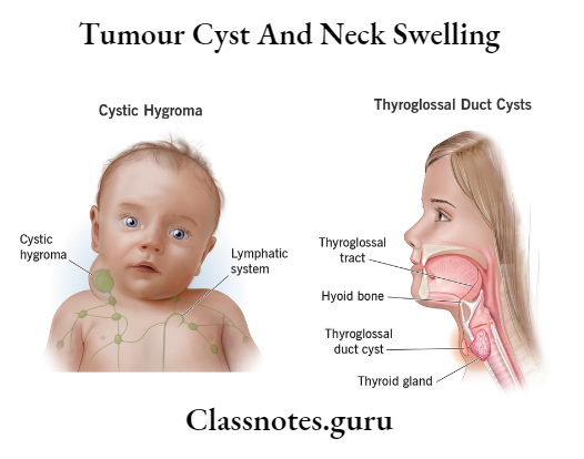
Question 9. Thyroglossal cyst
Answer:

Thyroglossal Cyst: It is a cystic swelling developed in the remnant of the thyroglossal tract

Thyroglossal Cyst Etiology: Develops from the ventral portion of the endoderm between the first and second branchial arch

Thyroglossal Cyst Clinical Features:

  • Age- common between 15-30 years of age n Sex- common in women h Pain in cyst
  • Size: 0.5 cm to 5 cm in diameter
  • Shape: spherical or oval
  • Cysfbecomes tender
  • It may develop anywhere along the thyroglossal tract
  • Firm or hard in consistency
  • Cyst moves with deglutition

Thyroglossal Cyst Management: Excision of the cyst

Question 10. Plunging goitre
Answer:

Plunging goitre

  • Plunging goitre is also called wandering goitre or diving goitre
  • It is freely movable above or below the sternal notch
  • It is usually not palpable
  • It is palpable during coughing, and sneezing due to increased intrathoracic pressure

Question 11. Hypercalcaemia
Answer:

Hypercalcaemia

Hypercalcaemia is a condition in which the calcium level in the blood is above normal

Hypercalcaemia Causes:

  • Overactive parathyroid glands
  • Cancer
  • Calcium and vitamin D supplements

Hypercalcaemia Clinical features

  1. Excessive thirst and frequent urination
  2. Nausea, vomiting, constipation
  3. Bone pain. Muscular weakness
  4. Confusion, lethargy, depression
  5. Palpitation, cardiac arrhythmia

Question 12. Gynaecomastia
Answer:

Gynaecomastia

It is an endocrine disorder in which a noncancerous increase in size of male breast tissue occurs

Gynaecomastia Causes:

  • Altered ratio of Estrogen to androgen
  • Diseases like liver disease, kidney failure, low testosterone
  • Certain medications

Gynaecomastia Clinical features

  • Male breast enlargement with soft, compressible and mobile subcutaneous chest tissue
  • Enlargement may occur on one side or both
  • Asymmetry of chest tissue occurs

Diseases Of Thyroid And Parathyroid Glands Viva Voce

Among simple goitre, nodular goitre is more prone to malignant transformation

Infections And Diseases Of The Larynx And Nasopharynx Question And Answers

Infections And Diseases Of The Larynx And Nasopharynx Important Notes

  1. Arteries supplying tonsils
    • Anterior tonsillar
    • Posterior tonsillar
    • Ascending pharyngeal
    • Superior tonsillar
    • Inferior tonsillar
  2. Acute tonsillitis
    • It is generalised inflammation of the tonsils
    • Causative organisms
      • Viral
      • Bacterial
    • Acute Tonsillitis Types
      • Acute catarrhal tonsillitis
      • Acute follicular tonsillitis
      • Acute parenchymatous tonsillitis
      • Acute membranous tonsillitis
    • Acute Tonsillitis Clinical features
      • Sore throat
      • Difficulty or painful swallowing
      • Earache
      • Enlai’ged and congested tonsils
      • Cervical lymphadenopathy
  3. Quinsy
    • Collection of pus in peritonsillar space is called quinsy
    • Causative organisms
      • Streptococcus pyogenes
      • Staphylococcus aureus
      • Anaerobes
    • Quinsy Clinical features
      • Severe pain in throat
      • High temperature
      • Dysphagia
      • Dribbling of saliva
      • Trismus
      • Torticollis
      • Bulging of tonsillar, peritonsillar, and palatal region
      • Cervical lymph nodes are tender and enlarged
  4. Ryle’s tube
    • It is one meter long tube made up of red rubber or plastic
    • It has got markings at different levelsInfections And Diseases Of The Larynx And Nasopharynx Ryle's Tube
  5. Obstructive lesions of oesophagus
    • Oesophageal stricture
    • Oesophageal cancer
    • Lower oesophageal ring
    • Dysphagia
    • Scleroderma
    • Achalasia


Infections And Diseases Of The Larynx And Nasopharynx Long Essays

Question 1. Describe signs, symptoms, and treatment of carcinoma of larynx
Answer:

Carcinoma of Larynx: It is a malignancy involving true vocal cords and anterior and posterior commissures

Carcinoma of Larynx Clinical Features:

  • Carcinoma of Larynx Types
    • Supraglottic
      • Throat pain
      • Dysphagia
      • Referred ear pain
      • Lump in the neck
      • Hoarseness of voice
      • Weight loss
      • Respiratory obstruction
      • Persistent cough
      • Haemoptysis
      • Halitosis

Read And Learn More: General Surgery Question and Answers

    • Glottic
      • Hoarseness of voice
      • Stridor
    • Subglottic
      • Stridor
      • Hoarseness of voice

Carcinoma of Larynx Investigations:

  • Chest X-ray- to rule out lung disease
  • Lateral veiw of the neck- to detect extension of tumor
  • CT scan- to detect
    • Extension of tumour
    • Cartilage invasion
    • Nodal metastasis
  • Direct laryngoscopy
    • For staging of tumour
  • Microlaryngoscopy
  • Biopsy- to confirm diagnosis

Carcinoma of Larynx Treatment:

  • Radiotherapy
    • Indicated in early lesions when the vocal cords are not involved
  • Surgery
    • Done to
      • Preserve voice
      • Prevent permanent tracheostoma
      • Allow adequate resection of tumour
    • Total laryngectomy
      • Structures removed are
        • Entire larynx
        • Hyoid bone
        • Pre-glottic space
        • Strap muscles
        • Tracheal rings
  • Combined therapy

Question 2. Discuss the differential diagnosis of obstructive lesions in the oesophagus
Answer:

Various differential diagnosis of obstructive lesions in the oesophagus are as follows:

  • Oesophageal stricture
    • It is complication of acid reflux
    • Oesophagus Causes
      • GERD
      • Zollinger- Ellison syndrome
      • Trauma from nasogastric tube placement
      • Chronic acid exposure
    • Oesophagus Features
      • Progressive dysphagia
  • Oesophageal Cancer
    • Oesophageal Cancer Clinical features
      • Progressive dysphagia
      • Insidious in onset
      • Retrosternal discomfort
      • Indigestion
      • Weight loss
      • Mild anaemia
      • Hoarseness of voice
    • Oesophageal Cancer Treatment
      • Radiotherapy
      • Surgery
        • Resection
        • Intubation
        • Laser photocoagulation
        • Diathermy
  • Lower oesophageal ring
    • Schatzki’s ring
    • It is an oesophageal web in its lowermost part
    • It contains only mucosa and submucosa
    • Covered by squamous epithelium- above and columnar epithelium- below
    • Appears at the Squamocolumnar junction
    • It is asymptomatic
  • Dysphagia lusoria
    • It is caused by compression of the oesophagus from any congenital vascular abnormality
    • The abnormality may be in
      • Aberrant right subclavian artery
      • Double aortic arch
      • Right aortic arch with left ligament- martyrium
  • Scleroderma
    • It is a collagen vascular disease of unknown etiology
    • Scleroderma Features
      • Induration of skin
      • Fibrous replacement of the smooth muscle of internal organs
      • Dysphagia
      • Severe heartburn
    • Scleroderma Treatment
    • Treat gastro-oesophageal reflux disorder
    • Use of H2 inhibitors like cimetidine or ranitidine
  • Achalasia
    • In it peristalsis is absent
    • Lower oesophageal sphincter fails to relax during swallowing
    • Achalasia Features
      • Dysphagia
      • Regurgitation
      • Weight loss
    • Achalasia Treatment
      • Use of calcium channel antagonist
      • Mechanical dilatation
      • Oesophago cardiomyotomy

Question 3. Mention the indications for tracheostomy. Describe the steps of the operation
Answer:

Tracheostomy: It is a procedure of making an opening in anterior wall of trachea and converting it into a stoma on skin surface

Tracheostomy Indications:

  • To bypass obstructions
    • Infections
    • Trauma to larynx
    • Tumour
    • Foreign body
    • Laryngeal edema
    • Congenital anomaly
    • Supraglottic or glottic pathologic condition
  • Retained secretions
    • Comatose patient
    • Respiratory muscle paralysis
    • Painful cough
    • Aspiration of pharyngeal secretions
  • Respiratory insufficiency
    • Emphysema
    • Chronic bronchitis
  • Facial fractures
  • To provide a long-term route for mechanical ventilation
  • Prophylactic
  • Severe sleep apnoea

Tracheostomy Steps:

  • Position the unconscious patient in a supine position with the neck extended
  • Vertical skin incision is given n Veins are ligated n Muscles are separated
  • Subcutaneous fat is removed with electrocautery to aid in exposure
  • Thyroid gland is retracted upwards n Trachea is exposed
  • 4% xylocaine is infiltrated into the trachea
  • Suction secretions and blood out of the lumen
  • n Place appropriate size tracheostomy tube with an inflated cuff
  • Secure it to the skin with 4-0 permanent sutures
  • Attach a tracheostomy collar
  • Place a sponge soaked in iodine between the skin and the flange for 24 hours

Question 4. Discuss the types, postoperative management, and complication of tracheostomy.
Answer:

Tracheostomy Types:

  • Emergency
  • Elective
  • Permanent

Tracheostomy Postoperative Management:

  • The site should be kept clean and dry to minimize infection
  • Monitor
    • Bleeding
    • Breathing difficulty
    • Displacement of tube
    • Subcutaneous emphysema
  • Irrigate with normal saline regularly
  • Suctioning done regularly in every Vi hour
  • Use of humified oxygen
  • Use of mucolytic agents
  • Deflate cuff every hour for 5 minutes

Tracheostomy Complications:

  • Immediate complications
    • Bleeding
    • Apnoea
    • Pneumothorax
    • Injury to adjacent structures- recurrent laryngeal nerve, vessels, and oesophagus
    • Postobstructive pulmonary edema
    • Endotracheal tube ignition
  • Early complication
    • Bleeding- due to increased blood pressure
    • Plugging of mucus
    • Tracheitis
    • Cellulitis
    • Displacement of tube
    • Subcutaneous emphysema
  • Intermediate complications
    • Secondary infection
    • Blockage of tube
  • Late complications
    • Tracheal stenosis
    • Scar
    • Tracheomalacia
    • Tracheoesophageal fistula
    • Tracheocutaneous fistula
    • Granulation

Infections And Diseases Of The Larynx And Nasopharynx Upper respiratory tract

Infections And Diseases Of The Larynx And Nasopharynx Short Essays

Question 1. Retropharyngeal abscess
(or)
Chronic retropharyngeal abscess
Answer:

Retropharyngeal Abscess: The collection of pus in retropharyngeal space is called a retropharyngeal abscess

Retropharyngeal Abscess Types:

Infections And Diseases Of The Larynx And Nasopharynx Retropharyngeal Abscess Types

Question 2. Peritonsillar abscess
(or)
Quinsy
Answer:

Peritonsillar abscess

The collection of pus in peritonsillar space is called quinsy

Peritonsillar abscess Etiology:

  • Acute tonsilitis
  • De novo
  • Causative organisms
    • Streptococcus pyrogens
    • Staphylococcus aureus
    • Anaerobic organisms

Peritonsillar abscess Clinical Features:

  • High-grade fever with chills
  • Malaise
  • Headache
  • Neck pain
  • Throat pain
  • Dysphagia
  • Change in voice
  • Oralgia
  • Odynophagia
  • Nausea
  • Constipation
  • Trismus
  • Halitosis
  • Ipsilateral earache

Peritonsillar abscess Treatment:

  • Conservative
    • 4 fluids
    • Systemic antibiotics
    • Analgesics
    • Antipyretics
  • Surgical treatment
    • Needle aspiration- to drain the abscess
    • Incision and drainage for larger abscess
    • Tonsillectomy

Peritonsillar abscess Complications:

  • Parapharyngeal abscess
  • Laryngeal oedema
  • Septicaemia
  • Endocarditis
  • Lung abscess
  • Nephritis
  • Brain abscess
  • Jugular vein thrombosis

Question 3. Acute tonsilitis
Answer:

Acute tonsilitis

It is inflammation of the tonsil

Acute tonsilitis Types:

  • Acute catarrhal tonsillitis
    • Seen in viral infections
  • Acute follicular tonsilitis
    • Crypts are filled with purulent material
  • Acute parenchymal tonsilitis
    • Tonsils are inflamed and enlarged
  • Acute membranous tonsilitis
    • Exudates from crypts form a membrane over the surface

Acute tonsilitis Etiology:

  • Hemolytic streptococci
  • Staphylococci
  • Pneumococci

Acute tonsilitis Clinical Features:

  • Sore throat
  • Dysphagia
  • Fever
  • Earache
  • Headache
  • Abdominal pain
  • Body ache
  • Malaise
  • Fetid breath
  • Coated tongue

Acute tonsilitis Treatment

  • Bed rest
  • Increased fluid intake
  • Analgesics
  • Antibiotics

Infections And Diseases Of The Larynx And Nasopharynx Short Answers

Question 1. Quinsy
(or)
Peritonsillar abscess
Answer:

Quinsy

Collection of pus in peritonsillar space is called quinsy

Peritonsillar abscess Etiology:

  • Acute tonsilitis
  • De novo
  • Causative organisms
    • Streptococcus pyogens
    • Staphylococcus aureus
    • Anaerobic organisms

Peritonsillar abscess Clinical Features:

  • High-grade fever with chills
  • Malaise
  • Headache
  • Neck pain
  • Throat pain
  • Dysphagia
  • Change in voice
  • Oralgia
  • Odynophagia
  • Nausea
  • Constipation
  • Trismus
  • Halitosis
  • Ipsilateral earache

Question 2. Tracheitis
Answer:

Tracheitis

It is the inflammation of trachea

Tracheitis Causative Organism:

  • Staphylococcus aureus
  • Clinical Features:
  • Fever
  • Stridor
  • Tachypnoea
  • Respiratory distress
  • High WBC count
  • Cough

Tracheitis Treatment:

Systemic antibiotics

Question 3. Pharyngitis
Answer:

Pharyngitis

It is an infection of the pharynx

Pharyngitis Etiology:

  • Infections
  • Allergy
  • Trauma
  • Toxins
  • Neoplasia

Pharyngitis Clinical Features:

  • It can be acute or chronic
  • Enlarged tonsils
  • Difficulty in swallowing and breathing
  • Cough
  • Fever

Question 4. Arterial supply to tonsils
Answer:

Arterial supply to tonsils

Infections And Diseases Of The Larynx And Nasopharynx Arterial Supply To Tonsils

Question 5. Collar stud abscess
Answer:

Arterial supply to tonsils

  • It is an acute suppurative infection of a digit presenting as a stud-like blister
  • It results when a cold abscess which is deep to deep fascia ruptures through the deep fascia and forms another swelling in the subcutaneous plane which is fluctuant

Collar stud abscess Treatment:

  • Simple incision does not resolve the case
  • Nondependent aspiration avoids formation of sinus
  • Systemic antibiotics are preferreds

Question 6. Pharyngocele
Answer:

Pharyngocele

  • Pharyngocele refers to the lateral pharyngeal wall herniation

Pharyngocele Clinical features

  • Dysphagia
  • Lateral cervical mass
  • Valsalva maneuver
  • It is diagnosed by pharyngoesophageal swallow

Question 7. Endotracheal intubation
Answer:

Endotracheal intubation

  • Endotracheal intubation secures the airway by placing a tube into the trachea either via nose, mouth, or tracheostomy
  • This tube has an inflatable cuff
  • Once the tube is placed into the trachea, the cuff is inflated
  • This prevents the aspiration of debris
  • This tube is connected to an anaesthetic machine to allow the delivery of oxygen, nitrous oxide, and inhalation anesthesia
  • A throat pack is used as a supplement to the cuff to prevent aspiration of blood, saliva, and debris

Question 8. Nasogastric intubation
Answer:

Nasogastric intubation

  • Nasogastric intubation refers to insertion of nasogastric tube through the nose into the stomach

Nasogastric intubation Uses:

  • In acute gastric dilatation
  • To aspirate gastric contents in intestinal obstruction
  • To diagnose GI bleeding
  • To provide enteral feeding in comatose patients

Question 9. Acute glottic edema
Answer:

Acute glottic edema

  • Acute glotticedemaia a rare condition when not associated with an infectious disease or other clinical symptoms
  • It is one of the complication of prolonged orotracheal intubation

Acute glottic edema Clinical Features:

  • Hoarseness of voice
  • Cough
  • Short expiration
  • Swelling in the throat

Acute glottic edema Treatment:

  • Application of ice externally
  • Holding small pieces of ice in the back part of mouth and frequently swallowing a small piece
  • Alternate hot and cold application

Question 10. Ryle’s tube
Answer:

Ryle’s tube

  • It is also called nasogastric tube
  • At the end of this tube there are lead shots
  • After introducing within the stomach its position is confirmed by pushing 5-10 ml of air and auscultating in the epigastrium
  • It is a long tube having 3 marks
  • When the tube is passed upto 1st mark it enters stomach
  • Usually it is passed upto 2nd mark

Ryle’s tube Uses:

  • In acute gastric dilatation
  • To aspirate gastric contents in intestinal obstruction
  • To diagnose GI bleeding
  • To provide enteral feeding in comatose patients

Question 11. Signs and symptoms of acute pharyngitis
Answer:

Signs and symptoms of acute pharyngitis

  • Acute pharyngitis is related to sore throat
  • Symptoms are:
    • Sore throat
    • Running nose
    • Sneezing
    • Cough
    • Headache
    • Body aches
    • Muscle pain
    • Fever
    • Malaise
    • Fatigue
    • Nausea
    • Loss of appetite

Question 12. Causes of acute tonsillitis
Answer:

Causes of acute tonsillitis

  • Bacteria causing acute tonsillitis are
    • Haemolytic streptococci
    • Staphylococci
    • Pneumococci

Question 13. Singer’s nodule
Answer:

Singer’s nodule

  • It is vocal cord nodule
  • Results from repetitive overuse or misuse of the voice
  • This causes irritation of vocal cords
  • They are hard, rough, callous-like growth
  • They can small as pinhead size or large as pea
  • Site: midpoint of vocal folds
  • Occasionally associated with abnormal blood vessels
  • Women between 20-50 years of age are more prone to develop

Singer’s Nodule Clinical features

  • Hoarseness of voice
  • Shooting pain in ears
  • Coughing
  • Tiredness

Singer’s Nodule Treatment: Vocal rest

Infections And Diseases Of The Larynx And Nasopharynx Viva Voce

  1. Tonsils drains into jugulodigastric lymph nodes
  2. High tracheostomy can cause tracheal stenosis
  3. Mid-tracheostomy is ideal
  4. Low tracheostomy impinges the suprasternal notch
  5. Tracheostomy is ideal for intermittent positive pressure ventilation


Ulcers Question And Answers

Ulcers Importance Notes

1. Different ulcers

Ulcer Different Ulcers

2. Painless and painful ulcer

Ulcer Painless And Painful Ulcer

3. Edge of ulcer in different ulcers

Ulcer Edge Of Ulcer In Different Ulcers


Ulcers Short Essays

Question 1. Nonhealing ulcers

Answer:

Nonhealing ulcers are

  1. Venous ulcer: It is commenest ulcer of the leg
    • Venous Ulcer Etiology:
      • Abnormal venous hypertension in the lower third of the leg, ankle, and dorsum of foot
    • Venous Ulcer Treatment:
      • Elevation of affected limb
      • Passive movement of limb
      • Active movement of calf muscles
      • Application of blue line bandage
      • Systemic antibiotics
  2. Diabetic Ulcer:
    • Diabetic Ulcer Etiology:
      • Slight injury in glucose-laden tissues
      • Ischaemia
      • Infection
      • Peripheral neuritis
    • Diabetic Ulcer Sites:
      • Toes and feet
      • Sole
      • Leg
    • Diabetic Ulcer Feature:
      • Ulcer is deep and spreading
    • Diabetic Ulcer Treatment:
      • Diabetic control
      • Antibiotics- to control infection
      • Excision of ulcer
  3. Tuberculous Ulcer:
    • Tuberculous Ulcer Etiology:
      • Bursting of cold abscess
    • Tuberculous Ulcer Features:
      • Oval in shape
      • Multiple in number
      • Has thin reddish blue and undermined edge
      • It is usually shallow
      • Mild pain occurs
      • Presence of slight induration
    • Tuberculous Ulcer Treatment
      • Antitubercular treatment
      • Excision and grafting

E:\Flow Charts\General Surgery\Ulcers Upper Gastrointestinal Bleeding.png

Read And Learn More: General Surgery Question and Answers

Question 2. Define and classify ulcers.
Answer

Ulcers Definition: Ulcer is a break in the continuity of the covering epithelium

Ulcers Classification

1. Clinical classification

Ulcer Clinical Classification

2. Pathological classification

  • Nonspecific
    • Traumatic
      • Mechanical – dental ulcer of tongue
      • Physical – x-ray of bum
      • Chemical – application of caustics
    • Arterial
      • Atherosclerosis, Raynaud’s disease, Buerger’s disease
    • Venous – in post phlebitis limb
      • Neurogenic
      • Infective – pyogenic
      • Tropical – in people living in tropical countries
      • Crvopathic – due to cold injur)
      • Martorell’s ulcer – hypertensive
      • Bazin’s ulcer
      • Diabetic ulcer
      • Miscellaneous
  • Specific ulcers
    • Tubercular ulcer
    • Syphilitic ulcer
    • Actinomycosis
    • Meleney’s ulcer
  • Malignant
    • Epithelioma
    • Rodent ulcer
    • Malignant melanoma

Question 3. Tubercular ulcer
Answer:

Tubercular ulcer

  • Such ulcer develops due to bursting of cold abscess
  • Clinical features
  • Shape – oval in shape
  • Border – irregular crescentic
  • Number – multiple in number
  • Edge – thin reddish blue and undermined
  • Pain – slight pain is present
  • Floor – pale granulation tissue seen on the floor
  • Base – slight induration is present

Tubercular ulcer Treatment

  • Antitubercular drugs
  • Excision and skin grafting in nonhealing ulcer

Question 4. Rodent ulcer
Answer:

Rodent ulcer

  • Basal cell carcinoma is called rodent ulcer
  • Common sites are inner and outer canthus of the eye, the eyelids bridge of the nose, and around the nasolabial fold.
  • Most common pattern is a nodule-ulcerative lesion a slow-growing small nodule that undergoes central with pearly, rolled margins.
  • Tumour enlarges in size by burrowing and by destroying the tissues locally like a rodent and hence the name “rodent ulcer”.

Question 5. Marjolin’s ulcer
Answer:

Marjolin’s ulcer

  • It is squamous carcinoma arising in a chronic benign ulcer or scar
  • It is a long-standing venous ulcer

Marjolin’s ulcer Features:

  • Slow-growing malignant lesion
  • Edges may be everted and raised
  • It is a painless ulcer
  • There is no lymphatic metastasis

Marjolin’s ulcer Treatment:

Wide excision of the lesion along with a margin of at least 1 cm

Question 6. Diabetic ulcer
Answer:

Diabetic ulcer Etiology:

  • Slight injury in glucose-laden tissues
  • Ischaemia
  • Infection
  • Peripheral neuritis

Diabetic ulcer Sites:

  • foes and feet
  • Sole Leg Feature:
  • An ulcer is deep and spreading

Diabetic ulcer Treatment:

  • Diabetic control
  • Antibiotics- to control infection
  • Excision of ulcer

Question 7. Tropical ulcer
Answer:

Tropical ulcer

  • These ulcers occur in the legs and feet of persons living in tropical countries
  • Caused by Vincent’s organisms

Tropical ulcer Etiology:

  • Malnutrition
  • Anaemia
  • Avitaminosis
  • Rheumatoid arthritis

Tropical ulcer Features:

  • Edges are slightly raised
  • Presence of discharge
  • Remains of the same size for many months and years
  • It destroys the surrounding tissues
  • Pustule develops
  • This burst forms painful ulcers
  • The ulcer heals leaving a scar

Question 8. Snail track ulcer
Answer:

Snail track ulcer

  • Snail track ulcers are oral ulcers in syphilitic patients
  • They appear in 3-6 weeks after the development of chancre
  • These are small, round, and superficial erosions
  • These coalesce to form ulcers
  • Ulcers are narrow, curved, and shallow

Question 9. Arterial ulcer
Answer:

Arterial ulcer

  • Arterial ulcers are caused due to
    • Peripheral arterial disease
    • Inadequate circulation
    • Atherosclerosis
    • Trauma
    • Infection
  • It is a painful ulcer
  • Ulcers tend to be punched out
  • It destroys the whole skin and deep fascia
  • Expose the tendons on the floor of the ulcer
  • Sites involved are
    • Anterior and lateral aspects of leg
    • On the toe
    • Dorsum of the foot
    • Heel

Ulcers Viva Voce

  1. Marked induration of the edge is a characteristic feature of carcinoma.
  2. A leather slough on the floor of the ulcer is seen in the Gummatous ulcer.
  3. The black mass on the floor suggests malignant melanoma.
  4. Callous ulcer shows no tendency toward healing.

Tumour Cyst And Neck Swelling Question And Answers

Tumour Cyst And Neck Swelling Long Essays

Question 1. Describe the various types of cysts of mandible and discuss in detail about their management.
Answer:

Types Of Cysts Of Mandible Classification:

Epithelial Cyst:

  1. Odontogenic cysts
    • Developmental cysts
      • Primordial cyst
      • Gingival cysts of infants
      • Dentigerous cyst
      • CEOC
    • Inflammatory cysts
      • Radicular cyst
      • Residual cyst
      • Paradental cyst
  2. Nonodontogenic cysts
    • Naso palatine cyst
    • Globulomaxillary cyst
    • Nasolabial cyst

Non-Epithelial Cyst:

  • Simple bone cyst
  • Traumatic bone cyst
  • Solitary bone cyst
  • Aneurysmal bone cyst

Read And Learn More: General Surgery Question and Answers

Tumour Cyst And Neck Swelling Cyst Types And Treatment

Question 2. Describe clinical features, diagnosis, and management of tumor of maxilla.
Answer:

Tumour of Maxilla: It is relatively uncommon, well-circumscribed, odontogenic neoplasm characterized by the formation of multiple duct-like structures by the neoplastic epithelial cells

Tumour of Maxilla Clinical Features:

  • Age- common in young age
  • Sex: females are commonly affected
  • Site- commonly occurs in maxillary anterior region
  • It is slow enlarging tumour
  • Small, bony hard swelling is present in maxillary anterior region
  • Causes elevation of the upper lip on the involved side
  • Displacement of regional teeth
  • Mild pain and expansion of the cortical plates
  • Severe expansion of bone in severe cases
  • Associated with unerupted teeth
  • It also has extra osseous involvement in the anterior maxillary gingiva
  • Produces solitary painless, asymptomatic nodular swelling on the gingiva

Tumour of Maxilla Diagnosis:

  • Clinically- slow enlarging bony hard swelling present in the maxillary anterior region
  • Radiographically
    • Shows well-circumscribed, unilocular, radiolucent area with few small radiopaque foci
  • Microscopically
    • Spindle-shaped, neoplastic odontogenic epithelial cells appears to proliferate in multiple duct-like patterns
    • These ducts are lined by tall columnar cells
    • Central lumen is filled with eosinophilic coagulum

Tumour of Maxilla Management

  • It is treated by surgical enucleation
  • The associated tooth is removed

Question 3. What are the different types of haemangiomas? Describe treatment of each type.

Answer:

Haemangioma is a developmental malformation of blood vessels

Haemangioma Types:

  1. Capillary haemangioma
    • Arises from capillaries
    • Mainly there are three varieties of it
      • Strawberry angioma
      • Port wine stain
      • Salmon patch
    • Management:
      • Most of the lesions disappears on its own
      • If it persists following measures are undertaken
        • Excision of the lesion with skin grafting
        • Carbon dioxide snow application
        • Injection of hot water or hypertonic saline
        • Radiotherapy
        • Injection of steroid
  2. Venous haemangioma:
    • Also called cavernous haemangioma
    • Arises from veins
    • It consists of multiple dilated venous channels
    • It is a spongy swelling
    • Management:
      • Conservative treatment
        • Injection of sclerosing agent
        • Cautery
      • Surgery
        • Ligation of feeding vessels
        • Excision of the esion
        • Diathermy to control hemorrhage
  3. Arterial haemangioma:
    • Also called plexiform haemangioma
    • Arises from arteries
    • It is a type of congenital arteriovenous fistula
    • There is pulsatile swelling of arteries and the veins become arterialized
      • Management:
        • Ligation of feeding vessels
        • Therapeutic embolization of the feeding arteries
        • Excision of lesion with diathermy

Question 4. Describe the methods of spread of carcinoma, grading, and staging of carcinoma in general.
Answer:

Spread of Tumours: It is by 2 ways.

  1. Local invasion/direct spread:
    • Benign tumours:
      • Form encapsulated/circumscribed masses
      • These expand and push aside the surrounding normal tissues without actually invading, infiltrating/metastasizing.
      • Malignant tumours: They also enlarge by expansion.
      • These tumours invade via the route of least resistance
      • Often cancers extend through tissue spaces, via lymphatics, blood vessels, and perineural spaces and may penetrate bone.
      • More commonly, tumours invade thin-walled capillaries and veins than thick-walled arteries.
  2. Metastasis/Distant spread:
    • Metastasis is defined as spread of tumor by invasion in such a way that discontinuous secondary tumour mass/masses are formed at the site of lodgement.
    • Benign tumours do not metastasise while all malignant tumours with a few exceptions like gliomas of the CNS and basal cell carcinoma of skin can metastasize.

Routes of Metastasis:

1. Lymphatic spread: In general, carcinomas metastasise by lymphatic route

  • The involvement of lymph nodes by malignant cells may be of two forms.

Tumour Cyst And Neck Swelling Routes Of Metasis Lymphatic Spread

2. Haemategenous spread:

  • Sarcomas spread through hematogenous spread
  • Common site for blood borne metastasis are
    • Lung,
    • Breast,
    • Thyroid,
    • Kidney,
    • Liver
    • Prostrate and
    • Ovary.
    • spread

Tumour Cyst And Neck Swelling Routes Of Metasis Haematagenous Spread

3. Various other routes:

Tumour Cyst And Neck Swelling Various Other Routes

  • TNM STAGING:
    • T- size of tumour
    • N- lymph node involvement
    • M- distant metastasis

Tumour Cyst And Neck Swelling Routes Of Metasis Various Other Routes Stating

  • T0- No tumour present
  • Tis- Carcinoma in situ
  • T1 – Tumour 2 cm or less
  • T2- Tumour between 2-4 cm
  • T3- Tumour more than 4 cm
  • T4- Tumour invading adjacent structures
  • NO- No node involvement
  • Nl- ipsilateral lymph node involvement of 3 cm or less
  • N2- ipsilateral lymph node involvement of more than 3 cm or less than 6 cm
  • N3- Contralateral node involvement
  • MO- no metastasis
  • Ml- metastasis present

Question 5. What are the epidermal malignant lesions of skin? Describe the clinical features and outline management of squamous cell cacinoma What are the premalignant conditions of the skin?

Answer:

Epidermal malignant lesions of skin?

  • Epidermal malignant lesions of skin are
    • Basal cell carcinoma
    • Squamous cell carcinoma
    • Verrucous carcinoma
    • Malignant melanoma
    • Spindle cell carcinoma
    • Primary intra-alveolar carcinoma

Squamous Cell Carcinoma:

It is the most common epidermal malignant neoplasm derived from the stratified squamous epithelium

Squamous Cell Carcinoma Clinical Features:

  • Age: common in old age
  • Sex: Common in males
  • Sites involved are:
    • Lower lip
    • Lateral border of tongue
    • Floor of the mouth
    • Soft palate
    • Gingiva or alveolar ridge
    • Buccal mucosa
  • Associated with oral leukoplakia and erythroplakia
  • Initial symptoms are
    • Asymptomatic
    • White or red, varigated patch
    • Nodule or fissure over oral mucosa
    • Painless
  • Later symptoms
    • Fast enlarging
    • Exophytic or invasive ulcer
    • Persistent induration around periphery
    • Presence of superadded candidal infections
    • Painful lesions due to secondary infections
    • Fixation to the underlying tissues
    • Trismus
    • Invasion of the alveolar bone
    • Enlarged regional lymph nodes
    • Pathological fractures of the jaw bone

Squamous Cell Carcinoma Management:

  • Surgical excision
  • Radiotherapy
  • Chemotherapy

Premalignant Conditions:

  • It is defined as the generalised state of the body which is associated with a significantly increased risk of cancer
  • They are
    • Oral submucous fibrosis
    • Sideropenic dysphagia
    • Syphilis
    • Oral lichen planus

Question 6. Discuss in detail about etiology, pathology, clinical features, investigations, and management of basal cell carcinoma.
Answer:

Basal cell carcinoma

  • Also called as rodent ulcer. Most common tumour.
  • It is locally invasive, slow-growing tumour of middle-aged individuals which rarely metastasizes.

Basal Cell Carcinoma Etiology:

  • Prolonged exposure to strong sunlight
  • UV rays
  • Arsenic used in skin ointments.
  • Dysregulation of PTCH pathway.
  • Inherited effects of PTCH gene causing BCC, Gorlin syndrome.

Basal Cell Carcinoma Pathology:

  • It is characterized by neoplastic proliferation of basaloid epithelial cells in the form of multiple solid islands or strands
  • These cells arises from the basal cell layer of the epidermis
  • Cells in the periphery are columnar in shape
  • Central cells may be polyhedral, oval, round, or even spindle-shaped
  • Fibrous connective tissue contains large number of elastic fibres

Basal Cell Carcinoma Clinical Features:

  • Majority of lesions occur on the face, usually above a line joining lobe of the ear and the angle of the mouth.
  • Common sites are inner and outer canthus of the eye, the eyelids bridge of the nose, and around nasolabial fold.
  • Most common pattern is a nodule-ulcerative lesion a slow-growing small nodule that undergoes central with pearly, rolled margins.
  • Tumour enlarges in size by burrowing and by destroying the tissues locally like a rodent and hence the name “rodent ulcer”.

Basal Cell Carcinoma Investigations:

  • Histopathological examination of the lesion

Basal Cell Carcinoma Treatment:

  • Tumours are usually treated with complete local excision.
  • BCC responds well to radiation. Radiation is indicated in elderly patients with extensive lesions, does – 4000 – 6000 gy units.
  • Cryosurgery
  • Local chemotherapy
  • Laser beam destruction of the tumour.Tumour Cyst And Neck Swelling C ystic Hygroma And Thyroglossal Duct Cysts

Question 7. Discuss clinical features and management of jaw tumours.
(or)
Classify the tumours of the alveolar bone and describe its clinical features and management of it.
Answer:

Tumours Classification:

1. Benign Tumours:

  1. Epithelial origin
    • Ameloblastoma
    • CEOT
    • AOT
  2. With mesenchymal origin
    • Ameloblastic fibroma
    • CEOC
    • Odontoma
  3. With epithelial and mesenchymal origin:
    • Odontogenic fibroma
    • Myxoma

2. Malignant Tumours:

  1. Odontogenic carcinoma
    • Malignant ameloblastoma
    • Primary intraosseous carcinoma
  2. Odontogenic sarcoma
    • Ameloblastic fibro asrcoma
    • Ameloblastic fibro dentinosarcoma
  3. Odontogenic carcinosarcoma

Jaw Tumours Clinical Features:

  • Commonly occurs in the mandible
  • Slow enlarging, painless mass occurs
  • Bony hard swelling of the jaw
  • Expansion and distortion of the cortical plates
  • Pain
  • Paresthesia
  • Mobility of the regional teeth
  • Presence of cyst
  • Pathological fractures may occur
  • The overlying mucosa appears normal
  • Larger lesions may perforate the cortical plates

Jaw Tumours Management:

  • Enucleation

Jaw Tumours Technique:

Tumour Cyst And Neck Swelling Tumours Jewelling Flowchart

  • Currettagc

Question 8. Describe the classification, clinical features, and treatment of odontoma.
Answer:

Odontoma

Odontomes are common hamartomatous odontogenic lesions with limited growth potential

Odontoma Types:

  1. Complex odontome:
    • Consists of a massof haphazardly arranged enamel, dentin, and cementum
  2. Compound odontome:
    • Consists of collections of numerous small, discrete, tooth-like structures

Odontome Clinical Features:

  • Age: Children and young adults
  • Sex: Both
  • Site: Compound- in maxilla
  • Complex- mandible

Odontome Presentation:

  • Small asymptomatic lesion
  • Expansion of cortical plates
  • Displacement of teeth
  • Impacted or retained deciduous teeth
  • Pain, inflammation
  • Ulceration
  • Fistula formation

Radiographic Features:

  • Compound- A bag of teeth appearance
  • Complex- sunburst appearance

Radiographic Treatment:

  • Surgical enucleation

Question 9. Describe clinical features, diagnosis, and treatment of ameloblastoma.
Answer:

Ameloblastoma Clinical Features

  • Sex: Both sex
  • Age- 1st to 7th decade

Ameloblastoma Types:

  1. Solid or multicystic ameloblastoma
    • Slow-growing locally invasive tumours
    • High recurrence rate
  2. Unicystic type
    • Lesion can be enucleated
    • Rarely seen in maxilla
    • Recurrence rate is low
  3. Peripheral ameloblastoma
    • Does not invade bone
    • Treated in the early stages of development

Ameloblastoma Features:

  • Swelling and pain in the region
  • Inflammation
  • Tension
  • Dental trauma
  • Ulceration of mucosa
  • Loosening of teeth
  • Epitaxis
  • Nasal obstruction

Radiological Features:

  • Unilocular or multi locular radiolucency
  • Soap bubble appearance of the lesion
  • Border is clear
  • Resorption of the teeth

Radiological Management:

  • Forintraosseous- marginal resection
  • Segmental resection
  • Aggressive resection

Jackson’s Guidelines:

  • Tumour confined to maxilla without orbit floor involvement- Partial maxillectomy
  • Tumour involving orbital floor- Total maxillectomy
  • Tumour involving orbital contents- Total max-lobectomy.
  • Tumour involving skull bone- Neurological procedure

Question 10. Describe the pathology, clinical features, and management of adamantinoma.
Answer:

Adamantinoma: It is a relatively uncommon, well-circumscribed, odontogenic neoplasm characterized by the formation of multiple duct-like structures by the neoplastic epithelial cells

Adamantinoma Clinical Features:

  • Age- common in young age
  • Sex: females are commonly affected
  • Site- commonly occurs in maxillary anterior region
  • It is slow enlarging tumour
  • Small, bony hard swelling is present in maxillary anterior region
  • Causes elevation of the upper lip on the involved side
  • Displacement of regional teeth
  • Mild pain and expansion of the cortical plates
  • Severe expansion of bone in severe cases
  • Associated with unerupted teeth
  • It also has extra osseous involvement in the anterior maxillary gingiva
  • Produces solitary painless, asymptomatic nodular swelling on the gingiva

Adamantinoma Pathology:

  • Spindle-shaped, neoplastic odontogenic epithelial cells appears to proliferate in multiple duct-like patterns
  • These ducts are lined by tall columnar cells
  • Central lumen is filled with eosinophilic coagulum

Adamantinoma Management

  • It is treated by surgical enucleation
  • The associated tooth is removed

Question 11. Discuss the clinical features, management of cysts of the jaw.
Answer:

Cysts of The Jaw: It is pathological cavity containing fluid, semi-fluid or, gas, which is usually lined by epithelium and is not formed by the accumulation of pus

Cysts of The Jaw Management:

Enucleation of cyst:

    • Smaller single cyst through intraoral approach
    • Unilocular lesions through marginal excision
    • Large multilocular lesions

Tumour Cyst And Neck Swelling Cysts Of Jaw

Cysts of The Jaw Marsupialization:

  • It refers to creating a surgical window in the wall of the cyst and evacuation of the cystic contents

Question 12. Name the swellings arising from the jaw. Describe clinical features, diagnosis, and treatment of dentigerous cyst.
Answer:

Dentigerous Cyst Clinical Features

  • Sex: Common in males
  • Age: 1st and 3rd decade
  • Site: Mandibular 3rd molar, maxillary canines, maxillary 3rd molar
  • Expansion of bone
  • Facial asymmetry
  • Displacement of adjacent teeth
  • Resorption of adjacent teeth

Radiological Features:

  • The unilocular, well-defined radiolucency
  • Margins- sclerotic

Radiological Types:

  • Central- covering the crown of an unerupted tooth
  • Circumferential- covering the crown from all the sides
  • Lateral- covering crown from side

Radiological Management:

  • Marsupialization- in children
  • Enucleation – in adults

Question 13. Describe the midline swellings of neck. Discuss etiology, clinical features, and management of thyroglossal cyst.
Answer:

Midline Swellings of The Neck:

  • Ludwig’s angina
  • Enlarged submental lymph nodes
  • Sublingual dermoid
  • Thyroglossal cyst
  • Subhyoid bursitis
  • Goitre
  • Lipoma
  • Retrosternal goitre
  • Thymic swellings
  • Bony swelling arising from manubrium sterni

Thyroglossal Cyst:

It is a cystic swelling developed in the remnant of the thyroglossal tract

Thyroglossal Cyst Etiology:

Develops from the ventral portion of the endoderm between the first and second branchial arch

Thyroglossal Cyst Clinical Features:

  • Age- common between 15-30 years of age
  • Sex- common in women
  • Pain in cyst
  • Size: 0.5 cm to 5 cm in diameter
  • Shape: spherical or oval
  • Cyst becomes tender
  • It may develop anywhere along the thyroglossal tract
  • Firm or hard in consistency
  • Cyst moves with deglutition

Thyroglossal Cyst Management:

Excision of the cyst

Tumour Cyst And Neck Swelling Short Essays

Question 1. Dermoid cyst
Answer:

Dermoid cyst

  • Dermoid cyst is a cyst that lies deep to the skin and is lined by the skin
  • It is lined by squamous epithelium and contains toothpaste-like material

Dermoid cyst Types:

  • Sequestration dermoid
  • Implantation dermoid
  • Tubule-dermoid
  • Teratomatousdermoid

Tumour Cyst And Neck Swelling Dermoid Cyst

Question 2. Sublingual dermoid
Answer:

Sublingual dermoid

  • Sublingual dermoid is a congenital swelling
  • It is formed at the point of fusion of the two mandibular arches
  • It develops from the secretion of the sequestrated surface ectoderm at the fusion site

Sublingual Dermoid Clinical Features:

  • Present below or above the hyoid bone
  • Causes swelling of the floor of the mouth
  • Color- whitish in color
  • It is opaque
  • Age: 10-25 years of age
  • Sex: both sex are equally affected
  • It is painless swelling
  • Rarely the contents become infected and the cyst becomes painful and tense

Sublingual Dermoid Types:

  • Median variety
    • Supramylohyoid variety
      • Midline swelling on the floor of the mouth
    • Inframylohyoid variety
      • Swelling is present in the submental region resulting in double chin appearance
  • Lateral variety
    • Supramylohyoid variety
      • Opaque cystic swelling in the floor of the mouth
    • Inframylohyoid variety
      • Cystic swelling present in the submandibular region

Sublingual Dermoid Treatment:

  • Total excision
    • Supramy lohyoidvarities- approached through floor of the mouth
    • Infra mylohyoid varieties approached through the neck by curved incision

Question 3. Preauricular dermoid
Answer:

Preauricular dermoid

It is type of sequestration dermoid

Preauricular dermoid Etiology:

  • Imperfect fusion of 1st and 2ndbranchial arches in formation of the auricle communicating with an ear pit on the surface

Preauricular dermoid Clinical Features:

  • Painless swelling
  • Slow growing
  • Size- more than 2 cm in diameter
  • Ovoid or spherical in shape
  • Soft in consistency
  • Has smooth surface

Preauricular dermoid Complications

  • Infection
  • Suppuration
  • Ulceration
  • Pressure symptoms to the surrounding structures

Preauricular dermoid Treatment

  • Surgical excision

Question 4. Postauricular dermoid and External angular dermoid
Answer:

Postauricular dermoid

  • It is type of sequestration dermoid
  • Site: behind the ear- at the site of fusion of the mesodermal hillocks

External angular dermoid

  • It is type of sequestration dermoid
  • It is present above the outer canthus of the eye- at the line of fusion of the frontonasal and maxillary processes

Question 5. Lipoma
Answer:

  • Lipoma is benign neoplasm of adipose tissue
  • It is composed of mature fat cells

Lipoma Clinical Features:

  • Age: above 40 years of age
  • Sex: both sex are equally affected
  • It is soft, well-defined, movable mass
  • Smooth-surfaced
  • Nodular, pedunculated or sessile lesion
  • These are painless swelling
  • Color
    • Superficial lesions- yellow in color
    • Deep lesions- pink in color
  • Size: less than 3 cm in diameter
  • It floats in aqueous solution

Lipoma Pathology:

  • It presents well-circumscribed areas of proliferating mature fat cells within loose areolar tissue stroma
  • Cells has round, vacuolated clear cytoplasm
  • Lobules of cells are separated by fibrous tissue septa
  • May contain benign lip blasts which results in soap bubble appearance

Lipoma Treatment: Surgical excision

Question 6. Basal cell carcinoma
Answer:

Basal cell carcinoma Etiology:

  • Prolonged exposure to strong sunlight
  • UV rays
  • Arsenic used in skin ointments.
  • Dysregulation of PTCH pathway.
  • Inherited effects of PTCH gene causing BCC, gorlin syndrome.

Basal cell carcinoma Clinical Features:

  • Majority of lesions occur on the face, usually abore a line joining lobe of the ear and the angle of the mouth.
  • Common sites are the inner and outer canthus of the eye, eyelids bridge of the nose, and around the nasolabial fold.
  • Most common pattern is a nodule-ulcerative lesion a slow-growing small nodule that undergoes central with pearly, rolled margins.
  • Tumour enlarges in size by burrowing and by destroying the tissues locally like a rodent and hence the name “rodent ulcer”.

Basal cell carcinoma Investigations

  • Histopathological examination of the lesion

Basal cell carcinoma Treatment:

  • Tumours are usually treated with complete local excision.
  • BCC responds well to radiation. Radiation is indicated in elderly patients with extensive lesions, does – 4000 – 6000 gy units.
  • Cryosurgery
  • Local chemotherapy
  • Laser beam destruction of the tumour

Question 7. Premalignant lesions of the oral cavity
Answer:

Premalignant lesions of the oral cavity

  • It is defined as a morphologically altered tissue in which cancer is more likely to occur than its apparently normal counterpart.
  • They are
    • Leukoplakia
      • It is a white patch or plaque which cannot be scrapped off or stripped off easily and cannot be characterized clinically or pathologically as any other disease
      • They can be nonpalpable, faintly translucent, white areas
      • Many lesions are thick, fissured, indurated
      • Size is variable
      • Surface may be smooth or finely wrinkled
      • Color- white or grayish or yellowish-white in color
    • Erythroplakia
      • It is red patch or plaque which cannot be characterized clinically or pathologically as any other condition and which has no apparent cause
      • It has clearly defined margins
      • Types
        • Homogenous erythroplakia
        • Erythroplakia interspersed with patches of leukoplakia
        • Speckled erythroplakia
    • Stomatitis nicotine
      • It is tobacco-related keratosis
      • It affects both hard and soft palate
      • There is hyperkeratosis of the epithelium and inflammatory swelling of the palatal mucous glands
      • Surface has elevated keratotic rings
    • Chronic candidiasis
      • It is fungal infection
      • Seen in the palatal mucosa of the denture-wearing persons
      • Common in females
      • Appears as bright red, edematous, velvety area

Question 8. Epulis
Answer:

Epulis

  • Epulis is a swelling situated on the gum
  • It arises from the alveolar margin of the jaw

Epulis Types:

  • Fibrous epulis
    • Arises from the periosteum at the neck of an incisor or premolar tooth
    • It is a firm nodule at the junction of the gum and tooth
    • Polypoid in shape
    • Slow growing tumour
    • Adjacent teeth are slightly separated and loosen

Epulis Complications:

Fibrosarcoma

Epulis Treatment:

  • Excision
  • Granulomatous epulis
  • It is a mass of granulation tissue around a carious tooth
  • It looks bright red in color
  • It is soft to firm in consistency
  • It easily bleeds on touch
  • Associated with carious teeth
  • Regional lymph nodes are enlarged and tender
    • Extraction of the carious tooth
    • Replacement of ill-fitted denture
    • Maintenance of oral hygiene
    • Removal of granulation tissue
    • Diathermy of granulation tissue
    • Myeloid epulis
      • It is an osteoclastoma and arises from the underlying bone
      • Overlying mucosa becomes hyperaemic, edematous and soft to touch
      • It is sessile
      • Rapidly growing
      • Plum colored
      • Adjoining teeth are loosen and separated

Epulis Complication:

  • Ulceration
  • Serious haemorrhage

Epulis Treatment:

  • Currettage of small swelling
  • Radical excision of large swellings
  • Carcinomatous epulis
    • It is epithelioma of the gum
    • Present as lump or ulcer
    • Painful lesion
    • Invades bone
    • Regional lymph nodes are involved
    • Adequate resection of the lesio
    • Radiotherapy

Question 9. Burkitt’s tumour
Answer:

Burkitt’s tumour

  • It is a distinctive type of B-cell lymphoma caused by Epstein-Barr virus (EBV) infection.
  • 3 sub-groups of Burkitt’s lymphoma are:
    • African endemic
    • Sporadic
    • Immunodeficiency associated.
  • Etiology – EBV infection and immune suppression.

Burkitt’s tumour Features:

  • Disease affects children and adolescents
  • Involves extranodal sites, particularly the jaw, gastrointestinal tract, and gonads.

Histological appearances:

Tightly packed lymphoblasts interspersed with phagocytic macrophages which impart a starry-sky appearance in histological sections.

Question 10. Adamantium
Answer:

Adamantium

It is relatively uncommon, well-circumscribed, odontogenic neoplasm characterized by the formation of multiple duct-like structures by the neoplastic epithelial cells

Adamantium Clinical Features

  • Age- common in young age
  • Sex: females are commonly affected
  • Site- commonly occurs in maxillary anterior region
  • It is slow enlarging tumour
  • Small, bony hard swelling is present in maxillary anterior region
  • Causes elevation of the upper lip on the involved side
  • Displacement of regional teeth
  • Mild pain and expansion of the cortical plates
  • Severe expansion of bone in severe cases
  • Associated with un erupted teeth
  • It also has extra osseous involvement in the anterior maxillary gingiva
  • Produces solitary painless, asymptomatic nodular swelling on the gingiva

Adamantium Management:

  • It is treated by surgical enucleation
  • The associated tooth is removed

Question 11. Epidermoid cyst
Answer:

Epidermoid cyst

These are small, hard lumps that develop under the skin

Epidermoid cyst Causes:

  • Excessive keratin production Acne
  • Excessive sun exposure

Epidermoid cyst Clinical Features:

  • Site involved
    • Face
    • Head
    • Neck
    • Back
    • Genitals
  • Slow growing
  • Painless swelling a Size- 1/2 -2 inch in diameter
  • Color- tan to yellow in color
  • Filled with thick smelly matter

Epidermoid cyst Treatment:

  • It heals on its own
  • In case of infection- antibiotics and steroid injection is used
  • Rarely removed for cosmetic purposes

Epidermoid Cyst Prognosis: If drained it recurs again

Question 12. Adenoma
Answer:

Adenoma

  • It is benign tumour of glandular tissue
  • It consists of dense mass of acini lined by exuberant epithelium which may be columnar or cuboidal

Adenoma Types:

  • Fibroadenoma
    • It occurs in breast
    • The specialised connective tissue surrounding the glandular epithelium is involved
    • It occurs due to excessive stromal reaction
    • It is further divided into two types
      • Hard pericanalicula rfibroadenoma
      • The ducts are surrounded by dense connective tissue
    • Soft intracanlicular fibroadenoma
      • Loose connective tissue is impinged into the ducts
    • The connective tissue shows rapid growth and enlarges to a great extent
  • Cystadenoma
    • Adenoma forms cystic spaces into which papillary in growth of neoplastic epithelium occurs
    • It is called cystadenoma
    • Sites involved
      • Ovary
      • Pancreas
      • Parotid gland
      • Kidney
  • Adenoma Types
    • Serous cystadenoma- serous-producing adenoma
    • Pseudomucinous cystadenoma- mucin-producing adenoma

Question 13. Sequestration dermoid
Answer:

Sequestration dermoid

  • It is congenital type of dermoid cyst formed by inclusion of epithelium at the line of embryonic fusions

Sequestration dermoid Common Sites:

  • At the midline of the body
  • External angular
  • Post auricular
  • On the skull at the fusion of skull bones
  • At the midline of face

Sequestration dermoid Clinical Features:

  • It is congenital variety
  • Painless swelling
  • Slow growing
  • Size- more than 2 cm in diameter
  • Ovoid or spherical in shape
  • Soft in consistency
  • Has mooth surface

Sequestration dermoid Complications:

  • Infection
  • Suppuration
  • Ulceration
  • Pressure to the surrounding structures

Sequestration dermoid Treatment:

  • Complete excision of the cyst

Question 14. Implantation dermoid
Answer:

Implantation dermoid

  • It is acquired dermoid
  • It arises from indriven epithelium beneath the skin due to puncture injury

Implantation dermoid Common Sites:

  • Palm of the hand
  • Any part of finger
  • Sole

Implantation dermoid Clinical Features:

  • Swelling in the finger or palm n Painful cyst
  • Cyst is tensed n Consistency is firm to hard
  • There may be scar over the skin

Implantation dermoid Complication:

  • Infection
  • Suppuration
  • Bursting

Implantation dermoid Treatment:

Complete excision of the cyst

Question 15. Sternomastoid tumour
Answer:

Sternomastoid tumour

  • It is also called congenital torticollis

Sternomastoid tumour Etiology:

  • Infarction of the central portion of one sternomastoid muscle
  • Congenital anomaly
  • Infarction of the sternomastoid branch of the superior thyroid artery
  • Trauma

Sternomastoid tumour Clinical Features:

  • Age- several days to several months after birth
  • A lump is produced
  • Site: in the middle of the sternomastoid muscle
  • Torticolis- head is turned to one side
  • It is painful lesion
  • Size: 1-2 cm in diameter
  • It is fusiform in shape
  • Surface is smooth
  • It is firm in consistency
  • Regional lymph nodes are normal

Sternomastoid tumour Treatment:

  • At birth
    • Manipulation of head of the child
  • After the development of torticollis
    • Use of brace
    • Sternomastoid muscle is divided
    • Subcutaneous tenotomy
      • Deep fascia or neighboring muscles are divided

Tumour Cyst And Neck Swelling Short Answers

Question 1. Nasopalatine cyst
Answer:

Nasopalatine cyst

  • A nasopalatine duct cyst is a relatively common, nonodontogenic intraosseous, cystic lesion arising within the nasopalatine duct or incisive canal

Nasopalatine cyst Clinical Features:

  • Age- fourth, fifth and sixth decade of life
  • Sex: Males are commonly affected
  • It is small, painful, fluctuant swelling present in the midline of the anterior part of the hard palate near the opening of the incisive foramen
  • Causes pressure sensation on the floor of the nose and displacement of the upper central incisors
  • There may be purulent discharge
  • Swelling in the soft tissue between upper central incisors occurs
  • The regional teeth are vital

Nasopalatine cyst Treatment:

Surgical excision

Question 2. Neurofibroma
Answer:

Neurofibroma

  • It is the tumour arising from the connective tissue of the nerve sheath

Neurofibroma Types:

  • Local or solitary neurofibroma
    • Found in the subcutaneous tissue
    • Usually seen in the extremities
    • Cranial nerves may be involved
  • Generalised neurofibromatosis/ Von Recklinghausen’s disease
    • In it there are multiple neurofibromas arising from the cranial, spinal, and peripheral nerves
    • It comprises of two types
      • Type 1- Von Recklinghausen’s disease
      • Type 2- acoustic neurofibromatosis
  • Plexiform neurofibromatosis
    • It is an excessive overgrowth of neural tissue in the subcutaneous fat
    • Common in branches of trigeminal nerve
  • Elephantiasis neurofibromarosa
    • Severe form of plexiform neurofibromatosis affecting subcutaneous nerves of the limbs
  • Cutaneous neurofibromatosis
    • These are multiple subcutaneous nodules, sessile or pedunculated over the chest, abdomen, or back

Question 3. Spread of malignant tumours
Answer:

1. Lymphatic spread: In general, carcinomas metastasise by the lymphatic route

  • Involvement of lymph nodes by malignant cells may be of two forms.

2. Haemategenous spread:

  • Sarcomas spread through hematogenous spread
  • Common site for blood-borne metastasis are
    • Lung
    • Breast
    • Thyroid
    • Kidney
    • Liver
    • Prostrate and
    • Ovary.

Question 4. Glomus tumour
Answer:

Glomus tumour

  • Glomus tumour is rare, benign, and painful tumour of the skin and subcutaneous tissue
  • Occurs in the extremities
  • It is circumscribed
  • Blue or reddish in color
  • Size- not more than 1 cm in diameter
  • Pain occurs spontaneously or with pressure or trauma
  • Pain is stabbing and burning type
  • Pain is caused by dilated glomus vessels pressing on the numerous nerve endings

Glomus tumour Treatment:

Surgical excision of the lesion

Question 5. Carcinoid tumour
Answer:

Carcinoid tumour

They are rare tumors of the neuroendocrine system

Carcinoid tumour Causes:

  • Hereditary
  • Neurofibromatosis
  • Tuberous sclerosis
  • Lymphoma

Carcinoid tumour Risk Factors:

  • Diabetes
  • Gastritis
  • Smoking
  • Obesity

Carcinoid tumour Clinical Features:

  • Slow growing
  • Patient remains asymptomatic for many years
  • Individuals above 60 years are affected
  • Sites involved are
    • GIT
    • Lung
    • Pancreas
    • Kidney
    • Ovaries
    • Testicles
  • Spread to liver, lymph nodes, and bone
  • Carcinoids of lung cause
    • Cough
    • Breathlessness
    • Chest pain
    • Wheezing
    • Tiredness
    • Weight gain
  • Carcinoid of stomach
    • Weight loss
    • Pain
    • Fatigue

Question 6. Malignant melonoma
Answer:

Malignant melonoma

Malignant melanomal is a tumour arising from pigment-forming cells i.e., melanoblast which are desired from the neural crest.

Malignant melonoma Etiology: Unknown but there is role of excessive exposure of white skin to sunlight.

Malignant melonoma Common site:

  • Oral and anagenital mucosa,
  • Oesophagus,
  • Conjuctiva,
  • Orbit,
  • Leptomeninges.

Malignant Melanoma Clinical features:

  • Flat/slightly elevated nerves which has variegated pigmentation, and irregular borders and has undergone secondary changes of ulceration, bleeding and increase in size.
  • Depending upon clinical course and prognosis, cutaneous malignant melanomas are of 4 types.
    • Lentigo maligna melanoma
    • Superficial spreading melanoma
    • Aeralcontigenous melanoma
    • Nodular melanoma.
  • Spread: Metastatic spread in very common via lymphatics.

Malignant Melanoma Treatment: 

  • Surgery – main modality.
  • Palliative and supportive – other modalities.

Question 7. Potato tumour
Answer:

Potato tumour

  • Carotid body tumour is called potato tumour because
    • It looks yellow or orange on cut surface
    • It is firm, homogenous, and compact tumor
    • Well capsulated
  • It is adherent to the carotid artery

Potato tumour Clinical Features:

  • Age- 40-60 years of age
  • Sex: Both sex are equally affected
  • There is slow growing painless swelling at the bifurcation of common carotid artery
  • Horner’s syndrome may be present
  • Patient may suffer from transient cerebral ischaemia

Potato tumour Treatment: Surgical excision

Question 8. Osteoclastoma
Answer:

Osteoclastoma

Osteoclastoma is rare tumour of jaw

Osteoclastoma Clinical Features:

  • Age- 25-40 years of age
  • Sex: males are commonly affected
  • It is rapidly growing tumour
  • Mandible is commonly affected
  • There is an expansion of cortical plates
  • It is painless lesion

Osteoclastoma Differential Diagnosis:

  • Giant cell reparative granuloma
  • Adamantionoma

Question 9. Cavernous haemangioma
Answer:

Cavernous haemangioma

  • Arises from veins
  • It consists of multiple dilated venous channels
  • It is a spongy swelling

Cavernous haemangioma Management: Conservative treatment

  1. Injection of sclerosing agent
  2. Cautery
    • Surgery
      • Ligation of feeding vessels
      • Excision of the lesion
      • Diathermy to control haemorrhage

Question 10. Clarke’s level of invasion of malignant melonoma
Answer:

Clarke’s level of invasion of malignant melonoma

  • Malignant melanoma can be staged according to Clarke’s level of invasion
    • Level 1- all tumour cells are above the basement membrane
    • Level 2- Tumour extends into the papillary but not the reticular dermis
    • Level 3- Tumour cells reach the interface between the papillary and reticular dermis
    • Level 4- tumour cells reaches the reticular dermis
    • Level 5- tumour cells invade the subcutaneous fat

Tumour Cyst And Neck Swelling Clark's Levels

Question 11. Microscopic picture of squamous cell carcinoma
Answer:

Microscopic picture of squamous cell carcinoma

It is characterised by malignant cells

  • These cells show variable degree of differentiation
  • Cells invade through the basement membrane into the dermis

Squamous Cell Carcinoma Arrangement:

  • Cells are arranged in concentric layers called epithelial pearls
  • They contain keratin material in the center of the cell masses
  • Cells are separated by lymphocytes

Question 12. Hamartoma
Answer:

Hamartoma

  • It is a developmental malformation consisting of a tumour-like overgrowth in which tissues of a particular part of the body are arranged haphazardly
  • It is present since birth
  • Growth is co-ordinated
  • It may regress on its own
  • It may be multiple- like neurofibroma
  • It grows along with its surrounding
  • It is associated with a chromosomal abnormality

Hamartoma Examples

  • Vascular hamartoma- haemangioma
  • Benign nevus
  • Angiomatous syndromes
  • Glomus tumour
  • Lymphangioma
  • Neurofibroma
  • Skeletal hamrtoma
  • Adenoma sebaceum

Question 13. Osteoid osteoma
Answer:

Osteoid osteoma

Osteoid osteoma are benign intraosseous neoplasms

Osteoid osteoma Clinical Features:

  • Occurs among young individuals
  • Males are commonly affected
  • Develop in long bones
  • Causes bony expansion
  • Lesion is painful
  • Size: 1 cm in diameter
  • It don’t grow further

Osteoid osteoma Treatment:

  • Surgical excision
  • Curettage

Question 14. Fibrous epulis
Answer:

Fibrous epulis

  • Fibrous epulis is common type of epulis
  • It arises from the periosteum at the neck of an incisor or premolar tooth
  • It is a firm nodule at the junction of the gum and tooth
  • Polypoid in shape
  • Slow growing tumour
  • Adjacent teeth are slightly separated and loosen

Fibrous epulis Complications: Fibrosarcoma

Fibrous epulis Treatment: Excision

Question 15. Branchial cyst
Answer:

Branchial cyst

It is congenital cyst

Branchial cyst Clinical Features:

  • Age- symptoms appears at the age between 20-25 years
  • Sex: equal in both sex
  • Appears as painless swelling
  • Site- upper and lateral part of the neck deep to the upper third of the sternocleidomastoid
  • It is round or oval in shape
  • Size– 5-10 cm in diameter
  • Soft in consistency
  • Regional lymph nodes are not palpable

Branchial cyst Complications:

  • Recurrent infections
  • Branchial fistula

Branchial cyst Treatment: Excision

Question 16. Digastric triangle
Answer:

Digastric triangle

  • Boundaries
  • Anteroinferior – anterior belly of digastric
  • Posteroinferior – posterior belly of digastric
  • Superior or base
    • Base of mandible
    • Line joining angle of the mandible to mastoid process
    • Roof
      • Skin
      • Superficial fascia containing
    • Platysma
    • Cervical branch of facial nerve
    • Ascending branch of transverse cutaneous nerve of neck
      • Deep fascia
    • Floor
      • Anterior – mylohyoid muscle
      • Posterior – Hyoglossus muscle
      • Floor – middle constrictor of pharynx

Question 17. Types of neck dissections
Answer:

Types of neck dissections

  • Neck dissection is a major surgery done to remove lymph nodes that contain cancer
  • Types: based on the amount of tissue and lymph nodes removed
    • Radical neck dissection
      • AI the tissues on the side of the neck from the jawbone to the collarbone is removed
      • The muscles, nerve, salivary gland, and major blood vessel in this area are all removed
    • Modified radical neck dissection
  • It is most common type
    • All lymph nodes are removed
    • Less tissue is removed
    • Nerves, blood vessels, or muscles are spared
  • Selective neck dissection
    • Fewer lymph nodes are removed
    • Muscles, nerve, and blood vessels are saved

Question 18. Ganglion cyst
Answer:

Ganglion cyst

  • It is a cystic swelling occurring in relation to tendon sheath or synovial sheath or joint capsule
  • Contains clear gel-like fluid

Ganglion cyst Common sites

  • Dorsum of wrist
  • Flexor aspect of wrist
  • Around ankle joint

Ganglion cyst Clinical features

  • Well-localized, smooth, soft, cystic swelling
  • It is non-tender, mobile and transilluminate
  • Often pain, tenderness, and restricted joint movements may be present

Ganglion cyst Treatment: Excision

Tumour Cyst And Neck Swelling Viva Voce

  1. Infection is the commonest complication of cyst
  2. Multiple cysts is a characteristic feature of sebaceous cyst


General Principles Of Operative Surgery Question And Answers

General Principles Of Operative Surgery Long Essays

Question 1. What is sterilisation? What are the methods of sterilizing surgical materials? Mention the merits and demerits.

Answer:

Sterilisation:

  • It is defined as a process by which an article, object or surface is free of all micro-organisms in the vegetative form and in the spore state.

Sterilisation Physical Methods

General Principles Of Operative Surgery Sterilisation Physical Methods

Sterilisation Chemical agents

General Principles Of Operative Surgery Sterilisation Chemical Methods

General Principles Of Operative Surgery Short Essays

Question 1. Autoclave.

Answer:

Autoclave

Autoclaving is the process of sterilization by saturated steam under high pressure above 100°C temperature.

Autoclave Principle:

  • Water boils when its vapour pressure equals that of the surrounding atmosphere.
  • When the atmospheric pressure is raised then the boiling temperature is also increased.
  • At normal pressure water boils at 100oC but when pressure inside a closed vessel increases, the temperature at which water boils also increases.
  • When steam comes into contact with a cooler surface, it condenses to water and gives up its Latent heat to the surface.

Read And Learn More: General Surgery Question and Answers

Sterilisation conditions:

General Principles Of Operative Surgery Sterilisation Conditions

Sterilisation control:

  • Spores of bacillus stearothermophilus.
  • Brown’s tubes.
  • Thermocouples.
  • Autoclave tapes.

General Principles Of Operative Surgery A Simple Autoclave

Autoclave Uses:

  • Used for sterilisation of
    • Culture media.
    • Rubber articles.
    • Syringes and surgical instruments
    • OT gowns and dressing materials.
    • Endodontic instruments.

Question 2.Absorbable suture material.

Answer:

Absorbable suture material

Various processes like hydrolysis and proteolytic enzymatic degradation break down absorbable sutures.

  • They are made from either natural or synthetic polymers.

Absorbable Suture Material Classification:

General Principles Of Operative Surgery Absorbable Suture Material Classification

Question 3. Cryosurgery.

Answer:

Cryosurgery

Cryosurgery is the deliberate destruction of tissue by the application of extreme cold.

  • Barnard introduced it.
  • Temperature used for it is -20°C to -180°C.

Cryosurgery Methods Employed Are:

  • Spray freeze technique.
  • Applicator technique.
  • Cryoprobe method.
  • Thermocouple method.

Cryosurgery Technique:

  • Anaesthetize the area.
  • Freeze the exposed nerve for 2 min.
  • Wait for 5 min.
  • Next freeze it for another 3 min.

Cryosurgery Uses:

  • Keratotic lesions.
  • Granulomatous lesions.
  • Hyperplastic lesions.
  • Vascular and pigmented lesions.
  • Salivary gland lesions.
  • Gingival lesions.
  • Periodontal diseases.

Cryosurgery Effects:

  • Dehydration.
  • Denaturation of lipid molecules.
  • Necrosis of tissues, capillaries, small arterioles and venules.
  • Allows destruction of tumoral or any other pathological tissues.

Question 4. Antiseptics

Answer:

Antiseptics

It is chemical that is applied to living tissues such as mucous membrane to reduce the number of micro organism present, through inhibition of their activity

Antiseptics Requirements:

  • A broad spectrum of activity
  • Fast acting
  • Not effected by physical factors
  • Non-toxic
  • Surface compactable
  • Easy to use
  • Odourless
  • Economical

Antiseptics Agents:

  1. Alcohols: ethyl alcohol
  2. Iodophors povidone iodine
    • Hepatitis virus not suspectible to it
    • Formulated as 1% iodine solution
    • Not stable at high temperature
  3. Hexachlorophene
    • Toxic
    • Used in patients who are sensitive to iodine
  4. Chlorhexidine 0.075%

Question 5. Sutures

Answer:

Sutures

Ideal requirements

  • Should have
    • Adequate strength
    • Good knot-holding property
    • Least reactive
    • Easy to handle
    • Less memory
    • Easily available
    • Cost-effective

Sutures Classification

General Principles Of Operative Surgery Digested By Body Enzymes

Coated or non-coated: Coated with biologically inert non-resorbable compound

General Principles Of Operative Surgery Short Answers

Question 1. Antisepsis.

Answer:

Antisepsis

Antisepsis is defined as the prevention of infection by inhibiting the growth and multiplication of bacteria in wounds or tissues.

  • Antiseptics are substances that kill or prevent the growth of micro-organisms.
  • English physician Sir John Pringle first used the word antiseptic.
  • Commonly used antiseptics are:
    • Alcohol.
    • I-propanol.
    • Isopropanol.
    • Boric acid
    • Brilliant green.
    • Chlorhexidine gluconate.
    • Hydrogen peroxide.

Question 2. Asepsis.

Answer:

Asepsis

Asepsis means precautions taken before any surgical procedure, against development of infection.

  • Elimination of infection is the goal of asepsis.

Examples:

  • Washing the hands prior to delivery reduces puerperal fever.
  • Wearing gloves before any procedure.
  • Cleaning the operative site with iodine.
  • Sterilisation of instruments.

Question 3. Disinfection.

Answer:

Disinfection

Disinfection means the destruction of all pathogens or organisms capable of producing infection but not necessary spores.

  • It is less effective than sterilisation.
  • It only reduces the number of microorganisms.
  • It doesn’t need any strict protocol.
  • It is commonly used in daily life.
  • Thus, it prevent the spread of infection.

Question 4. Sterilisation.

Answer:

Sterilisation Definition:

  • It is defined as a process by which an article, object or surface is free of all micro-organisms in the vegetative and spore form.

Sterilisation Classification:

General Principles Of Operative Surgery Sterilisation

Sterilisation Uses:

  • Sterilisation of materials or instruments used in surgical and diagnostic procedures.
  • For media and reagents used in the microbiology laboratory.

Question 5. Sterilisation of hot air.

Answer:

Sterilisation of hot air

It is a method of sterilization using dry heat.

  • It is most widely used.

Temperature and Time:

  • 160°C for 2 hours.

Sterilisation of hot air Advantages:

  • Safer to use – as it does not require water and high pressure.
  • Suitable to work in laboratory.
  • Smaller in size.
  • Rapid method.

Sterilisation of hot air Disadvantages:

  • Some organisms may not killed by it

Sterilisation of hot air Uses:

  • Used for sterilisation of
    • Glasswares like glass syringes, flasks and test tubes.
    • Surgical instruments like scalpels, and scissors.
    • Chemicals such as liquid paraffin, fats.

Question 6. Incineration.

Answer:

Incineration

Incineration is a waste treatment process that involves the combustion of organic substances contained in waste material.

  • It converts waste into ash, fuel gas and heat.
  • It is used to destroy soiled dressings, bedding, bandages, etc.
  • Pathogens and toxins can be destroyed be high temperatures by it.
  • An incinerator, and a furnace is used for it.

Incineration Types:

  • Moving grate
  • Fixed grate
  • Rotary kiln.
  • Fluidized bed.

Question 7. Cidex,

Answer:

Cidex

Cidex is one of the brand name of glutaraldehyde.

  • It is effective against mycobacterium, fungi and viruses.

Cidex Advantages:

Doesn’t effect the lens of the instrument.

Cidex Disadvantages:

  • Toxic, oils liquid.
  • Harmful if inhaled or swallowed.
  • Irritating to eyes and respiratory tract.
  • Can cause severe damages to skin and eyes.

Cidex Uses:

  • Used to sterilise cystoscopes, bronchoscopes, endotracheal tubes and metallic instruments,

Question 8. Diathermy.

Answer:

Diathermy

Diathermy is a therapeutic treatment most commonly prescribed for joint conditions.

  • In it, a high-frequency electric current is delivered via shortwave, microwave or ultrasound to generate deep heat in body tissues.

Diathermy Effects:

  • Promotes blood flow.
  • Improves flexibility in stiff joints.
  • Decreases pain and inflammation.
  • Causes blood to coagulate and vessel to be sealed off or centerised.
  • Removes tumour cells.

Diathermy Types:

  • Shortwave diathermy.
  • Microwave diathermy.
  • Ultrasound diathermy.

Diathermy Side-effects:

  • Causes extreme heat in metal devices such as bone pins, dental fillings and metal sutures.
  • Causes burns in adjacent tissues.
  • Can cause shock or burn.

Diathermy Uses:

  • Arthritis.
  • Back pain.
  • Arthralgia.
  • Fibromyalgia.
  • Muscle spasms.
  • Neuralgia.
  • Bone injuries.
  • Bursitis.
  • In treatment of tumour.

Question 9. Cryosurgery

Answer:

Cryosurgery

  • Cryosurgery is the deliberate destruction of tissue by the application of extreme cold
  • It was introduced by Barnard
  • Temperature used is -20 degree C to -180 degree C
  • Technique
    • Anaesthesize the area
    • Freeze the exposed nerve for 2 min
    • Wait for 5 min
    • Next freeze for other 3 min

Cryosurgery Uses:

  • Keratotic lesions
  • Granulomatous lesions
  • Hyperplastic lesions
  • Vascular and pigmented lesions
  • Salivary gland lesions
  • Gingival lesions
  • Periodontal lesions

Question 10. Types of biopsy

Answer:

Types of biopsy

It is the removal of part of tissue for the purpose of histological examination and analysis

Biopsy Types:

1. Punch Biopsy: Sample is obtained with the help of punch

Biopsy  Indications:

  • Mucosal lesions
  • Inaccessible areas

2. Incisional Biopsy:

Indication: large lesions

  • Tumours Edge biopsy is taken where the tumour cells can be compared with the normal cells

3. Excisional Biopsy:

Indication: small lesions

Entire lesion is excised in single sitting and sent for histological examination

4. Needle Biopsy: FNAC

Indication: cystic cavity:

23-26 gauge needle is used to aspirate the contents of the lesion

Question 11. Drains

Answer:

Drains

Need for Drainage:

  • Obliterate dead space
  • Removal of foreign bodies
  • Evacuate fluid/ blood/ urine from body

Drains Indications:

  • Abscess cavities
  • Surgical defects
  • Urinary retention
  • Gastric lavage
  • Osteomvelitic lesions

Drains Functions:

  • Escape of fluids from body
  • Introduce drugs into the body

Drains Types:

  1. Simple rubber catheter
  2. Corrugated rubber drain
  3. Infant feeding tube
  4. Foley’s catheter
  5. Nasogastric tube

Question 12. Name suture materials

Answer:

Suture Materials Classification:

Absorbable: Digested by body enzymes
/\
/\
/\
/\
Natural Synthetic

Non-absorbable: Cannot be digested
/\
/\
/\
Natural Synthetic

Coated or non-coated: Coated with biologically inert non-resorbable compound


Types of Fracture Short Question and Answers

Fractures General Principles Important Notes

  1. Fracture – it is loss of continuity of bone
  2. Types of fractures
    Fractures General Principles Types Of Fractures
  3. Stages of healing of fracture
    • Stage of hematoma formation
    • Stage of cellular proliferation
    • Stage of callus formation
    • Stage of new bone formation
    • Stage of remodeling
  4. Brain injuries
    Fractures General Principles Brain Injuries
  5. Sequele of contusion and laceration
    • Post traumatic amnesia
    • Cerebral irritation
    • Post contusional syndrome
    • Traumatic epilepsy of Jacksonian type
    • Cerebral compression
  6. Lucid interval
    • In case of extradural haemorrhage when the hematoma has reached a considerable size it causes a sufficient rise in intracranial pressure to cause cerebral compression
    • This causes unconsciousness due to pressure on the reticular system of the midbrain
    • The time taken to form such a big hematoma is known as a lucid interval
  7. Complications of head injury
    • Early complication
      • Leakage of CSF
      • Aerocele
      • Meningitis
      • Fat embolism
      • Brain stem injury
      • Posterior fossa injury
      • Pituitary failure
    • Late complication
      • Chronic subdural hematoma
      • Post-traumatic epilepsy
      • Headache
      • Hydrocephalus

Read And Learn More: General Surgery Question and Answers

Fractures General Principles Short Answers

Question 1. Clicking jaw

Answer:

Clicking jaw Causes:

  • Normal jaw mechanics
  • Temporomandibular joint disorders
  • Masticatory muscles disorders
  • Maxillo-mandibular alignment disorder
  • Occlusal discrepancies
  • Bruxism

Question 2. Black eye

Answer:

Black eye

  • Feature of Lefort 2 fracture

Black eye Appearance:

  • Presence of bilateral circumorbital edema
  • Presence of bilateral circumorbital ecchymosis

Black eye Diagnosis:

  • Difficult due to rapid development of swelling of eyelids

Question 3. Lefort I fracture

Answer:

Lefort I Clinical Features:

  • Oedema of lower part of face
  • Ecchymosis in buccal vestibule
  • Bilateral epitaxis
  • Mobility of upper teeth
  • Disturbed occlusion
  • Pain
  • Upward displacement of fragment- telescopic fracture
  • ‘Cracked cup’ sound on percussion of upper teeth
  • ‘Guerin sign’- ecchymosis in the greater palatine region

Lefort I Management:

  1. Reduction
    • Reduction of impacted fragment with the help of disimpaction forceps (Rowe’s and William’s forceps)
    • Placement of Rowe’s forceps:
    • The straight blade is placed into the nostrils
    • The curved blade is placed over the palate
    • Placement of William’s forceps:
    • Placed over the buccal aspect
    • Displaces maxilla in mesiodistal direction
  2. Fixation:
    • Zygomatic suspension fixation is done
    • Holes are drill over the zygomatic arch
    • Pass wire through it
    • Bring it up to the arches
    • Twisted over are arch bars
    • 3.Inter Maxillary Fixation
    • IMF done for 3-4 weeks

Question 4. Extradural hematoma

Answer:

Extradural hematoma

  • It is the hemorrhage in the space outside the dura mater but inside the skull

Extradural Hematoma Causes:

  • Injury to main trunk of the middle meningeal artery
  • Injury to middle meningeal vein
  • Bleeding in the posterior cranial fossa
  • Fractures of the anterior fossa
  • Bleeding from one of the venous sinuses

Extradural Hematoma Clinical Features:

  • Bleeding in the epidural spaces
  • They can quickly expand and compress the brain stem
  • Unconsciousness
  • Abnormal posture
  • Abnormal pupil responses to light

Extradural Hematoma Treatment:

  • Blood may be aspirated surgically to remove the mass and reduce the pressure on the brain
  • Hematoma is evacuated through a burr hole or craniotomy

Question 5. Subdural hematoma

Answer:

Subdural hematoma

  • Subdural hematoma is a type of hematoma in which blood gathers within the outermost meningeal layer between the duranater which adheres to the skull and the arachnoid mater enveloping the brain

Subdural Hematoma Causes:

  • Laceration of the cortex
  • Rupture of superior cerebral veins

Subdural Hematoma Clinical Features:

  • Severe brain damage
  • No definite lucid interval
  • Early unconsciousness
  • Cerebral compression
  • When subdural hematoma is less dramatic and delayed by several days, it is called sub acute subdural hematoma
  • When subdural hematoma appears further late, it is called chronic subdural hematoma

Subdural Hematoma Treatment:

  • Extensive craniotomy
  • Hemorrhagic vessels are secured and ligated
  • Hematoma are cleared off
  • Subdural Hematoma

Fractures General Principles Subdural Hematoma

Question 6. Temporomandibular Dislocation

Answer:

Temporomandibular Dislocation

  • Dislocation refers to the condition in which the condyle is placed anterior to the articular eminence with collapse of the articular space

Temporomandibular Dislocation Clinical Features:

  • Pain
  • Inability to close the mouth
  • Tense masticatory muscles
  • Difficulty in speech
  • Excessive salivation
  • Open bite
  • Protuding chin
  • Deviation of the lower jaw

Question 7. An immediate complication of fracture

Answer:

An immediate complication of fracture

  • Anesthesia
    • Anaesthesia of lower lip occurs in case of neuropraxia, axonometric or, neurotmesis
  • Malunion
    • Improper alignment of the fracture ends leads to malunion
  • Infection
    • It may be initiated as a localized abscess but later progresses to osteomyelitis
  • Superior orbital fissure syndrome
    • Hematoma within the fissure causes damage to the 3rd, 4th and 6th cranial nerves
  • Nonunion
    • It is lack of bony fusion of the fractured ends
  • Delayed union
    • If the fracture does not heal in 4-6 weeks, it is called delayed healing
    • It is temporary condition and can be corrected
  • Derangement of occlusion
    • If there is traumatic occlusion it is corrected by selective grinding of teeth
  • Ankylosis of TMJ
    • Prolonged immobilization causes ankylosis
  • Other complications
    • Diplopia
    • Enophthalmos
    • Blockade of nares
    • Anosmia
    • epiphora

Question 8. Head injury management

Answer:

Head injury management

  • Management of the head injury depends on Glasgow Coma Scale
    • Less than 8 score- indicate severe injury
    • Score 9-12- moderate injury
    • Score 13-15- mild injury
  • Measures includes
    • Examination of the wound
    • Continued ventilation
    • Intensive care unit management of intracranial pressure
    • Oxygenation
    • Frequent neurological examination
    • CT scan

Question 9. Artificial Respiration

Answer:

Artificial Respiration

  • Artificial respiration is required whenever there is arrest of breathing which occurs during
    • Accidents
    • Drowning
    • Asphyxia
    • Gas poisoning

Artificial Respiration Purpose:

  • Ventilation of alveoli
  • Stimulation of respiratory centers

Artificial Respiration Methods:

  • Manual method
    • Mouth to mouth method
    • Holger Neilson method
  • Mechanical methods
    • Drinker’s method
    • Ventilation method

Question 10. Cardiopulmonary resuscitation

Answer:

Cardiopulmonary resuscitation

  • Cardiopulmonary resuscitation is done by external cardiac compression which is a rhythmic application of pressure over the lower half of the sternum

Cardiopulmonary Resuscitation Steps:

  • Position yourself in kneeling position on the side of the patient
  • Place the heel of one hand on the position of pressure
  • Place the heel of other hand on top of first one and interlock the fingers
  • Apply pressure to depress the sternum at least 11/2 – 2 inches
  • The rate of compression should be 60 per min.
  • Apply 15 compressions on the chest followed by 2 full ventilation
  • Repeated to form 4 complete cycles in a minute

Cardiopulmonary Resuscitation Effects:

  • Pressure in the thorax increases
  • Thorax causes the blood from the periphery to flow back into the heart and refill the chambers
  • This increases cardiac output

Question 11. Malunion

Answer:

Malunion

  • Improper alignment of the fractured ends leads to malunion
  • Usually it does not require any treatment
  • But if it affects patient’s occlusion, function, and esthetics it should be treated

Malunion Treatment:

  • Osteotomy of fragment segments
  • Realignment
  • Fixation
  • Use of elastics to correct malocclusion

Question 12. Depressed fractured skull

Answer:

Depressed fractured skull

  • Depressed fractured skull can be of two types
    • Open
    • Closed
  • It may lead to
    • Dural tear
    • Pressure on the cerebral cortex
    • Underlying hemorrhage
    • Epilepsy
    • Pressure on dural venous sinuses

Depressed Fractured Skull Treatment:

  • Shaving of the head
  • Detect neurological deficit
  • Debridement of the scalp
  • A burr hole is made by the side of the fractured portion
  • Through it an elevator is introduced and the underlying dura is gently separated
  • The depressed fragments are lifted up and dura is inspected

Question 13. Cerebral concussion

Answer:

Cerebral concussion

  • It is a type of brain injury

Cerebral Concussion Features

  • Temporary physiological paralysis of function without organic structural damage
  • Transient loss of consciousness, dizziness, or mild confusion followed by complete recovery
  • May last from 1 min to hours or even a day

Question 14. causes of nasal bleeding

Answer:

causes of nasal bleeding

  • Trauma
  • Exposure to warm, dry air for long time
  • Nasal and sinus infection n Allergic rhinitis
  • Nasal foreign body
  • Vigorous nose blowing
  • Deviated nasal septum
  • Cocaine use
  • Use of anti-coagulant
  • Hypertension
  • Bleeding disorders

Fractures General Principles Viva Voce

  1. The mass of new bone formation at the site of fracture is known as a callus
  2. Crepitus is a sensation of grating which may be felt or heard
  3. Perkin’s formula helps to estimate the time required for union of fracture and consolidation
  4. If time taken for union for fracture is unduly prolonged it is called a delayed union
  5. When bony union cannot takes place naturally without operation it is called non-union
  6. Meningitis is very common complication of skull fractures
  7. Post-traumatic amnesia is loss of memory for events after the occurrence of trauma
  8. Retrograde traumatic amnesia means loss of memory for events before the occurrence of the accidents


Types of Fracture Short Essays

Fractures General Principles Short Essays

Question 1. Compound fracture

Answer:

Compound fracture

  • Compound fracture or open fracture involves wounds that communicate with the exterior and gets contaminated

Compound Fracture Classification:

  • Type 1
    • The wound is smaller than 1 cm
    • It is clean wound
  • Type 2
    • The wound is longer than 1 cm
    • It is clean and without any soft tissue damage
  • Type 3
    • The wound is longer than 1 cm with extensive soft tissue damage

Compound Fracture Management:

  • Aims
    • To prevent infection
    • To allow tire fracture to heal
    • To restore function
  • Treatment
    • Control of hemorrhage
    • Antibiotic administration
      • Cefazolin or clindamycin is preferred for type 1 and type 2
      • Metronidazole is preferred for type 3
    • Tetanus vaccination
    • Irrigation and debridement of the wound
    • Incision and drainage in case of type 3

Question 2. Pathological fractures

Answer:

Pathological fractures

  • Pathological fracture is one that occurs due to underlying diseases like
  1. Tumors:
    • Giant cell tumour
    • Bone cysts
  2. Infections:
    • Acute osteomyelitis
  3. Metabolic bone diseases
    1. Hyperparathyroidism
    2. Osteoporosis
    3. Paget’s disease

Pathological fractures Common Sites Involved:

  • Vertebral fractures
  • Fractures of the neck of femur
  • Colles fracture of the wrist

Pathological fractures Diagnosis:

  • Laboratory investigation
    • To rule out the systemic diseases present
    • ESR estimation
    • Total blood count
  • A comminuted bone scan is done
  • Biopsy

Read And Learn More: General Surgery Question and Answers

Question 3. Fracture healing

Answer:

Stages of Fracture Healing:

  • Stage of inflammation
    • Occurs soon after the fracture
    • Trauma to the blood vessels of the periosteum, endosteum, bone marrow, and Haversian system occurs
    • As a result hematoma formation occurs
    • This causes hypoxia and necrosis of the fragment ends
    • There is acute inflammatory reaction with edema at the site
    • Pleuripotent cells produce osteoblast, fibroblast, and chondroblasts
    • Granulation tissue is formed
    • Hematoma gets organized
  • Soft callus formation
    • There is formation of subperiosteal fibrous tissue with fibrocartilagenous and cartilagenous components
    • This is called callus
    • It is soft at this stage
  • Hard callus formation
    • The endosteal and periosteal blood supply improves
    • The callus gets converted into woven bone
    • This immature bone is called hard callus
  • Stage of remodeling
    • There is a continuous process of deposition and resorption of bone
    • The immature bone gets converted into mature lamellar bone

Question 4. Nonunion

Answer:

Nonunion

Lack of bony fusion of fractured ends

Nonunion Etiology:

  • Inadequate fixation
  • Infection of the fracture
  • Lack of adequate blood supply
  • Excessive periosteal stripping
  • Pathological fractures

Nonunion Features:

  • Pain
  • Difficulty in occlusion
  • Difficulty in mastication
  • Abnormality mobility of fractured fragments

Nonunion Management:

  • Expose the site
  • Graft the space
  • Stabilize the fractured ends
  • Fixation
  • Immobilization

Question 5. Dislocation and subluxation

Answer:

Dislocation and subluxation

  • Dislocation refers to the condition in which the condyle is placed anterior to the articular eminence with collapse of the articular space
  • Subluxation is the partial dislocation

Dislocation and Subluxation Clinical Features

  • Pain
  • Inability to close the mouth
  • Tense masticatory muscles
  • Difficulty in speech
  • Excessive salivation
  • Open bite
  • Protruding chin
  • Deviation of the lower jaw

Dislocation and Subluxation Management:

  • Reassure the patient
  • Sedative drugs
  • Pressure and massage the area
  • Manipulation
  • The operator grasp the patient’s mandible
  • Thumb is placed over occlusal surfaces of lower molars
  • Fingertips are placed below the chin
  • Downward pressure is placed over posteriors
  • This overcomes spasm of muscles
  • Backward pressure is applied which pushes entire mandible posteriorly
  • Immobilization is done

Question 6. General management of patient with head injury

Answer:

General management of patient with head injury

  • Management of the head injury depends on Glasgow Coma Scale
    • Less than 8 score- indicate severe injury
    • Score 9-12- moderate injury
    • Score 13-15- mild injury
  • Measures includes
    • Examination of the wound
    • Continued ventilation
    • Intensive care unit management of intracranial pressure
    • Oxygenation
    • Frequent neurological examination
    • CT scan

Fractures General Principles General Management Of Patient With Head Injury

Types Of Fracture Long Essays

Fractures General Principles Long Essays

Question 1. Lefort’s classification of fractures of the maxilla

Answer:

Lefort 1

Maxilla Clinical Features:

  • Oedema of lower part of face
  • Ecchymosis in buccal vestibule
  • Bilateral epitaxis
  • Mobility of upper teeth
  • Disturbed occlusion
  • Pain
  • Upward displacement of fragment- telescopic fracture H ‘Cracked cup’ sound on percussion of upper teeth
  • ‘Guerin sign- ecchymosis in the greater palatine region

Maxilla Management:

  1. Reduction
    • Reduction of the impacted fragment with the help of disimpaction forceps (Rowe’s and William’s forceps)
    • Placement of Rowe’s forceps:
      • A straight blade is placed into the nostrils
      • A curved blade is placed over the palate
    • Placement of William’s forceps
      • Placed over the buccal aspect
      • Displaces maxilla in mesiodistal direction
  2. Fixation:
    • Zygomatic suspension fixation is done
    • Holes are drilled over the zygomatic arch
    • Pass the wire through it
    • Bring it up to the arches
    • Twisted over are arch bars
  3. Inter Maxillary Fixation
    • IMF done for 3-4 weeks

Lefort 2 Clinical Features:

  • Cross edema of middle third of the face.
  • Ballooning of face
  • Black eye
  • Lengthening of face
  • Bilateral subconjunctival hemorrhage
  • Depressed nasal bridge.
  • Anterior open bite
  • Bilateral epistaxis
  • Loss of occlusion
  • Difficulty in mastication and speech
  • Airway obstruction
  • CSF leak
  • Paraesthesia of cheek
  • Step deformity

Read And Learn More: General Surgery Question and Answers

Lefort 2 Management:

    • Reduction – reduction of the fragments through disimpaction forceps.
    • Fixation Zygomatic suspension fixation is done,
    • Inter – maxillary fixation
      • It is done for 3-4 weeks.

E:\Flow Charts\General Surgery\Types of Fracture Fractures General issues.png

Lefort’s 3 Clinical Features:

  • Ballooning of face
  • Panda facies
  • Racoon eyes
  • Bilateral subconjunctival hemorrhage
  • Lengthening of face
  • Separation of sutures
  • ‘Dish face’ deformity
  • Enophthalmus
  • Diplopia
  • Deviation of the nasal bridge
  • Epitaxis
  • CSF rhinorrhoea

Lefort’s 3 Management:

Bilateral frontomalar suspension

Application of arch bars

Intraosseous wiring

Question 2. Discuss the management of maxillofacial injuries

Answer:

Management of Maxillofacial Injuries:

1. Primary assessment

  • Check for airway
  • Bilateral anterior mandibular fractures have the risk of the tongue falling back, check for it
  • Orotracheal intubation is carried out
  • Hemorrhage is controlled
  • Anterior and posterior nasal packing is used

2. Secondary assessment

Fractures General Principles Secondary Assessment

3. Radiography

Fractures General Principles Radiology

Principles of Management:

  1. Reduction
    • Restoration of fractured fragments to their original position
    • Brought by
      • Closed reduction
      • Open reduction
  2. Fixation
    • Fractured fragments are fixed
    • This prevents displacement of the fragments
    • Consists of
      • Direct fixation
      • Indirect fixation
  3. Immobilization
    • The fixation device is retained in position till a bony union is obtained
    • It depends on the type of fracture and bone involved.

Question 3. Classify fractures of the face and discuss the management of each type of fracture

Answer:

Definition: Fracture is defined as a sudden break in the continuity of bone and it may be complete/incomplete

Fracture Classification:

  1. Lefort’s classification
    • Lefort 1
    • Lefort 2
    • Lefort 3
  2. Erich’s classification
    • Horizontal fracture
    • Pyramidal fracture
    • Transverse fracture
  3. Depending on the zygomatic bone
    • Sub zygomatic
    • Supra zygomatic
  4. Depending on level
    • Low level
    • Mid-level
    • High level

Fracture Management:

  • Open reduction
    • In it, the fractured fragments are surgically exposed and visualized
    • Indications
      • Dislocation of the condyle into the middle cranial fossa
      • Dislocation of condyle into the external auditory canal
      • Lateral extracapsular displacement
      • Inability to obtain the desired occlusion
      • Bilateral subcondylar fractures in edentulous
      • Bilateral subcondylar fractures associated with comminuted fractures
      • Consists of
        • Exposure of the site
        • Detachment of the bone from all muscle attachments
        • Reinserting
        • Fixation of the segment
  • Closed reduction
    • In it, the fractured fragments are not openly visualized for anatomical alignment
    • Consists of
      • Manipulation of joint
      • Intermaxillary fixation for 10 days
      • Mobilization of the jaw
    • Indications
      • Fractures of the condylar neck that are not displaced
      • Fractures of the condyle in children
      • Intracapsular fractures
  • Fixation
    • The anatomically aligned fragments are then held in place by devices to fix it in that position
    • It is divided into
      • Nonrigid
      • Semi-rigid
      • Rigid
  • Immobilization
    • The fragments are retained without any movement for at least 4-6 weeks
    • It enables callus formation and healing of fragments

Types of Fracture Types of fracture

Question 4. Classify fractures. Describe the treatment of fractured mandible and clinical features.

Answer:

Fractured Mandible  General Classification:

  1. Simple/ closed
    • Doesn’t communicate with the exterior
  2. Compound
    • It communicates with the exterior
  3. Comminuted
    • Bone is crushed into pieces
  4. Complex
    • Involvement of vital structures
  5. Impacted
    • One fragment is driven into other
  6. Greenstick
    • Fracture of one fragment and bending of other
  7. Pathological
    • Superimposition of disease

Management of Fractured Mandible:

  • Closed Reduction and Indirect fixation:

1. Wiring:

  1. Essig’s wiring
    • Used to stabilize dentoalveolar structures
    • Steps:
      • Move the luxated teeth back to the position
      • Adapt wire to the teeth
      • Pass the wire’s one end buccally and the other lingually
      • Join both ends
      • Pass small wires interdentally and fix it
      • Twist it, cut it, and adjust it interdentally
  2. Gilmer’s wiring
    • The pre-stretched wire is passed around the individual tooth
    • Both ends are brought together and twisted
    • Repeat for each tooth
    • Repeat for both the arches
    • Final twisting of mandibular and maxillary wires
    • Twist cut it, and adapt interdentally
  3. Risdon’s wiring
    • Pass the wire around both the 2nd molar
    • Both ends are twisted together
    • Repeat for each tooth
    • Both the base wires are bought to the midline
    • Twisted together
    • Cut it
    • Adapt it to the neck of the teeth
  4. Eyelet wiring
    • Prepare loops in the center of wire
    • Two tails of the wire are passed interdentally
    • One end is passed around the distal tooth from lingually to buccally
    • Another end is passed around the mesial tooth lingually to buccally
    • Twist both ends
    • Cut it short
  5. Multiloop wiring
    • Adapt solder wire around the buccal surface of the tooth
    • Adapt wire buccally from the last molar to the midline
    • Pass the other end distal to the 2nd molar over the lingual side
    • Pass interdentally bring it to the buccal side by passing under the wire
    • Now pass it from buccal to lingual
    • Round it around the tooth
    • Repeat the same procedure

2. Arch Bar Fixation:

  • It is a method of indirect fixation used in the management of mandibular fractures
    • Open Reduction and Direct Fixation:
      • Transosseous wiring or osteosynthesis
      • Plating using compression plates
      • Lag screw fixation
      • Titanium or stainless steel mesh fixation

Fractured mandible Clinical Features:

  • Change in the contour of the face
  • Lacerations
  • Ecchymosis of the floor of the mouth
  • Occlusal disturbances
  • Step deformity of the mandible
  • Pain and tenderness rismus
  • Deviated mouth opening
  • Anesthesia and paraesthesia of the lower lip and chin

Question 5. Clinical signs, symptoms, and general principles of treatment of fractures.

Answer:

Treatment of Fractures Clinical Features:

  • Pain at or near the site of fracture
  • Tenderness or discomfort on gentle pressure over the area
  • Swelling
  • Loss of sensation
  • The injured part cannot move normally
  • The contracting muscles may cause the broken ends of the bone to override
  • Irregularity of the bone
  • Crepitus may be heard or felt
  • Unnatural movement at the site of fracture

Principles of Fracture Management:

  1. Reduction
    • Restoration of fractured fragments to their original position
    • Brought by
      • Closed reduction
      • Open reduction
  2. Fixation
    • Fractured fragments are fixed
    • This prevents displacement of the fragments
    • Consists of
      • Direct fixation
      • Indirect fixation
  3. Immobilization
    • The fixation device is retained in position till a bony union is obtained.
    • It depends on the type of fracture and bone involved.

Diseases Of The Arteries Veins And Lymphatic Question and Answers

Diseases Of The Arteries Veins And Lymphatic System Long Essays

Question 1. Describe the clinical features, diagnosis, and treatment of thromboangitis obliterans.

Answer:

Thromboangitis Obliterans/Buerger’s Disease:

  • It is the inflammatory reaction in the arterial wall with the involvement of the neighboring vein and nerve, terminating in thrombosis of the artery.

Thromboangitis Obliterans Clinical Features:

  • Age/sex – 20 – 40 years males.
  • Pain while walking at the arch of the foot
  • Pain increases when muscle is exercised
  • Postural colour changes appear followed by trophic changes.
  • Gradually ulceration and gangrene occurs.
  • Gangrene starts from one digits and then involves the entire foot.
  • BP – normal in normal limb, reduced in diseased limb.

Thromboangitis Obliterans Diagnosis:

  • Arteriography.
    • Large arteries shows abrupt areas of occlusion surrounded by extensive collateral circulation.
    • It gives ‘tree roof or ‘spider legs’ in appearance.
    • Peripheral arteries gives a ‘cork screw’ appearance.

Thromboangitis Obliterans Treatment:

  1. Conservative treatment.
    • Quit smoking.
    • Prostaglandin therapy to prevent platelet aggregation.
  2. Surgical treatment
    • Microvascular transplantation of free grafts
    • Amputation – to remove gangrenous area.

Question 2. Define gangrene. Describe the types, clinical features, and management of wet gangrene.

Answer:

Gangrene: Gangrene is death of a portion of the body with putrefaction.

Gangrene Types:

  1. Dry gangrene due to slow occlusion of arteries.
  2. Wet gangrene – due to sudden occlusion of arteries.

Wet Gangrene: It is characterized by moist and oedematous limb.

Wet Gangrene Clinical Features:

  • The part is cold, pulseless, swollen and oedematous
  • Color changes varies-dark red, green, purple and black depending on hydrogen produced by bacteria.
  • Skin becomes raised into blebs containing foul-smelling fluid.
  • There is no line of demarcation present
  • Crepitus may be present.

Read And Learn More: General Surgery Question and Answers

Wet Gangrene Management:

  1. General treatment
    • Nutritious diet
    • Control of diabetes
    • Relief of pain.
  2. Local treatment.
    • Conservative treatment
      • Part should be kept dry
      • Part may be kept elevated
      • Part should be protected.
    • Surgical treatment.
      • Amputation – major amputation is necessary.

Question 3. Discuss the clinical features and management of diabetic gangrene.

Answer:

Diabetic Gangrene: Diabetes makes limbs more liable to gangrene formation.

Diabetic Gangrene Clinical features:

  • Pain and ulceration of foot
  • Loss of sensation.
  • Absence of peripheral pulse.
  • Change of colour and temperature.
  • There may be abscess formation.
  • Dry gangrene occurs frequently in old diabetic patients while moist gangrene in young diabetics.

Diabetic Gangrene Management:

  1. Conservative treatment
    • Diabetic control.
    • Drugs used – vasodilators, dipyridamole, low-dose aspirin.
    • Care of foot – keep it dean and dry.
    • Antibiotics – in case of infections.
    • Use of micro-cellular rubber footwear.
  2. Surgical treatment.
    • Amputation of the part.

Question 4. Describe the signs, symptoms, and treatment of varicose veins of leg.

Answer:

Varicose vein: When a vein becomes dilated, elongated, and tortuous, the vein is said to be varicose

Varicose vein Clinical features:

  1. Symptoms:
    • Visible distension of superficial veins.
    • Tired and acting sensation in affected limb.
    • Sharp pain.
    • Ankle swelling towards evening.
    • Skin over the varicosities may itch and pigmented
    • Eczema of affected skin.
  2. Signs:
    • Varicose eczema.
    • Hemosiderin pigmentation.
    • Atrophie blanche.
    • Lipodermatosclerosis.
    • Oedema.
    • Ulceration.

Varicose vein Treatment:

  1. Palliative treatment:
    • Avoid prolonged standing.
    • Apply elastic stocking from toes to the thigh.
    • Elevation of lower extremities.
    • Exercise like bicycle riding.
  2. Operative treatment:
    • Saphenous stripping.
      • It involves removal of all or part the saphenous vein’s main trunk.
    • Ambulatory phlebectomy.
      • Vein ligation
      • Cryosurgery.

Question 5. Describe the clinical features, diagnosis, and etiology, treatment of tuberculosis cervical lymphadenitis.

Answer:

Tuberculosis cervical lymphadenitis:

  • Tuberculous cervical lymphadenitis refers to lymphadenitis of tire cervical lymph nodes associated with tuberculosis.

Tuberculosis cervical lymphadenitis Clinical features:

  • Commonly found in children and young adults.
  • Presence of chronic, painless, enlarging, or persistent mass.
  • Nodes are firm and rubbery which later becomes matted.
  • Skin become adhered to the mass and may rupture
  • Systemic symptoms includes.
    • Fever with chills.
    • Weight loss
    • Malaise

Tuberculosis cervical lymphadenitis Diagnosis:

  • Positive tuberculin test.
  • Chest radiograph
  • CT scan
  • FNAC
  • Acid-fast bacilli staining
  • Mycobacterial culture.

Tuberculosis cervical lymphadenitis Treatment:

  1. Anti-tubercular drugs:
    • Injection streptomycin – 0.5 – lg 1M daily.
    • INH – 300 mg/ day.
    • PAS – 5 – 15 g/day.
    • It is continued for at least 1 and half years.
  2. Supportive treatment.
    • Vitamin supplements.
    • High protein diet
  3. Surgery.
    • Removal of lymph nodes
    • Incision and drainage of abscess.

Etiology:

  • It is caused by Mycobacterium tuberculosis.
  • It has 4 pathological stages.
    • Stage 1 – lymphoid hyperplasia.
      • There is formation of tubercles and granulomas without caseation necrosis.
    • Stage 2 and 3 – caseation necrosis.
      • Caseation necrosis in the affected lymph nodes occurs.
      • There is the destruction of capsule of lymph nodes and adherence of multiple nodes with periadenitis.
    • Stage 4 – There is rupture of caseous material into surrounding soft tissue.
      • There is abscess cavity formation.

Question 6. Discuss the differential diagnosis of cervical lymphadenopathy.

Answer:

The differential diagnosis of cervical lymphadenopathy

Diseases Of The Arteries Veins And Lymphatic System Differential Diagnosis Of Cervical Lymphadenopathy

Question 7. What are the methods of spread of carcinoma? Describe the block dissection of neck.

Answer:

Methods of spread of carcinoma:

  1. Through lymphatic system.
    • It is called embolization
  2. Through bloodstream.
    • Malignant cells can break off from the tumour and travel through the bloodstream until they find a suitable place to start forming a new tumor.
    • Sarcomas spread through the bloodstream.
  3. Through local invasion.
    • Tumours invade the surrounding normal tissue.
  4. Through implantation or inoculation.
    • It occurs rarely.
    • Can happens accidentally when a biopsy is done or when cancer surgery is performed.
    • Malignant cells actually drip from a needle or an instrument.

Block Dissection of Neck:

  • The main goal of the procedure is to remove the entire ipsilateral lymphatic structures.

block dissection of neck Procedure:

  • Incisions are made.
  • Crile’s T incision
  • Martin’s double Y incision
  • Ward’s Y incision

Two horizontal incisions.

Skin flaps are reflected

Fibro-areolar tissue of posterior triangle are dissected away from trapezius.

The lower end of the sternomastoid muscle is divided

Internal jugular vein is separated and divided

Above again sternomastoid muscle is transected.

The submandibular salivary gland is dissected

Spinal accessory nerve is divided in 2 places.

Transection of jugular vein.

Skin is closed with suction drainage.

block dissection of neck Structures removed:

  • Lymph nodes – submental, submandibular, upper and lower deep cervical groups, posterior cervical group and supraclavicular group.
  • Sternomastoid muscle.
  • Internal jugular vein.
  • Submental and submandibular salivary glands.
  • Spinal accessory nerve.
  • Branches of external carotied artery.

Diseases Of The Arteries Veins And Lymphatic System Block Dissection Of Neck


Diseases Of The Arteries Veins And Lymphatic System Important Notes

  1. Buerger’s disease and Raynaud’s diseaseDiseases Of The Arteries Veins And Lymphatic System Buerger's Disease And Raynaud's Disease
  2. Varicose veins
    • Develop in the calf when the veins above are normal
    • More frequent in people who stand during their work
    • Often develop during pregnancy under the influence of Estrogen and progesterone which cause the smooth muscle in the vein wall to relax
    • Complications
      • Superficial thrombophlebitis
      • Deep vein thrombosis
      • Venous ulceration
  3. Lymph nodes in different diseases
    • Soft, elastic and rubbery – Hodgkin’s disease
    • Firm, discrete – syphilis
    • Stony hard – secondary carcinoma
    • Matted – tuberculosis
  4. Draining lymph nodes in different diseaseDiseases Of The Arteries Veins And Lymphatic System Draining Lymph Nodes In Different Disease
  5. Intermittent claudication
    • It is most common complication of the limb due to chronic arterial occlusion
    • Features
    • Cramp like pain felt in the muscles during exertion and gradually disappears within minutes upon cessation of activity
    • Pain is due to accumulation of excessive P substance in the muscles
    • Boyd’s classification
      • Grade 1 – pain disappears if the patient continues to walk
      • Grade 2 – pain continues but the patient can still walk with effort
      • Grade 3 – pain compels the patient to take rest
  6. Indications of sympathectomy
    • Rest pain and minor ulceration
    • Buerger’s disease
    • Raynaud’s disease
    • Senile gangrene
  7. Types of gangreneDiseases Of The Arteries Veins And Lymphatic System Types Of Gangrene
  8. Complications of varicose veins
    • Thrombophlebitis
    • Pigmentation
    • Eczema
    • Ankle flare
    • Venous ulcer
    • Flaemorrhage
    • Periostitis
    • Calcification
    • Equinus deformity
  9. Virchow’s triad – considered in etiology of venous thrombosis which includes
    • Stasis
    • Injury to the vessel wall
    • Hypercoagulability of blood

Read And Learn More: General Surgery Question and Answers

Diseases Of The Arteries Veins And Lymphatic System Short Essays

Question 1. Cervical rib.

Answer:

Cervical rib

Cervical rib is an extra rib present in the neck.

Cervical Rib Types:

  • Type 1 – Free end of the cervical rib is expanded into a hard, bony mass.
  • Type 2 – complete cervical rib extending from C7 vertebra to manubrium.
  • Type 3 – Incomplete cervical rib – partly bony and partly fibrous.
  • Type 4 – Complete fibrous band.

Cervical Rib Clinical Features:

  • Common in females.
  • Dull aching pain.
  • Hand of the affected side is colder and paler
  • Numbness of the fingers.
  • Bruit is heard.
  • Hard mass may be visible and palpable.
  • Seonsory and motor disturbances in the area

Cervical Rib Treatment:

  1. Conservative.
    • Shoulder girdle exercise.
    • Correction of faulty posture.
  2. Surgery.
    • Excision of cervical rib.
    • Removal of thrombus if present

Diseases Of The Arteries Veins And Lymphatic System Four Types Of Cervical Rib

Question 2. Aneurysm.

Answer:

Definition: Dilatation of a localized segment of the arterial system is known as aneurysm.

Aneurysm Types:

  1. True aneurysm – contains all three layers of arterial wall.
    • It is further classified into
      • Fusiform aneurysm
      • Saccular aneurysm
      • Dissecting aneurysm
  2. False aneurysm – It has a single layer of fibrous tissue as the wall of the sac.
  3. Arteriovenous aneurysm.

Aneurysm Clinical Features:

  • Elderly patients are commonly affected.
  • Pain
  • Expansile pulsatile mass
  • Severe ischaemia
  • Bruit is heard.

Aneurysm Causes:

  1. Congenital
  2. Acquired.
    • Trauma
    • Infections
    • Atherosclerosis

Aneurysm Treatment:

  • Repair of the aneurysm with graft.

Question 3. Arteriovenous aneurysm or Arteriovenous fistula.

Answer:

Arteriovenous aneurysm

Communication between an artery and adjacent vein leads to an arteriovenous aneurysm.

Arteriovenous Fistula Causes:

  1. Congenital
  2. Acquired – trauma
  3. Iatrogenic.

Arteriovenous Fistula Clinical Features:

  1. Systemic effects.
    • Increased cardiac output.
    • Increased heart rate
    • Increased systolic pressure
    • Cardiac hypertrophy.
    • Decreased peripheral resistance.
  2. Local effects.
    • Aneurysmal dilatation.
    • Extensive collateral circulation.
    • Bruit can be heard
    • Veins are enlarged.

Arteriovenous Fistula Treatment:

  1. Congenital lesions-excision.
  2. Acquired lesions.
    • Reconstructive
    • Ligation of involved artery
    • Selective intra-arterial embolization.

Question 4. Venous Ulcer.

Answer:

Venous Ulcer Causes:

  • Varicose veins
  • Increased venous hydrostatic pressure.

Venous Ulcer Clinical Features:

  • Located on the medial side of lower 1/3rd of leg
  • It is shallow and superficial
  • Painless
  • Pain occurs if it is infected.
  • Skin around the ulcer is pigmented
  • Shows evidence of healing.

Venous Ulcer Treatment:

  1. Conservative treatment:
    • Elevation of affected limb.
    • Movement of limb
    • Apply of firm elastic bandage.
    • Cleaning of ulcer.
    • Antibiotic administration.
  2. Surgical
    • Sclerotherapy.
    • Split skin graft.
    • Ligation.

Question 5. Thrombophlebitis.

Answer:

Thrombophlebitis

  • It is superificial vein thrombosis.
  • It occurs more often in varicose veins or after intravenous infusion.

Thrombophlebitis Clinical Features:

  • Painful cord-like inflamed area.
  • Redness
  • Tenderness
  • Local induration.

Thrombophlebitis Treatment:

  1. Conservative treatment:
    • Hot bath or compression
    • Application of crepe bandage
    • Use of anti-coagulant
    • Use of aspirin.
    • Intravenous infusion of antibiotics
  2. Surgical treatment.
    • Ligation of the involved vein.

Question 6. Cystic hygroma.

Answer:

Cystic hygroma

It is the most common form of lymphangioma.

Cystic Hygroma Clinical Features:

  • Common in neck region.
  • Mostly seen in children.
  • Painless swelling.
  • Pain occurs when it is infected.
  • Fluctuation and fluid thrill are present.
  • Swellings are translucent.
  • Regional lymph node enlarges in presence of infection.

Cystic Hygroma Treatment:

  • Complete excision.

Question 7. Hodgkin’s lymphoma.

Answer:

Definition: It is a malignant neoplasm of the lymphoreticular system.

Hodgkin’s Lymphoma Clinical Features:

  • Age – 30 – 50 years
  • Sex- More common in males,
  • Generalised Iymphodenopathy.
  • Site involved- lymph nodes in the neck, axilla, mediastinal, para-aortic, and inguinal.
  • Nodes are firm without matting.
  • Fever with rigors.
  • Malaise, weight loss, and pallor.
  • Itching of skin.
  • Abdominal pain.
  • Bony pain.
  • Ascites.
  • Superior vena caval obstruction.

Hodgkin’s Lymphoma  Investigation:

Diseases Of The Arteries Veins And Lymphatic System Hodgkin's Lymphoma Investigation

Hodgkin’s Lymphoma  Treatment:

  • Radiotherapy – for stage 1 and 2
  • Chemotherapy – for stage 3 and 4

Question 8. Staging of Hodgkin’s disease.

Answer:

Stage 1:

  • Lymph node involvement in one anatomical region.
  • Example: palpable left supraclavicular nodes.

Stage 2:

  • Involvement of two or more lymph nodes on the same side of the diaphragm.
  • Example: Left supraclavicular and left axillary node.

Stage 3:

  • Involvement of lymph node on both sides of the diaphragm.
  • Example: Left supraclavicular and left inguinal lymph nodes.

Stage 4:

  • Diffuse involvement of one or more extra lymphoid organs with or without lymph node involvement.

Question 9. Varicose ulcer

Answer:

Varicose ulcer

  • It is type of venous ulcer
  • Cause
    • Abnormal venous hypertension in lower third of leg, ankle, and dorsum of foot
  • Features
    • Shallow and superficial
    • Doesn’t penetrate deep fascia
    • Usually painless
    • Associated with varicose veins
    • Skin around the ulcer is pigmented

Diseases Of The Arteries Veins And Lymphatic System Viva Voce

  1. The commonest type of lymphoma is Hodgkin’s lymphoma
  2. Application of warmth will increase the symptoms of arterial occlusion
  3. Venous ulcers are commonest ulcers of the legs
  4. Continuous machinery murmur indicates presence of an arteriovenous fistula
  5. Synthetic grafts are used in aortoiliac occlusion
  6. Vein grafts are used in femero-popliteal occlusion
  7. Majority of the pulmonary emboli originates in the lower extremity


Diseases Of The Arteries Veins And Lymphatic System Short Answers

Diseases Of The Arteries Veins And Lymphatic System Short Answers

Question 1. Aneurysm of aorta.

Answer:

Aneurysm of aorta

It is an abnormal enlargement of the wall of the aorta.

Aneurysm of aorta Types:

  1. Abdominal aortic eneurysm.
    • Aneurysm occurring in the section of the aorta that runs through abdomen.
  2. Thoracic aorta aneurysm.
    • It is aneurysm occurring in the chest area.
  3. Thoracoabdominal aortic aneurysm.
    • Involves aorta as it flows through both the abdomen and chest.

Aneurysm of aorta Features:

  • Pain in the jaw, neck, upper back, or chest
  • Coughing.
  • Hoarseness of voice.
  • Difficulty breathing.
  • Pulsating enlargement.

Question 2. Mycotic aneurysm.

Answer:

Mycotic aneurysm

  • Mycotic aneurysm is an aneurysm arising from bacterial infection of the arterial wall.
  • It is caused by streptococcus pneumonia.

Mycotic aneurysm Symptoms:

  • Fever
  • Leucocytosis
  • Palpable mass.

Question 3. Cricoid aneurysm/Aneurysmal bone cyst.

Answer:

Cricoid aneurysm

Circoid aneurysm involves the bone anywhere in the body including the jaws.

Cricoid aneurysm Clinical Features:

  • Age – 10 – 19 years of age.
  • Sex – occurs commonly in females.
  • Rapid, enlarging, diffuse, firm swelling occurs.
  • Swelling is painful.
  • Perforation of cysts causes profuse bleeding.
  • Paraesthesia.

Question 4. Signs of Aneurysm.

Answer:

Signs of Aneurysm

  • Expansile pulsation in the course of artery.
  • Pulsation diminishes when pressure is applied
  • Compressible swelling.
  • Thrill is palpable over swelling
  • Bruit is heard.

Read And Learn More: General Surgery Question and Answers

Question 5. Arteriography.

Answer:

Arteriography

  • It is most reliable method of determining the state of main arterial tree.
  • It gives information about.
    • Size of lumen of artery.
    • Course of artery.
    • Constriction and dilatation of arteries.
    • Condition of collateral circulation.

Arteriography Methods:

  1. Retrograde percutaneous catheterization.
  2. Direct arterial puncture.

Diseases Of The Arteries Veins And Lymphatic Lymphatic System

Question 6. Embolism.

Answer:

Embolism Definition: Embolism is the partial/complete obstruction of some part of the cardiovascular system by any mass carried in the circulation.

Embolism Types:

  1. Depending upon the matter in the emboli.
    • Solid emboli.
    • Liquid emboli.
    • Gaseous emboli.
  2. Depending upon whether infected or not
    • Sterile
    • Septic.
  3. Depending upon the source of emboli.
    • Cardiac
    • Arterial
    • Venous
    • Lymphatic
  4. Depending upon the flow of blood.
    • Paradoxical embolus.
    • Retrograde embolus.

Question 7. Pulmonary embolism.

Answer:

Pulmonary embolism Definition: Pulmonary embolism is the most common and fatal form of venous thromboembolism in which there is occlusion of the pulmonary arterial tree by thrombotic emboli.

Etiology:

  • Varicosities in superifical veins of legs.

Pulmonary embolism Complication:

  • Acute corpulmonale
  • Chronic corpulmonale
  • Pulmonary hypertension
  • Pulmonary infarction.
  • Pulmonary haemorrhage.
  • Sudden death.

Question 8. Raynaud’s disease.

Answer:

Raynaud’s disease Definition: It is a condition characterized by episodic attacks of vasospasm in response to cold exposure or emotional stimuli.

Raynaud’s disease Phases:

  1. Intense pallor
  2. Cyanosis
  3. Rubor.

Etiology:

  • Unknown etiology.
  • Secondary to systemic diseases like
  • Buerger’s disease.

Question 9. Subclavin steal syndrome.

Answer:

Subclavin steal syndrome

  • It is a condition in which there is atherosclerotic stenosis of the subclavian artery proximal to the site of origin of the vertebral artery.

Subclavin steal syndrome Clinical features:

  • Reduction in pressure in subclavian beyond the stenosis.
  • Retrograde blood flow
  • Syncopal attack.
  • Visual disturbances.
  • Decreased pulse and blood pressure
  • Localised bruit inthe supraclavicular space.

Question 10. Trendelenburg’s test.

Answer:

Trendelenburg’s test

  • Trendelenburg’s test is used to determine the incompetency of the saphenofemoral valve.
  • It can be performed in two ways.
  1. Patient is placed in a recumbent position.
    • Legs are raised
    • Sapheno-femoral junction is compressed with thumb of the clinician and the patient is asked to stand up quickly.
    • Pressure is released.
    • If the varies fill very quickly, it indicates a positive Trendelenburg’s test.
  2. Patient is placed in recumbent position.
    • Legs are raised
    • Sapheno-femoral junction is compressed and patient is asked to stand up quickly.
    • Pressure is maintained for 1 minute.
    • Gradual filling of varices indicated positive Trendelenburg’s test.

Question 11. Commando’s operation.

Answer:

Commando’s operation Indication: When carcinoma of tongue is fixed to the mandible

Commando’s operation Steps:

  • Hemiglossectomy.
  • Hemimandibulectomy.
  • Removal of floor of the mouth.
  • Radical neck dissection.

Commando’s operation Structure removed:

  • Fat, fascia, lymphatics.
  • Lymph nodes – submental, submandibular deep cervical nodes, posterior group of nodes.
  • Submandibular salivary gland.
  • Sternomastoid.
  • Internal jugular vein.
  • Spinal accessory nerve.

Question 12. Clinical staging of Hodgkin’s lymphoma.

Answer:

Clinical staging of Hodgkin’s lymphoma

Stage 1: Involvement of single lymph node.

Stage 2: Involvement of 2/ more lymph node on same side of diaphragm.

Stage 3: Involvement of 2/more lymph nodes on both sides of the diaphragm.

Stage 4: Diffuse involvement of extra-lymphoid organs with or without lymph node involvement.

Question 13. Histological classification of Hodgkin’s lymphoma.

Answer:

Histological classification of Hodgkin’s lymphoma

  1. Type 1 – Lymphocyte predominant type.
    • Reed Sternberg (RS) cells are scanty; scattered among large number of matured lymphocytes.
  2. Type 2 – mixed cellularity.
    • There is significant number of eosinophils, neutrophils, plasma cells, and atypical histio- cytesalongwith. RS cells and lymphocytes.
  3. Type 3 – Nodular sclerosis.
    • Presence of broad collagen bands separating the lymphoid tissue.
  4. Type 4 – lymphocyte depletion.
    • Lymphocytes are few
    • Presence of malignant appearing histiocytes.

Question 14. Non-Hodgkin’s lymphoma.

Answer:

Non-Hodgkin’s lymphoma

It is a group of primary malignancy of lymph-reticular tissue.

Non-Hodgkin’s lymphoma Classification:

  1. Histological.
    • Lymphocyte predominant
    • Mixed cellularity
    • Nodular sclerosis.
    • Lymphocyte depletion.
  2. Based on the prognosis.
    • Nodular – favorable prognosis.
    • Diffuse-unfavorable prognosis.

Non-Hodgkin’s lymphoma Clinical features:

  • Extranodal involvement.
  • Fever with night sweats
  • Weight loss
  • Local invasion of adjacent structures
  • Regional lymphadenopathy.

Non-Hodgkin’s lymphoma Management:

  • Staging laparotomy is required
  • Splenectomy.

Question 14. Lymphadenitis.

Answer:

Lymphadenitis

Lymphadenitis is the inflammation of lymph nodes.

Lymphadenitis Clinical features:

  • Site involved – lymph nodes under neck, in the axilla, or in the groin.
  • Lymph nodes are enlarged.
  • Firm, painful enlargement occurs
  • Hyperaemic overlying skin.
  • Fever

Lymphadenitis Treatment:

  • Analgesic
  • Antibiotic
  • Abscess drainage.

Question 15. Lymphosarcoma.

Answer:

Lymphosarcoma Definition: It is defined as a malignant neoplastic disorder of the lymphoid tissue characterized by proliferation of atypical lymphocytes and their localization. In various parts of the body.

Lymphosarcoma Clinical Features:

  • Age – common in children.
  • Lymph nodes involved – in neck, mediastinum, and abdomen.
  • Extra-nodal involvement – spleen, tonsil, pharynx, bowel.
  • Enlargement of lymph nodes.
  • Constitutional symptoms – fever, loss of weight, anemia, anorexia, weakness.
  • The overlying skin is shiny and tense
  • Surface is irregular.

Lymphosarcoma Treatment:

  • Radiotherapy.
  • Chemotherapy – in case of diffuse involvement.

Question 16. Microscopic appearance of tuberculous lymphadenitis.

Answer:

Microscopic appearance of tuberculous lymphadenitis

  • Tubercles are seen consisting of epithelial cells and gaint cells with peripherally arranged nuclei.
  • Next lymphocytes with darkly stained nuclei and scanty cytoplasm appears.
  • As the disease progresses caseation necorsis occurs.
  • Thus, in the center of follicle caseation occurs
  • This is surrounded by gaint cells, epitheloid cells, zone of chronic inflammatory cells, and fibroblasts.

Diseases Of The Arteries Veins And Lymphatic System Microscopic Appearance Of Tuberculuous Lymphadentitis

Question 17. Malignant secondary lymph node.

Answer:

Malignant secondary lymph node

Can occur commonly from malignant melanoma.

Malignant secondary lymph node Clinical Features:

  • Site
  • Painless swelling
  • Constitutional symptoms – anorexia, weight loss, weakness.
  • Lymph nodes are irregular, and discrete.
  • They fuse to form a large mass.
  • Nodes are usually hard.
  • Gradually they gets fixed to the surrounding structures.

Diseases Of The Arteries Veins And Lymphatic System Malignant Secondary Lymph Node Clinical Features

Question 18. Use of MRI ion head and neck.

Answer:

Use of MRI ion head and neck

  • For study of TMJ deformities in the sagittal plane.
  • To evaluate various spaces in head and neck region.
  • For nasopharynx, skull base, tongue pathology.
  • Posturgical evaluation of TMJ.
  • To identify and localize orofacial soft tissue lesions.
  • Provides image of salivary gland parenchyma.

Question 19. Lymphatic drainage of tongue.

Answer:

Lymphatic drainage of tongue

Diseases Of The Arteries Veins And Lymphatic System Lymphatic Drainage Of Tongue

Question 20. Waldeye’s ring.

Answer:

Waldeyer’s ring consists of

  • Pharyngeal tonsil-posteriorly and above
  • Tubal tonsil – laterally and above
  • Lingual tonsil – Inferiorly.
  • Submandibular nodes
  • Retropharyngeal nodes
  • Submental nodes
  • Jugulodigastric nodes
  • Jugular chains of nodes.
  • Retropharyngeal node
  • Tubal tonsil Palatine tonsil Lingual tonsil
  • Jugular chain of nodes

Diseases Of The Arteries Veins And Lymphatic System Waldeyer's Lymphatic Ring

Question 21. Causes of wet gangrene

Answer:

Causes of wet gangrene

  • Gangrene from acute inflammation
  • Long-standing venous thrombosis
  • Bed sores
  • Gas gangrene

Question 22. Stages of tubercular lymphadenitis

Answer:

Stages of tubercular lymphadenitis

Diseases Of The Arteries Veins And Lymphatic System Stages Of Tubercular Lymphadenitis