Male And Female Reproductive Organs Question And Answers
Question 1. Describe about the external and internal features, lobes, and relations of the prostate.
Answer:
Prostate
- Accessory sex gland in the male
- It secretes acid phosphatase, citric acid, fibrinolysin, prostate specific antigens, PGs, amylase, etc.
- These secretions contribute to the bulk of the seminal fluid
- Paraurethral glands of skene are homologous to the prostate in female.
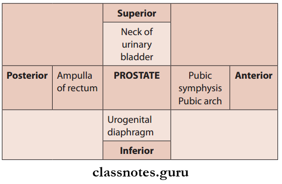
Prostate Location
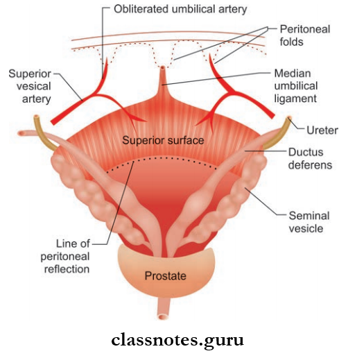
- In the lesser pelvis between the neck of the urinary bladder above and the urogenital diaphragm below:
- Anteriorly related to the lower part of the pubic symphysis and the upper part of pubic arch
- Posteriorly related to the ampulla of the rectum
Read And Learn More: Abdomen And Pelvis
Relations Of The Prostate:
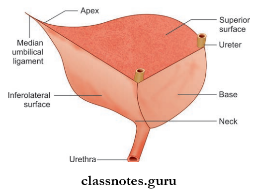
- Shape: Inverted cone, with broad base—directed upward and pointed apex directed downward
- Weight: 3 g
- Length: 3 cm
- Breadth: 4 cm
- Thickness: 2 cm
Reproductive System Important Questions
Prostate External Features
- The prostate has an apex, four surfaces—anterior, posterior, and two inferolateral surfaces and a base
- Apex
- Directed downward
- It is continuous with the neck of the urinary bladder
- The junction between the base and neck of the urinary bladder is marked by a circular groove
- Numerous venules of the prostatic plexus and the urinary bladder are present inside the groove
- Base is pierced in the median plane by the urethra approximately at the junction of its anterior 1/3rd and posterior 2/3rd.
- Anterior surface
- Narrow and convex from side to side
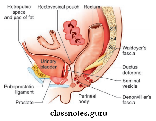
- Location: 2 cm behind the pubic symphysis
- This 2 cm gap is filled up by retropubic fat and forms the retropubic space of Retzius.
- The lower end of the anterior surface (just a little above the apex) is pierced by the urethra.
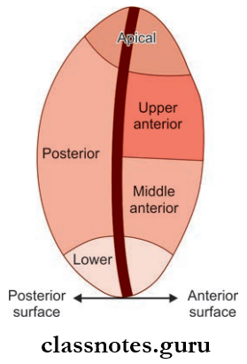
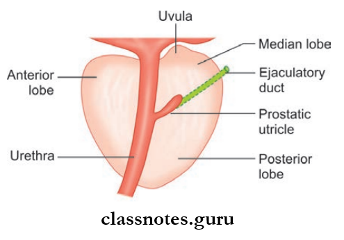
- Posterior Surface
- It is triangular in shape
- Posteriorly, it is separated from the ampulla of the rectum by the fascia of Denonvilliers
- The posterior surface is divided into an upper smaller area and a lower larger area by a transverse sulcus passing through it
- The transverse sulcus is pierced on each side by the ejaculatory duct
- A lower large area is again divided by a posterior median sulcus into two lateral lobes
- The upper smaller area is called the median lobe
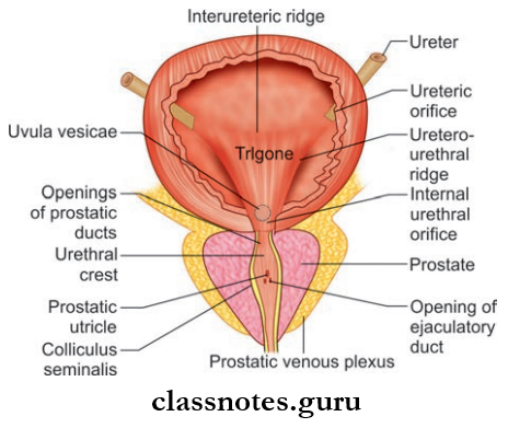
- The median lobe is wedge-shaped, its base has an elevation known as uvula vesicae and is composed mainly of glandular tissue.
- Lobes Of Prostate
Human Reproductive System Question Answers
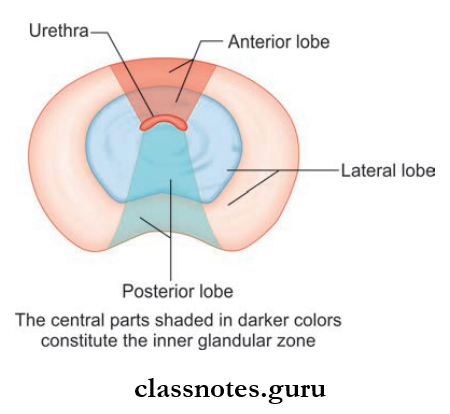
The Prostate Is Incompletely Divided Into Five Lobes:
- Anterior lobe
- Posterior lobe
- Median lobe
- Two lateral lobes
1. Anterior Lobe (isthmus)
- Situated in front of the urethra
- Bridges the two lateral lobes in front of the urethra
- Devoid of glandular tissue, so adenomas never occur here
2. Posterior Lobe
- Situated behind urethra and below the ejaculatory duct
- Connects the two lateral lobes behind the urethra
- Contains glandular tissue and is the main site of carcinoma
3. Median Lobe
- Lies posterior and superior to prostatic utricle and ejaculatory ducts
- Utricle lies within lobe
- Common site for adenoma
4. Two Lateral Lobes (right and left)
- Contains numerous glands
- Also can become a site for carcinoma.
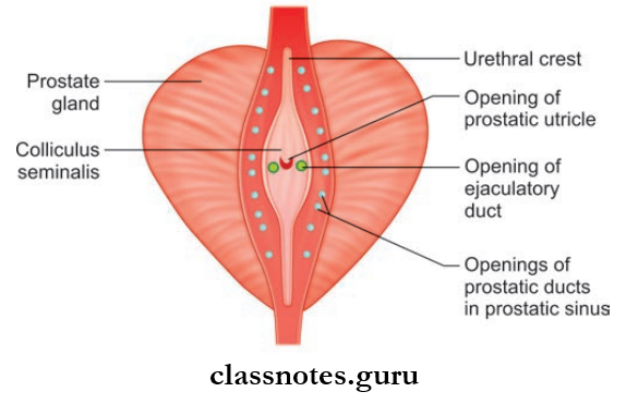
Internal Features Of Prostate
- Prostatic urethra—it passes through the gland
- There are openings for ducts of prostatic follicles into the prostatic sinuses of the prostatic urethra
- Right and left ejaculatory ducts pass through the gland and open into the prostatic urethra
- Prostatic utricle saw inside the prostate.
Male And Female Reproductive System MCQs With Answers
Question 2. Classify the zones of the prostate.
Answer:
Prostate Gland Has Been Divided Into Different Zones On The Basis Of Its Composition By Mcneal. They Are:
- Peripheral Zone
- Forms about 70% of the fraction of gland
- Location: Subcapsular portion of posterior aspect of mprostate gland which surrounds distal urethra
- Most common site of prostatic carcinomas.
- Central Zone
- Forms about 25% of the fraction of gland
- It is the part of the gland surrounding the ejaculatory duct.
- Transitional Zone
- Approximately 5% of the fraction of gland
- This is the region of the gland responsible for benign prostatic hypertrophy.
- Anterior Fibromuscular Zone
- Area devoid of glandular components
- Composed of muscle and fibrous tissue.
Question 3. Write briefly on the capsules of the prostate.
Answer:
The Prostate Has A True Capsule And A False Capsule:
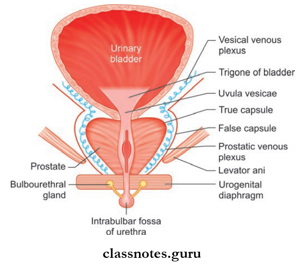
True Capsule Of Prostate:
- Formed by condensation of connective tissue stroma of the gland and is continuous with stroma of gland
- True Capsule Of Prostate is devoid of venous plexus
- The true Capsule Of the Prostate is adherent to the prostate.
False Capsule Of Prostate:
- Located outside true capsule
- False Capsule Of Prostate is derived from pelvic fascia
- False Capsule Of Prostate covers the prostate gland and urinary bladder
- False Capsule Of Prostate contains prostatic venous plexus on each side
- Anteriorly it is continuous with puboprostatic ligaments
- And posteriorly with true ligaments of bladder and rectovesical fascia.
Applied Anatomy Of Prostate
- During BPH, the prostate gets an additional pathological capsule
- Adenoma compresses the peripheral part of the gland which acts as the third capsule.
Reproductive Organs NEET Questions
Question 4. What are the supports of the prostate?
Answer:
- Puboprostatic ligaments—on the anterior aspect
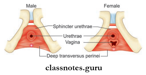
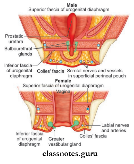
- Urogenital diaphragm
- The rectovesical fascia of Denonvilliers—on the posterior aspect.
Question 5. Describe the blood supply, lymphatic drainage, and nerve supply of the prostate. What are the communications of prostatic venous plexus and what is its importance?
Answer:
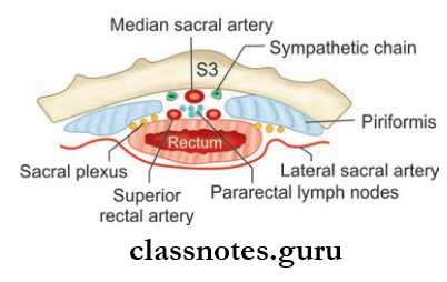
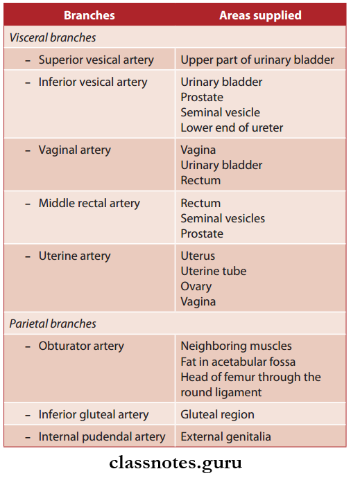
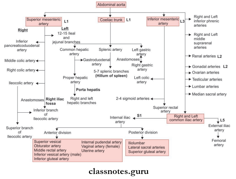
- Arterial Supply Of Prostate
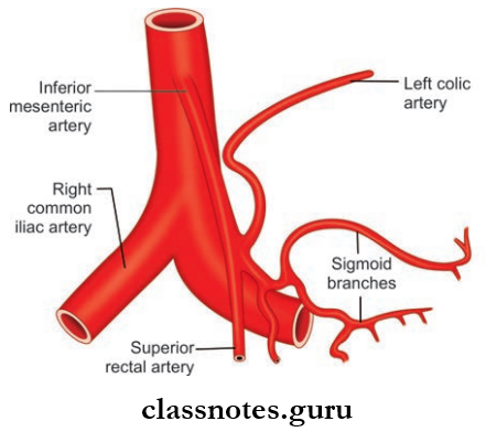
- Inferior vesical artery
- Middle rectal artery
- Internal pudendal artery
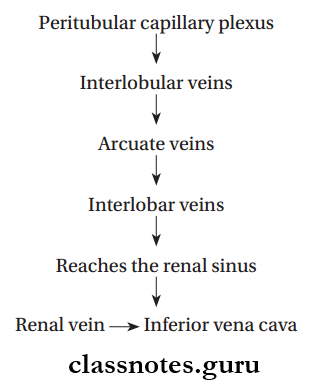
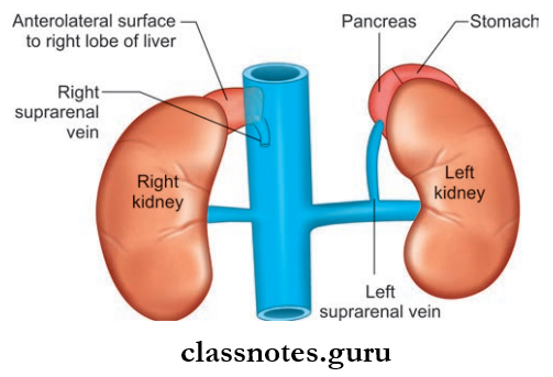
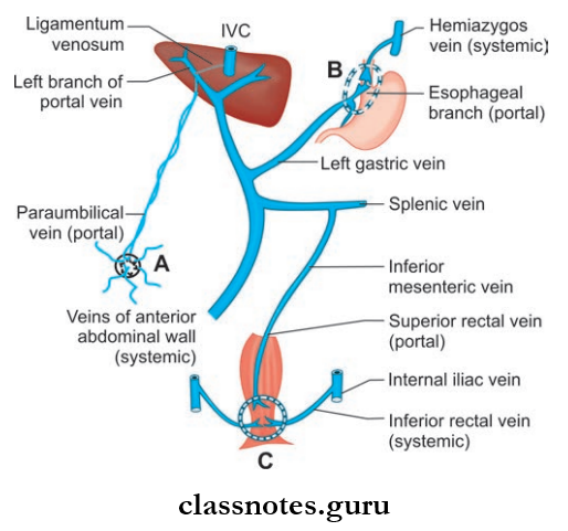
- Venous Drainage Of Prostate
- Veins of prostate form the prostatic venous plexus
- They drain into the inferior vena cava through iliac veins
- They also have valveless communications with internal and external vertebral plexus through which they drain into the intracranial dural venous sinuses.
- Applied: Carcinoma prostate can metastasise to the heart via internal iliac veins and vertebral column and through internal and external vertebral plexus.
- Lymphatic Drainage Of Prostate
- Lymphatics Drain:
- Mainly into internal iliac nodes and sacral group of lymph nodes
- Partly into external iliac nodes.
- Nerve Supply Of Prostate
- Sympathetic Supply: From L1, L2 spinal segments through superior hypogastric plexus
- Parasympathetic Supply: From S2, S3, and S4 spinal segments by pelvic splanchnic nerves.
Reproductive System Viva Questions
Question 6. Briefly mention the age changes in the prostate.
Answer:
The Age Changes In The Prostate
- Childhood: Small
- Puberty: Sudden increase in size
- 20–30 Years: Marked proliferation
- 30–45 Years: Size remains constant and involution starts
- 45 Years And Above: May be enlarged (BPH) or reduced (senile atrophy).
Question 7. Write a note on the development of the prostate.
Answer:
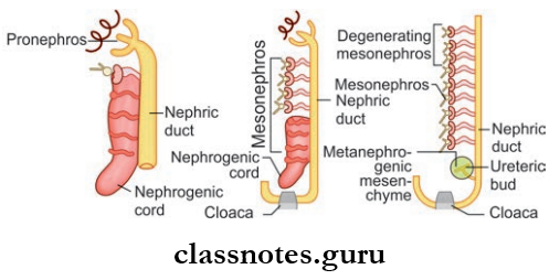
Prostate Develops From A Large Number Of Buds Arising From The Prostatic Urethra
- Buds arising from the endodermal part of the prostatic urethra form the glandular part of the prostate
- Buds arising from the mesodermal part of the prostatic urethra form the stroma of the prostate.
Question 8. Write a note on the location, size, and position of the uterus. Describe the different parts of the uterus.
Answer:
Uterus
- Also called hysteria
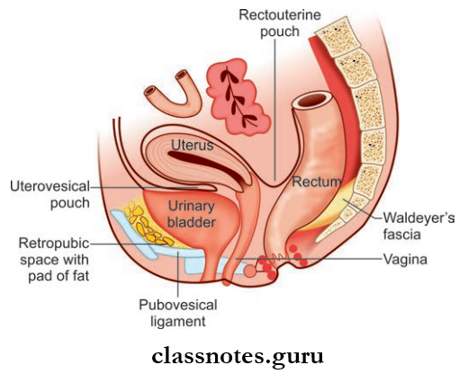
- Location Of Uterus: In the lesser pelvis between the bladder and rectum
- Shape Of Uterus: Pearshaped, flattened anteroposteriorly backward
- Size Of Uterus:
- Length: 7.5 cm
- Breadth: 5 cm
- Height/thickness: 2.5 cm
- Weight: 30–40 g
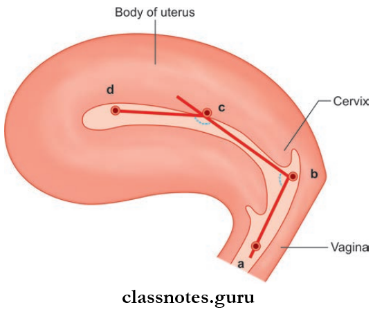
The Normal Position Of The Uterus: Uterus normally lies in the position of anteversion and anteflexion
- Anteversion—the long axis of the uterus forms an angle of about 90 degrees with the long axis of the vagina. This forward angle is known as the angle of anteversion
- Anteflxion—the long axis of the body of the uterus is bent forward at the level of internal os on the axis of the cervix. They make an angle of 170 degrees with each other. This position is known as anteflexion and the angle is known as the angle of anteflexion.
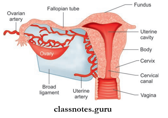
Parts Of Uterus
- Body: Upper 2/3rd (extends from fundus to isthmus)
- Cervix: Lower 1/3rd
The Junction Between the Body And Cervix Is Called The Isthmus.
- Body It includes:
- Fundus
- Two Surfaces:
- Anterior/vesical
- Posterior/intestinal
- Two lateral borders—right and left.
- Fundus
- Free upper end of the uterus
- Convex domelike in appearance
- It lies above the openings of the uterine tubes
- It is covered by peritoneum.
Two Surfaces Of Uterus
- Anterior Surface/Vesical Surface
- Flat surface
- Directed downward and forward
- It is covered with the peritoneum up to the isthmus
- From this point, the peritoneum sets reflected onto the upper surface of the urinary bladder and becomes the ureterovesical pouch.
- Posterior Surface/Intestinal Surface
- Convex
- Directed upward and backward
- It is covered by the peritoneum
- It also forms the anterior wall of the rectouterine pouch.
Two Lateral Borders Of Uterus:
- Rounded and convex
- The nonperitoneal part, since it provides attachment to the broad ligament of the uterus
- The uterine tube opens in the upper end of the lateral border
- The round ligament of the uterus is attached anteroinferior to the opening of the uterine tube and the ligament of the ovary is attached posteroinferior to the opening of the uterine tube.
Cervix Of Uterus
- Lower cylindrical part
- Length: 2.5 cm
- Wider in the middle
- Its lower part projects into the anterior wall of the vagina, dividing the cervix into two parts:
- Supravaginal Part—upper part
- Vaginal Part—lower part
- Vaginal Fornices: Space between the vaginal part of the cervix and the vaginal wall.
External OS Of Uterus : Opening of Cervix into Vagina
- Nulliparous Women: Small and circular
- Multiparous Women: Large and transverse
Internal Os Of Uterus: Opening of the body of the uterus into cervix
- Uterine Cavity Proper/Cavity Of The Body—triangular in shape
- Cervical Canal/Cavity Of The Cervix—spindleshaped.
Reproductive System Short Questions And Answers
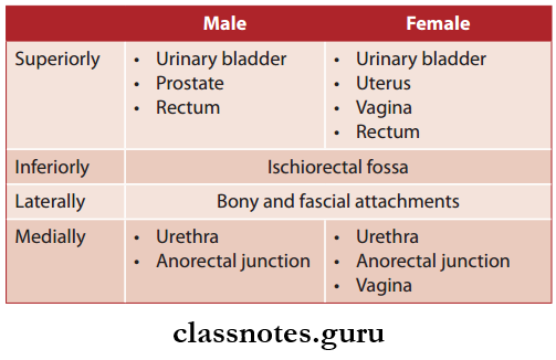
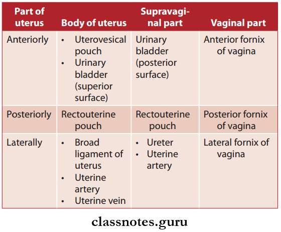
Question 9. What are the relations of different parts of the uterus?
Answer:
The Relations Of Different Parts Of The Uterus
Question 10. Classify the ligaments of uterus. Write a note on broad ligament.
Answer:
Ligaments Of Uterus classified Into Two Types:
- False Ligaments: Peritoneal folds, provide no support to uterus
- True Ligaments: Fibromuscular bands, provide support to the uterus.
1. False Ligaments Of Uterus
- Broad Ligaments (Right And Left)
- Folds of peritoneum
- Have two layers—anterior and posterior
- Connects the uterus to the lateral pelvic wall.
- False Ligaments Of Uterus External Features
- When the bladder is full, it has:
- Two Surfaces—anterior and posterior
- Four Borders—upper, lower, medial, lateral.
- Parts of Broad Ligament
- Mesosalpinx—the part between the uterine tube and the ligament of ovary
- Mesometrium—part below the ligament of the ovary
- Mesovarium—it attaches the posterior layer of the ligament with the ovary
- Suspensory ligament of ovary
- Part of the broad ligament extends from the upper pole of the ovary and infundibulum of the uterine tube to the lateral wall of the lesser pelvis.
- Contents of Broad Ligament
- Uterine tube
- Ligaments—round ligament of uterus, ligament of ovary
- Arteries—uterine artery, ovarian artery
- Nerves—uterovaginal plexus, ovarian plexus
- Vesicular appendices
- Lymphatics
- Anterior Ligaments—ureterovesical fold of peritoneum
- Posterior Ligaments—rectovaginal fold of peritoneum.
2. True Ligaments Of Uterus
- Round ligament of uterus
- Transverse cervical ligaments
- Uterosacral ligaments.
Question 11. Write briefly about the blood supply, lymphatic drainage, and nerve supply of uterus.
Answer:
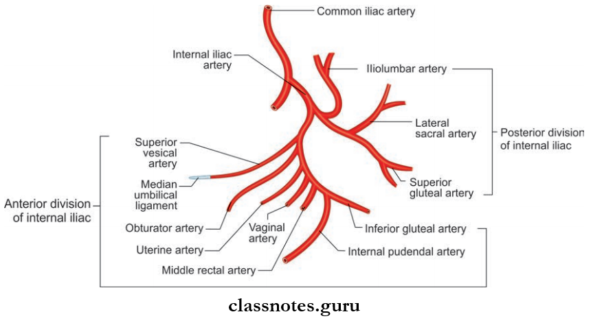
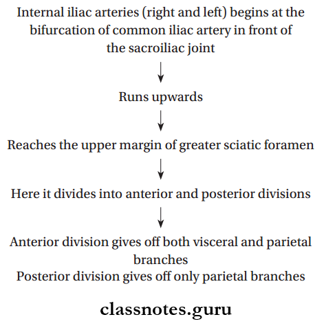
Arterial Supply Of Uterus
- Two Uterine Arteries:
- Branch of the anterior division of the internal iliac artery
- It ascends along the side of the uterus
- Terminates by anastomosing with the ovarian artery.
- Two Ovarian Arteries
- Branch of abdominal aorta.
Venous Drainage Of Uterus
- Veins correspond to arteries
- They form venous plexus
- Drains into uterine and vaginal veins and then to internal iliac veins.
Lymphatic Drainage Of Uterus
- Fundus and upper part of the body—pre and paraaortic lymph nodes
- The lower part of the body—external iliac nodes
- Cervix:
- Lateral part—external iliac nodes obturator nodes
- Posterolaterally—internal iliac nodes
- Posteriorly—sacral nodes.
NCERT Class 12 Biology Chapter Reproductive System Questions
Nerve Supply Of Uterus
- Sympathetic Supply: Derived from T12–L2 spinal segments
- Parasympathetic Supply: Derived from S2–S4 spinal segments.
Question 12. What are the supports of the uterus? Write briefly about some of the important supports.
Answer:
- They keep the uterus in position
- And prevent it from sagging down.
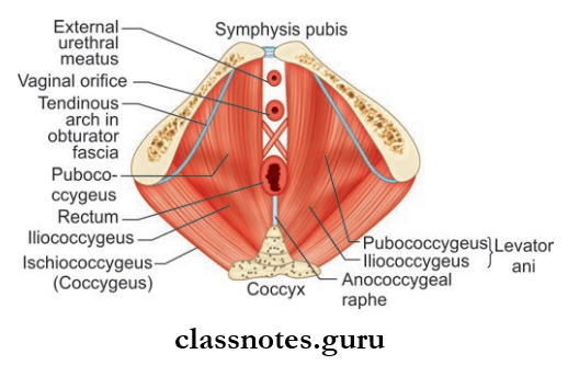
Primary Supports Of Uterus
- Muscular
- Pelvic diaphragm
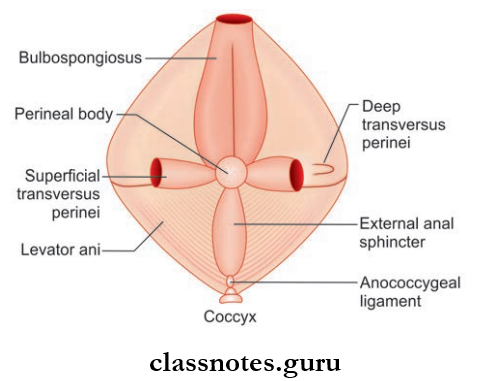
- Perineal body
- Urogenital diaphragm.
- Fibromuscular
- Pubocervical ligaments
- Transverse cervical ligaments of Mackenrodt
- Uterosacral ligaments
- Round ligament of uterus.
- Visceral
- Urinary bladder
- Vagina
- Uterine axis.
Secondary Supports Of Uterus
- Broad ligaments
- Uterovesical fold of peritoneum/anterior ligament
- The rectovaginal fold of the peritoneum/posterior ligament.
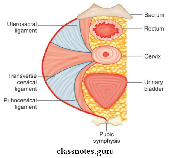
- Pubocervical ligaments Of Uterus
- Derived from the endopelvic fascia
- Connects cervix to the posterior surface of the pubis
- Corresponds to puboprostatic ligaments in males.
- Transverse cervical ligaments Of Uterus
- Fanshaped fibromuscular band
- Derived from the endopelvic fascia
- Present on both sides
- Extent: From the lateral aspect of the cervix and upper vaginal wall to the lateral pelvic wall
- They form a hammock, which supports the uterus.
- Uterosacral ligaments Of Uterus
- Two In Number
- Derived from the endopelvic fascia
- Extent: From cervix to posterior aspect of 5–2 and 5–3 vertebrae
- They are enclosed within rectouterine folds of the peritoneum
- The ligaments pull the cervix backward against the forward pull of the round ligament.
- Round Ligament Of The Uterus
- Length: 10–12 cm
- Lives between the 2 layers of broad ligaments
Course Of Uterus:
Uterine Axis
- Anteversion position prevents the uterus from sagging down through the vagina
- Because of the anteversion, during an increase in intraabdominal pressure, the uterus gets pushed against urinary bladder and pubic symphysis.
Question 13. Write a note on the development of the uterus.
Answer:
The Development Of The Uterus
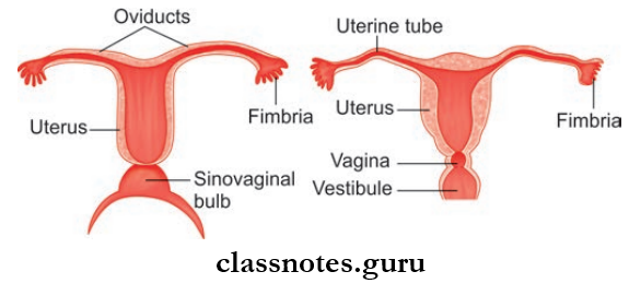
- Paramesonephric ducts (Müllerian ducts) get fused to form a uterovaginal canal
- Epithelium of uterus is developed from the uterovaginal canal
- Myometrium is formed from the surrounding mesoderm
- Unfused horizontal parts of two paramesonephric ducts partially get embedded in the substance of myometrium to form ‘fundus of uterus’
- Soon after, the cervix is also recognized as a separate region
- Didelphys uterus: Complete duplication of uterus
- Unicornuate uterus: Onehalf of uterus absent.
Reproductive System Exam Questions And Answers
Question 14. Explain in detail about the uterine tubes and mention its development.
Answer:
The Uterine Tubes
- Also known as fallopian tubes
- Length: 10 cm
- Location: Upper free margin of broad ligament of uterus.
Uterine Tubes External Features: It has two ends and four parts
Uterine Tubes External Features Ends
- Medial End: Opens into lateral angle of the uterine cavity
- Lateral End:
- Also called infundibulum
- Funnelshaped
- It has a number of figerlike projections called fimbriae
- One fimbriae is longer and is attached to a tubal pole of the ovary. It is called ovarian fimbriae.
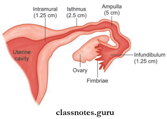
Uterine Tubes External Features Parts: From lateral to medial, the fallopian tube is divided into
- Infundibulum
- Ampulla
- Isthmus
- Intramural
1. Infundibulum
- Funnelshaped
- Length: 1 cm
- Lateral: most end
2. Ampulla
- Length: 5 cm
- Lateral 2/3rd of tube
- Widest and longest part
- Thinwalled and tortuous
3. Isthmus
- Narrow and rounded
- Medial 1/3rd of tube
4. Intramural/Uterine Part
- Length: 1 cm
- Lies within the walls of the uterus
- Opens at the superior angle of the uterine cavity.
Blood Supply Of Uterine
- Arterial Supply
- Medial 2/3rd of tubeuterine artery
- Lateral 1/3rd of tubeovarian artery
- Venous Drainage
- Uterine vein
- Ovarian vein.
- Lymphatic Drainage
- Internal iliac lymph nodes
- Pre and paraaortic lymph nodes.
- Nerve Supply
- Sympathetic Supply
- Though ovarian and superior hypogastric plexus
- Derived from T–10 to L–2 spinal segments.
- Parasympathetic Supply
- Medial part – derived from S2, S3, S4 spinal segments through pelvic splanchnic nerves
- Lateral part – derived from vagus nerve.
Development Of Uterine Tube: Derived from unfused parts of paramesonephric ducts.
Male And Female Reproductive Organs Questions And Answers
Male And Female Reproductive Organs Multiple Choice Questions And Answers
Question 1. Anatomically how many lobes are noticed in the prostate gland?
- 2 lobes
- 4 lobes
- 3 lobes
- 5 lobes
Answer: 4. 5 lobes
Question 2. Which lobe of prostate is more prone to malignancy?
- Anterior lobe
- Median lobe
- Lateral lobe
- Posterior lobe
Answer: 2. Median lobe
Question 3. Which best describes the prostate gland?
- Surrounds the base of the bladder and pierces the membranous urethra
- Is most prone to cancer in the inferiorposterior region and lateral lobes
- Has a superior lobe that is most readily palpable by digital rectal exam
- Is supplied mainly by the superior rectal artery
Answer: 2. Is most prone to cancer in the inferiorposterior region and lateral lobes
Question 4. Lymph from the uterine body drains into __________ nodes:
- Superficial inguinal
- External iliac
- Internal iliac
- Lumbar
Answer: 2. External iliac
Question 5. Which statement most accurately describes the body of the uterus?
- The thick, convex superior portion
- Inferiormost portion
- Contains most of the uterine cavities
- Has a tapered region leading to the cervix called the uterine cornu
Answer: 3. Contains most of the uterine cavities
Question 6. Which ligament transmits the uterine arteries from the internal iliac to the uterus?
- Uterosacral ligament
- Round ligaments of the uterus
- Suspensory ligament of the ovary
- Transverse cervical ligament
Answer: 4. Transverse cervical ligament