
Biliary Apparatus Question And Answers
Question 1. Classify biliary apparatus. Mention briefly about hepatic ducts.
Answer:
Biliary Apparatus
Biliary Apparatus Can Be Classified Into:
- Intrahepatic biliary apparatus
- Extrahepatic biliary apparatus
The intrahepatic part drains the bile secreted by hepatocytes to the outside, i.e. into extrahepatic part, which stores the bile in the gallbladder and transports it into 2nd part of the duodenum
Read And Learn More: Abdomen And Pelvis
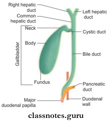
1. Intrahepatic Part: It consists of
- Bile canaliculi
- Bile ductules
- Interlobular bile ducts
Biliary Apparatus Important Questions
2. Extrahepatic Part: It consists of:
- Right and left hepatic ducts
- Common hepatic duct
- Gallbladder
- Cystic duct
- Common bile duct
Right And Left Hepatic Ducts:
- They emerge at the porta hepatis after the union of their respective interlobular ducts
- Arrangement of structures at porta hepatis (Posterior to anterior)
- Mnemonic: VAD (Vein artery duct)
- Vein
- Artery
- Duct (hepatic duct).
Common Hepatic Duct:
- Length: 3.5 cm
- Formation: By the union of right and left hepatic ducts at the right end of porta hepatis
- Course: It runs downward for about 2.5–3 cm and unites with the cystic duct at an acute angle
- And forms the common bile duct
- It also forms the left boundary of Calot’s triangle.
Question 2. Write in detail about the gallbladder.
Answer:
Gallbladder:
- The Gallbladder is an elongated pear-shaped organ
- The Gallbladder is the reservoir of bile
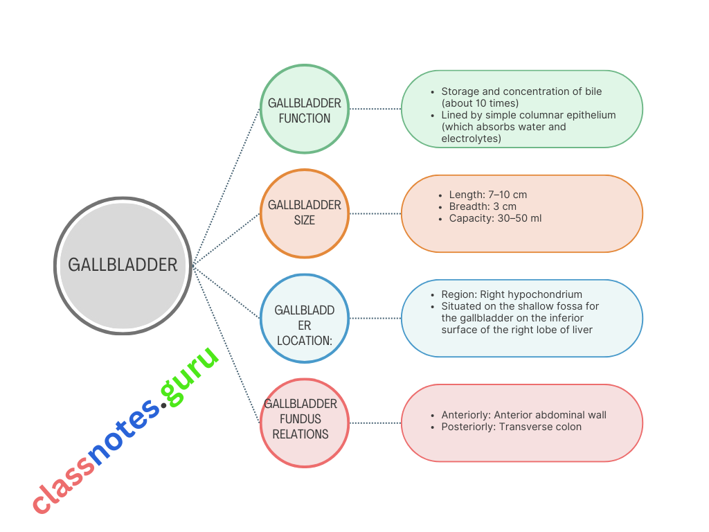
Gallbladder Extent: Right end of porta hepatis to the inferior margin of the right lobe of the liver
Gallbladder Parts: It can be anatomically divided into fundus, body and neck
Gallbladder Fundus
- Most anterior and expanded part of the gallbladder, completely surrounded by peritoneum
- It projects from the inferior border of the liver and lies very near to anterior abdominal wall (fundus corresponds to the tip of 9th costal cartilage)
Biliary Apparatus Anatomy MCQs
Gallbladder Fundus Relations
- Anteriorly: Anterior abdominal wall
- Posteriorly: Transverse colon
Gallbladder Body
- Gallbladder is the continuation of fundus
- Gallbladder is directed upwards, backward, and to the left
- The superior surface of a body of the gallbladder is in contact with the inferior surface of the liver at the fossa of the gallbladder, this part is devoid of the peritoneum
- The inferior surface is covered by the peritoneum
- Its upper end is narrow and is continuous with the neck at the right end of the porta hepatis
Gallbladder Body Relations
- Superior Surface: Inferior surface of the liver
- Inferior Surface: Transverse colon, 1st and 2nd parts of duodenum.
Gallbladder Applied Anatomy
Due to the close association of the body of the gallbladder with the transverse colon and duodenum, gallstones can sometimes ulcerate through its wall into the duodenum or transverse colon
Neck Of Gallbladder: The narrowest part of the gallbladder
Gallbladder Course:

- The posteromedial wall of neck shows a projection called Hartman’s pouch
- The Lumen of neck has a spiral wall
- Inferiorly neck is related to fist part of duodenum.
Hartman’s Pouch: A spheroid or conical pouch at the junction of the neck of the gallbladder and the cystic duct Gall-stones commonly get stuck at the Hartman’s pouch
Biliary Apparatus Viva Questions
Question 3. Write a note on the cystic duct.
Answer:
Cystic Duct
- Cystic Duct is S-shaped
- Length: 3–4 cm
Cystic Duct Course:
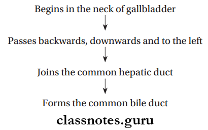
Cystic Duct Provides A Two-Way Passage: Receives bile from the common hepatic duct and sends bile out via the common bile duct.
Question 4. Write a note on the common bile duct. Mention the important anatomical features of the intraduodenal part common bile duct.
Answer:
Common Bile Duct
- Length: 7.5 cm
- Diameter: < 7 mm
- Formation: By the union of the cystic duct with common hepatic duct near the porta hepatis
Common Bile Duct Course:
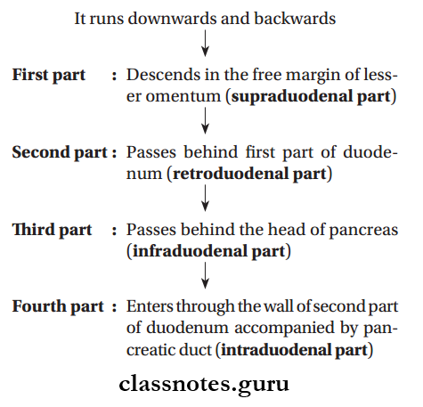
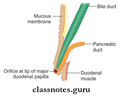
- The narrowest part of common bile duct
- Has a very oblique course
- The pancreatic duct and common bile duct unite to form, a hepatopancreatic ampulla or ampulla of Vater in the wall of the duodenum, very close to the summit of major duodenal papilla
- Distal end of the ampulla is constricted
- Ampulla is surrounded by the hepatopancreatic sphincter or sphincter of Oddi
- The sphincter is made up of circular muscles.
Cystic Duct Relations
- Supraduodenal Part
- Anteriorly: Liver
- Posteriorly: Portal vein and epiploic foramen
- Left: Hepatic artery
- Infraduodenal Part
- Anteriorly: A groove in the upper and lateral parts of the posterior surface of the head of the pancreas
- Posteriorly: Inferior vena cava
- Right: 2nd part of duodenum
- Retroduodenal part
- Anteriorly :1st part of duodenum
- Posteriorly: Inferior vena cava
- Left: Gastroduodenal artery
Biliary System Question Answers
Question 5. Briefly describe the blood supply, lymphatic drainage, and nerve supply of the biliary apparatus.
Answer:
The Blood Supply, Lymphatic Drainage, And Nerve Supply Of The Biliary Apparatus
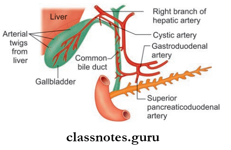
Biliary Apparatus Arterial Supply
- Gallbladder: Cystic artery—right branch of hepatic artery
- Common Bile Duct:
- The upper part from the descending branches of the cystic artery
- The lower part from the ascending branches of the superior pancreaticoduodenal artery
- Also minor contribution by hepatic artery proper.
Biliary Apparatus Venous Drainage
- The superior surface of the gallbladder is drained by veins which directly enter into the liver substance and open in to hepatic ducts
- Rest of the gallbladder drained by one or two cystic veins into the right branch of the portal vein
- Lower part of bile duct drains into portal veins.
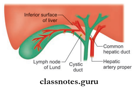
Biliary Apparatus Lymphatic Drainage
- Gallbladder, cystic duct, hepatic duct, the upper part of common bile—cystic nodes of Lund, node of the anterior border of the epiploic foramen
- Lower part of common bile duct—lower hepatic nodes, upper pancreaticosplenic nodes.
Biliary Apparatus Nerve Supply
- Sympathetic Supply: T7–T9
- Parasympathetic Supply: Both vagus and right phrenic nerve.
Question 6. Write a short note on Calot’s triangle and its importance.
Answer:
Calot’s Triangle
- Also known as cystohepatic triangle
- An inverted triangle (apex facing downwards)
Calot’s Triangle Boundaries
- Superiorly: Inferior surface of liver
- Right: Cystic duct
- Left: Common hepatic duct
Calot’s Triangle Contents
- Right hepatic artery
- Cystic artery
- Cystic lymph nodes of Lund.
- Applied Anatomy
- During cholecystitis, the cystic nodes of Lund are found enlarged.
Biliary Tract Anatomy Questions
Biliary Apparatus Multiple Choice Questions And Answers
Question 1. The sphincter of Oddi consists of a following number of small sphincters:
- 2
- 3
- 5
- 6
Answer: 2. 3
Question 2. The Fundus of the gallbladder is related to the tip of:
- Right 8th costal cartilage
- Right 9th costal cartilage
- Left 8th costal cartilage
- Left 9th costal cartilage
Answer: 2. Right 9th costal cartilage