Specialized Radiographic Techniques Long Essays

Specialized Radiographic Techniques Long Essays

Question 1. Enumerate techniques for TMJ imaging. Describe any two in detail.
Answer.

Techniques For TMJ Imaging:

  • Transcranial projection:
    • Structure Seen:
      • Useful in detecting arthritis of the articular surfaces
      • To evaluate the joint’s bony relationship
    • Film Position:
      • The cassette is placed against the patient’s ear and centered over the TMJ of interest
      • It is placed parallel to the sagittal plane
    • Patient’s Position:
      • The sagittal plane must be vertical
      • The ala tragus line should be parallel to the floor
      • The view is taken with
        • Open mouth
        • Rest position
        • Closed mouth
    • Central Ray:
      • It differs according to the technique
      • Postauricular
        • Point of entry is 1/2 “behind and 2” above the auditory meatus
      • Grewcock approach
        • The path of entry is through point 2 above the auditory meatus
      • Gill’s approach
        • Point of entry is 1/2 “anterior and 2” above the auditory meatus
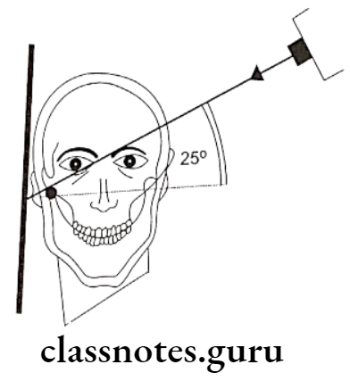
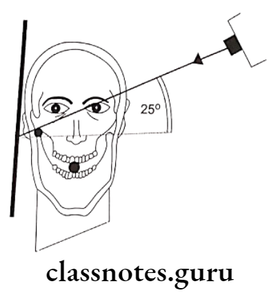
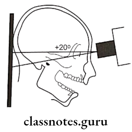
    • Angulation: +20º To +25º
    • Point Of Exit: TMJ of interest

Specialized radiographic techniques long essay
Oral Radiology Specialized Radiographic Techniques Transcranial projection 1

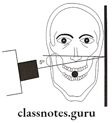
A – Transcranial projection, the central ray is oriented at 25º positive angle from the opposite side and anteriorly 20º, centered over the TMJ of interest, mouth closed
Oral Radiology Specialized Radiographic Techniques Transcranial projection 2

B – Transcranial projection, the central ray is oriented at 25º positive angle from the opposite side and anteriorly 20º, centered over the TMJ of interest, mouth open

  • Tranpharyngeal projection:
    • Structures Seen:
      • The medial surface of the condylar head and neck
    • Film Placement:
      • The cassette is placed flat against the patient’s ear
      • It is centered to a point 1/2″ anterior to the external auditory meatus, over the TMJ of interest

Read And Learn More: Oral Radiology Question and Answers

    • Position of the Patient:
      • The sagittal plane should be vertical and parallel to the film
      • The film is centered to a point 1/2 “anterior to the external auditory meatus
      • The occlusal plane should be parallel to the transverse axis of the film
      • The patient should open his mouth
    • Central Ray:
      • It is directed from the opposite side cranially at an angle of -5 to -10 degrees posteriorly
      • It is directed through the Mandibular notch of the opposite side below the base of the skull to the TMJ of interest

Oral Radiology Specialized Radiographic Techniques Transpharyngeal projection 1

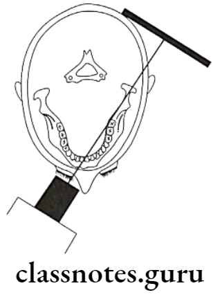
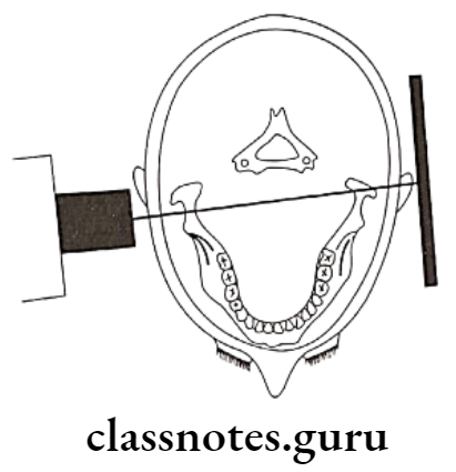
A – Transpharyngeal projection. The central ray is oriented superiorly 5º to 10º and posteriorly approximately 10º, centered over the TMJ of interest. The mandible is positioned at the maximal opening
Oral Radiology Specialized Radiographic Techniques Transpharyngeal projection 2

Long essay on advanced radiographic techniques

B – Transpharyngeal projection, showing positioning from above, showing the X-ray beam aimed slightly posteriorly across the pharynx

  • Transorbital Projection:
    • Structures Seen:
      • The anterior view of TMJ
    • Film Placement:
      • The film is positioned behind the patient’s head at an angle of 45 degrees to the sagittal plane
    • Position of the Patient:
      • The Sagittal Plane should be vertical
      • The canthomeatal line should be 10 degrees to the horizontal
      • Head tipped downwards
      • The mouth should be widely open
    • Central Ray:
      • Directed to the joint of interest at an angle of +20 degree
      • Strike the cassette at a right angle
        • Point of entry
        • A pupil of the same eye
        • Medial canthus of the same eye
        • Medial canthus of the opposite eye

Oral Radiology Specialized Radiographic Techniques Transorbital projection 1

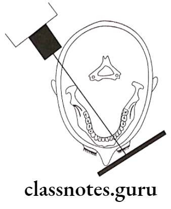
A – Transorbital projection, the central ray is oriented downward approximately 20º and laterally approximately 30º through the contralateral orbit, centered over the TMJ of interest
Oral Radiology Specialized Radiographic Techniques Transorbital projection 2

B – Transorbital projection, positioning from above, showing the cassette behind the condyletown’sray beam aimed across the orbit

  • Reverse towne’s projection:
    • Structures Seen:
      • Condylar head and neck
    • Film placement:
      • The cassette is placed perpendicular to the floor
    • Position of the Patient:
      • The sagittal plane should be vertical and perpendicular to the film
      • Lips should be centered on the film
      • Only the patient’s forehead should touch the film
      • The patient is asked to keep his mouth wide open
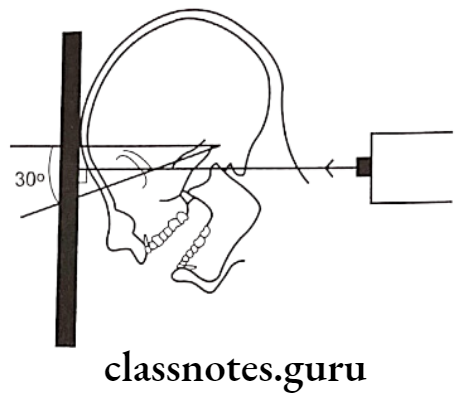
      • Angulation is -30º to the film
    • Central Ray:
      • It is directed through the midsagittal plane at the level of the mandible
      • It is perpendicular to the film

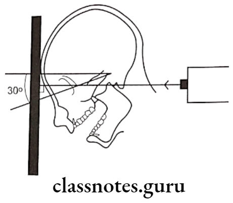
Oral Radiology Specialized Radiographic Techniques Diagram for the positioning of Reverse Townes projection

Diagram for the positioning of Reverse Towne’s projection, the radiographic baseline is 30º to the film and the X-ray is directed perpendicular to the film

Essay on special imaging techniques in radiology

Question 2. Describe sialography in detail.
Or
Describe indications, contraindications, and technique of sialography.
Or
Describe sialography and write on its signs in various salivary gland disorders.
Answer.

Sialography Indications:

  • Detection of calculus or foreign bodies
  • Determination of the extent of destruction of the gland secondary to obstructing calculi or foreign bodies
  • Detection of fistula, diverticula, or strictures
  • Determination and diagnosis of recurrent swellings and inflammatory processes
  • Demonstration of a tumor and the determination of its location, size, and origin
  • Selection of a site for biopsy
  • Outline of the plane of the facial nerve
  • Detection of residual stones
  • Sialography can be used for therapeutic procedures

Sialography  Contradictations:

  • Patients with knowacutesitivity of iodine
  • During the presence of avute inflammation
  • It may interfere with subsequent thyroid function tests

Sialography  Technique:

  • Identification of duct:
    • The parotid duct is located at the base of the papilla in the buccal mucosa adjacent to the first or second molar
    • The area over the mucosa should be dried with a small sponge
    • The submandibular duct orifice is situated on the summit of the small papilla at the side of the lingual frenum
  • Exploring of the duct:
    • The duct can be explored with a lacrimal probe
    • In the case of the submandibular duct, the probe should pass through the length of the floor of the mouth to the level of the posterior border of the mylohyoid muscle i.e. about 5cm
    • Eversion of the cheek should be done in case of parotid duct
    • By it, the duct is adequately enlarged
  • Introduction of cannula:
    • The sialographic cannula is inserted into the duct so that the tissue stop presses firmly into the orifice to prevent dye reflux
  • Introduce contrasting media:
    • Liquid-soluble or water-soluble agents are slowly introduced
  • Amount of the agent:
    • Submandibular gland: 0.5 – 0.75 ml
    • Parotid gland 0.76 – 1ml
  • Radiograph is taken:
    • Occlusal view/ lateral oblique view is sialolith delineate the submandibular gland
    • view itith is better viewed in an occlusal view
    • AP viewit is used for both the glands
    • It demonstrates the medial and lateral gland structures
  • Evacuation:
    • After the radiography is taken, the cannula should be removed
    • The patient is instructed to chew gum or the lemon slice and then asked to rinse
    • This is done to stimulate the gland and cause excretion of the dye

Radiographic Appearance In Different Disorders:

  • Parotid Gland – tree in winter appearance
  • Submandibular gland – Bush in winter appearance
  • Sjogren’s syndrome – Cherry blossom appearance
    • Results in thinning of individual ducts and decrease in number of ducts
  • Malignant tumor – Ball holding in hand appearance
  • Stones within duct
  • Results in filling defect distal to the site of obstruction
  • There can be dilatation of the duct proximal to the obstruction

Specialized dental radiography procedures essay

Question 3. Enumerate various radiographic techniques for the diagnosis of fracture of the mandible.
Answer.

Radiographic Techniques For Diagnosis Of Fracture Of Mandible:

  • Orthopantomogram:
    • It is a technique for producing a single tomographic image of the facial structures that induce both the maxillary and mandible dental arches and their supporting structures
    • Principle:
      • This is based on the curvilinear variant of conventional tomography
      • The movement of the tube head and the film produces images through the process known as tomography
      • Curvilinear tomography is also based on the principle of reciprocal movement of an X-ray source and an image receptor around a central point or plane called an image layer
    • Procedure:
      • Explain the procedure to the patient
      • Make the patient remove all the accessories that may interfere with the image
      • Position the patient such that he is in the focal trough
      • Instruct the patient to look straight
      • The patient is positioned such that dental arches are located in the middle of the focal trough
      • Mid sagittal plane is kept perpendicular to the floor
      • The patient back and spine are adjusted in an erect position
      • The occlusal plane is adjusted such that the Frankfort plane is parallel to the floor
      • This is done by placing the central incisor into a notched incisal device with a lead marker
      • Center the lower border of the mandible on the chin rest and is equidistant
      • Instruct the patient to position the tongue on the palate
      • Exposure the film
      • Process it as usual
  • Lateral oblique view:
    • Anterior body of the mandible:
      • Structures Seen:
        • Anterior body of mandible
        • Position of teeth in that region
      • Film Placement:
        • The cassette is placed flat against the patient’s cheek
        • It is centered over the body of the mandible overlying the canine
      • Position of the Patient:
        • The ala tragus line should be parallel to the floor
        • The mandible iscassetteded slightly
        • The inferior border of the casette should be parallel to the lower border of the mandible
        • The sagittal plane is tilted so that it is 5º to the vertical and rotated 30º from the true lateral poscassettehe nose and chin should approximate the casette
      • Central Ray:
        • Directed from 2 cm below the angle of the mandible opposite to the side of interest
        • The beam is directed upward from -10º to -15º
        • It is centered on the anterior body of the mandible
        • The beam is directed perpendicular to the horizontal plane of the film

Oral Radiology Specialized Radiographic Techniques Diagram for the positioning of lateral oblique projection 1

Advanced radiographic imaging methods essay

Diagram for the positioning of lateral oblique projection for the anterior body of the mandible, the film is in contact with the cheek at the canine area, and the X-ray beam aims at the canine area, through the radiographic keyhole

    • Posterior Body:
      • Structures Seen:
        • Body of the mandible
        • Position of teeth in that area
        • Ramus of the mandible
        • The angle of the mandible
      • Film Placement:
        • The cassette is placed against the patient’s cheek
        • It is centered over the body of the mandible
        • The cassette is placed parallel to the body of the mandible
      • Position of the Patient:
        • The ala tragusprotruded parallel to the floor
        • The mandible is protuded slightly
        • The inferior border of the cassette should be parallel to the lower border of the mandible and below it
        • The sagittal plane is tilted to 5º to the vertical
        • The head is rotated 10º to 15º from the true lateral position
      • Central Ray:
        • It is directed from 2 cm below the angle of the mandible opposite to the side of interest
        • The beam is directed upwards (-10º to -15º)
        • It is centered on the body of the mandible

Oral Radiology Specialized Radiographic Techniques Diagram for the positioning of lateral oblique projection 2

The diagram for the positioning of lateral oblique projection for the posterior body of the mandible film is in contact with the cheek at the premolar area, and the X-ray beam aims at the premolar area, through the radiographic keyhole

  • Ramus of Mandible:
    • Structures Seen:
      • Ramus from the angle of the mandible to the condyles
    • Film Placement:
        • The cassette is placed against the patient’s cheek
      • It is centered over the ramus of the mandible
      • It should be parallel to the ramus
    • Position of the Patient:
      • The ala tragus line should be parallel to the floor
      • The mandible protrudes slightly
      • The inferior border should be parallel to the lower border of the mandible and below it
      • The sagittal plane is tilted 10º to the vertical
      • The head is rotated 5º from the true lateral
    • Central Ray:
      • It is directed from 2 cm below the angle of the angle of the mandible opposite to the side of interest to a point posterior to the third molar region
      • The beam is directed upwards (-10º to -15º)
      • It is centered on the ramus of the mandible
  • Posteroanterior or anteroposterior view:
    • Film Placement:
      • The cassette is placed perpendicular to the floor
    • Position of the Patient:
      • The mid-sagittal plane should be perpendicular to the plane of the film
      • The patient’s head is extended so that only the chin touches the cassette
      • The cassette is centered around the acanthion
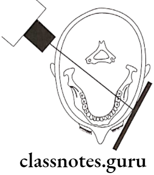
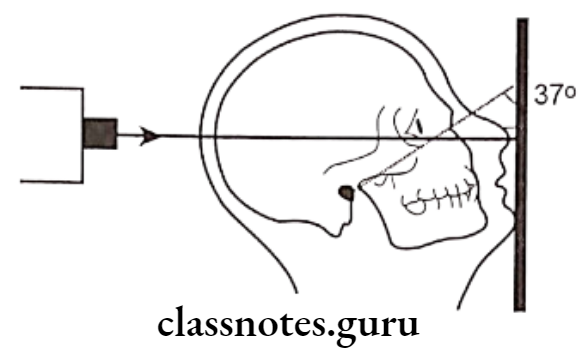
      • The canthomeatal line should be 37º to the plane of the film
      • The line from the external auditory meatus to the mental protuberance should be perpendicular to the film
    • Central Ray:
      • It is directed perpendicular and to the midpoint of the film
      • It enters from the vertex and exists from the acanthion

Oral Radiology Specialized Radiographic Techniques Diagram for the positioning of PA Water projection

Extraoral radiographic techniques long essay

Diagram for the positioning of PA water’s projection, the radiographic baseline is at 37º to the film, and the X-ray is perpendicular to the film

  • Reverse Towne view:
    • Film Placement:
      • The cassette is placed perpendicular to the floor
    • Position of the Patient:
      • The sagittal plane should be vertical and peronndicular to the film
      • Lips should be centered to the film
      • Only the patient’s forehead should touch the film
      • The patient is asked to keep his mouth wide open
      • Angulation is -30º to the film
    • Central Ray:
      • It is directed through the midsagittal plane at the level of the mandible
      • It is perpendicular to the film

Oral Radiology Specialized Radiographic Techniques Diagram for the positioning of Reverse Townes projection

Specialized X-ray techniques essay

Diagram for the positioning of Reverse Towne’s projection, the radiographic baseline is 30ºto the film, and the X-ray is directed perpendicular to the film

Red And White Lesions Short Essays

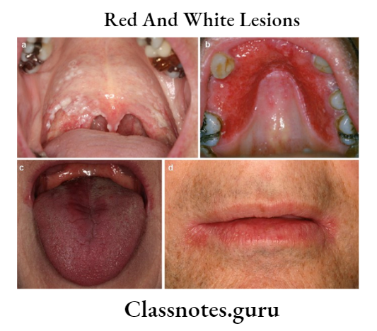
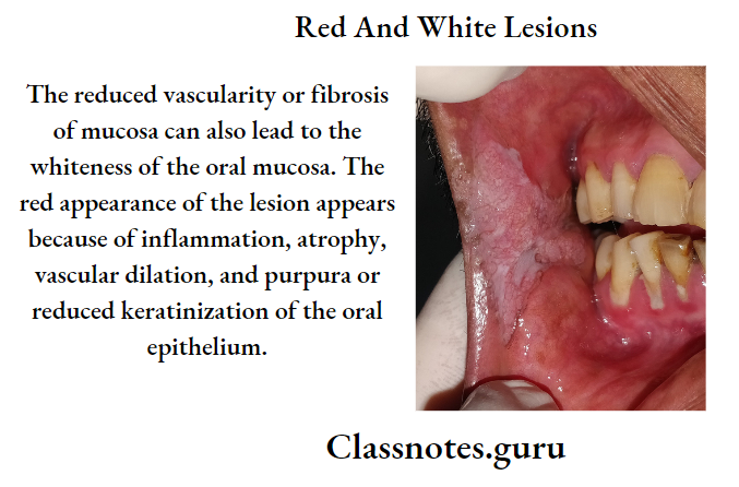
Red And White Lesions

Question 1. TNM staging
Answer:

TNM Staging

TNM Staging is the staging of malignancy which measures 3 major parameters of cancer

  • T- The Size Of The Tumor
    • N- lymph node involvement
    • M-distant metastasis
  • T- Primary Tumor
    • The tx-primary tumor cannot be assessed
    • To-No evidence of primary tumor
    • This- carcinoma in situ o Ti- Tumour size- 2 cm or less in diameter
    • T2– Tumour size- 2-4 cm in diameter
    • T3– Tumour size- more than 4 cm in diameter
    • T4– Tumour invades adjacent structures
  • N- Regional Lymph Node
    • Nx – Regional lymph node cannot be assessed
    • N0-No regional lymph node metastasis
    • N1– Metastasis in a single ipsilateral lymph node, 3 cm or less in dimension
    • N2– Metastasis in the single ipsilateral lymph node, more than 3 cm but less than 6 cm
    • N2a– Metastasis in the single ipsilateral lymph node, 3-6 cm in dimension
    • N2b – Metastasis in multiple ipsilateral lymph nodes, not more than 6 cm
    • N2c– Metastasis in bilateral or contralateral lymph nodes, not more than 6 cm
    • N3 – Metastasis in the lymph node, more than 6 cm in dimension
  • M- Distant Metastasis
    • Mx – The presence of distant metastasis cannot be assessed
    • M0 – No distant metastasis Mi – Presence of metastasis
    • M1– Presence of metastasis

Read And Learn More: Oral Medicine Question and Answers

Question 2. Treatment of cancer
Answer:

Aims Of Cancer Treatment:

  • Cure of the patient
  • Palliation
  • Preservation of function
  • Cosmetic function
  • Treatment of lymph node
  • Treatment of advanced tumors

Role Of Chemotherapy:

    • Cisplatin is the most effective drug
    • In advanced cases, chemotherapy is given before surgery or radiotherapy
    • This is called induction chemotherapy

Radiotherapy:

    • Radiotherapy preserves anatomical parts and functions
    • 6500-7500 cGy units are required to eradicate cancer

Role Of Surgery:

    • It may be in the form of wide excision or wide excision with removal of the bone
    • Radical neck dissection is done in case of lymph node involvement

Question 3. Lichenoid reactions
Answer:

Lichenoid Reactions

Lichenoid Reactions has a clinical picture similar to lichen planus

Lichenoid Reactions Etiology:

  • Disorders: lichen planus
  • Drugs:
    • Antimicrobial: tetracycline
    • Anti par asltit thlornquine
    • Antihypertensive methyl dopa
    • Anti ills gold

Lichenoid Reactions Clinical Features:

    • Lichenoid mm positive over oral mucosa
    • Lichenoid dermatitis: over the skin
    • Lichenoid gingivitis.: over gingiva

Lichenoid reactions Management: discontinuation of the drug

Question 4. Erythroplakia.
Answer:

Erythroplakia

Erythroplakia is a red patch or plaque in the oral mucosa which cannot be characterized clinically or pathologically as any other condition and which has no apparent cause

Etiology:

  • Use of tobacco
  • Alcohol
  • Candida infection
  • Idiopathic

Erythroplakia Clinical Features:

  • Age: a fifth-seventh decade of life
  • Sex: both sexes are equally affected
  • Site
    • The floor of the mouth
    • Retromolar area
    • Buccal mucosa
    • Gingiva
    • Tongue
    • Soft palate
  • Presentation
  • It appears as a small or extensive reel lesion
  • It has well-defined borders

Erythroplakia Types:

  • Homogeneous
    • Has uniform red patches all over
  • Erythroplakia with interspersed patches of leukoplakia
    • Has a few white leukoplakic patches along with a red patch
  • Speckled leukoplakia
    • It is characterized by the presence of soft irregular, raised, erythematous areas with a granular surface

Erythroplakia Differential Diagnosis:

  • Candidiasis: Lesson can be rubbed off
  • Denture Stomatitis: The commonly involved site is the palate
  • Tuberculosis: Present of tubercular ulcers
  • Histoplasmosis:  It is common in farmers

Erythroplakia Management

  • Elimination of the causative agent
  • Mucosal stripping of the lesion
  • Laser ablation
  • Electrocoagulation
  • Cryotherocoagulation
  • Maintenance by periodic recall visits every 3 months

Red And White Lesions.

Question 5. Diagnosis of oral lichen planus.
Answer:

Diagnosis Of Oral Lichen Planus

  • Clinical
    • The presence of bilateral interlacing white striae
    • Presence of Wickham striae and Koebner phenomenon
  • Laboratory Diagnosis
    • Hyperorthokeratosis
    • Hyperparakeratosis
    • Acanthosis with intercellular edema
    • Civatte bodies
    • The sawtooth appearance of the rete pegs
  • Immunofluorescence
    • Positive reactions with IgA, IgM, and IgG antisera
    • Presence of subepithelial deposits of fibrinogen and antigenically related substances

Question 6. Atrophic candidiasis.
Answer:

Atrophic Candidiasis Synonym: Antibiotic sore mouth

Atrophic Candidiasis Clinical Features:

  • Site: tongue, tissue underlying the prosthesis
  • Presentation
    • The lesion appears red or erythematous
    • Patients usually have vague pain or a burning sensation
    • Lesion reveals a lew white thickened foci, that are rubbed off leaving a painful surface
    • It closely resembles erosive lichen planus and erythroplakia

Atrophic Candidiasis  Differential Diagnosis:

  • Chemical Burn: History of chemicals
  • Drug Reaction: Diminished host response
  • Syphilitic Mucous Patch– Skin lesion is also present
  • NecroticUlcer And Gangrenous Stomatitis: Ulcer is deeper
  • Traumatic Ulcer: History of trauma present

Atrophic Candidiasis  Management:

  • Elimination Of Causative Agent:
    • Replacement of denture
    • Relining of denture
    • The denture must be cleaned thoroughly and regularly
    • It should be left out of out of the mouth at night in a hypochlorite solution
      • Topical application
  • Clotrimazole:
    • It is an effective topical treatment when dissolved in the mouth for five minutes daily
    • Nystatin Preparation:
      • Dissolves only in the mouth for 5 minutes a day.
  • Amphotericin B:
      • 5-10 ml of oral solution was used as a rinse.

Question 7. Erosive lichen planus.
Answer:

Erosive Lichen Planus

  • It clinically exhibits a mixture of erythematous, ulcerated, and white pseudomembranous areas
  • A faint white zone resembling radiating striae is frequently seen at the junction where the erosive area meets the normal epithelium
  • Most of the lesions develop on the buccal mucosa and the vestibule
  • Patients often complain of severe pain and burning sensation at the time of taking hot and spicy food
  • Patients may restrict themselves to only the bland liquid diet
  • Palpation of the affected mucosa often elicits pain and bleeding
  • The areas of mucosa where the lesion has already healed up exhibit melanotic hyperpigmentation.


Red And White Lesions Long Essays

Oral Medicine Red And White Lesions Long Essays

Question 1. Classify red and white lesions of the oral cavity. Write etiology, clinical features, and management of oral submucous fibrosis and candidiasis.

Answer.

Oral Cavity Classification:

  1. Red lesions:
  • Inflammatory Conditions:
    • Inflammation associated with traumatic injury
    • Mechanical- cheek biting, ill-fitted denture
    • Chemical- aspirin, formoterol
    • Thermal- hot food, hot beverages
    • Radiation- mucositis
    • Infection
      • Bacterial
      • Scarlet fever
      • Gonococcal stomatitis
      • Vincent infection
      • Fungal
      • Atrophic candidiasis
      • Angular cheilitis
      • Viral
      • Measles
      • Herpes simplex infection
      • Herpes zoster
      • Herpangina
      • Chickenpox
      • Allergic
      • Pyogenic granuloma
      • Giant cell epulis
      • Pregnancy tumor
  • Congenital:
    • Hemangioma
    • Sturge-Weber syndrome
    • Median rhomboid glossitis
    • Geographic tongue
  • Vascular Diseases:
    • Purpura
    • Polycythemia
    • Agranulocytosis
    • Leukemia

Read And Learn More: Oral Medicine Question and Answers

  • Dermatological:
    • Pemphigus
    • Erythema multiforme
    • Steven Johnson’s syndrome
    • Lichen planus
    • Psoriasis
  • Other Diseases:
    • Uremic stomatitis
    • Diabetes stomatitis
    • Scurvy
    • Pernicious anemia
  • Premalignant And Malignant Lesions:
    • Atrophic leukoplakia
    • Erythroplakia
    • Carcinoma in situ
    • Kaposi’s sarcoma

2. White Lesions:

  • Variation In Structure And Appearance Of Normal Mucosa
    • Leukoedema
    • Fordyce’s granules
    • Linea alba
  • A White Lesion With Precancerous Potential
    • Leukoplakia
    • Erythroplakia
    • Lupus erythematous
    • Carcinoma in situ
    • Lichen planus
  • White Lesion Without Precancerous Potential
    • Traumatic lesions
    • Focal epithelial dysplasia
    • White sponge nevus
    • Stomatitis nicotine
    • Hairy leukoplakia
  • Nonkeratotic Lesion
    • White hairy tongue
    • Burns
    • Pemphigus
    • Desquamative gingivitis
    • Candidiasis
    • Koplik’s spots

Oral Submucous Fibrosis:

  • An insidious chronic disease affecting any part of the oral cavity and sometimes the pharynx.
  • Although occasionally preceded by and /or associated with vesicle formation, it is always associated with juxtaepithelial inflammation reaction followed by fibroelastic changes of lamina pro¬pria with epithelial atrophy leading to stiffness of oral mucosa and causing trismus and inability to eat

Etiology:

  • Use of chilies due to the presence of capsaicin in it
  • Tobacco- acts as a local irritant
  • Lime used along with betel nut
  • Nutritional deficiency- Vitamin B complex deficiency
  • Defective iron metabolism
  • Bacterial infections
  • Collagen disorders like scleroderma, rheumatoid arthritis
  • Immunological disorders like raised ESR and globulin levels
  • Altered salivary composition- Increase salivary pH, Increase salivary amylase, low levels of calcium, increase in alkaline phosphatase
  • Genetic susceptibility

Oral Submucous Fibrosis Clinical Features:

  • Age and sex: fourth-decade males are more affected
  • Initially patient complaints of a burning sensation on having spicy and hot food
  • It is followed by vesicle formation
  • There may be excessive or reduced salivation
  • Gradual stiffness of the mucosa occurs
  • Oral mucosa becomes blanched
  • Palpation of the mucosa reveals many vertical fibrous bands
  • There may be difficulty in deglutition
  • Referred pain in the ear
  • Depapillation of tongue
  • Restricted movement of the floor of the mouth
  • Shrunken uvula
  • Restricted mouth opening

Oral Submucous Fibrosis Management:

  • Quit the habit
  • Antioxidant- Oxyace-1 capsule/ day
  • Multivitamin therapy
  • Steroid- Betnovate 0.12%
  • Tumeric application
  • Intralesional injection of Hyaluronidase- 1500 U
  • Physiotherapy
  • Splitting of fibrous bands
  • Laser

Oral Submucous Fibrosis Candidiasis:

  • Candidiasis is the most common fungal infection of the oral cavity

Etiology/Causative Organisms:

  • Candidiasis is caused by
  1. Mainly Candida albicans
  2. Other species- C.tropicalis, C.glabrata, C. Krusie, C.pscudotropicalia

Causative Organisms Clinical Features:

Candidiasis may range from mild superficial mucosal involvement to severe, fatal, disseminated form

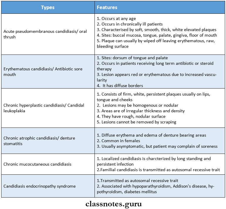
Oral medicine Red And White lLsions oral cavity clinial types and features

Causative Organisms Management:

1. Removal Of Causative Agents

  • Replacement or relining of denture

2. Topical Applications

  • Clotrimazole- oral troche- 10 mg tablet dissolved in mouth five times daily
  • 1% gentian violet
  • Nystatin preparations
  • Vaginal tablets- one tablet, 1,00,000 units dissolved in mouth 3 times a day
  • Oral pastilles 2,00,000 units dissolved slowly in mouth 5 times a day
  • Oral suspension- 1,00,000 units/cc, 1 teaspoon is mixed with 1/4 cup of water, used as an oral rinse
  • Amphotericin B- 0.1 mg/ml used as rinse
  • Mycostatin cream- 1 lac unit or lactose-containing vaginal tablet

3. Systemic Treatment

  • Nystatin- 250 mg TDS for 2 weeks followed by 1 troche per day for the third week
  • Ketoconazole- 200 mg tablet once daily
  • Fluconazole-100 mg tablet once daily for 2 weeks
  • Itraconazole-100 mg capsules for 14 days.

Red And White Lesions

Question 2. Enumerate oral precancerous lesions and discuss differential diagnosis of oral leukoplakia

Answer:

Precancerous Lesions:

  • It is defined as morphologically altered tissue in which cancer is more likely to occur than its normal counterparts
  • For example,
  • Leukoplakia
  • Erythroplakia
  • Mucosal changes associated with smoking habits
  • Carcinoma in situ
  • Bowens disease
  • Actinic keratosis

Differential Diagnosis Of Leukoplakia::

1. Lichen Planus

  • There is the presence of Wickham’s striae

2. Chemical Burn

  • Gives a history of burn
  • Painful lesion
  • Appears as an irregularly shaped, white pseudonym-brane-covered lesion
  • Gentle lateral pressure causes the white material to slide away exposing painful central red ulceration

3. Syphilitic Mucous Patches

  • Presence of split papule or condyloma latum

4. White Sponge Nevus

  • White Sponge Nevus occurs soon after birth or at least by puberty
  • White Sponge Nevus is widely distributed over the oral mucous membrane
  • The familial pattern is seen

5. Discoid Lupus Erythematosus

  • Central atrophic area with a small white dot and slightly elevated border zone or radiating white striae

6. Psoriasis

  • Positive Au spitz’s sign
  • Skin lesions are present

7. Leukoedema

  • Occurs on buccal mucosa covering most of the oral surface of the cheek and extending onto labial mucosa
  • The presence of a faint milky appearance with a folded and wrinkled pattern

8. Hairy Leukoplakia

  • Associated with AIDS patients
  • Corrugated leukoplakic lesions occurring on the lateral and ventral surface of the tongue

9. Verruca Vulgaris

  • Commonly occurs in the oral cavity as a small,, raised white lesion, more than 0.5 cm in diameter

10. Verrucous Carcinoma

  • Lesions are elevated

11. Cheek Biting Lesion

  • The presence of local irritants as a causative factor
  • Removal of cause relieves the condition

12. Electrogalvanic White Lesion

  • Disappears when different metal restorations are replaced with composite restorations

Question 3. Define premalignant lesion conditions. Describe oral lichen planus in detail.

Answer:

Premalignant Lesion:

  • It is defined as morphologically altered tissue in which cancer is more likely to occur than its nor¬mal counterparts
  • For example,
  • Leukoplakia
  • Erythroplakia
  • Mucosal changes associated with smoking habits
  • Carcinoma in situ
  • Bowen’s disease
  • Actinic keratosis

Preremalignant Condition:

  • It is defined as a generalized state or condition associated with a significantly increased risk for cancer development
  • For example:
    • Oral submucous fibrosis
    • Syphilis
    • Sideropenic dysplasia
    • Dyskeratosis congenital
    • Lupus erthymetosis

Oral Lichen Planus:

  • Oral Lichen Planus is a relatively common dermatological disorder occurring on skin and oral mucous membranes and refers to lace-like patterns produced by symbolic algae and fun-gal colonies on the surface of rocks in nature

Etiology:

  • Cell-mediated immune response:
  • It is associated with wadi lymphocyte epidermal interactions
  • Autoimmunity:
    • The activated T lymphocytes also secrete gamma interferon which induces keratinocytes to increase differentiation

Immune deficiency:

  • There is decreased serum levels of IgG, IgA, or IgM in lichen planus

Genetic factor:

  • Oral Lichen Planus has a familial trait

Infection:

  • Oral Lichen Planus may be caused due to Spirochetes and rod-like bodies

Psychogenic Factor

  • Stress may be related to the occurrence of lichen planus

Habits:

  • Oral Lichen Planus Is associated with tobacco chewing and betel nut chewing habits

Miscellaneous:

  • Oral Lichen Planus Is associated with deficiency of Vitamin HI, HO, and C and also associated with electric potential difference, anemia, and patient with secondary syphilis

Oral Lichen Planus Types:

  • Reticular
  • Papular
  • Plaque
  • Atrophic
  • Classical
  • Erythematous
  • Ulcerative
  • Hypertrophic
  • Erosive
  • Bullous
  • Annular
  • Actinic
  • follicular
  • Linear

Oral Lichen Planus Clinical Features:

  • Site:
    • Buccal mucosa
    • Tongue
    • Lips
    • Gingiva
    • The floor of the mouth
    • Palate
    • Presentation
  • Initially, there is a burning sensation of the oral mucosa
  • It appears as radiating white and grey velvety thread-like papules in a linear, angular or retiform arrange¬ment forming typical lacy, reticular patterns, rings, and streaks
  • Wickham’s striae- tiny white elevated dots are present at the intersection of white lines
  • It may be superimposed to candidal infections

Oral Lichen Planus Types Explanation:

  • Reticular Type:
    • It is bilateral
    • It consists of slightly elevated fine whitish lines that produce lace-like patterns
  • Papular Type:
    • Whitish elevated lesions of 0.5-1 mm in size
    • It is well seen in keratinized areas
    • They are seen at the periphery of the reticular pattern
  • Plaque Type:
    • It is soon on the dorsum of the tongue and buccal mucosa
    • It loads lo disappearance of papillae
    • It spreads In concentric peripheral growth
  • Atrophic Type:
    • It appears a smooth, red, poorly defined area
    • The attached gingival is frequently involved
    • Patient may complain of pain and burning In the areas of Involvement
  • Bullo Us Type:
    • It consists of vesicles and bullae which are short-lived
    • They rupture leaving an ulcerated surface
  • Hypertrophic Form:
    • It appears as a well-circumscribed, elevated white le¬sion resembling leukoplakia
  • Annular Form:
    • It appears as a round or ovoid, white outline with either pink or reddish-pink center

Oral Lichen Planus Differential Diagnosis:

  • Leukoplakia:
    • Absence of Wickham’s striae
  • Candidiasis:
    • Pseudomembrane can be rubbed off
  • Lupus Erythematous:
  • It has the feathery appearance of a lesion

Oral Lichen Planus Management:

  • Removal of the causative agent
  • Steroid therapy.
  • Steroid spray.
    • Use of beclomethasone dipropionate spray, triamcinolone acetonide in gel or cream form over small and moderately sized painful lesions
  • Steroid coating:
    • Some painful lesions are treated with topical application of topical steroids
  • Topical delivery
  • Prednisolone:
    • 30 mg/day for the first week
    • 15 mg/day for the second week
    • 5 mg/day for the third and final week
  • Intralesional injection of methylprednisolone 40 mg/ml
  • Topical application of fluocinolone acetonide for 4 weeks
    • Nystatin and ketoconazole for treating fungal infections
    • Vitamin supplements
  • Surgical therapy:
    • Cryosurgery and cauterization
  • Psychotherapy:
    • Tranquilizers are tried to reduce anxiety
  • Dapsone therapy:
    • To control the lymphocytes-mediated progress of lichen planus
  • PUVA therapy
    • High-intensity long-wave ultraviolet (PUVA) light is used as a therapeutic agent
  • Fluocinonide
    • It is used as an adhesive base
  • Symptomatic therapy
    • As topical analgesic, topical anesthetic, and antihistaminic

Question 4. Classify white lesion. Describe in detail the etiology, classification, clinical features, and management of leukoplakia.

Answer:

White Lesion Classification:

1. Variation In Structure And Appearance Of Normal Mucosa

  • Leukoedema
  • Fordyce’s granules
  • Linea alba

2. White Lesion With Precancerous Potential:

  • Leukoplakia
  • Erythroplakia
  • Lupus erythematosus
  • Carcinoma in situ
  • Lichen planus

3. White Lesion Without Precancerous Potential

  • Traumatic lesions
  • Focal epithelial dysplasia
  • White sponge nevus
  • Stomatitis nicotine
  • Hairy leukoplakia

4. Nonkeratotic lesion

  • White hairy tongue of Burns
  • Pemphigus
  • Desquamative gingivitis
  • Candidiasis
  • Koplik’s spots

Leukoplakia Definition:

  • Leukoplakia is a whitish patch or plaque that cannot be characterized, clinically or pathologically, as any other disease and which is not associated with any other physical or chemical causative agent except the use of tobacco.

Etiology:

  • Tobacco
    • Smokeless tobacco
    • Smoking tobacco
  • Alcohol
  • Chronic irritation
  • Candidiasis
  • Galvanism
  • Vitamin deficiency
  • Xerostomia
  • Nutritional deficiency
  • Hormones: sex hormones
  • Drugs: Anticholinergic, antimetabolic
  • Virus: herpes simplex and HIV
  • Idiopathic

Leukoplakia Clinical Features:

  • Age and sex: in older age males it occurs commonly i.e in the age of 35-45 years
  • Sites
    • Buccal mucosa
    • Commissures
    • Lip
    • Tongue
  • Presentation
    • Oral leukoplakia often represents solitary or multiple white patches
    • They can be nonpalpable, faintly translucent, white areas over the mucosa
    • Many lesions can be thick, fissured, indurated, or papillomatous in nature
    • The size of the lesion varies from a small well-localized patch measuring about a few mm in diameter to a dif¬fuse large lesion, covering a wide mucosal surface
    • The surface of the lesion may be smooth or finely wrinkled or even rough on palpation, and the lesion cannot be removed by scraping
    • The lesions are usually white or grayish or yellowish-white in color and in some cases, due to the heavy use of tobacco, they may take a brownish-yellow color
    • Some lesions may exhibit a pumice-like surface, which occurs due to the presence of multiple discrete keratotic striae, on the surface of these lesions
    • Leukoplakia of the floor of the mouth sometimes has an ebbing tide pattern of appearance
    • The thickness of the patch may vary from only faint to considerably thick
    • In most of cases, these lesions are asymptomatic, however in some cases, they may cause pain, a feeling of thickness and a burning sensation, etc
  • Leukoplakia Types:
    • Homogenous leukoplakia
    • Ulcerative leukoplakia
    • Nodular or speckled leukoplakia

Staging Of Leukoplakia:

According To Size, Clinical Aspect, And Pathological Features

Size: It is denoted by L

  • L1: size is less than 2 cm
  • L2: size is in the range of 2-4 cm
  • L3: size is more than 4 cm
  • Lx: size is not specified

Clinical Aspect: it is denoted by C

  • C1– homogenous
  • C2– nonhomogenous
  • Cx-not specified

Pathological Features: It is denoted by P

  • P1– no dysplasia
  • P2– mild dysplasia
  • P3– moderate dysplasia
  • P4– severe dysplasia
  • Px– not specified

Site: it is denoted by S

  • S1– all sites excluding floor of the mouth, tongue
  • S2– floor of the mouth and tongue
  • S3-not specified

Leukoplakia Management:

  • Elimination of etiological factor
  • Prohibition of smoking
  • Removal of chronic irritants
  • Elimination of other etiologic factors like syphilis, alcohol dissimilar metal restoration, etc
  • Conservative treatment
  • Vitamin therapy:
    • Vitamin A: ‘3000 to 300000 H1 tor 3 months
    • Vitamin A + Vitamin K therapy to inhibit metabolic degradation
  • Nystatin therapy
    • it is given o00000 ID twin’ daily pins .’0’V borax glycerol or t ‘V gentian violet or month rinses with chlorogenic solution
  • Panthenol
    • Lingual tablets and oral sprays may be used against glossitis and glossodynia
  • Ksttvgen:
  • it can be helpful

Surgical Management:

  • Conventional surgery
    • The affected area is undermined and disserted from the underlying tissue
    • Sliding unusual dap is prepared for covering the wound
    • After proper mobilization of the unusual dap, it is advanced, and multiple interrupted Mark silk sutures are used
    • Post-operative application of ioehags at the sife is advised to minimize bleeding and swelling:
  • Cryosurgery
    • Tissue is exposed to extreme cold to produce irreversible cell damage
    • Liquid nitrogen or pressurized nitrogen oxide is used at -d0” Celsius
  • Fulguration
    • It’s a technique in which there is the destruction of tissues by high voltage current and action is controlled by movable electrodes

Leukoplakia Laser:

  • CO2 lasers are most commonly used in oral lesions due to their great affinity for any tissue with high water content and their minimum penetration depth. U\ tVd- 0. 3 mm in oral tissue
  • It is used in the form of
    • Biopsy
    • Laser peel
    • Ablation

Leukoplakia Miscellaneous:

  • Radiation therapy is only used in neoplastic tissues
  • Chemotherapeutic agents-
    • Bleomycin and human fibroblast interferon are used

Question 5. Classify white lesion. Describe in detail the clinical features, differential diagnosis & management of oral submucous fibrosis.

Answer:

Classification Of White Lesions:

  • Variation In Structure And Appearance Of Normal Mucosa
    • Leukoedema
    • Fordyce’s granules
    • Linea alba
  • A White Lesion With Precancerous Potential
    • Leukoplakia
    • Erythroplakia
    • Lupus erythematosus
    • Carcinoma in situ
    • Lichen planus
  • White Lesion Without Precancerous Potential
    • Traumatic lesions
    • Focal epithelial dysplasia
    • White sponge nevus
    • Stomatitis nicotine
    • Hairy leukoplakia
  • Nonkeratotic Lesion
    • White hairy tongue
    • Burns
    • Pemphigus
    • Desquamative gingivitis
    • Candidiasis
    • Koplik’s spots

Oral Submucous Fibrosis Clinical Features:

  • Age And sex: Third – fourth-decade males are more affected
  • Gradual stiffness of the mucosa occurs
  • Oral mucosa becomes blanched
  • Palpation of the mucosa reveals many vertical fibrous bands
  • There may be difficulty in deglutition
  • Referred pain in the ear
  • Depapillation of tongue
  • Restricted movement of the floor of the mouth
  • Shrunken uvula
  • Restricted mouth opening

Oral Submucous Fibrosis Differential Diagnosis:

  • Scleroderma
  • Chemical burns

Oral Submucous Fibrosis Management:

  • Quit the habit
  • Antioxidant- Oxyace- 1 capsule/ day
  • Multivitamin therapy
  • Steroid-Betnovate 0.12%
  • Tumeric application
  • Intralesional injection of Hyaluronidase- 1500 U
  • Physiotherapy
  • Splitting of fibrous bands
  • Laser

Instruments In Operative Dentistry Short Question and Answers

Instruments Short Answers

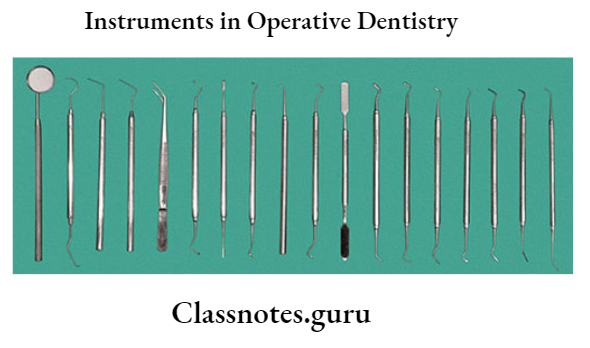
Instruments In Operative Dentistry.

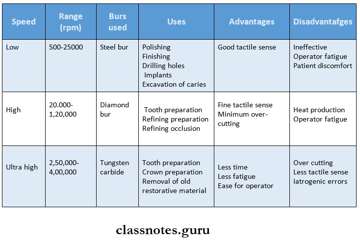
Question 1. Slow Speed.

Answer:

Slow Speed Range:

  • 5002500 rpm

Slow Speed Bur Used:

  • Steel bur

Slow Speed Uses:

  • Polishing
  • Finishing
  • Drilling holes
  • Implants
  • Excavation of caries

Slow Speed Advantages:

  • Good tactile sense

Slow Speed Disadvantages:

  • Inefficient
  • Operator fatigue
  • Patient discomfort

Question 2. 245 bur.

Answer:

  • It is nonstandard carbide bur
  • Designed to combine rounded corners with flat ends
  • ADA size number 330L
  • ISO size number 008
  • Head diameter 0.80 mm
  • Head length 3 mm
  • Shape pear, elongated

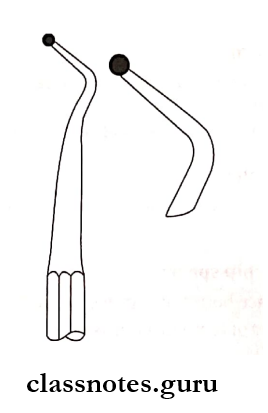
Question 3. Enamel Hatchet.

Answer:

  • Paired instrument
  • Blade angle 4590°
  • Bevel Unibevel/Bibeveled

Conservative And Operative Dentistry Instruments Hatchet

Enamel Hatchet Use:

  • Unibevelled instrument
  • Cleaving of enamel
  • Planning of dentinal walls

1. Bibevelled:

  • Use in a chopping motion
  • Refine line and point angle

Question 4. AngleFormer.

Answer:

  • Type of excavator, mono-angled instrument
  • Cutting edge angle 8085°
  • Paired instrument
  • Blade Beveled on sides as well as at the end
  • Manner of using Push or pull motion
  • Use Establish retention form in DFG restoration
  • Planning the gingival cavosurface margin

Question 5. High Speed.

Answer:

  • Speed 20,000 1,20,000 RPM
  • Burs used diamond burs with lubricant

High Speed Uses:

  • Tooth preparation
  • Refining tooth preparation
  • Refining occlusion

High Speed Advantages:

  • Fine tactile sense
  • Minimum over cutting

High-Speed Disadvantages:

  • Heat production
  • Not fit for larger preparation
  • Operator fatigue

Question 6. Dental Bur.

Answer:

Dental Bur Definition:

Dental Bur is defined as a rotary cutting instrument with cutting heads of various shapes and two or more sharp-edged blades, used as a rotary grinder

Dental Bur Classification:

1. According To Their Mode Of Attachment To The Handpiece

  • Latch type
  • Friction grip type

2. According To Their Composition

  • Stainless steel
  • Tungsten carbide
  • Combination

3. According To Their Motion

  • Right bur Revolves clockwise
  • Left bur Revolves anticlockwise

4. According To The Length Of Their Head

  • Long
  • Short
  • Regular

5. According To Their Use

  • Cutting burs
  • For finishing and polishing

6. According To Their Shapes

  • Round
  • Pear shaped
  • Inverted cone
  • Wheel shaped
  • Tapering fissure
  • Straight fissure

Question 7. Grasps are used with Hand Instruments.

Answer:

1. Modified Pen Grasp:

  • Middle finger pad placed over shank
  • Index finger Bend over middle phalanges and placed over the middle finger
  • Thumb Placed on the opposite side of the instrument

Modified Pen Grasp Advantages:

  • Creates tripod effect
  • Enhances instrument control stabilizers instru¬ment

Modified Pen Grasp Uses:

  • Commonly used in mandibular teeth

2. Inverted Pen Grasp:

  • Similar to a modified pen grasp
  • Palm faces toward the operator

Inverted Pen Grasp Uses:

  • Lingual surface of maxillary anterior
  • Occlusal surface of maxillary posteriors

3. Palm And Thumb Grasp:

  • The shaft placed on the palm of the hand and grasped by four fingers
  • The thumb is free to control movements

Palm And Thumb Grasp Use:

  • Holding handpiece
  • Cutting incisal retention for Class 3

4. Modified Palm And Thumb Grasp:

  • Provides more control to avoid slipping of instrument
  • Used for maxillary anterior

Question 8. Speed in operative Density

Answer:

Conservative And Operative Dentistry Instruments Speed in operative density

Question 9. Rake angle.

Answer:

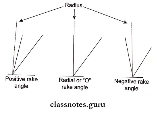
  • It is the angle between the rake face and the radial line
    • Positive Rake Angle Radial line is ahead of rake face
    • Negative Rake Angle Rake face is ahead of radial line
    • Zero Rake Angle Rake face and radial line coincide

Conservative And Operative Dentistry Instruments Three types of rake angles

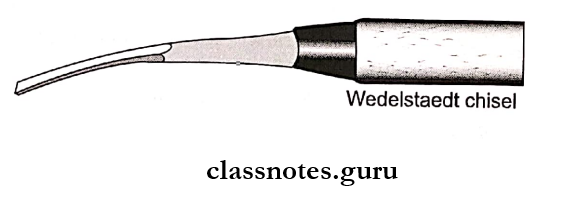
Question 10. Wedel Staedt Chisel.

Answer:

  • Have slightly curved shanks
  • Used on anterior teeth
  • It is a single plane, unibevel instrument
  • Used for cleaving undermined enamel

Conservative And Operative Dentistry Instruments Wedelestaedt chisel

Question 11. Balancing of Hand Instrument.

Answer:

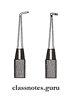
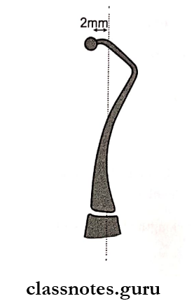
  • Balancing is achieved by providing angles in the shank of the instrument so that the cutting edge is within 23 mm of the long axis of the instrument
  • It provides better access
  • It is also called contra angling

Conservative And Operative Dentistry Instruments Blacing of an instrument

Question 12. Spoon Excavator.

Answer:

  • It is a modified hatchet
  • Double ended instrument
  • BladeSpoon, claw or disk-shaped
  • Use Removal of caries and debris
  • Manner of use Scooping motion

Question 13. Advantages and Disadvantages of Burs.

Answer:

Burs Advantages:

  • Precise
  • Easy to control
  • Tactile perception
  • Removal of debris

Burs Disadvantages:

  • Pain
  • Vibration
  • Noise production
  • Pulpal damage
  • Over cutting

Instruments In Operative Dentistry Short Essays

Instruments Short Essays

Question 1. Classify hand-cutting instruments.

Answer:

According To Gv Black:

1. Cutting Instruments.

  • Hand
  • Hatchet
  • Chisel
  • Hoe

2. Condensing Instruments Pluggers.

3. Plastic Instruments.

  • Plastic filling instrument
  • Cement carriers
  • Carvers

4. Finishing And Polishing Instruments.

  • Orangewood sticks
  • Polishing points

5. Isolation Instruments

  • Saliva ejector, evacuating tips.

6. Miscellaneous

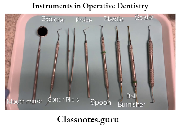
  • Mouth mirror, probe.

Question 2. Hand-cutting instruments.

Answer:

1. Excavators:

Excavators Types:

  • Hatches:
    • The cutting edge of the blade is directed in the same plane as that of the long axis of the handle
    • It is beveled
      • Uses:
        • Used in anterior teeth for preparing retentive areas
        • Sharpening of line angles
        • In preparation for a direct gold restoration
  • Hoes:
    • The cutting edge of the blade is perpendicular to the axis of the handle
      • Uses:
        • Planing tooth preparation walls
        • For forming line angles
  • Angleformers:
    • Monoangled instrument
    • The cutting edge is at 90 degrees to the bladder
      • Uses:
        • Sharpening line angles
        • Creates retentive features in dentin in gold restoration preparation
  • Spoon Excavator:
    • Blades are slightly curved and cutting edges are either circular or clawlike
      • Uses:
        • To remove caries
        • Carves amalgam or direct wax pattern

2. Chisels:

  • Used for cutting enamel
  • Grouped into

1. Straight, Slightly Curved Or Bangle chisels:

  • It has a straight shank and blade with a bevel on only one side
  • The edge is perpendicular to the axis of the handle

2. Enamel Hatchet:

  • Paired instrument
  • Blade angle 4590°
  • Bevel Unibevel/Bibeveled

Enamel Hatchet Use:

  • Unibevelled Instrument:
    • Cleaving of enamel
    • Planning of dentinal walls
  • Beveled:
    • Use in a chopping motion
    • Refine line and point angle

3. Gingival Margin Trimmer:

  • It is a modified hatchet with opposite curva¬ture and bevels
  • Paired instrument Distal and mesial
  • Instrument formula 7585 Mesial GMT, 95100 Distal GMT

Gingival Margin Trimmer Use:

  • Planning of gingival cavosurface margin
  • Removal of unsupported enamel
  • Bevel axiopupal line angle in Class 2

Question 3. Bur design.

Answer:

  • The design of the bur includes the following

1. Blade Or Cutting Edge:

  • It is in contact with the horizontal line or face

2. Tooth Face:

  • The sides of the tooth head of the cutting edge in the direc¬tion of the rotation is the tooth face

3. Back Of The Tooth:

  • The opposite of the bur tooth is the back of the tooth

4. Rake Angle:

  • Rake Angle is the angle between the rake face and the radial line
    • Positive Rake Angle Radial line is ahead of the rake face
    • Negative Rake Angle The Rake face is ahead of the radial line
    • Zero Rake Angle Rake face and radial line coincide

5. Clearance Angle:

  • Clearance Angle is the angle between the back of the tooth and the work
  • Mostly it is straight and clearly defined

6. Tooth Angle:

  • Tooth Angle is the measurement between the face and the back

7. Flute Or Chip Space:

  • Flute or chip space is the space between successive teeth
  • The number of teeth in a bur is 6 or 8

Instruments in Operative Dentistry,.

Question 4. Instrument Nomenclature.

Answer:

  • Order Purpose of instrument Example: Excavator
  • Suborder Manner of use Example: Push or pull
  • Class Form of working end Example: Hatchet, chisel
  • Subclass shape of the shank Example: Monoangle

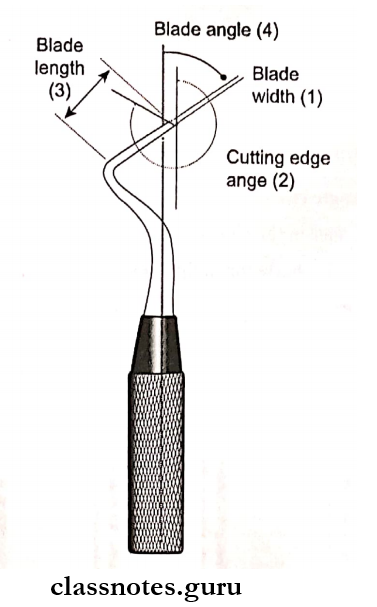
Question 5. Instrument Formula.

Answer:

Unit-1:

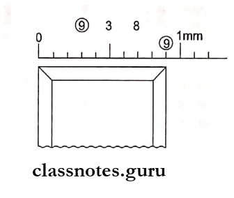
  • Blade width
  • Represents the width of the blade in tenths of a millimeter

Unit-2:

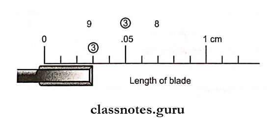
  • Blade length
  • Expressed in millimeter

Unit-3:

  • Blade angle
  • The angle formed between the blade and the long axis of the instrument
  • Expressed in 100th of a circle

Unit-4:

  • Cutting edge angle
  • The angle formed between the cutting edge and the long axis of the handle

Conservative And Operative Dentistry Instruments Instrument formula

Conservative And Operative Dentistry Instruments First digit of formula indicates width of blade of a millimeter

Conservative And Operative Dentistry Instruments Third number indicates length of blade in millimeters

Question 6. Chisels.

Answer:

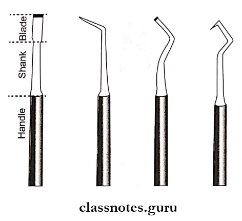
Chisels Types:

1. Straight Chisels:

  • Straight blade in line with handle and shank
  • Cutting edge on one side

2. Monoangle Chisels:

  • Blade angle to the shaft
  • Maybe medially or distally

3. Biangle Chisel:

  • Two angles between the shaft and blade
  • Unibevelledmedially or distally

4. Triangle Chisel:

  • 3 angles in the shank
  • Used for flattening the pulpal floor

Conservative And Operative Dentistry Instruments Chisel

Question 7. Excavators.

Answer:

  • Used for removal of caries
  • Refining internal line angles

Excavators Types:

1. Hatchet:

  • The blade of the hatchet is perpendicular to the shaft
  • The cutting edge is parallel to the shaft
  • Paired i.e. right and left
  • Used for delicate cutting

2. Hoe Excavators:

  • Single planed instrument
  • Unibevelled
  • Used with a push motion
  • Used for cutting axial walls

3. Spoon Excavators:

  • Paired instruments
  • Double planed instruments
  • Used for removal of the decayed dentin

4. Cleoid Excavator:

  • Blade resembles a claw
  • Used for amalgam carving, excavating decay in difficult areas

Conservative And Operative Dentistry Instruments Spoon excavator

Infectious Diseases Long Essays

Infectious Diseases Long Essays

Question 1. What is the differential diagnosis of ulcers over the penis? How do you treat syphilis?

Answer:

Differential Diagnosis Of Ulcers Over Penis:

  • Chancre
  • Chancroid
  • lymphogranuloma venereum
  • Genital herpes
  • Neoplasm

Syphilis:

  • Syphilis is a sexually transmitted disease

Syphilis Clinical Features:

  1. Primary syphilis
    • Incubation period- about 21 days
    • Chancre develops
    • Syphilis is a solitary, painless, indurated, non-tendered, ulcerated, or eroded lesion
    • Initially, it was a dull red macule
    • Later it becomes ulcerated
    • Regional lymphadenopathy
  2. Secondary syphilis
    • Appears about 6-8 weeks
    • Skin lesions appear as macules, papules, fol-locules, or, papulosquamous patches
    • Circinate lesions develop on the face
    • Headache
    • Fever, anorexia
    • Joint and muscle pain
    • Laryngitis, pharyngitis
    • Generalised lymphadenopathy
    • Lesions develop over the mucocutaneous junction
  3. Tertiary syphilis
    • Develops about 5-10 years after primary infection
    • Affects the skin, central nervous system, CVS, mucous membrane
    • Lesions are called gumma
    • Tertiary syphilis is localized, chronic granulomatous lesion with a nodular or ulcerated surface
    • Causes generalized paresis, dementia, and strokes
    • Bone lesions cause osteomyelitis and destruction of joints

infectious diseases long essay questions

Syphilis Treatment:

Infectious Diseases Differntial Diagnosis Of Ulcers Over Penis Treatment

Question 2. Describe clinical features, diagnosis, complications, and management of typhoid fever.
(or)
Discuss the etiology, clinical features, complications, and management of enteric fever.

Answer:

Typhoid Or Enteric Fever:

  • Typhoid is an acute systemic illness

Etiology:

  • Salmonella typhi
  • Salmonella paratyphi

Enteric Fever Clinical Features:

  • Incubation period- 10-14 days
  1. First 5-7 days
    • High-grade, remittent fever with chills
    • Headache
    • Aches
    • Malaise
    • Constipation
    • Leucopenia
    • Dry cough
    • Epistaxis
    • Relative bradycardia
  2. Between 1st and 2nd week
    • Rose spots develop over the trunk as small macules, red in color, and blanch on pressure
    • Splenomegaly
    • Bronchitis
    • Abdominal pain
    • Abdominal distension
    • Diarrhea
  3. After 2nd week
    • Confusion
    • Delirium
    • Toxaemia
    • Coma

Read And Learn More: General Medicine Question and Answers

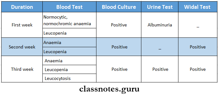
Enteric Fever Diagnosis:

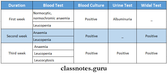
Infectious Diseases Typhoid Fever Diagnosis

Enteric Fever Complications:

  1. Intestinal complications
    • Hemorrhage
    • Paralytic ileus
    • Perforation
    • Peritonitis
  2. Extraintestinal complication
    • Meningitis
    • Cholecystitis
    • Pneumonia
    • Myocarditis
    • Bone and joint infection
    • Encephalopathy
    • Granulomatous hepatitis
    • Nephritis

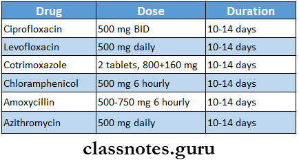
Enteric Fever Management

Infectious Diseases Thyroid Fever Management

long essays on communicable diseases

Question 3. Describe the etiology, pathology, clinical features, and management of cervical TB lymphadenitis.

Answer:

Tuberculous Lymphadenitis:

  • Tuberculous Lymphadenitis is defined as achronic specific granulomatous inflammation with caseation necrosis of the lymph node

Etiology:

  • Mycobacterium tuberculosis complex which includes M. tuberculosis, M. bovis, M. africanum, M. canetti, and M.caprae

Pathology:

  • Pathological stages of the disease are:
  1. Stage 1- lymphoid hyperplasia
    • Formation of tubercles and granulomas occurs without cassation necrosis
  2. Stages 2 and 3
    • Caseation necrosis in the affected lymph nodes occurs
    • It is followed by the destruction of capsules of lymph nodes
    • Periadenitis
  3. Stage 4
    • There is a rupture of caseous material into the surrounding soft tissue
    • Formation of abscess cavity

Tuberculous Lymphadenitis Clinical Features:

  • Persistent, painless swelling of the lymph nodes
  • Release of discharge of fluid
  • Fever
  • Weight loss
  • Fatigue
  • Night sweats

tuberculous LymphadenitisManagement:

  1. Anti-tubercular therapy
    • Isoniazid, Rifampicin, Pyrazinamide, and Ethambutol are given for 2 months
    • Followed by Isoniazid and Rifampicin for the next 4 months
  2. DOTS therapy
  3. Surgery- indicated in
    • Persistent fluctuant lesions
    • Failure with chemotherapy
    • Symptomatic relief

Question 4. Describe the etiology, clinical features, and management of intestinal amoebiasis.

Answer:

Intestinal Amoebiasis

Caused by entamoeba histolytic

  • E. histolytica can cause two types of pathological lesions as follows

1. Primary (or) Intestinal Amoebiasis:

Infectious Diseases Primary or intestinal amoebiasis

  • This condition is called amoebic dysentery.
  • These ulcers may be generalized (or) may be localized to the ileocaecal (or) sigmoidorectal region.
  • Occasionally ulcers may involve deeper tissues and cause perforation (or) peritonitis.
  • Erosion of blood vessels may lead to bleeding.
  • The superficial lesions generally heal without scarring.
  • Deep ulcers form scars leading to strictures, partial obstruction, and thickening of the gut wall.

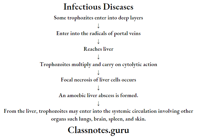
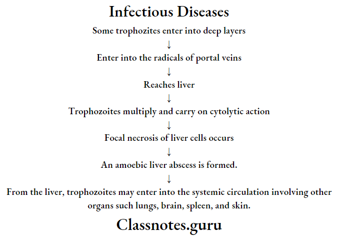
2. Extra-intestinal amoebiasis (or) secondary (or) metastatic lesions.

  • Amoebic liver abscess:

Infectious Diseases Primary or intestinal amoebiasis

long answer questions on infectious diseases

Question 5. Diphtheria etiology, Clinical Feature Management

Answer:

Etiology:

  • Etiology is caused by Corynebacterium diphtheria

Diphtheria-etiology Clinical Features:

  • Diphtheria-etiology is insidious at the onset
  • Moderate grade fever
  • Tachycardia
  • Sore throat
  • Formation of greyish-greenish pseudomembrane on the tonsils
  • Swollen neck- Bull neck
  • Tender lymphadenopathy
  • Nasal infection
  • Hoarseness of voice
  • Cough
  • Respiratory obstruction
  • Acute circulatory failure
  • Myocarditis
  • Nerve palsies
  • Dysphagia
  • Dysphonia
  • Paraesthesia in the limbs

infectious diseases long essays for mbbs

Diphtheria-Etiology Management:

  1. Antitoxin diphtheric serum (ADS)
    • The dose varies from 20,000 to 1,00,000 units depending on the duration and severity of the disease
    • In mild cases, a lesser dose may be used
  2. Antibiotics
    • Penicillin G-6,00,000 units every 12 hourly intravenously or
    • Amoxycillin 500 mg 8 hourly for 7-10 days
    • Patient allergic to penicillin
      • Erythromycin- 500 mg 6 hourly or
      • Azithromycin 500 mg daily or
      • Rifampicin 600 mg daily
  3. Tracheostomy

Infectious Diseases Short Essays

Question 1. Oral manifestations of diphtheria

Answer:

Oral Manifestations Of Diphtheria

  • Diphtheria is an infectious disease caused by Corynebacterium Diphtheriae
  • Oral Manifestations Of Diphtheria is characterized by local exudative inflammation of the upper respiratory tract and usually with the formation of pseudomembrane at the level of the nasopharynx
  • Oral manifestations
      • Sore throat
        • Formation of thick, fibrinous, greyish-green pseudomembrane on tonsils, larynx, pharynx
          • Exudation in tonsillar area
          • Hoarseness of voice
          • Paralysis of the soft palate
          • Dysphagia
          • Dysphonia
          • Cervical Lymphadenopathy

Infectious Diseases Short Notes

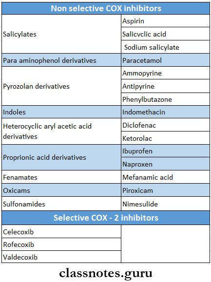
Question 2. NSAIDs

Answer:

NSAIDs Classification

Infectious Diseases NSAIDs Classification

Question 3. Oral manifestations of AIDS

Answer:

Oral Manifestations Of AIDS

  1. Bacterial infections
    • Tuberculosis
    • M. avium complex
    • Salmonellosis
  2. Viral infection
    • Herpes simplex
    • Varicella zoster
    • Epstein Barr virus
  3. Mycotic infections
    • Pneumocystis pneumonia
    • Candidiasis
    • Aspergillosis
    • Cryptococcosis
  4. Parasitic infection
    • Toxoplasmosis
    • Cryptosporidiosis
  5. Malignancies
    • Kaposi’s sarcoma
    • B cell lymphoma

Read And Learn More: General Medicine Question and Answers

Question 4. Human immunodeficiency virus (HIV)- post-exposure prophylaxis

Answer:

Human Immunodeficiency Virus (HIV)- Post-Exposure Prophylaxis

  • Following exposure, postexposure prophylaxis may be required depending upon the category of exposure and HIV status of the exposure source
  • Drugs used are:
  1. Zidovudine 300 mg BD
  2. Lamivudine 150 mg BD
  3. Protease inhibitors
    • Lopinavir 400 mg BD or 800 md OD
    • Ritonovir 100 mg BD or 200 mg OD
      • Drugs are started within the first 72 hours
      • It should be continued for 4 weeks
      • The injured site on the wound should be thoroughly washed with soap and water
      • Antiseptics are also used

Short essays on communicable diseases

Question 5. Gonorrhoea

Answer:

Gonorrhoea Definition:

  • Gonorrhoea is a communicable sexually transmitted disease of humans

Etiology:

  • Etiology is caused by Neisseria gonorrhoea

Gonorrhoea Clinical Features:

  1. In males
    • Dysuria
    • Increased frequency of micturition
    • Purulent discharge per urethra
    • Oedema of the penis
    • Erythema of the urethral meatus with purulent discharge
  2. In females
    • Dysuria
    • Vaginal discharge
    • Abnormal menstrual bleeding
    • Rectal discomfort
  3. In homosexuals
    • Asymptomatic
  4. In children
    • Conjunctivae
    • Involvement of the pharynx or the respiratory tract

Gonorrhoea Complications:

  • Acute epididymitis
  • Prostatitis
  • Periurethral abscess or fistula
  • Salpingitis
  • Bartholin’s abscess
  • Premature birth
  • Arthritis
  • Bacteraemia
  • Perihepatitis

Gonorrhoea Treatment:

  1. For uncomplicated gonorrhea
    • 2.4 g of procaine penicillin IM + 1 g of oral probenecid or
    • Oral amoxicillin 2-3 g + Oral probenecid 1 g or
    • Cotrimoxazole (400+80 mg) 8 tablets orally
  2. In penicillin-resistant gonorrhea
    • Ciprofloxacin-250-500 mg orally single dose or
    • Cefotaxime- 0.5-1 g IM as single dose or
    • Cefixime- 400 mg orally as a single dose or
    • Ceftriaxone-250 mg IM as single dose
  3. In gonorrhea with complications
    • Aqueous crystalline penicillin G-10 million units 4 daily for 5 days
    • Ciprofloxacin 500 mg twice daily for 5 days
    • Ceftriaxone 1 g 4 daily for 5 days

Question 6. Chickenpox

Answer:

Chickenpox

  • Chickenpox is caused by Varicella zoster virus
  • Chickenpox rarely occurs a second time
  • The virus enters the mucosa of the upper respiratory tract and spreads by droplets from the throat

Chickenpox Clinical Features:

  • Chickenpox involves young children and adults
  • The incubation period is 14-21 days
  • Malaise
  • Low-grade fever followed by a rash
  • Rash is macula papular
  • Heals by scabs formation

Chickenpox Sites Involved:

  • Skin lesions- over trunk and face then spread to peripheral parts of the body
  • Mucosal lesions- affects the mucosa of the pharynx and vagina

Chickenpox Complications:

  • Superinfection of skin due to frequent scratching
  • Encephalitis
  • Cerebellar ataxia
  • Myocarditis
  • Osteomyelitis
  • Septic arthritis
  • Septicaemia
  • Hepatitis
  • Pneumonia

Infectious Diseases Short Answer Questions

Question 7. Herpes simplex

Answer:

Herpes simplex

  • Herpes simplex is a DNA virus o It belongs to the herpesviridae family.
  • Man is the only natural host.
  • Herpes simplex virus has two types as follows.
  1. Herpes simplex type 1 – is usually associated with oral and ocular lesions.
  2. Herpes simplex type 2 – is usually associated with genital infections.
    • Lesions caused by herpes simplex depend on the site of infection, age, and immune status of the individual as follows:
    • Cutaneous infections: These include:
      • Fever
      • Blisters on the cheeks, chins around the mouth, (or) on the forehand
      • Napkin rash on the buttocks of children, – Herpetic with low in medical professionals.
    • Eczema herpeticum:
      • The generalized herpetic eruption is seen in children suffering from eczema.
    • Mucosal lesions:
      • Acute gingivostomatitis
      • Recurrent herpes labialis.
    • Ophthalmic lesions:
      • Keratoconjunctivitis
      • Follicular conjunctivitis.
    • Visceral lesions:
    • Nervous system:
      • HSV meningitis,
      • Sacral autonomic dysfunction.
    • Genital lesions:
      • Infection of cervix, vagina, vulva (females)
  3. Congenital lesions:
    • Subclinical or localized infection of skin mouth or eyes.
    • Besides primary infections, herpes viruses may present as latent infection, reactivation, and recrudescence.

Question 8. Herpes labialis

Answer:

Herpes Labialis

  • Herpes labialis occurs in patients with no prior infection with Herpes Simplex Virus-1

Herpes Labialis Clinical Features:

  • Age- children and young adults
  • Incubation period-5-7 days
  1. Prodromal generalized symptoms
    • Fever
    • Malaise
    • Headache
    • Nausea, vomiting
    • Painful mouth
    • Sore throat
    • Irritability
    • Excessive drooling of saliva
    • Lack of tactile sensation
    • Cervical lymphadenopathy
  2. Later symptoms
    • Numerous vesicle formations over keratinized mucosa
    • Vesicles are thin-walled
    • They contain clear fluid
    • They rupture leaving multiple, small, punctuate shallow painful ulcers of 2-6 mm
    • Ulcers are surrounded by a red ring of inflammation
    • Ulcers may become secondarily infected
    • Healing starts in about 3 days and is completed within 7-14 days

Herpes Labialis Treatment:

  • To prevent secondary infection- antibiotics are used
  • To control fever- Antipyretics are given

Question 9. Mumps

Answer:

Mumps

  • Mumps is an acute viral infectious disease characterized by unilateral (or) bilateral nonsuppurative enlargement of the salivary glands usually the parotid gland.
  • Submandibular and sublingual glands are occasionally involved.
  • Usually, children are affected. May also occur in adults.
  • Also known as epidemic parotitis

Mumps Causative Organism:

  • Organisms involved in paramyxovirus.

Mumps Route Of Infection:

  • Infection is acquired by inhalation (or) direct contact or through the conjunctiva.
  • The incubation period is 12 – 18 days.

Mumps Clinical Features:

  • Unilateral (or) bilateral swelling of parotid glands.
  • Fever, malaise
  • Local pain and tender
  • Involvement of extra parotid sites can cause more serious problems as follows.
    • CNS involvement may lead to
      • Meningitis,
      • Meningoencephalitis
      • Orchitis

Mumps Complications:

  • Orchitis
  • Oophoritis
  • Arthritis
  • Nephritis
  • Pancreatitis
  • Thyroiditis and myocarditis

Mumps Laboratory Diagnosis:

  • Isolation of viruses from CSF, saline (or) urine.
  • Serological tests with paired serum samples tested for the rise in the titer of antibodies by complement fixation test, ELISA, and haemagglutination tests.
  • Serum amylase levare els elevated in both parotitis and acute pancreatitis.

Mumps Treatment:

  • Treatment is conservative maintaining hydration. Prevention is using MMR vaccine

microbiology short notes on infections

Question 10. Diphtheria

Answer:

Diphtheria Clinical Features:

  • It is insidious at the onset
  • Moderate grade fever
  • Tachycardia
  • Sore throat
  • Formation of greyish-greenish pseudomembrane on the tonsils
  • Swollen neck- Bull neck
  • Tender lymphadenopathy
  • Nasal infection
  • Hoarseness of voice
  • Cough
  • Respiratory obstruction
  • Acute circulatory failure
  • Myocarditis
  • Nerve palsies
  • Dysphagia
  • Dysphonia
  • Paraesthesia in the limbs

Diphtheria Management:

1. Antitoxin diphtheric serum (ADS)

  • The dose varies from 20,000 to 1,00,000 units depending on {ho duration and severity of the disease
  • In mild cases, a lesser dose may be used

2. Antibiotics

  • Penicillin G-o.00,000 units every 12 hourly intravenously or
  • Amoxycillin 500 mg S hourly for 7-10 days
  • Patient allergic to penicillin
    • Erythromycin- 500 mg 6 hourly or
    • Azithromycin 500 mg daily or
    • Riftamoicin 600 mg daily

3. Tradieostomy

Question 11. Three malarial parasites

Answer:

Three Malarial Parasites

  • Malaria is a disease caused by four plasmodium speeds which are as follows:
  1. P. vivax
  2. P. Falciparum
  3. P. Malaria
  4. P. Ovale.

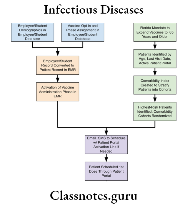
Infectious Diseases Employee student

Question 12. Widal test

Answer:

Widal Test

  • Widal Test is an agglutination test for detection of agglutinin H and O in patients with enteric fever

Widal test Procedure:

  • Mix equal volumes of serial dilutions of the serum and H and O antigens
  • Incubate in a water bath at 37 degrees C for 4 hours
  • Read after overnight refrigeration at 4 degrees C

Widal test Result:

  • A Titre upto 160 indicates enteric fever

Question 13. Chancre

Answer:

Chancre

  • Chancre is a typical lesion of primary syphilis that appears on genitals/at extragenital sites in 2-4 weeks after exposure to infection.
  • Initially, the lesion is a painless papule that ulcerates in the center so that the fully developed chancre is an indurated lesion with central ulceration accompanied by regional lymphadenitis.
  • Chancre heals without scarring, even in the absence of treatment.

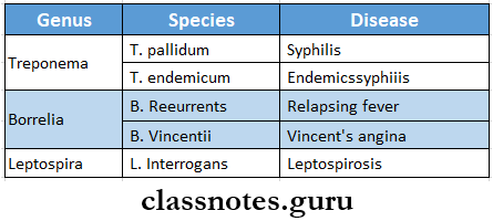
Question 14. Two spirochaetal diseases

Answer:

Two spirochaetal diseases

Infectious Diseases Two Spirochaetal Diseases

Short note on infectious diseases

Question 15. Oral manifestations of syphilis

Answer:

Oral manifestations of syphilis

  • Syphilis is a sexually transmitted disease caused by spirochaetes, treponema pallidum.
  • Syphilis is divided into 3 stages depending upon the period after which the lesions appear and the type of lesions.
  • They are primary, secondary, and tertiary.
  • Oral lesions occur in secondary syphilis known as mucous patches, on the tongue, gingiva, etc., or as a split papule on the lips, which are highly infectious.
  • Tertiary/late syphilis/gumma is a granuloma with central necrosis, noninfectious, and is most common on the tongue and palate.
  • Palatal perforation by ulcer after vigorous antibiotic use is known as the Herxheimer reaction.
  • In congenital/prenatal syphilis, the most constant finding is relatively short roots of mandibular permanent 1st molars, short maxilla, Hutchinson triad (teeth, eye/ear involved), hypoplasia of incisors and molars i.e., notched incisors and mulberry molars.
  • Treatment: Penicillin is the drug of choice.

Question 16. Congenital syphilis

Answer:

Congenital syphilis Major features are:

  • Hutchinson’s triad includes.
  • Hutchinson’s teeth-small, widely spaced, peg-shaped permanent teeth.
  • Notched central incisors
  • Intestinal keratitis with blindness and deafness from Stir cranial nerve injury.
  • Saddle shaped nose
  • Bony lesions like epiphysis and periostitis
  • Mucocutaneous lesions of acquired secondary syphilis
  • Diffuse fibrosis in the liver.

Question 17. VDRL test

Answer:

VDRL test

  • VDRL test is an abbreviated form of the Venereal Disease Research Laboratory test
  • VDRL test is the most widely used test for syphilis
  • VDRL test is a simple arid rapid test
  • VDRL test is a slide flocculation test

VDRL test Procedure:

  • Cardiolipin antigen is freshly prepared
  • A drop of this is added to a drop of inactivated patient’s serum on a slide
  • Mix it by shaking

VDRL test Result:

  • If floccules appear it indicates a positive test

Question 18. Prednisolone

Answer:

Prednisolone

  • Prednisolone has potent glucocorticoid and mild mineralocorticoid activity
  • Prednisolone is the most commonly used glucocorticoid

Prednisolone Uses:

  • Replacement therapy
  • Rheumatoid arthritis
  • Osteoarthritis
  • Acute gout
  • Allergic diseases
  • Bronchial asthma
  • Collagen diseases
  • Eye diseases
  • Renal diseases
  • Skin diseases
  • GIT diseases
  • Liver diseases
  • Malignancies
  • Organ transplantation

Prednisolone Adverse Effects:

  • HPA axis suppression
  • Cushing’s syndrome
  • Hyperglycemia
  • Susceptibility to infection is increased
  • Osteoporosis
  • Avascular necrosis
  • Peptic ulceration
  • Mental disturbances
  • Cataract and glaucoma
  • Delayed wound healing

Question 19. Metronidazole

Answer:

Metronidazole

  • Metronidazole is a nitroimidazole

Metronidazole Mechanism of Action:

  • Metronidazole is prodrug
  • Susceptible micro-organisms reduce the nitro group of metronidazole by nitroreductase
  • Convert it into a cytotoxic derivative
  • It binds to DNA
  • Inhibits protein synthesis

Metronidazole Uses:

  • Anaerobic infection
  • Amoebiasis
  • Trichomonas vaginitis
  • Giardiasis
  • H.pylori infection
  • Pseudomembranous colitis
  • Draetmcwlosis
  • Topical application

Metronidazole Adverse Reactions:

  • Nausea, anorexia, abdominal pain, metallic taste in mouth
  • Headache, dizziness
  • Stomatitis, glossitis
  • Furry tongue
  • Insomnia, ataxia, vertigo
  • Peripheral neuropathy
  • Pruritis, urticaria, skin rashes

Question 20. Aminoglycosides

Answer:

Aminoglycosides

  • Aminoglycosides are antibiotics with amino sugars in glycosidic linkages

Aminoglycosides Mechanism of Action:

  • Penetrate bacterial cell membrane
  • Bind to 30S ribosomes
  • Inhibits bacterial protein synthesis

Aminoglycosides Uses:

  • Tuberculosis
  • Subacute bacterial endocarditis
  • Plaque
  • Tularemia
  • Brucellosis

Aminoglycosides Adverse Effects:

  • Ototoxicity
  • Nephrotoxicity
  • Neuromuscular blockade

Aminoglycosides Examples:

  • Streptomycin
  • Kanamycin
  • Tobramycin
  • Neomycin
  • Gentamicin
  • Netilmicin

Short questions in infectious diseases

Question 21. Chloramphenicol

Answer:

Chloramphenicol

  • Chloramphenicol Is a broad spectrum and loll.

Chloramphenicol Mechanism of Action:

  • It binds to 50S ribosome
  • Inhibits transpeptidation reaction
  • Inhibits protein synthesis

Chloramphenicol Uses:

  • Typhoid lever
  • Bacterial meningitis
  • Anaerobic infections
  • Rickettsial infection
  • Kyo infections

Chloramphenicol Adverse Reactions:

  • Gastrointestinal disturbances- nausea, vomiting, diarrhea
  • Bone marrow depression
  • Gray baby syndrome
  • Hypersensitivity reactions
  • Superinfection

Question 22. Malaria

Answer:

Malaria is a disease caused by four plasmodium species which are as follows:

  1. P. vivax
  2. P. Falciparum
  3. Malaria
  4. P. Ovale.

Malaria Life Cycle:

The malarial parasites pass their life cycle in two hosts.

  1. Man intermediate host [a sexual development
  2. Femalanopheles mosquito delinitive host [sexual development].

Malaria Clinical Features:

  • Intermittent fever which is named malaria is caused by plasmodium
  • It consists of
    • Febrile paroxysm
      • It comprises of three stages
    • Anaemia
      • Microcytic or normocytic hypochromic anemia develops
    • Splenomegaly.
      • The spleen becomes enlarged and palpable
      • Black water fever and pernicious malaria are the most commonly seen complications in falciparum malaria.

Infectious Diseases Malaria Clinical Features

infectious disease short essay for mbbs

Question 23. Ludwig’s angina

Answer:

Ludwig’s angina

  • Ludwig’s angina was described by Wilhelm Fredrich Von Ludwig in 1836
  • Ludwig’s angina is rapidly spreading cellulitis involving simultaneously all three spaces i.e. Submandibular, sublingual, and submental spaces

Etiology:

  • Odontogenic infection
  • Traumatic injuries
  • Infective condition
  • Pathologic conditions

Ludwig’s angina Clinical Features:

  1. Generalised symptoms
    • Dehydration
    • Fever
    • Dysphagia
    • Dyspnoea
    • Hoarseness of voice
    • Stridor
  2. Extraoral features
    • Brawny hard swelling
    • Erythematous skin
    • Local rise in temperature
    • Drooling of saliva
    • Respiratory distress
  3. Intraoral features
    • Trismus
    • Raised floor of mouth
    • Airway obstruction
    • Increased salivation

Ludwig’s angina Management:

  • Airway maintenance
  • Removal of the causative factor
  • Administration of 4 fluids
  • Antibiotics
    • Penicillin G-2-4 million units TV 4-6 hourly
    • Gentamycin-80 mg IM BD
    • Metronidazole-400 mg 8 hourly
    • Erythromycin-600 mg 6-8 hourly
    • Amoxicillin-500 mg 6-8 hourly orally
      • Surgical management

Infectious Diseases Short Question And Answers

Infectious Diseases Short Question And Answers

Question 1. Four complications of viral hepatitis

Answer:

Four Complications Of Viral Hepatitis

  • Hepatic necrosis
  • Chronic hepatitis
  • Cirrhosis of liver
  • Hepatic failure
  • Hepatocellular carcinoma

Question 2. Infectious Mononucleosis

Answer:

Infectious Mononucleosis

  • Infectious Mononucleosis is an acute febrile illness associated with lymph node enlargement caused by Epstein Barr virus
  • Features
    • Affects adolescents and adults
    • The incubation period is 7-10 days
  • Clinical features
    • Fever, malaise
    • Conjunctival haemorrhage
    • Maculopapular rash
    • Sore throat
    • Lymphadenopathy
    • Splenomegaly

Infectious Mononucleosis Complications

  • Chronic fatigue
  • Hepatitis
  • Hemolytic anemia
  • Thrombocytopenia pleurisy
  • Myocarditis
  • Meningoencephalitis
  • Rupture of an enlarged spleen

infectious diseases short questions and answers

Question 3. Rubella

Answer:

Rubella

  • Rubella is a mild childhood disease
  • Rubella may be acquired congenital or postnatally

Rubella Features:

  • Infection is acquired by inhalation
  • Incutwbon synod-2-3 weeks
  • Fever, malaise
  • Headache
  • Mild conjunctivitis
  • Lymphadenopathy
  • Rasa develops on the forehead and face
  • Rubella spreads downward to the trunk and extremities
  • Rubella lasts for 1-5 days

Read And Learn More: General Medicine Question and Answers

Rubella Prevention:

  • Rubella is prevented by MMR vaccine

Question 4. Mumps

Answer:

Mumps

  • The incubation period is 12 – 18 days.

Mumps Clinical Features:

  • Unilateral (or) bilateral swelling of parotid glands,
  • Fever, malaise
  • Local pain and tender
  • The involvement of extra parotid sites can cause more serious problems as follows.
    • CNS involvement may lead to
      • Meningitis,
      • Meningoencephalitis
      • Orchitis

Question 5. Measles

Answer:

Measles Clinical Features:

  • Insidious in onset
  • Moderate fever
  • Tachycardia
  • Sore throat
  • Formation of greyish-greenish pseudomembrane on tonsils
  • Associated nausea and vomiting
  • Bull neck- swollen neck
  • Tender lymphadenopathy
  • Xasal infection
  • Hoarseness of voice
  • Cough
  • Respiratory obstruction
  • Toxaemi
  • Acute peripheral circulatory failure
  • Nerve paralysis
  • Dysphagia
  • Dysphonia
  • Paraesthesia

short Q&A on infectious diseases

Question 6. Prevention of measles

Answer:

Prevention Of Measles

  1. Active immunization
    • One injection of live attenuated measles vaccine along -with mumps and rubella vaccines, MMR vaccine to children over 1 year
  2. Passive immunization
    • Human normal immunoglobulin is used

Dose:

Infectious Diseases Prevention Of Measles

Question 7. Diphtheria

Answer:

Diphtheria Clinical Features:

  • Diphtheria is insidious at the onset
  • Moderate grade fever
  • Tachycardia
  • Sore throat
  • Formation of greyish-greenish pseudomembrane on the tonsils
  • Swollen neck- Bull neck
  • Tender lymphadenopathy
  • Nasal infection
  • Hoarseness of voice
  • Cough
  • Respiratory obstruction
  • Acute circulatory failure
  • Myocarditis
  • Nerve palsies
  • Dysphagia
  • Dysphonia
  • Paraesthesia in the limbs

Question 8. Ascariasis

Answer:

Ascariasis

  • Infection caused by Ascaris lumbricoides is known as ascariasis

Ascariasis Features:

  • Migrating larvae in the lungs cause dyspnoea, wheezing, cough, and signs of pneumonia
  • Loss of appetite
  • Malnutrition
  • Fever
  • Eosinophilic leukocytosis
  • Appendicitis
  • Liver abscess

Ascariasis Diagnosis:

  1. Stool examination- demonstrates adult worms or eggs
  2. Barium meal- Demonstrate adult worms
  3. Serodiagnosis- detects antibodies

Ascariasis Treatment:

  • Pyrantel pamoate-11 mg/kg given orally
  • Mebendazole-100 mg twice daily for three days

Question 9. Hookworm Disease
(or)
Ankylostomiasis

Answer:

Hookworm Disease

  • Hookworm disease is a symptomatic infection caused by Ankylostoma duodenal and Necatar Ameri- can
  • Occurs in all tropical and subtropical countries

Hookworm Disease Clinical Features:

  • Dermatitis
  • Lesions in the lungs- bronchitis and bronchopneumonia
  • Microcytic, hypochromic anemia
  • Epigastric pain
  • Dyspepsia
  • Vomiting
  • Diarrhea

Hookworm Disease Diagnosis:

  1. Blood examination
    • Eosinophilia
    • Microcytic, hypochromic anemia
  2. Stool examination
    • Shows occult blood

Hookworm Disease Treatment:

  • High protein diet
  • Oral iron preparations
  • Blood transfusion
  • Drugs used are- mabendazole, albendazole, pyrantelpamoate

important short questions in infectious diseases

Question 10. Amoebic dysentery

Answer:

Amoebic Dysentery

  • Amoebic dysentery is also known as intestinal amoebiasis
  • Amoebic dysentery is a condition in which the infection is confined to the intestine and is characterized by the passage of blood and mucus in the stool

Amoebic dysentery Types:

  1. Acute amoebic dysentery
    • Multiple ulcers occur
    • These ulcers are deep and extensive
    • Its complications are
      • Pericaecal and pericolic abscess
      • Amoebic appendicitis
      • Perforation
      • Generalized peritonitis
      • Gangrene of guts
      • Fistula
  2. Chronic amoebic dyssentery
    • A single latent ulcer is present in the caecum while multiple small superficial ulcers are scattered throughout the large intestine
    • Stricture formation occurs
    • Amoeboma in the caecum and large intestine develops

Question 11. Treatment of malaria

Answer:

Treatment Of Malaria

  1. General management
    • Use of analgesics and antipyretics to treat fever
    • Administration of intravenous fluids
  2. Treatment of acute attack
    • Chloroquine-600 mg followed by 300 mg in 6 hours and then 150 mg twice a day for 3 days
    • Chloroquine-resistant cases- Dihydrochloride 600 mg salt three times a day by mouth for 5 days
    • Followed by a single dose of sulphadoxine 1.5 g combined with pyrimethamine 75 mg

Question 12. Complication of malaria

Answer:

Complication Of Malaria

  1. Pernicious malaria
  2. Black water fever.

1. Pernicious Malaria:

  • Pernicious Malaria is a life-threatening condition

Pernicious Malaria Cause:

  • Pernicious Malaria is due to heavy parasitization.

Pernicious Malaria Clinical Features:

  • Pernicious Malaria is grouped into the following:

Infectious Diseases Complication Of malaria

2. Blackwater Fever:

  • This occurs in persons who are previously infected and had inadequate doses of quinine

Blackwater Fever Clinical Features:

  • Intravascular hemolysis,
  • Fever,
  • Haemoglobinuria
  • Vomiting and prostration with the passage of dark red (or) blackish urine hence called backwater fever.

Blackwater Fever Mechanism:

  • An autoimmune mechanism may be involved in hemolysis
  • Parasitized erythrocytes during previous infection act as antigen
  • Antibodies are formed against it
  • Due to this antigen-antibody reaction, massive destruction of erythrocytes occurs
  • There is excessive deposition of haemosiderin pigment in the liver, spleen, and kidneys.
  • Sequel of black water fever include
    • Circulatory failure
    • Renal failure,
    • Liver failure,
    • Anaemia.

Question 13. Complication of mumps

Answer:

Complication Of Mumps

  • Orchitis
  • Oophoritis
  • Arthritis
  • Nephritis
  • Pancreatitis
  • Thyroiditis and myocarditis.

infectious disease viva questions and answers

Question 14. Diagnosis of typhoid fever

Answer:

Diagnosis Of Typhoid Fever

Infectious Diseases Typhoid Fever Diagnosis

Question 15. Complications of typhoid fever

Answer:

Complications Of Typhoid Fever

  1. Intestinal complications
    • Hemorrhage
    • Paralytic ileus
    • Perforation
    • Peritonitis
  2. Extraintestinal complication
    • Meningitis
    • Cholecystitis
    • Pneumonia
    • Myocarditis
    • Bone and joint infection
    • Encephalopathy
    • Granulomatous hepatitis
    • Nephritis

Question 16. Clinical features of typhoid fever

Answer:

Clinical Features Of Typhoid Fever

  • Incubation period- 10-14 days
  1. First 5-7 days
    • High-grade, remittent fever with chills
    • Headache
    • Aches
    • Malaise
    • Constipation
    • Leucopenia
    • Dry cough
    • Epistaxis
    • Relative bradycardia
  2. Between 1st and 2nd week
    • Rose spots develop over the trunk as small macules, red in color, and blanch on pressure
    • Splenomegaly
    • Bronchitis
    • Abdominal pain
    • Abdominal distension
    • Diarrhea
  3. After 2nd week
    • Confusion
    • Delirium
    • Toxaemia
    • Coma

Infectious Diseases Types Of Diseases Transmission

Question 17. Lepromatous leprosy

Answer:

Lepromatous Leprosy

  • Leprosy caused by mycobacterium leprae is of 2 types wepromatous leprosy represents low resistance and tuberculoid leprosy represents high resistance.
  • Lepromatous leprosy is characterized by multiple symmetrical skin lesions that are hypopigmented and erythematous maculopapular/nodular.
  • Nerve involvement is present with less severe sensory distribution.
  • Histopathology involves the collection of foamy macrophages/lepra cells in the dermis separated from the epidermis by a clear zone.

Question 18. Lepra reaction

Answer:

Lepra Reaction

  • Lepra reaction/reactional leprosy:
  • There may be two types:
  1. Type 1 (Reversal reactions),
  2. Type 2 (Erythema, nodosum leprosum)

1. Type 1: the polar forms of leprosy donor undergo any change in clinical ami histopathological picture.

  • Borderline groups are unstable and may move across the spectrum in either direction with upgrading/downgrading of patients’ immune stale.
  1. Upgrading Read Ion: Characterized by increased cell-mediated immunity and occurs in patients of borderline Icpromalous (BL) type on treatment who upgrade/shift towards tuberculoid type.
  2. Downgrading Reaction: Characterized by lowering of cellular immunity and is seen in borderline tuberculoid (BT) type who downgrade/shift towards Icpromalous type.

2. Type 2: Occurs in Icpromalous patients after treatment. It is characterized by tender cutaneous nodules, fever, iridocyclitis, synovitis, and lymph node involvement.

Short questions on bacterial and viral infections

Question 19. Diagnosis of AIDS

Answer:

Tests for AIDS:

  1. ELISA( Enzyme-Linked Immunosorbent Assay)
    • It is a color reaction test
    • Method:
      • A serum containing antibodies is developed from the patient’s blood sample
      • Tests for AIDS is added to the ELISA plate
      • Wash off the inactive antibodies
      • A second layer of antibodies called conjugate is added
      • Excess antibodies are again washed off
      • A substrate is added to it
    • Result:
      • Color becomes darker- positive test
      • No color change- negative test
  2. Western blot
    • Method
      • Viral proteins from the patient’s blood sample are passed through a gel
      • The separated proteins are then passed through an electric current
      • Human serum is added
      • A chromogen is added to it
  3. Result:
    • A specific band of viral protein is detected

Question 20. Quinolones

Answer:

Quinolones

  • Quinolones are a group of synthetic antimicrobial agents

Quinolones Uses:

  • Uncomplicated UTI
  • Diarrhea

Quinolones Adverse Effects:

  • Hemolytic anemia
  • Allergic reactions
  • Headache
  • Myalgia
  • Drowsiness

Examples:

  • Nalidixic acid
  • Oxalinic acid
  • Cinoxacin

Question 21. Cephalosporins

Answer:

Cephalosporins

  • Cephalosporins are semi-synthetic antibiotics with a beta-lactam ring

Cephalosporins Uses:

  • Gram-negative infections
  • Surgical prophylaxis
  • Gonorrhea
  • Meningitis
  • Mixed aerobic and anaerobic infection
  • Typhoid
  • Nosocomial infection
  • Orodental infection

Cephalosporins Adverse Reactions:

  • Hypersensitivity reactions
  • Nephrotoxicity
  • Diarrhea
  • Bleeding
  • Low WBC count
  • Pain at the injection site
  • Disulfiram like reaction

Question 22. Triple vaccine, Dan PT

Answer:

Triple Vaccine, DPT

  • DPT is used as an active immunization
  • Triple vaccine, DPT is combination of Diphtheria toxoid, pertusis vaccine and tetanus toxoid

DPT Route Of Administration:

  • Intramuscular

DPT Dose:

  • Initial dose- 6 weeks
  • Three doses are completed at intervals of 4-6 weeks
  • Booster doses-18 months and 5 years

DPT Adverse Reactions:

  • Transient local inflammation
  • Fever
  • Occthe asional convulsions

DPT Advantages:

  • Minimizes the number of injections
  • Improves immune response

Question 23. Anti-amoebic drugs

Answer:

Anti-Amoebic Drugs

  • Anti-amoebic drugs are drugs used in amoebiasis

Anti-Amoebic Drugs Classification:

  1. Drugs effective in both intestinal and extra-intestinal amoebiasis
    • Metronidazole
    • Tinidazole
    • Secnidazole
    • Omid azole
    • Satranidazole
    • Emetine
  2. Drugs effective in intestinal amoebiasis
    • Diloxanide furoate
    • Quiniodochlo
    • Iodoquinol
    • Tetracyclines
  3. Drugs effective in extraintestinal amoebiasis
    • Chloroquine

Question 24. Quinsy

Answer:

Quinsy

  • Quinsy is an infection in connective tissue between the tonsil and the superior constrictor
  • Quinsy is also called peritonitis sellar abscess

Quinsy Fethe Features:

  • Acute pain in the throat
  • Pain radiates to the ear
  • Dysphagia
  • Nausea
  • Constipation
  • Poor oral hygiene
  • Body ache
  • Headache
  • Enlarged lymph nodes
  • Dyspnoea
  • Trismus
  • Deviation of uvula
  • Hoarseness of voice
  • Foul breath

Quinsy Management:

  • Antibiotics
  • Incision over the most prominent part
  • Analgesics
  • Warm saline gargles 4 fluids
  • Tonsillectomy

Infectious Diseases Short Case Questions

Question 25. Rabies prevention

Answer:

Rabies Prevention

  • Rabies vaccines are two types
  1. Neural
  2. Non-neural

1. Neural Vaccines:

  1. Semple vaccine:
    • The most widely used vaccine
    • Developed by Semple at Central Research Institute, Kasauli.
    • It is a 5% suspension of infected sheep brain and inactivated by 5% phenol at 37°C leaving no residual live virus.
  2. Beta Propiolactone (BPL) Vacine:
    • Modified sample vaccine
    • Instead of phenol, BPL is used as an activating agent.
  3. Infant brain vaccine:
    • Used widely in America to Reduce neurological complications.
      • Vaccination Schedules:
        • Nowadays it’s not used.
        • In the past, they were given subcutaneously on the anterior abdominal wall.
        • 7 – 14 injections depend on the degree of risk.

2.  Non-Neural Vaccines:

  1. Duck Egg Vaccine:
    • BPL is used as an inactivating agent
    • It has poor immunogenicity so not used now.
  2. Tissue culture vaccines:
    • Following cell culture vaccines are available in India.
      • Human diploid cell strain vaccine [HDCS]
        • The HDCS vaccine is prepared by growing rabies virus on human diploid cells and is inactivated with BPL.
        • This vaccine is highly antigenic and free of side effects
    • Purified chick embryo cell vaccine [PCEC]
      • PCEC is now widely used.
      • It is cheaper
      • It contains BPL inactivated flurry LEP strain
    • Purified Vero cell vaccine [PVC]
      • This vaccine is under study

Question 26. Antiviral drugs

Answer:

Antiviral Drugs

Infectious Diseases Antiviral Drugs

Question 27. BCG

Answer:

BCG

  • The Bacille Calmette Guerin (BCG) vaccine was prepared by Calmette and Guerin in 1921
  • Used for protection against tuberculosis infections

BCG Forms:

  • Liquid form
  • Freeze-dried form- commonly used

Dose and Administration:

  • 0.1 ml is administered intradermally soon after birth

BCG Result:

  • A small nodule develops at the injection site in about 2-3 weeks
  • it gradually increases in size upto 441 mm in diameter
  • It breaks into a shallow ulcer leaving a 4-8 mm diameter round scar

BCG Contraindications:

  • AIDS patients
  • Eczema
  • Pertussis
  • Measles
  • Patients on steroids

Question 28. Complications of Hepatitis B

Answer:

Complications Of Hepatitis B Are:

  1. Cirrhosis of liver
  2. Liver failure
  3. Hepatocellular carcinoma

Question 29. Chloroquine

Answer:

Chloroquine

  • Chloroquine is antimalarial drug
  • Chloroquine is a synthetic 4-arninoquinolone

Chloroquine Uses:

  • Malaria
  • Extra-intestinal amoebiasis
  • Photogenic reactions
  • Lepra reactions

Chloroquine Adverse Reactions:

  • Severe nausea and vomiting
  • Pruritis
  • Headache
  • Visual disturbances
  • Insomnia
  • Skin rashes
  • Cardiomyopathy
  • Peripheral neuropathy
  • Psychiatric problems
  • Blurring of vision
  • Confusion
  • Bleaching of hair

Question 30. Human insulins

Answer:

Human Insulins

  • Human insulin is produced by recombinant DMA technology
  • They are expensive
  • Absorbed rapidly

Human Insulins Forms:

  • Regular B Lente
  • Isophane insulin
  • Lente
  • Ultralente preparations

Human Insulins Advantages:

  • Less antigenic
  • More stable
  • Fewer chances of resistance
  • Less chances of lipodystrophy

Human Insulins indications:

  • Allergy to conventional preparations
  • Insulin resistance
  • Lipodystrophy at the injection site
  • Pregnancy

Infectious Diseases Important One Liners

Question 31. Four causes of lymphadenopathy

Answer:

Lymphadenopathy Causes:

  1. Inflammatory
    • Acute lymphadenitis
    • Chronic lymphadenitis
    • Granulomatous lymphaleukemiaeoplastic
    • Benign
    • Malignant
  2. Lymphatic leukemia
    • Autoimmune disorders
    • Juvenile rheumatoid arthritis
    • Collagen diseases like systemic lupus erythematosus, polyarteritis nodosa
  3. Generalised lymphadenopathy
    1. Tuberculosis
    2. Syphilis- secondary stage
    3. Infectious mononucleosis
    4. Sarcoidosis
    5. Toxoplasmosis
    6. Hodgkin’s disease
    7. Lymphosarcoma

Endocrine And Metabolic Diseases Short Essays

Question 1. Hypercalcemia

Answer:

Hypercalcemia Causes:

  • Conditions associated with hypercalcemia and increased PTH levels
    • Hyperparathyroidism
    • Chronic renal failure
  • Conditions associated with hypercalcemia and low PTH levels
    • Multiple myeloma
    • Sarcoidosis
    • Hyperthyroidism
    • Thiazide diuretics
    • Milk alkali syndrome
    • Familial

Hypercalcemia Clinical Features

  • Nausea, vomiting
  • Pain in abdomen
  • Dehydration
  • Hypotension

Hypercalcemia Treatment:

  • Avoid calcium-containing antacids
  • Replacement of fluid and electrolytes by 4-6 l of normal saline
    • To correct dehydration, hypotension, and calcium levels
  • Forced diuresis by saline and diuretic frusemide given 4 to depress tubular reabsorption of calcium
  • Drugs given include a generation bis- phosphonate ex: pamidronate given as a single 4 dose of 15-60 mg in normal saline
  • Calcitonin is used in emergency state
  • Cinacalcet is used orally in hypercalcemia crisis

Endocrine System Short Essay Questions

Question 2. Thyrotoxicosis

Answer:

Thyrotoxicosis

  • Thyrotoxicosis is a clinically toxic manifestation due to excess thyroid hormones

Thyrotoxicosis Causes:

  • Common causes
  • Grave’s disease
  • Toxic nodular goitre
    • Multinodular
    • Solitary nodule
  • Less common causes
    • Thyroiditis Drug-induced
    • Factitious
    • Iodine induced
  • Rarely
    • Pituitary or ectopic TSH
    • Thyroid carcinoma

Thyrotoxicosis Clinical Features:

  • Goitre
  • Weight loss, vomiting, diarrhea
  • Increased pulse rate, dyspnea, arrhythmia
  • Nervousness, restlessness, tremors, muscular weakness
  • Perspiration, clubbing, loss of hair, pre-tibial myxoedema
  • Amenorrhea, abortions, infertility, loss of libido
  • Lid lag, exophthalmos, diplopia, watering of eyes
  • Fatigue, heat intolerance, polydipsia

Question 3. Calcium homeostasis

Answer:

Regulation Of Calcium/ Hemostasis:

  • Calcium level is regulated by

Calcitriol

  • Calcitriol is the active form of vitamin D

Calcium Homeostasis Mechanism:

Diseases Of The Gastrointestinal System Gingival Induces Synthesis Of Calcium Binding Protein

  • Calcium homeostasis also promotes calcification and remodeling of bone.

2. Parathyroid hormones

  • Secreted by Parathyroid glands

Read And Learn More: General Medicine Question and Answers

Calcium Homeostasis Mechanism:

Endocrine And Metabolic Diseases Calcium Homeostasis Mechanism

Question 4. Hypoglycemia-clinical features and management

Answer:

Hypoglycaemia

  • Hypoglycaemia is defined as a fall in blood glucose concentration below 3.1 mmol/1

Hypoglycaemia Clinical Features:

  1. CVS symptoms
    • Palpitation
    • Tachycardia
    • Anxiety
    • Cardiac arrhythmias
  2. CNS symptoms
    • Tremors
    • Confusion
    • Headache
    • Tiredness
    • Difficulty in concentration
    • Slurred speech
    • Drowsiness
    • Convulsion
    • Coma
  3. GIT symptoms
    • Nausea
    • Vomiting
  4. Dermatological symptoms
    • Sweating
    • Hypothermia

Hypoglycaemia Management:

  1. In unconscious patients
    • Stop anti-diabetic medication
    • Administer 50 ml of 50% intravenous glucose
    • Intramuscular injection of 1 ml of glucagon
  2. Unconscious patients
    • Oral glucose intake
  3. In severe cases
    • 1 mg glucagon subcutaneously or intramuscularly and repeated if necessary after 10 minutes

Endocrine and metabolic disorders short answer

Question 5. Neurological complications of diabetes mellitus

Answer:

Neurological Complications Of Diabetes Mellitus

  • Diabetes can involve any part of the nervous system except the brain
  • Precipitating factors
  1. Poor glycaemic control
  2. Long control of diabetes

Pathological Changes:

  • Axonal degeneration of myelinated and nonmyelinated fibers
  • Patchy, segmental demyelination
  • Vasculopathy

Endocrine And Metabolic Diseases Neurological Complications Of Diabetes Mellitus

Question 6. Grave’s disease

Answer:

Grave’s Disease

  • Grave’s disease is a diffuse goiter affecting the whole functioning thyroid tissue causing hypertrophy and hyperplasia due to abnormal thyroid stimulants

Grave’s Disease Clinical Features

  • Grave’s Disease is a classical triad composed of
  1. Goitre
    • Diffuse and symmetric enlargement of thyroid
    • The gland surface is smooth
    • Bruit is heard
  2. Thyrotoxicosis
    • Excitability
    • Restlessness
    • Emotionally unstable
    • Insomnia
    • Muscle weakness
    • Fatigue
    • Myopathy
    • Heat intolerance
    • Increased sweating
    • Weight loss
    • Tachycardia
    • Palpitation
    • Increased cardiac output
    • Gynaecomastia
    • Increased libido
    • Pretibial myxoedema
  3. Exophthalmos
    • Protrusion of eyeballs

Grave’s Disease Treatment:

  1. Anti-thyroid drugs
    • To restore the patient to the euthyroid state
  2. Radioactive iodine
    • It destroys thyroid cells and reduces the mass of thyroid-functioning tissue
  3. Surgery
    • Reduce active thyroid mass

Question 7. Acromegaly- clinical features and complications

Answer:

Acromegaly:

  • Acromegaly occurs due to excess secretion of GH later in life after epiphyseal closure

Acromegaly Clinical Features:

  • Thick bones-larger hands and feet
  • Enlarged skull
  • Increased intracranial pressure
  • Headache
  • Photophobia
  • Visual disturbances
  • Hepatomegaly
  • Cardiomegaly
  • Osteoporosis
  • Arthralgia
  • Excessive sweating
  • Myalgia
  • Bowing of legs
  • Barrel shaped chest

Acromegaly Oral Manifestations:

  • Enlarged mandible
  • Class 3 malocclusion
  • Macroglossia D Thick lips
  • Proclamation of teeth
  • Hypercementosis
  • Large nose, ears, and prominent eyebrows
  • Periodontitis
  • Enlargement of maxillary air sinuses

Acromegaly Complications:

  • High blood pressure (hypertension)
  • Cardiovascular disease, particularly enlargement of the heart (Cardiomyopathy)
  • Osteoarthritis
  • Diabetes mellitus
  • Precancerous growths (polyps) on the lining of your colon
  • Sleep apnea is a condition in which breathing repeatedly stops and starts during sleep
  • Carpal tunnel syndrome
  • Reduced secretion of other pituitary hormones (hypopituitarism)
  • Uterine fibroids, benign tumors in the uterus
  • Spinal cord compression
  • Vision loss

Short notes on endocrine disorders

Question 8. Addison’s disease

Answer:

Addison’s Disease

  • Addison’s disease is due to progressive destruction of the three zones of the adrenal cortex and medulla with lymphatic infiltration

Addison’s Disease Clinical Features:

  1. Glucocorticoid insufficiency
    • Weight loss
    • Nausea and vomiting
    • Malaise
    • Weakness
    • Anorexia
    • Diarrhea
    • Constipation
    • Postural hypotension
    • Hypoglycaemia
  2. Mineralocorticoid deficiency
    • Hypotension
  3. Loss of androgens
    • Reduction of pubic and axillary hair in females
  4. Increased ACTH secretion
    • Pigmentation of exposed areas, pressure areas, mucous membranes, conjunctivae, and recently acquired scars

Question 9. Diabetes and surgery

Answer:

Diabetes And Surgery

  • Patients with diabetes need to maintain very good oral hygiene as they are prone to odontogenic, periodontal, and other infections
  • In well-controlled diabetics, dental procedures generally do not require any special precautions
  • Antibiotic coverage may be required
  • In uncontrolled diabetics, infections and wound healing may be a problem even after minor procedures
  • If major dental procedures or surgery is to be undertaken, prior control of diabetes by the physician is needed
  • If the patient is on only oral antihyperglycemic drugs, he should start insulin
  • Dental procedures may often reduce food intake and diabetics may go to hypoglycemia
  • Dose adjustments may be required

Question 10. Gestational diabetes mellitus

Answer:

Gestational Diabetes Mellitus

  • Gestational diabetes is defined as glucose intolerance that develops during pregnancy and usually cures after delivery
  • Persons affected are
  1. Older women
  2. Obese/overweight women
  3. Women with a history of delivering large babies
  4. Women with a history of gestational diabetes

Gestational Diabetes Mellitus Diagnosis:

  • All women during pregnane}7 should be screened based on random blood sugar tests in each trimester
  • 50 mg oral glucose is administered and serum glucose is measured at 60 min
  • If the glucose level is less than 140 mg, it is normal if not then the following step is taken
  • 100 mg of glucose is administered and glucose in the fasting state is measured at 1,2 and 3 hours
  • Deviation from normal levels indicates gestational diabetes

Normal Values:

Endocrine And Metabolic Diseases Diabetes And Surgery Normal Values

Endocrine and metabolic disorders short answer

Question 11. Cushing’s syndrome

Answer:

Cushing’s Syndrome

  • Cushing’s syndrome is caused by increased plasma glucocorticoid levels due to enhanced production of cortisol

Cushing’s Syndrome Clinical Features:

  • Age- common in 3rd and 4th decades of life
  • Females are more affected
  • Obesity
  • Truncal fullness
  • Moon facies
  • Buffalo hump
  • Purple striae are seen on the abdomen
  • Oligomenorrhoea and amenorrhoea
  • Impotence in males
  • Elevated blood pressure
  • Muscle weakness
  • Bone pain
  • Decreased glucose tolerance

Cushing’s Syndrome Investigations:

  • Screening test
  • Determine free cortisol in urine
  • Plasma ACTH measurement
  • Plasma ACTH levels of more than 200-500 Pg per ml indicate Cushing’s syndrome
  • Dexamethasone suppression test
  • Plasma values above 5 microgram/100 ml suggest Cushing’s syndrome

Cushing’s Syndrome Treatment:

  • Medical treatment
  • Metyrapone- dose- 2-6 g per day in divided dose
  • Aminoglutethimide-1-2 g per day
  • Surgical treatment
  • Pituitary irradiation
  • Adrenalectomy

Question 12. Eye signs on thyrotoxicosis

Answer:

Eye Signs On Thyrotoxicosis

  1. Exophthalmos
    • It means protrusion of the eyeball
  2. Spasm of the upper eyelid with lid retraction
  3. Proptosis with widening of the palpebral fissure
  4. Supraorbital and infraorbital swelling
  5. Congestion, edema, and chemosis of the conjunctiva
  6. Papilloedema
  7. Corneal ulceration
  8. External ophthalmoplegia
  9. Weakness of the extrinsic ocular muscles
  10. Other signs
    • Von Giraffe’s sign
      • When the patient is asked to look down, his upper eyelid fails to follow the rotation of the eyeball and thus lags behind
    • Joffroy’s sign
      • When the patient is asked to look upwards with the head fixed, there will be the absence of wrinkling on the forehead
    • Moebius sign
      • Failure of convergence of eyeballs
    • Dalrymple’s sign
      • Upper sclera is seen due to retraction of the upper eyelid
    • Stellwag’s sign
      • Absence of normal blinking

Endocrine And Metabolic Diseases Long Essays

Question 1. Describe the etiology, clinical features, and management of tetanus.

Answer:

Tetanus:

  • Tetanus is a disorder of neuromuscular excitability

Etiology:

  • Etiology is caused by exotoxin, tetanospasmin liberated by clostridium tetani

Etiology Clinical Features:

  • Trismus
  • Dysphagia
  • Risussardonicus- in it eyebrows and corners of the mouth are drawn up due to spasms of the muscles of the face and jaw
  • Opisthotonus- rigidity of the muscles of the neck and trunk
  • Elevation of temperature
  • Rapid pulse
  • Cyanosis

Endocrine and metabolic diseases long essays

Etiology Types:

  1. Acute tetanus
    • The incubation period is less than 10 days
  2. Chronic tetanus
    • The incubation period is about a month
  3. Delayed tetanus or latent tetanus
    • In it organism remains latent for many years
  4. Local tetanus
    • There is the presence of local contraction of muscles
  5. Cephalic tetanus
    • There is irritation or paralysis of the cranial nerves
  6. Bulbar tetanus
    • There is an extensive spasm of the muscles of deglutition and respiration
  7. Late tetanus
    • The inoculation period is prolonged
  8. Tetanus neonatorum
    • Recurrent tetanus

Read And Learn More: General Medicine Question and Answers

Etiology Management:

  1. Passive immunization
    • 1 Lac units of Anti-tetanus serum is given half intravenously and half intramuscularly
  2. Antibiotics
    • Penicillin injection of 10 lac units every 4 hours
    • Metronidazole
    • Tetracycline
  3. Wound care
    • Thorough cleansing and debridement
    • Removal of all necrotic tissues and foreign bodies
    • All stitches are removed
    • Blood clots, foreign bodies, and pus are cleared
    • The wound should be left open
  4. The patient is isolated to a quiet, dark, and well-ventilated room
  5. Further treatment depends on the severity of the cases
    • In mild cases
      • Prozac in -200 mg IM
      • Barbiturate, amylobarbitone, or injection diazepam-5-40 mg 4 times in a day
    • In moderate cases
      • Passage of nasogastric tube for feeding
      • Passage of self-retaining catheter to drain the bladder
      • Administration of intravenous fluid
      • Maintenance of intake and output chart
      • Tracheostomy
    • In very severe cases
      • Use of muscle relaxant, 40 mg turbo- marine initially 4 and then IM injections
      • Tracheostomy
      • Sedation with pentothal sodium
      • Nasogastric feeding
      • Elimination of visceral stimuli
      • Intravenous administration of fluid and electrolytes
      • Constant nursing

Long essay on endocrine system disorders

Question 2. Describe the etiology and clinical features of acromegaly with special experience in dental problems.

Answer:

Acromegaly:

  • Acromegaly occurs due to excess secretion of GH later in life after epiphyseal closure

Etiology:

  • Pituitary adenoma
  • Pancreatic islet cell tumors
  • Hypothalamic tumors
  • Bronchial carcinoid
  • Small cell carcinoma of Hung

Acromegaly Clinical Features:

  • Thick bones-larger hands and feet
  • Enlarged skull
  • Increased intracranial pressure
  • Headache
  • Photophobia
  • Visual disturbances
  • Hepatomegaly
  • Cardiomegaly
  • Osteoporosis
  • Arthralgia
  • Excessive sweating
  • Myalgia
  • Bowing of legs
  • Barrel shaped chest

Acromegaly Oral Manifestations:

  • Enlarged mandible
  • Class 3 malocclusion
  • Macroglossia
  • Thick lips
  • Proclination of teeth
  • Hypercementosis
  • Large nose, ears, and prominent eyebrows
  • Periodontitis
  • Enlargement of maxillary air sinuses

Endocrine And Metabolic Diseases Diagnosis

Metabolic diseases long answer questions

Question 3. Enumerate the proliferating factors, pathogenesis, investigations, and management of diabetic ketoacidosis.

Answer:

Diabetic Ketoacidosis:

  • Diabetic Ketoacidosis is a complication of diabetes mellitus

Diabetic Ketoacidosis Proliferating Factors:

  • Undetected or undiagnosed type 1 diabetes
  • Stress
  • Infection

Diabetic Ketoacidosis Pathogenesis:

Endocrine And Metabolic Diseases Diabetic Ketoacidosis Pathogenesis

Diabetic Ketoacidosis Investigations:

  • Diabetic ketoacidosis is confirmed by
    1. Hyperglycaemia
    2. Ketonaemia
    3. 1 leavy ketonuria
    4. Acidosis

Diabetic Ketoacidosis Management:

  1. Correction of hyperglycemia
    • Administration of regular insulin 0.1 U/k bolus followed by 0.1 U/kg/ hour by ccontnousIV infusion till the patient recovers
  2. Correction of dehydration
    • Normal saline infusion of 1 liter in the first hour
    • Then 1 litre over the next 4 hours
    • Then quantity is titrated
  3. Correction of acidosis
    • Use of sodium bicarbonate
  4. Potassium
    • 10-20 mEq/hour potassium chloride is added to the drip for rapid correction of hyperglycemia

Question 4. Classify diabetes mellitus. Discuss the complications and management of diabetes mellitus

Answer:

Diabetes Mellitus

  • Diabetes mellitus is defined as a heterogeneous metabolic disorder characterized by the common feature of chronic hyperglycemia with disturbance of carbohydrate, fat, and protein metabolism

Diabetes Mellitus Classification:

  1. Primary diabetes
    • Type 1- IInsulin-dependentdiabete mellitus
      • IImmunemediated
      • Nonimmune mediated
    • Type 2- NNoninsulin-dependentdiabetes mellitus
      • Obese
      • Non-obese
  2. Secondary diabetes
    • Pancreatic diabetes
    • Hormonal or endocrinal abnormalities
    • DDrug-inducedInsulin receptors antibodies
    • Genetic syndromes

Diabetes Mellitus Complications:

  1. Acute metabolic complications
    • Diabetic ketoacidosis
      • Develop in patients with severe insulin deficiency
      • Clinical Features:
        • Nausea, vomiting, anorexia
        • Deep and fast breathing
        • Mental confusion
        • Coma
    • Hyperosmolar hyperglycaemia nonketotic coma
      • It is a complication of type 2 diabetes mellitus
      • Caused by severe dehydration which leads to sustained hyperglycemia diuresis
      • Clinical Features:
        • High blood sugar
        • High plasma osmolality
        • Thrombotic and bleeding complications
    • Hypoglycaemia
      • Develop in type 1 diabetes mellitus
      • Clinical Features:
        • Permanent brain damage
        • Worsening of diabetic control
        • Rebound hyperglycaemia
  2. Late systemic complications
    • Atherosclerosis
      • Common in both type 1 and type 2 diabetes mellitus
      • Atherosclerosis may lead to
        • Myocardial infarction
        • Cerebral stroke
        • Gangrene of toes and feet
    • Diabetic microangiopathy
      • It is the abasementmembrane thickening of small blood vessels and capillaries of different organs and tissues
      • Occurs due to increased glycosylation of hemoglobin and other proteins
    • Diabetic nephropathy
      • It is a severe complication of diabetes mellitus
      • Occurs in both types
      • Features:
        • Asymptomatic proteinuria
        • Nephrotic syndrome
        • Progressive
        • Renal failure
        • Hypertension
    • Diabetic neuropathy
      • Effects all parts of the nervous system
    • Diabetic retinopathy
      • It is the cause of blindness
      • Other retinal complications include
        • Glaucoma
        • Cataract
        • Corneal disease
    • Infections
      • Diabetic patients are more susceptible to infections like tuberculosis, pyelonephritis, otitis, carbuncles and diabetic ulcers

Diabetes Mellitus Management:

  1. Diet management
    • The daily energy requirement for an individual is calculated according to age, weight, height, and activity
    • Calorie requirement:
      • For men- 36 kcal/kg
      • For women-34 kcal/kg
  2. Lifestyle modification
    • Physical exercise
    • Reduction of weight
    • Avoid alcohol and smoking
    • Behavioural exercises
  3. Oral hypoglycaemic drugs
    • Sulphonylureas
    • Meglitinides
    • Biguanides
    • Thiazolidinedione derivatives
    • Alpha-glucosidase inhibitors
  4. Insulin

Thyroid disorders long descriptive essay

Question 5. Discuss the complication of systemic ccorticosteroidtherapy with special reference to dental practice

Answer:

Complications Of Systemic Corticosteroids:

  1. Metabolic effects
    • Mood changes from euphoria to depression
    • Fluid retention and edema
    • Hypertension
    • Glucose intolerance
    • Osteoporosis
    • Increased susceptibility to infection
    • Reactivation of latent tuberculosis
    • Impaired wound healing
    • Gastric erosions
    • Masked perforation
    • HHemorrhagefrom stomach and duodenum
  2. Suppression of hypothalamic-pituitary-adrenal axis
    • It occurs with high-dose therapy
    • This makes it difficult to withdraw steroids

Measures To Reduce Side Effects:

  • Use of the lowest possible dose
  • Administer on alternate days rather than daily
  • Use of morning dose rather than any other time
  • Use of steroids for established cases
  • Monitor caloric intake to prevent weight gain and reduce sodium intake
  • Use of H2 receptor blockers or proton pump inhibitors
  • Provide high calcium intake and vitamin D

Question 6. Outline the conditions that produce pigmentation of oral mucosa. Discuss the clinical features of Addison’s disease.

Answer:

Causes Of Oral Pigmentation:

  • Autoimmune adrenalitis
  • Tubcrculousadrenalitis
  • Bilateral adrenalectomy
  • Drugs-ketoconazole
  • Metastases in the adrenal
  • Haemochromatosis
  • Amyloidosis
  • Histoplasmosis

Addison’s Disease:

  • Addison’s Disease is due to progressive destruction of the three zones of the adrenal cortex and medulla with lymphatic infiltration

Oral Pigmentation Clinical Features:

  1. Glucocorticoid insufficiency
    • Weight loss
    • Nausea and vomiting
    • Malaise
    • Weakness
    • Anorexia
    • Diarrhea
    • Constipation
    • Postural hypotension
    • Hypoglycaemia
  2. Mineralocorticoid deficiency
    • Hypotension
  3. Loss of aandrogensReduction of pubic and axillary hair in females
  4. Increased ACTH secretion
    • Pigmentation of exposed areas, pressure areas, mucous membranes, conjunctivae, and recently acquired scars

Adrenal gland disorders long essay

Question 7. Define hypo- and hyperthyroidism. Discuss clinical features, investigations, and treatment of hypothyroidism.

Answer:

Hyperthyroidism Definition:

  1. Hypothyroidism
    • V is a clinical condition caused by low levels of circulating thyroid hormones
  2. Hyperthyroidism
    • Hyperthyroidism is a clinical syndrome that results from exposure of the body tissues to excess circulating free thyroid hormones

Hypothyroidism Clinical Features:

  1. In new bom
    • Cretinism
    • Mental retardation
    • Poor growth
    • Difficult to nurse a baby
    • Dwarfism
  2. During childhood or adolescence
    • Juvenile hypothyroidism
    • Child appears young
    • Mental deficiency
    • Abdominal distension
    • Umbilical hernia
    • Prolapse of rectum
  3. In adults
    • Fatigue
    • Weight gain
    • Skin becomes thickened and puffy
    • Muscle cramps
    • Paraesthesia
    • Impaired speech
    • Reduced cardiac output
    • Elevation of blood pressure
    • Shortness of breath
    • Abdominal distension
    • Aches and pains
    • Anaemia
    • Alopecia
    • Menorrhagia, infertility
    • Constipation

Hypothyroidism Investigations:

  • Serum T3 and T4 levels- low
  • Serum TSH- high
  • Thyroid peroxidase antibodies- present
  • Serum cholesterol triglyceride- high
  • ECG- shows bradycardia, low amplitude of QRS, and ST-T changes
  • Blood- macrocytic anemia
  • X-ray chest- shows cardiomegaly
  • Photomtogram- delayed ankle jerk

Hypothyroidism Treatment:

  • Replacement of thyroid hormones by L-thyroxine
  • Initial starting dose-50-100 micrograms daily as a single dose empty stomach in the morning for the first 3-4 weeks
  • The dose is increased to 150 micrograms daily
  • The maximum dose is 300 micrograms in a day
  • In older persons or persons with ischaemic heart disease, a low dose of L-thyroxine 25 microgram/day is started and gradually increased

Pituitary diseases long answer question

Question 8. Discuss the etiology, clinical features, and management of thyrotoxicosis. Mention a note on thyroid function tests

Answer:

Thyrotoxicosis is a syndrome resulting from an increased level of free thyroxin

Thyrotoxicosis Clinical Features

  • Hyperactivity
  • Irritability
  • Heat intolerance
  • Palpitations
  • Fatigue
  • Weakness
  • Weight loss
  • Increased appetite
  • Tachycardia
  • Systolic hypertension
  • Presence of tremors
  • Cardiac arrhythmias
  • Excessive sweating
  • Exophthalmos

Thyrotoxicosis Management:

  1. General management
    • Rest
    • Nutritious diet
  2. Drug therapy
    • Carbimazole
    • Initial dose of 30 nag/ day, maintenance dose of 10-20 mg/day is given
    • Potassium perchlorate – 800 nag/day in divided doses
    • Sodium or potassium iodide 6-10 nag/day
  3. Surgical treatment
    • Subtotal thyroidectomy
  4. Radioiodine treatment
    • Iodine is given in ddosesof 8-10 millicuries

Thyroid Function Tests:

  1. Measurement of radioactive iodine uptake
    • The normal value is 30%
    • Higher values indicate thyrotoxicosis
    • It is no longer used because of
      • Lack of sensitivity
      • Lack of specificity
      • Time-consuming
      • Complicated procedures
  2. Measurement of total T3 and T4 in blood by radioimmunoassay
    • Widely used
    • Normal Values:
      • Serum T3 total- 70-200 ng/1
      • Serum T4 total- 5.5-13.5 microgram/1
    • Drawbacks:
      • Measure the total amount of hormone
      • Gets changed with the fluctuations in their binding protein
  3. Measurement of free T3 and T4 in blood
    • NORMAL VALUES
    • Serum free T3-1-2.6 nmol/1
    • Serum free T4-10-27 nmol/1
  4. Estimation of TSH
    • Normal value- 0.15-3.5 mIU/1
    • High levels of ooccurin primary hypothyroidism
    • Low levels ooccurin hyperthyroidism and hypopituitarism
  5. Ultrasound of thyroid
    • Measure the size and mass of the thyroid
    • Detects shape and nodularity of thyroid
  6. Thyroid scan
    • It is used to detect functioning and nonfunctioning nodules in the thyroid
    • I131 or 99mTc is used for
  7. Antithyroid antibodies
    • Detects autoimmune thyroid disorders
    • Stimulating antibodies and TSH receptor-blocking antibodies are raised in Grave’s disease
  8. Needle biopsy of thyroid
    • Detects malignancy

Parathyroid gland disorders long essay

Question 9. Discuss ethetiology, clinical features, and management of myxoedema

Answer:

Myxoedema:

  • Myxoedema indicates severe hypothyroidism

Etiology:

  1. Primary causes
    • Spontaneous atrophic hypothyroidism
    • Iodine deficiency
    • Following 1131 therapy
    • Post thyroidectomy
    • Hashimoto’s thyroiditis
    • Radiation
  2. Secondary causes
    • Destruction of the pituitary gland
    • Post-surgery
    • Post radiation
    • Tumour
    • Disorders of hypothalamus

Myxoedema Clinical Features:

  1. General symptoms
    • Age and sex- Middle-aged males are frequently affected
    • Weakness
    • Fatigue
    • Lethargy
    • Low blood pressure
    • Mental retardation
    • Dry coarse skin
    • Swelling of the face and extremities
    • Cold intolerance
    • Husky voice
    • Decreased sweating
    • Anorexia
    • Loss of memory
    • Hearing impairment
    • Arthralgia
    • Muscle cramps
    • Paraesthesia
  2. Oral manifestations
    • Peri-orbital puffiness
    • Loss of facial hair
    • Swollen tongue, lips, and eyelids
    • Difficulty in speech
    • Underdevelopment of maxilla and mandible

Myxoedema Management:

  • Replacement of thyroid hormones by L-thyroxine
  • Initial starting dose-50-100 micrograms daily as a single dose empty stomach in the morning for the first 3-4 weeks
  • The dose is increased to 150 micrograms daily the maximum dose is 300 micrograms every day
  • In older persons or persons with ischaemic heart disease, a low dose of L-thyroxine 25 microgram/day is started and gradually increased

Disorders of growth and metabolism long essay

Question 10. Describe etiology, clinical features, diagnosis, and treatment of hyperthyroidism.

Answer:

Hyperthyroidism:

  • Hyperthyroidism is a clinical syndrome that results from exposure of the body tissues to excess of circulating free thyroid hormones

Etiology:

  1. Common
    • Grave’s disease
    • Toxic nodular goitre
      • Multinodular
      • Solitary nodule
  2. Less common
    • Thyroiditis
    • Drug-induced
    • Self-induced
    • Iodine excess
  3. Rare causes
    • Pituitary or ectopic TSH
    • Thyroid carcinoma

Hyperthyroidism Clinical Features:

  1. Goitre
  2. Gastrointestinal symptoms
    • Weight loss
    • Diarrhea
    • Vomiting
  3. Cardiovascular symptoms
    • Sinus tachycardia
    • Exertional dyspnoea
    • Arrhythmias
    • Precipitation of angina
  4. Neuromuscular symptoms
    • Nervousness, irritability
    • Restlessness
    • Tremors of hands
    • Muscular weakness
    • Exaggerated tendon reflexes
  5. Dermatological symptoms
    • Increased sweating
    • Clubbing of fingers
    • Loss of hairs
    • Redness of palms
  6. Reproductive symptoms
    • Menstrual irregularity
    • Abortions
    • Infertility
    • Loss of libido
  7. Ophthalmological
    • Lid retraction
    • Wide palpebral fissures
    • Exophthalmos
    • Diplopia
    • Excessive watering of eyes
  8. Miscellaneous
    • Heat intolerance
    • Excessive thirst
    • Fatigue
  9. Oral manifestations
    • Early exfoliation of deciduous teeth
    • Premature eruption of permanent teeth
    • Alveolar bone atrophy
    • Increased susceptibility to oral infections

Hyperthyroidism Diagnosis:

  • T3 and T4 levels- elevated
  • TSH levels- low
  • 131I uptake is increased
  • Serum cholesterol level- low
  • ECG changes- shows tachycardia, arrhythmias, ST- T changes
  • USG- shows diffuse goitre
  • Increased BMR
  • Decreased urinary excretion of iodine

Hyperthyroidism Treatment:

  1. Drug therapy
    • Anti-thyroid drugs
      • Carbimazole
      • Propylthiouracil -100-150 mg
    • Beta-blockers
      • Propanolol-80-160 mg daily
      • Metoprolol-50 mg/ day
  2. Radioactive ablation of thyroid
  3. Subtotal thyroidectomy
  4. Treatment of relapse
    • Long-term drug therapy is used
    • Radioactive iodine therapy
  5. Management of ophthalmopathy
    • Use of 60 mg of prednisolone daily
  6. Management of dermopathy
    • Local betamethasone ointment or triamcinolone injection may be used

Obesity and metabolic syndrome long essay

Question 11. What are the causes of hypocalcemia? Describe clinical features and outline the treatment of tetany

Answer:

Hypocalcaemia:

  • Hypocalcaemia is a condition of low levels of calcium

Causes Of Hypocalcaemia:

  • Sepsis
  • Burns
  • Acute pancreatitis
  • Acute renal failure
  • Alkalosis
  • Drugs like diuretics, protamine, heparin, and glucagon
  • Toxic shock syndrome
  • Hypomagnesaemia
  • Plasmapheresis
  • Extensive transfusion
  • Malignancy

Tetany:

  • Tetany is a clinical condition characterized by low levels of ionized calcium leading to increased neuromuscular excitability

Tetany Clinical Features:

  1. In children
    • Characteristic triad- carpopedal spasm, stridor and convulsion
    • Carpopedal spasm- flexion at metacarpophalangeal joints and extension at interphase- large joints with other position of the thumb
    • Stridor- closure of the glottis
  2. In adults
    • Tingling sensation in peripheral parts of limbs or around the mouth
    • Painful carpopedal spasm
    • Rarely stridor and convulsions
  3. Signs
    • Trousseau’s sign
      • Raising the blood pressure above systolic level by inflation of the sphygmomanometer cuff produces carpal spasm within 3-5 minutes
    • Chvostek’s sign
      • A tap at the facial nerve at an angle of the aw produces twitching of facial muscles

Tetany Treatment:

  1. Treatment of hypocalcemia
    • Injection of 20 ml of 10% calcium gluconate
  2. Treatment of alkalosis
    • Intravenous administration of isotonic saline
    • Withdrawal of alkalies
    • Inhalation of 5% CO2in oxygen- to treat hyperventilation
    • Psychotherapy